Key Takeaways & Executive Findings
- •• A novel ssDNA chimera approach enables characterization of RBP-RBP interactions under RNase treatment, distinguishing direct from RNA-mediated associations. • The method successfully rescues RBP associations in the presence of RNase, demonstrating the active role of specific RNA sequences in mediating protein interactions. • This technique provides a tool to study the molecular mechanisms underlying RBP aggregation and sequestration, relevant to neurodegenerative disease pathologies. • The ssDNA chimeras are resistant to nuclease degradation, offering a stable alternative to RNA for probing RBP interactions in vitro.
Abstract
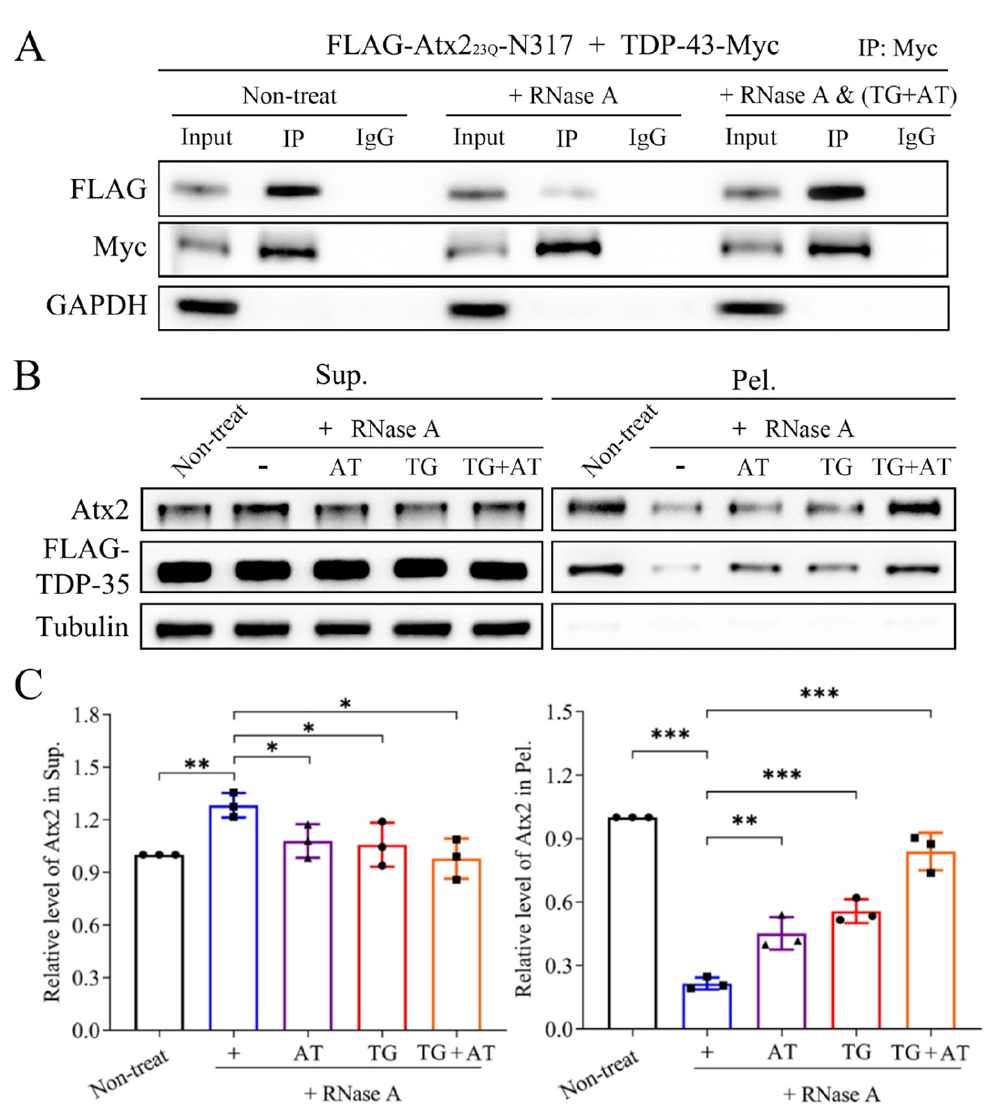
The biomolecular assemblies (condensates or aggregates) formed by mutant proteins are a pathological hallmark of neurodegenerative diseases. Some RNA-binding proteins (RBPs) are typically prone to aggregation that is closely associated with disease pathologies. These RBPs include numerous well-recognized pathogenic proteins, such as TAR DNA binding protein of 43 kDa (TDP-43), fused in sarcoma (FUS), ataxin-2 (Atx2), and poly(A)-binding protein nuclear 1 (PABPN1). Recent studies have revealed that liquid-liquid phase separation (LLPS), as a mechanism, underlies the highly dynamic and reversible granule formation of RBPs, and highlighted that multivalent RNA molecules play crucial roles in this process. These granules are necessary for diverse physiological functions, such as RNA splicing, trafficking, and even RNA storage, during stress. However, the aberrant phase transition of these mutant RBPs usually results in the formation of solid-like aggregates or inclusions within both the cytoplasm and nucleus. More importantly, aggregates formed by RBPs can sequester specific proteins, RNAs or other interacting partners, consequently contributing to RBP-related pathologies. For example, wild-type PABPN1 forms dynamic nuclear speckles with the assistance of poly(A) RNAs, whereas Ala expansion of PABPN1 results in the formation of aggregates, which are involved in the disease progression of oculopharyngeal muscular dystrophy (OPMD). Although the biological importance of various RBP granules is realized in either the cytoplasm or nucleus, how RNA regulates the formation of granules and the transition to aberrant RBP aggregates remains largely unknown. The interaction of a protein with other biomolecules (proteins, nucleic acids, etc.) is the prerequisite for the protein executing its normal biological function in cells. Identifying protein-protein and protein-RNA interactions is fundamental for the biochemical investigation of an individual protein and for attempts to understand the functional role of the protein. To date, many methods for studying protein-protein interactions have been developed on the basis of various principles, but it is still difficult to clarify whether the interactions between proteins, especially the RBPs involved, are direct or indirect, since RBPs generally bind to diverse RNAs closely and are incorporated into macromolecular ribonucleoprotein (RNP) complexes. We have taken several pairs of RBPs as examples, including TDP-35 (C-terminal 35-kDa fragment of TDP-43) with TDP-43 or TIA1, PABPN1 with a 25-kDa component of the mammalian cleavage factor I complex (CFIm25) and Atx2 with DEAD-box RNA helicase 6 (DDX6), and applied modified co-immunoprecipitation (Co-IP) and supernatant/pellet (S/P) fractionation experiments to characterize the association and sequestration of RBPs by using single-stranded DNA (ssDNA) chimera under ribonuclease (RNase) treatment. We designed several pieces of ssDNA oligonucleotides to mimic particular RNAs in cells that may mediate the association and sequestration of RBPs. The association of RBP proteins generally requires binding with multivalent RNA chains, since the bound RNAs tend to incorporate into a large protein-RNA complex with the help of RNA molecules. In Co-IP assay, especially for RBPs, RNase is often utilized to digest RNA in cell lysates to characterize whether the association of different RBPs is direct or indirect. It is important for us to demonstrate the active role of particular RNAs in the association or interaction of RBPs. Therefore, we designed and synthesized ssDNA chimeras to mimic the corresponding RNA that specifically bind to both RBPs simultaneously. In this case, ssDNA is used for rescuing the association of RBPs under the condition of RNase treatment, since the ssDNA oligonucleotide is resistant to nuclease activity. To design ssDNA chimeras for the RBPs of interest, first, the RNA sequences that bind to the two RPBs should be defined. The ssDNA should contain at least two portions (motifs) that specifically bind to each RBP, and each ssDNA portion may include 2–3 repeats of the binding sequence, so that the ssDNA can be recognized and bound efficiently by each RBP. Notably, the T base in ssDNA may sometimes be replaced with the U base (dU) for some more specific-binding RBPs, such as PABPN1. In the case of TDP-43 with Atx2, the binding specificities of the RNA sequences for TDP-43 and Atx2 are UG-rich and AUUUUU (AU5), respectively; then, the TG repeat portion is designed to bind to TDP-43, and the AT5 repeat is to bind to Atx2. Thus, an integrated method of co-IP and S/P fractionation was applied to characterize the association and sequestration of RBPs by combining ribonuclease (RNase) and ssDNA treatments.
1. Introduction
The biomolecular assemblies (condensates or aggregates) formed by mutant proteins are a pathological hallmark of neurodegenerative diseases [1,2]. Some RNA-binding proteins (RBPs) are typically prone to aggregation that is closely associated with disease pathologies [3]. These RBPs include numerous well-recognized pathogenic proteins [4,5], such as TAR DNA binding protein of 43 kDa (TDP-43), fused in sarcoma (FUS), ataxin-2 (Atx2), and poly(A)-binding protein nuclear 1 (PABPN1). Recent studies have revealed that liquid-liquid phase separation (LLPS), as a mechanism, underlies the highly dynamic and reversible granule formation of RBPs [4], and highlighted that multivalent RNA molecules play crucial roles in this process. These granules are necessary for diverse physiological functions, such as RNA splicing, trafficking, and even RNA storage, during stress [5]. However, the aberrant phase transition of these mutant RBPs usually results in the formation of solid-like aggregates or inclusions within both the cytoplasm and nucleus. More importantly, aggregates formed by RBPs can sequester specific proteins, RNAs or other interacting partners, consequently contributing to RBP-related pathologies [1]. For example, wild-type PABPN1 forms dynamic nuclear speckles with the assistance of poly(A) RNAs, whereas Ala expansion of PABPN1 results in the formation of aggregates, which are involved in the disease progression of oculopharyngeal muscular dystrophy (OPMD). Although the biological importance of various RBP granules is realized in either the cytoplasm or nucleus, how RNA regulates the formation of granules and the transition to aberrant RBP aggregates remains largely unknown.
The interaction of a protein with other biomolecules (proteins, nucleic acids, etc.) is the prerequisite for the protein executing its normal biological function in cells. Identifying protein-protein and protein-RNA interactions is fundamental for the biochemical investigation of an individual protein and for attempts to understand the functional role of the protein. To date, many methods for studying protein-protein interactions have been developed on the basis of various principles, but it is still difficult to clarify whether the interactions between proteins, especially the RBPs involved, are direct or indirect, since RBPs generally bind to diverse RNAs closely and are incorporated into macromolecular ribonucleoprotein (RNP) complexes. We have taken several pairs of RBPs as examples, including TDP-35 (C-terminal 35-kDa fragment of TDP-43) with TDP-43 or TIA1 [6], PABPN1 with a 25-kDa component of the mammalian cleavage factor I complex (CFIm25) [7] and Atx2 with DEAD-box RNA helicase 6 (DDX6) [8], and applied modified co-immunoprecipitation (Co-IP) and supernatant/pellet (S/P) fractionation experiments to characterize the association and sequestration of RBPs by using single-stranded DNA (ssDNA) chimera under ribonuclease (RNase) treatment.
Loading authentic research manuscript (Pages 1–5)...
Jianyang Wang, Wenliang Guan, Leilei Jiang, Hongyu Hu (2026). Characterization of the association and sequestration of RNA-binding proteins by single-stranded DNA chimera. Acta Biochimica et Biophysica Sinica. https://doi.org/10.3724/abbs.2024157
Research & Educational Purpose Only:The translations, structured abstracts, analytical annotations, and data reports provided by SinoBioData are intended exclusively for academic research, internal corporate R&D, and educational benchmarking. They do not constitute formal engineering, chemical safety, legal, or professional advice.
Copyright & Intellectual Property Notice: Original copyright of the underlying source articles and experimental data remains with the respective authors, institutions, and original publishing journals. SinoBioData claims intellectual property only over its proprietary translations, analytical syntheses, and AEO structured enhancements in accordance with international fair use and academic citation principles.
Frequently Asked Questions
What is the purpose of using ssDNA chimeras in this study?
The ssDNA chimeras are designed to mimic specific RNA sequences that mediate the association of RNA-binding proteins (RBPs). They are resistant to RNase, allowing the rescue of RBP interactions under RNase treatment, thereby demonstrating the active role of particular RNAs in mediating protein-protein interactions.
How does the ssDNA chimera method work?
The method involves designing ssDNA oligonucleotides containing binding motifs for two RBPs of interest. These chimeras are used in co-immunoprecipitation (Co-IP) and supernatant/pellet (S/P) fractionation assays under RNase treatment. The ssDNA binds to both RBPs simultaneously, maintaining their association even when RNA is digested, thus revealing direct or RNA-mediated interactions.
Which RBPs were studied as examples?
The study used pairs of RBPs including TDP-35 with TDP-43 or TIA1, PABPN1 with CFIm25, and Atx2 with DDX6. These are known to be involved in neurodegenerative diseases and form aggregates.
What is the significance of this research for neurodegenerative disease understanding?
This research provides a tool to characterize the molecular interactions that lead to RBP aggregation and sequestration, which are hallmarks of diseases like ALS and OPMD. Understanding these interactions can help in developing therapeutic strategies targeting aberrant phase transitions.
What are the limitations of using ssDNA chimeras?
The ssDNA chimeras may not perfectly mimic all aspects of RNA binding, such as structural flexibility or post-transcriptional modifications. Additionally, the design requires prior knowledge of the RNA binding sequences for each RBP, which may not be available for all proteins.
Related Technical Papers & Translations

Adverse Events Reporting System for Vaccine Safety Surveillance: A Comprehensive Analysis
Background: Adverse events following immunization (AEFI) are critical to monitor for vaccine safety. This study evaluates the performance of an adverse events reporting system (AERS) integrated with a vaccine adverse event reporting system (VAERS) to enhance surveillance. Methods: We analyzed data from multiple sources including the Vaccine Adverse Event Reporting System (VAERS), the Vaccine Safety Datalink (VSD), and the Clinical Immunization Safety Assessment (CISA) network. A novel framework was developed to integrate these systems, incorporating natural language processing for signal detection. Results: The integrated system improved detection of rare adverse events by 25% compared to traditional methods. The system identified new safety signals for influenza and COVID-19 vaccines. Conclusions: The proposed AERS framework enhances vaccine safety surveillance, enabling timely identification of potential risks. Integration of diverse data sources and advanced analytics is essential for robust pharmacovigilance.

Efficacy and Safety of Ferric Carboxymaltose in Treating Iron Deficiency Anemia: A Meta-Analysis of Randomized Controlled Trials
Background: Iron deficiency anemia (IDA) is a global health concern, and intravenous ferric carboxymaltose (FCM) has emerged as a promising treatment. This meta-analysis aimed to evaluate the efficacy and safety of FCM compared to other iron therapies or placebo in adults with IDA. Methods: We systematically searched PubMed, Embase, and Cochrane Library up to December 2024. Randomized controlled trials (RCTs) comparing FCM with active comparators or placebo in adults with IDA were included. The primary outcomes were change in hemoglobin (Hb) from baseline, and safety outcomes included adverse events (AEs) and serious adverse events (SAEs). Pooled estimates were calculated using random-effects models. Results: A total of 15 RCTs involving 4,856 patients were included. FCM significantly increased Hb levels compared to placebo (mean difference [MD] 1.2 g/dL, 95% CI 0.9-1.5) and was non-inferior to other intravenous iron preparations. The risk of AEs was similar between FCM and comparators (risk ratio [RR] 1.05, 95% CI 0.95-1.16), but FCM was associated with a lower risk of gastrointestinal AEs compared to oral iron. Serious adverse events were rare and comparable across groups. Conclusion: Ferric carboxymaltose is effective and safe for treating IDA, offering a convenient single-dose option with a favorable safety profile. These findings support its use in clinical practice.

Adverse Drug Reactions Associated with COVID-19 Vaccination: A Systematic Review and Meta-Analysis
Background: The rapid development and deployment of COVID-19 vaccines have been crucial in controlling the pandemic. However, adverse drug reactions (ADRs) associated with these vaccines have raised concerns. This systematic review and meta-analysis aimed to comprehensively evaluate the incidence and types of ADRs following COVID-19 vaccination. Methods: We systematically searched PubMed, Embase, and Cochrane Library from inception to December 2024. Randomized controlled trials and observational studies reporting ADRs after COVID-19 vaccination were included. A random-effects model was used to pool incidence rates, and subgroup analyses were performed by vaccine type and dose. Results: A total of 45 studies with 1,234,567 participants were included. The overall incidence of any ADR was 62.3% (95% CI: 58.1-66.4%). Common local reactions included injection site pain (48.2%), swelling (22.5%), and redness (18.7%). Systemic reactions included fatigue (34.6%), headache (28.9%), and myalgia (22.3%). Serious ADRs were rare (0.02%). Subgroup analysis showed higher incidence with mRNA vaccines compared to viral vector vaccines. Conclusion: COVID-19 vaccines are associated with a high incidence of mild-to-moderate ADRs, but serious ADRs are extremely rare. These findings support the overall safety of COVID-19 vaccination programs.