🧬 SinoBioData Academic Portal
šŸ“š Peer-Reviewed Translated Literature

All Biomedical & Clinical Articles (Page 36)

Browse complete peer-reviewed translations from top Chinese biomedical, oncology, and genomics journals. Read verified previews and download full authentic clinical reports.

Published Research Papers

Showing 24 of 1542 peer-reviewed translated articles (Page 36 of 65)

Urine-derived stem cells in kidney disease: progress, challenges, and future directionsGraphical AbstractVerified
Stem Cell Research & Therapy2026

Urine-derived stem cells in kidney disease: progress, challenges, and future directions

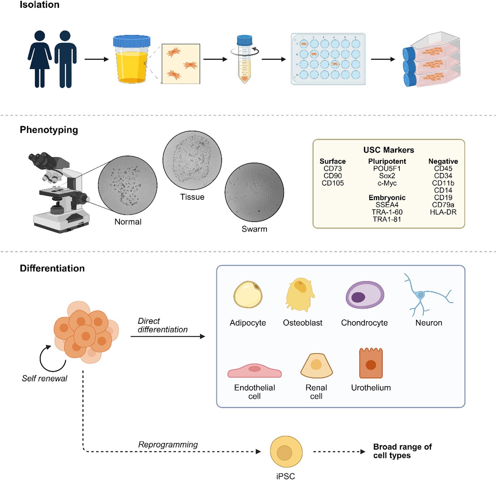
Chronic kidney disease (CKD) is a major global health burden with limited treatment options that address the underlying causes of fibrosis or promote regeneration. Urine-derived stem cells (USCs) have emerged as a promising tool in regenerative nephrology, offering a non-invasive and accessible source of multipotent cells with therapeutic potential. Sharing key properties with mesenchymal stem cells, USCs demonstrate paracrine activity, immunomodulation, and efficient extracellular vesicle (EV) production, and have shown anti-fibrotic, anti-inflammatory, and pro-regenerative effects in preclinical models of acute and chronic kidney injury. Recent advances in biomaterials and delivery technologies, including scaffold-free cell sheets and engineered EVs, have further enhanced the potential of USC-based therapies. However, challenges remain, particularly regarding functional integration, delivery optimization, and donor variability. This review summarizes the current progress in USC-based kidney therapy, identifies key limitations, and outlines future directions to support the translation of USC-based interventions into clinical practice.
Read Full Abstract10.1186/s13287-026-05040-2
Engineering hypoimmune stem cell-derived beta cellsGraphical AbstractVerified
Stem Cell Research & Therapy

Engineering hypoimmune stem cell-derived beta cells

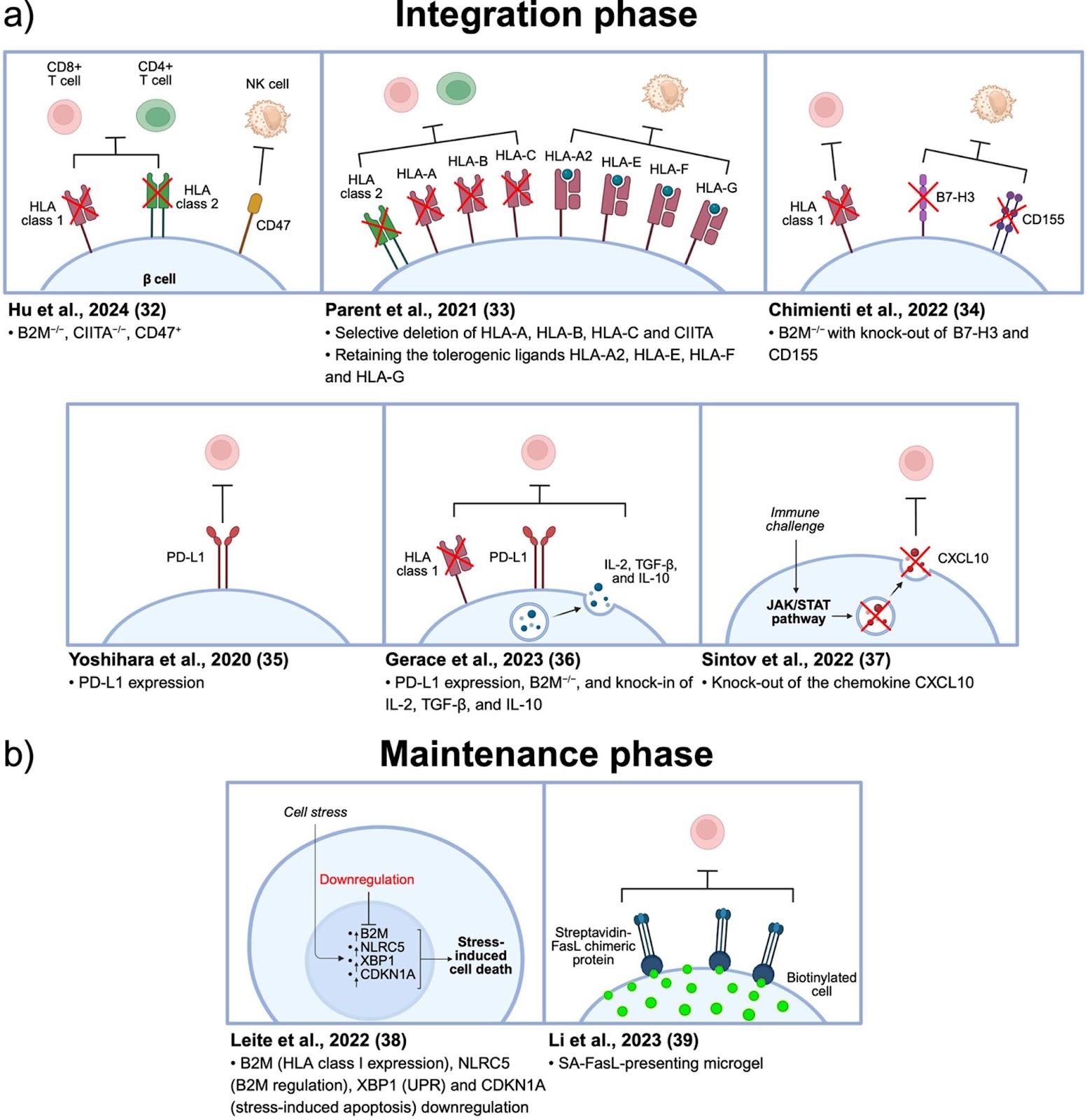
In type 1 diabetes (T1D), autoimmune targeting destroys insulin-producing β cells in the pancreas, creating a chronic state of insulin deficiency. Islet transplantation presents a regenerative cell therapy approach that can re-establish insulin production and intrinsic glycemic control. However, islet transplantation is currently limited by a lack of cadaveric human islet donors and a requirement for life-long immune suppression following transplant. Developments in stem cell maturation and differentiation protocols have enabled production of insulin-producing cells ā€˜on demand’, thereby addressing the pancreatic donor tissue shortage. Continued reliance on immune suppression to avoid graft rejection, however, can result in opportunistic infection and malignancy, thus remaining a major obstacle for wide-spread application of insulin-producing β cell transplantation. As such, there has been significant interest in identifying alternative strategies for avoiding graft rejection without immune suppression including encapsulation and co-transplantation of accessory immunomodulating cells. However, these approaches are limited by incomplete immune isolation as well as concerns over maintenance of effector function and graft survival in vivo, respectively. Genetically engineering hypoimmune stem cell-derived β cells has thus emerged as a promising strategy for improving islet transplantation outcomes. These approaches leverage our understanding of pathways involved in immune regulation to selectively protect the transplanted insulin-producing cells without affecting systemic immune function. This review will summarize recent bioengineering approaches for generating hypoimmune stem cell-derived β cells. It will also discuss relevant safety concerns and potential genetic targets for future investigation that take inspiration from the development of immune evasive primary islets and chimeric antigen receptor (CAR) T cells.

Read Full Abstract10.1186/s13287-025-04745-0
Therapeatic evaluation and single cell analysis of adipose stromal vascular fraction isolation from a commercial cell separation systemGraphical AbstractVerified
Stem Cell Research & Therapy

Therapeatic evaluation and single cell analysis of adipose stromal vascular fraction isolation from a commercial cell separation system

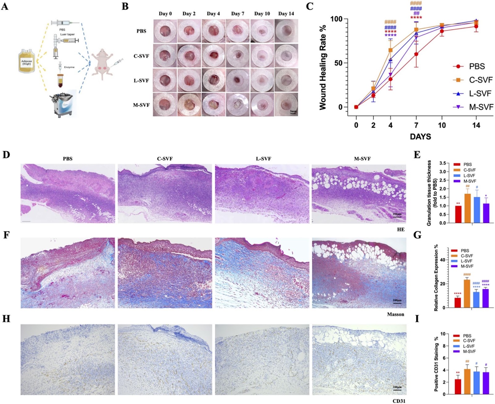
Background In the field of regenerative therapy, the stromal vascular fraction (SVF) extracted from adipose tissue has been widely recognized for its significant benefits. However, the cellular composition and therapeutic effect of SVF products prepared via different methods are unclear. Methods SVF cells were obtained via three approaches: (1) generation of the SVF via mechanical emulsification (M-SVF), (2) generation of the SVF via laboratory enzymatic digestion (L-SVF), and (3) generation of the SVF via commercial cell separation systems (C-SVF). We evaluated their healing effects on mouse wounds. Additionally, we utilized single-nucleus RNA sequencing (snRNA-seq) technology to explore the cellular composition of the C-SVF. Results The cell yield of C-SVF was comparable to that of L-SVF. During in vitro culture, C-SVF exhibited enhanced proliferation and a reduced proportion of apoptotic cells. In a mouse wound model, the application of C-SVF facilitated the closure of mouse wounds and improved collagen remodeling and angiogenesis in the wound area. Additional snRNA-seq analysis revealed that APOE+ adipose-derived stem cells and immune cells, especially M2 anti-inflammatory macrophages, are enriched in C-SVF, which together promote wound repair, and that APOE+ adipose-derived stem cells (ADSCs) and immune cells, especially M2 anti-inflammatory macrophages, are enriched in C-SVF, which jointly regulate and promote wound repair. Conclusion A commercial extraction system is an effective method for isolating viable SVF cells enriched with APOE+ ADSCs and M2 macrophages.

Read Full Abstract10.1186/s13287-025-04732-5
Long-term outcomes of mesenchymal stem cell therapy in severe COVID-19 patients: 3-year follow-up of a randomized, double-blind, placebo-controlled trialGraphical AbstractVerified
Stem Cell Research & Therapy

Long-term outcomes of mesenchymal stem cell therapy in severe COVID-19 patients: 3-year follow-up of a randomized, double-blind, placebo-controlled trial

Background The long-term effects and outcomes of human mesenchymal stem cell (MSC) therapy in patients with severe coronavirus disease 2019 (COVID-19) remain poorly understood. This study aimed to evaluate the extended safety and efficacy of MSC treatment in severe patients with COVID-19 who participated in our earlier randomized, double-blind, placebo-controlled clinical trial, with follow-up conducted over 3 years. Methods One hundred patients with severe COVID-19 were randomized to receive either an MSC infusion (n=65, 4Ɨ10^7 cells/dose, on days 0, 3, and 6) or a placebo, with both groups receiving the standard of care. At 36 months post-MSC therapy, patients were followed up to long-term safety and efficacy, particularly the effects of MSC therapy on persistent COVID-19 symptoms. Evaluated outcomes included lung imaging results, 6-min walking distance (6-MWD), pulmonary function test results, quality of life scores based on the Short Form-36 (SF-36) health survey, Long COVID symptoms, new-onset comorbidities, tumor marker levels, and rates of COVID-19 reinfection. Results Three years post-treatment, 46.94% (23/49) of patients in the MSC group and 34.48% (10/29) in the placebo group showed normal findings on computed tomography (CT) images (odds ratio [OR]=1.68, 95% confidence interval [CI]: 0.65–4.34). The general health (GH) score from the SF-36 was higher in the MSC group (67.0) compared to the placebo group (50.0), with a difference of 12.86 (95% CI: 1.44–24.28). Both groups showed similar results for total lung severity scores (TSS), 6-MWD, pulmonary function tests, and Long COVID symptoms. No significant differences between groups were observed in new-onset complications (including tumorigenesis) or tumor marker levels. After adjusting for China’s dynamic zero-COVID-19 strategy, severe acute respiratory syndrome coronavirus 2 (SARS-CoV-2) reinfection rates were 53.06% (26/49) in the MSC group and 67.86% (19/28) in the placebo group (OR=0.54, 95% CI: 0.20–1.41). Conclusions These findings support the long-term safety of MSC therapy in patients with severe COVID-19 over 3 years. MSC treatment may offer potential benefits for lung recovery and improved quality of life in patients experiencing Long COVID symptoms. Trial registration: ClinicalTrials.gov, NCT04288102. Registered 28 February 2020, https://clinicaltrials.gov/study/NCT04288102.

Read Full Abstract10.1186/s13287-025-04148-1
Effects of miR-210-3p/SDF2 and miR-31-5p/FGF7 from hypoxic endometrial exosomes on UCB-MSC proliferation, migration, and differentiationGraphical AbstractVerified
Stem Cell Research & Therapy

Effects of miR-210-3p/SDF2 and miR-31-5p/FGF7 from hypoxic endometrial exosomes on UCB-MSC proliferation, migration, and differentiation

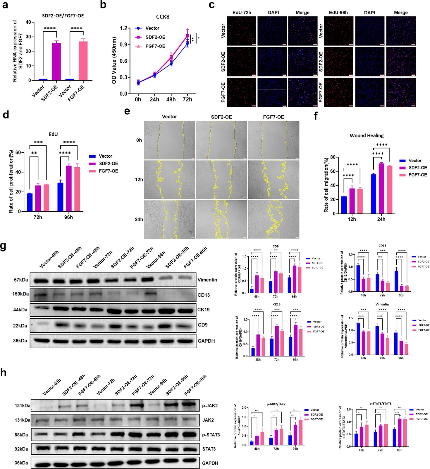
Background Mesenchymal stem cells (MSCs) isolated from umbilical cord blood (UCB) exhibit significant therapeutic efficacy in endometriosis; however, the molecular mechanisms governing their regulation remain incompletely elucidated. This study delves into the regulatory functions of miR-210-3p and miR-31-5p, which are secreted via exosomes from hypoxia-damaged endometrial epithelial cells, in modulating the behavior of UCB-MSCs. Methods UCB-MSCs were transfected with specific inhibitors targeting miR-210-3p and miR-31-5p. Proliferation and migratory capacities were quantified using CCK8, EdU incorporation, Transwell, and scratch wound healing assays. Western blotting was employed to assess the expression of endometrial epithelial markers (CD9 and CK19) and stromal markers (Vimentin and CD13), alongside the phosphorylation status of JAK2 and STAT3. Dual-luciferase reporter assays were conducted to validate SDF2 and FGF7 as direct targets of miR-210-3p and miR-31-5p, respectively. Results Suppression of miR-210-3p and miR-31-5p significantly augmented the proliferative and migratory abilities of UCB-MSCs, while simultaneously enhancing their differentiation into endometrial epithelial cells and attenuating their transition into stromal cells. Concurrently, the phosphorylation levels of JAK2 and STAT3 were markedly elevated. Overexpression of SDF2 and FGF7 further amplified the proliferative, migratory, and epithelial differentiation capacities of UCB-MSCs, accompanied by heightened activation of the JAK2/STAT3 signaling pathway. Notably, SDF2 overexpression and FGF7 overexpression effectively counteracted the inhibitory effects exerted by miR-210-3p and miR-31-5p mimics on UCB-MSC proliferation, migration, and epithelial differentiation, mediated through the modulation of JAK2/STAT3 signaling. Conclusion miR-210-3p and miR-31-5p orchestrate the functional dynamics of UCB-MSCs by targeting SDF2 and FGF7, respectively, through the JAK2/STAT3 pathway. These findings unveil novel mechanistic insights into the regenerative potential of UCB-MSCs, offering promising avenues for therapeutic advancements in endometriosis.

Read Full Abstract10.1186/s13287-025-04621-x
Phase separation participates in the genetic regulation mechanism of hematopoietic stem cells: potential therapeutic methodsGraphical AbstractVerified
Stem Cell Research & Therapy

Phase separation participates in the genetic regulation mechanism of hematopoietic stem cells: potential therapeutic methods

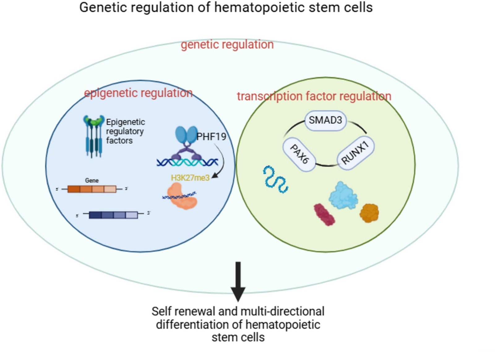
Hematopoietic stem cells (HSCs) are the primitive cells that give rise to common precursors for all blood cell lineages. Abnormalities in their number and/or function are important factors leading to the decline of immune function and the occurrence of various systemic diseases. Phase separation refers to a physicochemical mechanism in which intracellular liquid-liquid phase separation (LLPS) forms membrane-less organelles. It participates in various physiological activities and is related to the occurrence of diseases. Studies have shown that the functional activity of HSCs is regulated by complex mechanisms, and phase separation is closely related to these complex mechanisms such as genetic regulation, epigenetic regulation, microenvironment regulation, gene expression, autophagy degradation, and cell proliferation. With the deepening of research, the importance of phase separation in the pathogenesis and treatment of diseases such as leukemia and tumors has gradually emerged, but the deep mechanism of its regulation of HSCs genetic regulation still lacks exploration, and the direction of clinical targeted therapy is not yet clear. Here, we will summarize and elaborate the genetic regulation mechanism of HSCs, discuss the relationship between phase separation and the functional regulation of HSCs, and analyze the possibility of phase separation participating in the genetic regulation of HSCs to treat diseases, in order to provide help for the clinical implementation of targeted therapy for HSCs regulation.

Read Full Abstract10.1186/s13287-025-04350-1
Erythropoietin delivery through kidney organoids engineered with an episomal DNA vectorGraphical AbstractVerified
Stem Cell Research & Therapy

Erythropoietin delivery through kidney organoids engineered with an episomal DNA vector

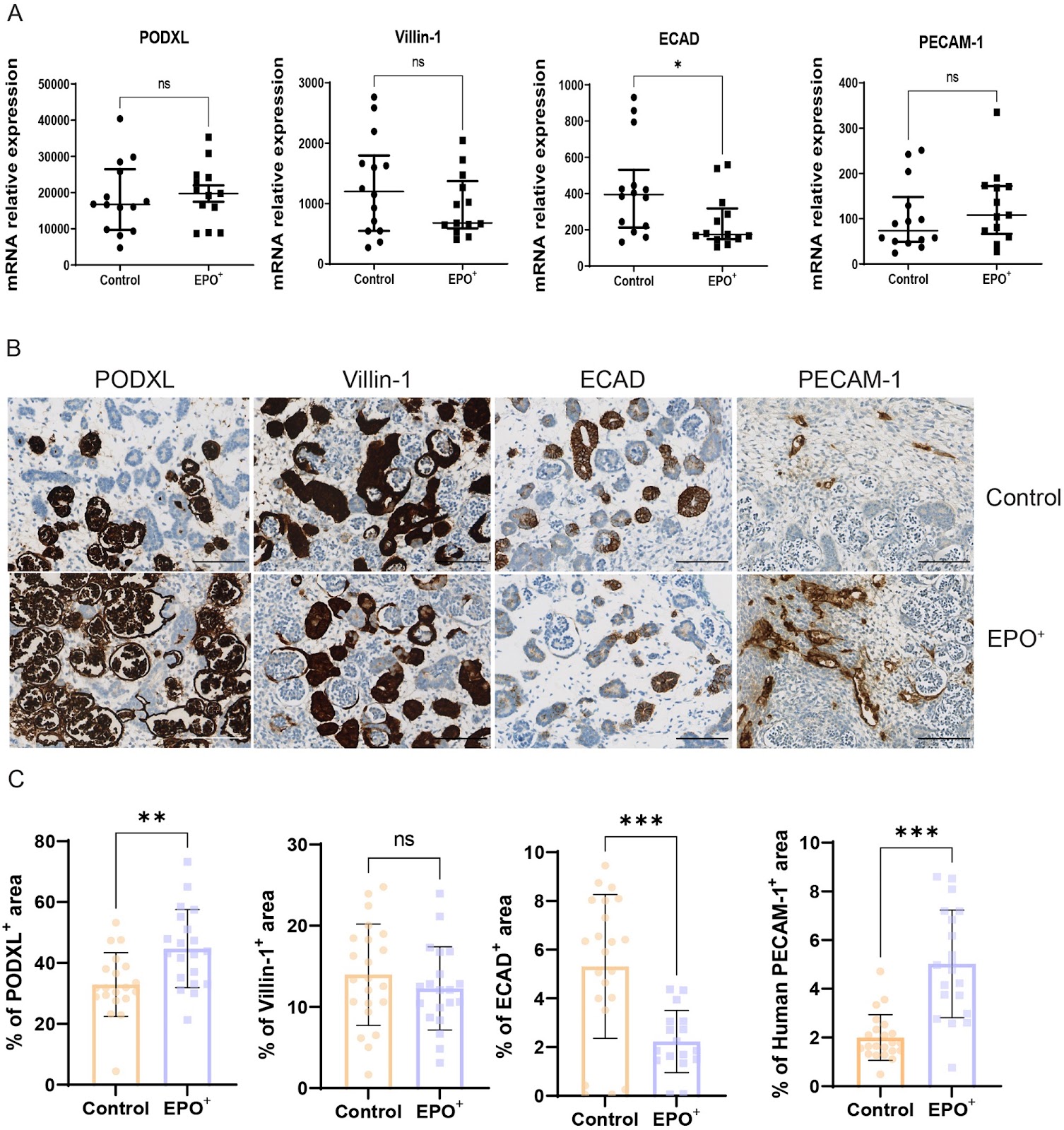
Background The kidney’s endocrine function is essential for maintaining body homeostasis. Erythropoietin (EPO) is one of the key endocrine factors produced by the kidney, and kidney disease patients frequently experience anemia due to impaired EPO production. In the present study we explored the potential of human induced pluripotent stem cell (iPSC)-derived kidney organoids to restore EPO production. Methods EPO secretion by kidney organoids was examined under 1% and 20% oxygen levels. To increase the EPO secreting capacity of kidney organoids, iPSC were genetically engineered with a non-integrating scaffold/matrix attachment region (S/MAR) DNA vector containing the EPO gene and generated EPO-overexpressing (EPO+) kidney organoids. To assess the physiological effects of EPO+ organoids, 2–8 organoids were implanted subcutaneously in immunodeficient mice. Results Kidney organoids produced low amounts of EPO under 1% oxygen. EPO S/MAR DNA vectors persisted and continued to robustly express EPO during iPSC expansion and kidney organoid differentiation without interfering with cellular proliferation. EPO+iPSC demonstrated efficient differentiation into kidney organoids. One-month post-implantation, EPO+ organoids displayed continuously elevated EPO mRNA levels and significantly increased endothelial cell numbers compared to control organoids. Hematocrit levels were notably elevated in mice implanted with EPO+ organoids in an organoid number-dependent manner. EPO+ organoids furthermore influenced bone homeostasis in their hosts, evidenced by a change in trabecular bone composition. Conclusion Kidney organoids modified by EPO S/MAR DNA vector allow stable long-term delivery of EPO. The observed physiological effects following the implantation of EPO+ organoids underscore the potential of gene-edited kidney organoids for endocrine restoration therapy.

Read Full Abstract10.1186/s13287-025-04282-w
β-Sitosterol preconditioning enhances the resistance of BMSCs and chondrocyte to oxidative stress and promotes cartilage repair in osteoarthritisGraphical AbstractVerified
Stem Cell Research & Therapy

β-Sitosterol preconditioning enhances the resistance of BMSCs and chondrocyte to oxidative stress and promotes cartilage repair in osteoarthritis

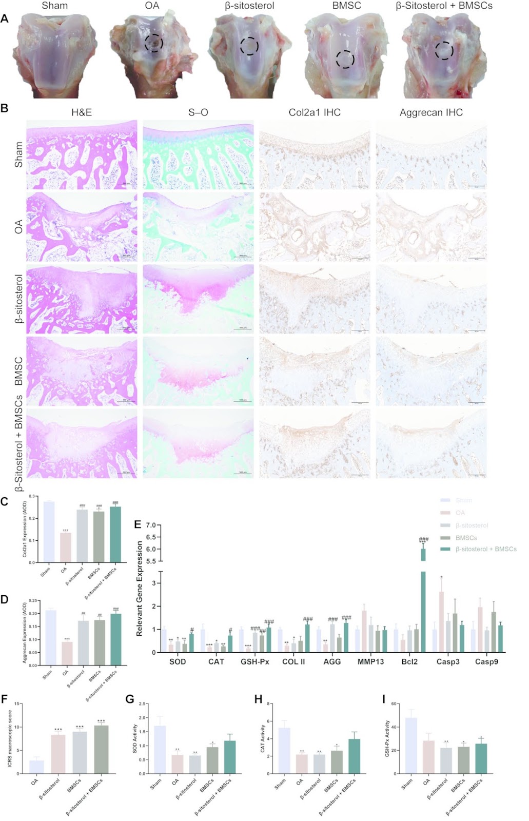
Background Osteoarthritis (OA) is a joint disorder that severely affects patients’ mobility, overall health, and ability to perform daily activities. Despite advancements in therapeutic strategies, stem cell-based therapies for OA still face challenges, particularly in enhancing the antioxidative capacity of stem cells to improve therapeutic outcomes. Therefore, this study aimed to explore the potential of β-sitosterol in this context. Methods This study evaluated the protective effects of β-sitosterol on bone marrow-derived mesenchymal stem cells (BMSCs) and chondrocytes under oxidative stress conditions and assessed its potential in promoting cartilage repair in a rabbit OA model. Cell viability, gene expression, oxidative stress markers, and mitochondrial function were examined. In vivo therapeutic effects were evaluated through histological and immunohistochemical analyses. Results The results revealed that β-sitosterol significantly enhanced BMSC viability, upregulated the expression of Col2a1 and aggrecan, while inhibiting MMP13 expression. Furthermore, β-sitosterol effectively alleviated oxidative stress and preserved mitochondrial function in BMSCs. Notably, BMSCs pretreated with β-Sitosterol exhibited a higher potential for facilitating cartilage regeneration in the OA model, as evidence by histopathological analysis. Conclusions These findings suggest that β-sitosterol possesses significant antioxidative and chondroprotective properties, which enhance the therapeutic efficacy of BMSCs in addressing OA-related cartilage damage.

Read Full Abstract10.1186/s13287-025-04613-x
The local pulsatile parathyroid hormone delivery system induces the osteogenic differentiation of dental pulp mesenchymal stem cells to reconstruct mandibular defectsGraphical AbstractVerified
Stem Cell Research & Therapy

The local pulsatile parathyroid hormone delivery system induces the osteogenic differentiation of dental pulp mesenchymal stem cells to reconstruct mandibular defects

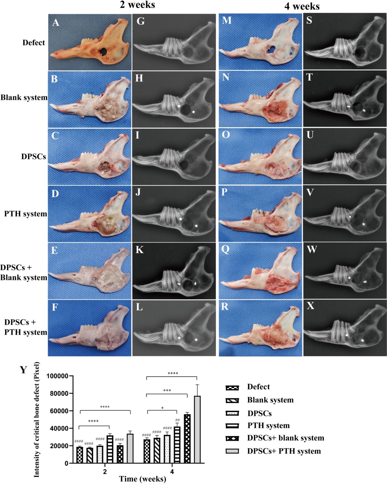
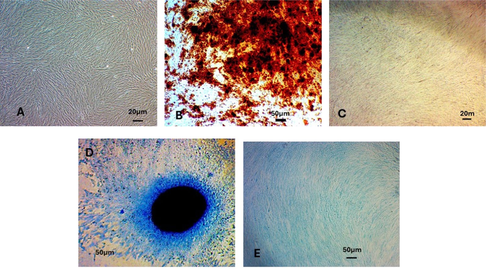
Background Tumors and injuries often lead to large mandibular defects. Accelerating the osteogenesis of large bone defect areas is a major concern in current research. In this study, dental pulp mesenchymal stem cells (DPSCs) were used as seed cells, and the local pulsatile parathyroid hormone (PTH) delivery system was used as an osteogenic-inducing active ingredient to act on DPSCs and osteoblasts, which were applied to the jaw defect area to evaluate its therapeutic effect on bone regeneration. Methods Pulsatile delivery systems, both with and without PTH, were developed following the protocols outlined in our previous study. In vitro, the biocompatibility of the pulsatile delivery system with DPSCs was assessed using the Cell Counting Kit-8 (CCK8) assay and live/dead cell staining. Osteogenic differentiation was evaluated through alkaline phosphatase staining and alizarin red staining. In vivo, critical bone defects with a diameter of 10 mm were created in the mandibles of white rabbits. The osteogenic effect was further assessed through gross observation, X-ray imaging, and histological examination. Results In vitro experiments using CCK8 assays and live/dead cell staining demonstrated that DPSCs successfully adhered to the surface of the PTH pulsatile delivery system, showing no significant difference compared to the control group. Furthermore, alkaline phosphatase staining and Alizarin Red staining confirmed that the localized pulsatile parathyroid hormone delivery system effectively induced the differentiation of DPSCs into osteoblasts, leading to the secretion of abundant calcium nodules. Animal studies further revealed that the PTH pulsatile delivery system promoted the osteogenic differentiation of DPSCs, facilitating the repair of critical mandibular bone defects.

Read Full Abstract10.1186/s13287-025-04258-w
Spatial self-organization of cancer stem cell niches revealed by live single-cell imagingGraphical AbstractVerified
Stem Cell Research & Therapy

Spatial self-organization of cancer stem cell niches revealed by live single-cell imaging

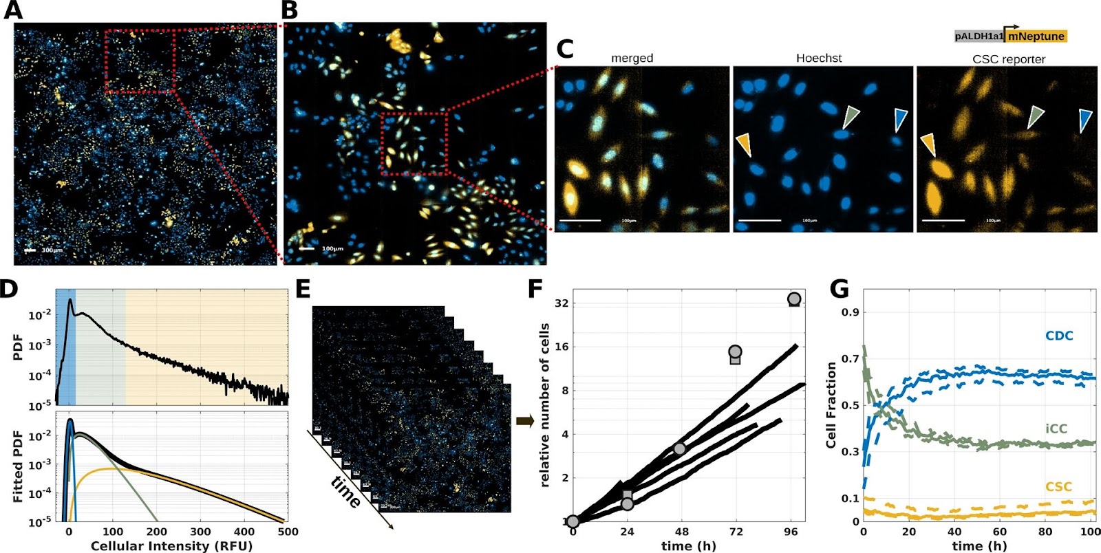
Background: Phenotypic plasticity is a major factor in tumor heterogeneity and treatment resistance. In particular, cancer stem cells (CSCs) represent a small subpopulation within tumors that possesses self-renewal and tumor-forming capabilities. Understanding reprogramming, maintenance, and lineage properties of CSCs requires dedicated tools to disentangle the respective influences of phenotypic inheritance and cell-cell interactions. Methods: Here, we set up ultra-wide field microscopy to image breast cancer cell lines expressing a stemness fluorescent reporter over several days. The fluorescent reporter distinguishes three phenotypes: CSCs, cancer differentiated cells (CDCs), and intermediate/transiting cancer cells (iCCs). Results: Spatial statistics indicate significant zonation in which CSCs cluster together and are spatially separated from CDCs, forming patterns resembling niches. Surprisingly, single-cell time series reveal spontaneous reprogramming events from CDC to CSC even in unperturbed populations. We identify that such transitions are prone to arise during the cell cycle. Moreover, lineage analysis shows that the phenotype is partially inherited from ancestor cells. However, such heredity is not sufficient to explain the spatial properties of the cell population, which also depend on cell-cell interactions. Indeed, we find that phenotypic transitions of cancer cells are influenced by the phenotypic state of neighboring cells. Reprogramming into CSCs is respectively promoted and inhibited by the presence of CSCs and CDCs in the neighborhood. Conclusions: Altogether, our results disentangle how phenotypic inheritance and intercellular interactions orchestrate the spatio-temporal self-organization of cancer cell heterogeneity, maintaining a subpopulation of CSCs within niches.

Read Full Abstract10.1186/s13287-025-04681-z
A meta-analysis on application and prospect of cell therapy in the treatment of diabetes mellitusGraphical AbstractVerified
Stem Cell Research & Therapy

A meta-analysis on application and prospect of cell therapy in the treatment of diabetes mellitus

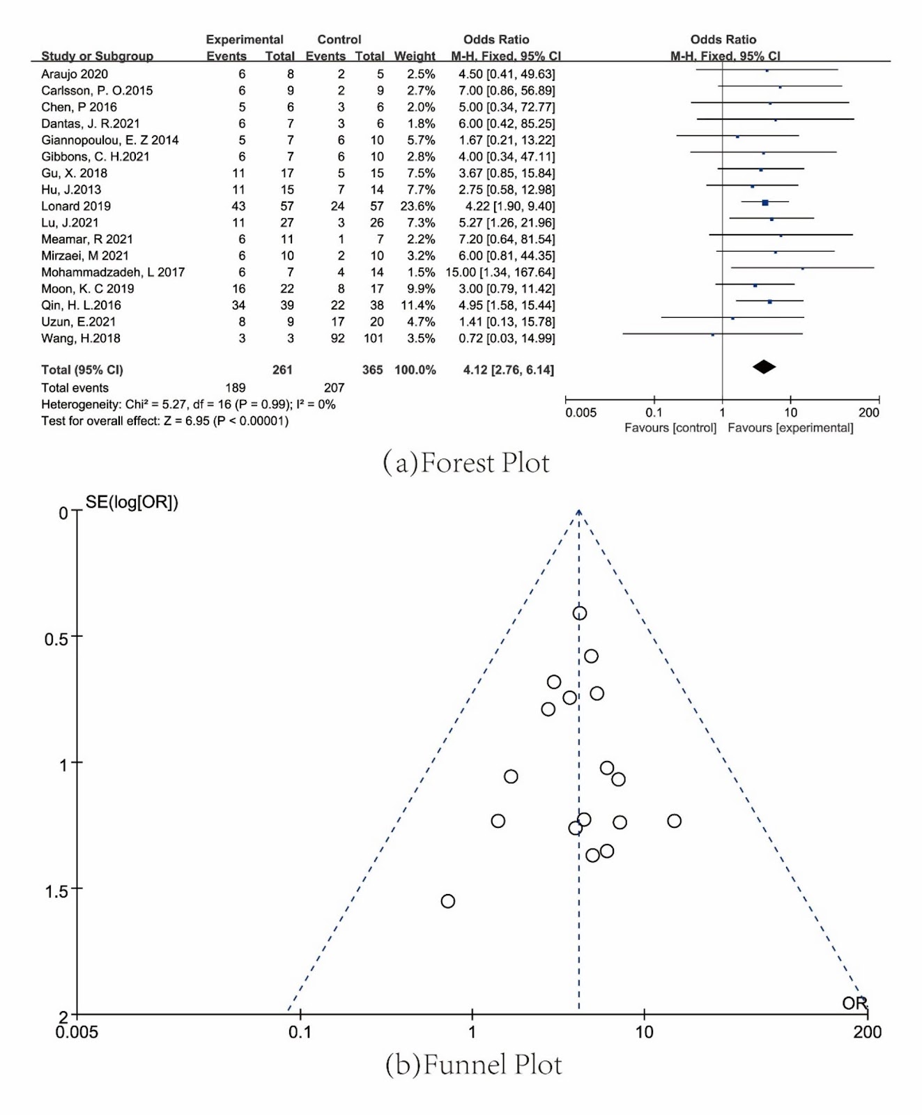
Objective Diabetes mellitus (DM) is a grave autoimmune disorder because of no insulin self-generation. Currently, mainly clinical methods exist, serious adverse effects leading to stem cell therapy are considered. The mesenchymal stem cells (MSCs), require high differentiation capacity and are judged as crucial in DM treatment. The meta-analysis aimed to systemically analyze the particular types of MSCs which play a more important role in DM and which DM is treated more effectively. Method A systematic review was conducted on the published literature, clinical trials and observational studies, utilizing databases such as PubMed, Embase, Cochrane and clinicaltrial.gov. RevMan software was adopted to draw Forest Plot and Funnel Plot, and subgroup analysis were employed to evaluate heterogeneity between different groups. Results We identified the meta-analyses of 34 unique random controlled trials and divided our own systematic reviews into 8 groups. The MSCs were associated with placebo (OR=2.79, 95% CI [1.63, 4.75]), Standard Clinical Treatment (SCT) (OR=4.12, 95% CI [2.76, 6.14]), and monocyte (OR=6.52, 95% CI [3.56, 9.48]). The comparison between Autologous MSCs and Allogenic MSCs (OR=4.64, 95% CI [3.42, 6.31]), Autologous BMMSCs and other MSCs (OR=5.28, 95% CI [3.64, 7.66]), Allogenic ASCs and UCMSCs (OR=3.54, 95% CI [1.83, 6.86]), Type I DM and Type II DM (OR=3.10, 95% CI [1.79, 5.38]), intravenous injection and other injections (OR=4.81, 95% CI [3.34, 6.94]), diabetic foot ulcers and diabetic neurological disease (OR=3.88,,95% CI [2.53,5.95]). Conclusion Current evidence suggests that MSCs hold significant potential for treating DM, demonstrating considerably high safety and efficacy. MSCs exhibit higher therapeutic benefits compared to monocytes, with autologous MSCs offering better clinical outcomes than allogenic sources. MSCs (BMMSCs) proved more effective than other types of MSCs. However, no significant differences were observed between adipose-derived MSCs (ASCs)

Read Full Abstract10.1186/s13287-025-04377-4
Purine metabolism in bone marrow microenvironment inhibits hematopoietic stem cell differentiation under microgravityGraphical AbstractVerified
Stem Cell Research & Therapy

Purine metabolism in bone marrow microenvironment inhibits hematopoietic stem cell differentiation under microgravity

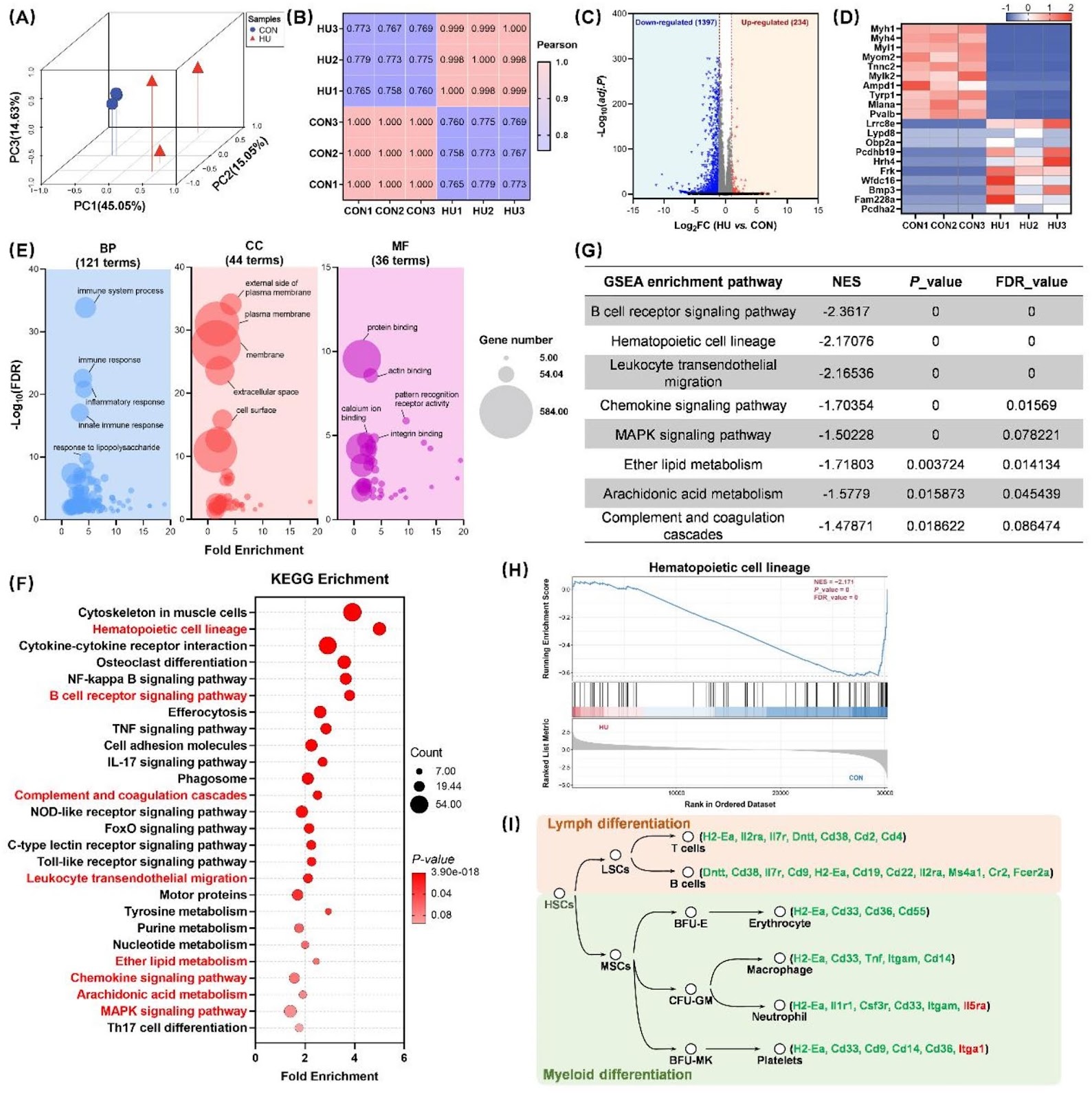
Background Spaceflight and microgravity environments have been shown to cause significant health impairments, including bone loss, immune dysfunction, and hematopoietic disorders. Hematopoietic stem cells (HSCs), as progenitors of the hematopoietic system, are critical for the continuous renewal and regulation of immune cells. Therefore, elucidating the regulatory mechanisms governing HSC fate and differentiation in microgravity environments is of paramount importance. Methods In this study, hindlimb unloading (HU) was employed in mice to simulate microgravity conditions. After 28 days of HU, cells were isolated for analysis. Flow cytometry and colony-forming assays were utilized to assess changes in HSC proliferation and differentiation. Additionally, transcriptomic and untargeted metabolomic sequencing were performed to elucidate alterations in the metabolic pathways of the bone marrow microenvironment and their molecular regulatory effects on HSCs fate. Results Our findings revealed that 28 days of HU impaired hematopoietic function, leading to multi-organ damage and hematological disorders. The simulated microgravity environment significantly increased the HSCs population in the bone marrow, particularly within the long-term and short-term subtypes, while severely compromising the differentiation capacity of hematopoietic stem/progenitor cells. Transcriptomic analysis of HSCs, combined with metabolomic profiling of bone marrow supernatants, identified 1,631 differentially expressed genes and 58 metabolites with altered abundance. Gene set enrichment analysis indicated that HU suppressed key pathways, including hematopoietic cell lineage and MAPK signaling. Furthermore, integrated analyses revealed that metabolites affected by HU, particularly hypoxanthine enriched in the purine metabolism pathway, were closely associated with hematopoietic cell lineage and MAPK signaling pathways. Molecular docking simulations and in vitro experiments confirmed that hypoxanthine interacts directly with core molecules within these pathways, influencing their expression. Conclusions These findings demonstrate that hypoxanthine in the bone marrow supernatant acts as a signaling mediator under microgravity, influencing HSCs fate by modulating hematopoietic cell lineage and MAPK signaling pathways. This study offers novel insights into the impact of microgravity on HSC fate and gene expression, underscoring the pivotal role of bone marrow microenvironmental metabolic changes in regulating key signaling pathways that determine hematopoietic destiny.

Read Full Abstract10.1186/s13287-025-04213-9
Multi-function of adipose-derived stem cells on gut disorder: from bench to bedsideGraphical AbstractVerified
Stem Cell Research & Therapy

Multi-function of adipose-derived stem cells on gut disorder: from bench to bedside

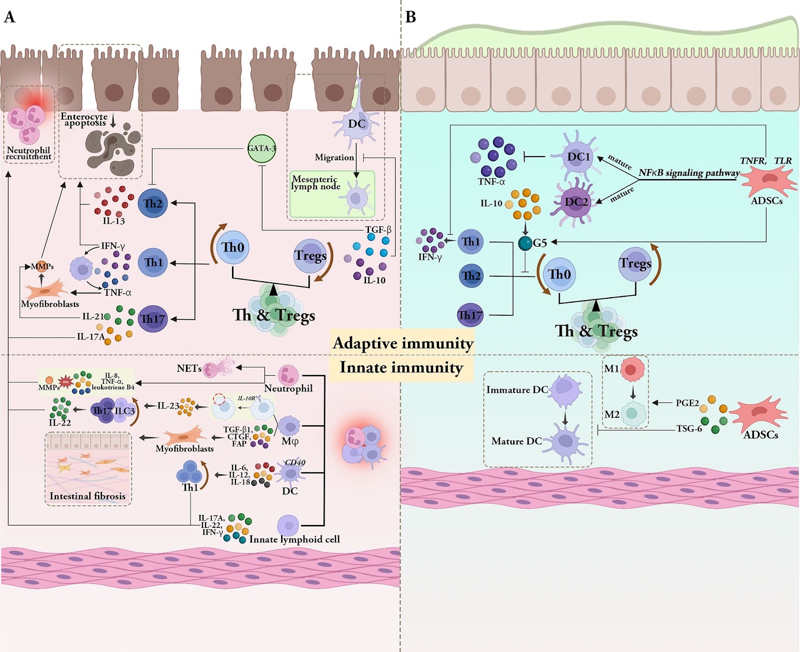
Adipose-derived stem cells (ADSCs) are a specific type of mesenchymal stem cells (MSCs) obtained easily from adipose tissue (AT). Compared with MSCs, ADSCs are easier to obtain, have fewer ethical issues, and have a higher proliferative capacity, which makes them a promising type of stem cell in regenerative medicine. ADSCs possess impressive capabilities in cell regeneration as well as differentiation, making them promising candidates for injury repair, tissue regeneration and alleviation of inflamed tissues. At present, most clinical studies on ADSCs focus on the treatment of wounds, multiple sclerosis, soft tissue trauma, aging, diabetes, Parkinson’s disease, bone and cartilage regeneration, stroke, and spinal cord injury, while its clinical applications in the gastrointestinal tract are relatively few. Therefore, this review summarizes the findings of preclinical experiments, clinical trials, and areas that may require further development of ADSCs in the treatment of digestive disorders, including inflammatory bowel disease (IBD), colorectal cancer (CRC), colorectal fibrosis, hepatocellular carcinoma, hepatic fibrosis, gastric cancer (GC), gastrostomy closure and radiation-induced proctitis. The review is concluded by discussing the goals for improvement and future directions for ADSCs before large-scale clinical application.

Read Full Abstract10.1186/s13287-025-04549-2
Induction of the p21/CDK6 pathway and alteration of the immune microenvironment by the stem cell marker CBX3 in melanomaGraphical AbstractVerified
Stem Cell Research & Therapy

Induction of the p21/CDK6 pathway and alteration of the immune microenvironment by the stem cell marker CBX3 in melanoma

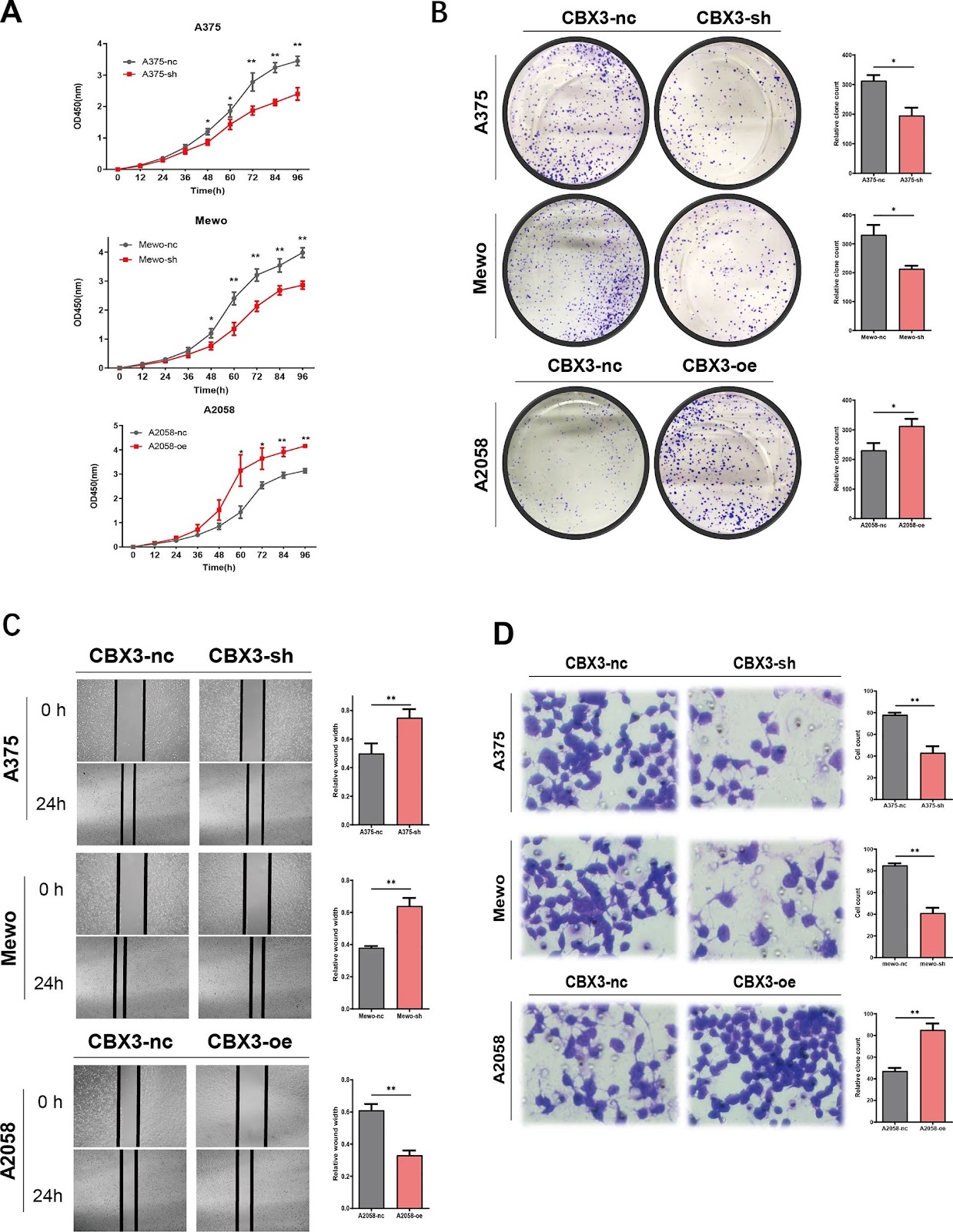
Background As one of the stem cell markers, chromobox protein homolog 3 (CBX3) participates in multiple signaling pathways that affect the progression of various tumors. However, the role of CBX3 in melanoma remains unclear, and the mechanisms by which CBX3 may regulate immunotherapy outcome remain largely unknown. Methods We used the Cancer Genome Atlas, Genotype-Tissue Expression portal, and Gene Expression Omnibus database to estimate CBX3 expression and its prognostic effect in melanoma. The role of CBX3 in proliferation and migration of melanoma cells were examined using the CCK8, cloning, wound healing, and transwell assays. The effect of CBX3 on melanoma tumorigenesis was assessed using an in vivo animal model. The role of CBX3 in cell cycle was examined using flow cytometry, and expression levels of cell cycle-related genes and proteins in cells with altered CBX3 levels were analyzed using qPCR and western blotting. The function of CBX3 in the immune microenvironment of melanoma was studied using single-cell RNA sequencing and public databases. Results We found that CBX3 was highly expressed in melanoma with poor prognosis. CBX3 promoted the proliferation and migration of melanoma cells in vivo and in vitro. Functional analysis revealed that CBX3 regulates cell cycle, as it accelerated the G1 to S transition, decreased p21 expression, and increased CDK6 expression. Finally, single-cell sequencing and immune-related assays showed that CBX3 is immunogenic and can change the immune microenvironment of melanoma. Conclusions We conclude that the stem cell marker, CBX3 activates the p21/CDK6 pathway and alters the immune microenvironment in melanoma.

Read Full Abstract10.1186/s13287-025-04179-8
Highly efficient XIST reactivation in female hPSC by transient dual inhibition of TP53 and DNA methylation during Cas9 mediated genome editingGraphical AbstractVerified
Stem Cell Research & Therapy

Highly efficient XIST reactivation in female hPSC by transient dual inhibition of TP53 and DNA methylation during Cas9 mediated genome editing

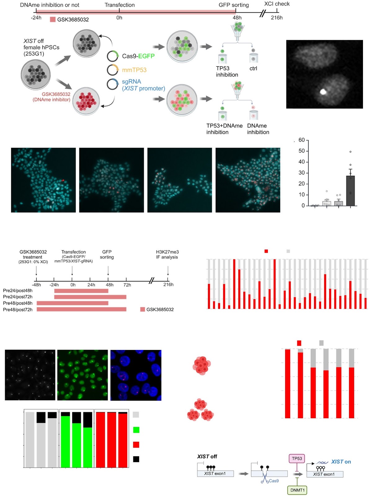
The irreversible erosion of X-chromosome inactivation (XCI) due to repression of the long non-coding RNA XIST presents a major challenge for disease modeling and raises safety concerns for the clinical application of female human pluripotent stem cells (hPSCs) due to the aberrant overexpression of X-linked genes. While Cas9-mediated non-homologous end joining (NHEJ) targeting the XIST promoter can induce DNA demethylation and restore XCI by reactivating XIST, its efficiency remains low. Here, we introduce a highly efficient strategy for XIST reactivation by combining TP53 inhibition with suppression of DNA methylation maintenance during Cas9-mediated NHEJ. This dual-inhibition approach increased the proportion of XIST-positive hPSCs from ~5 to ~43.7%, providing a robust method for stabilizing XCI in female hPSCs for diverse applications.

Read Full Abstract10.1186/s13287-025-04501-4
Blastocyst complementation: current progress and future directions in xenogeneic organogenesisGraphical AbstractVerified
Stem Cell Research & Therapy

Blastocyst complementation: current progress and future directions in xenogeneic organogenesis

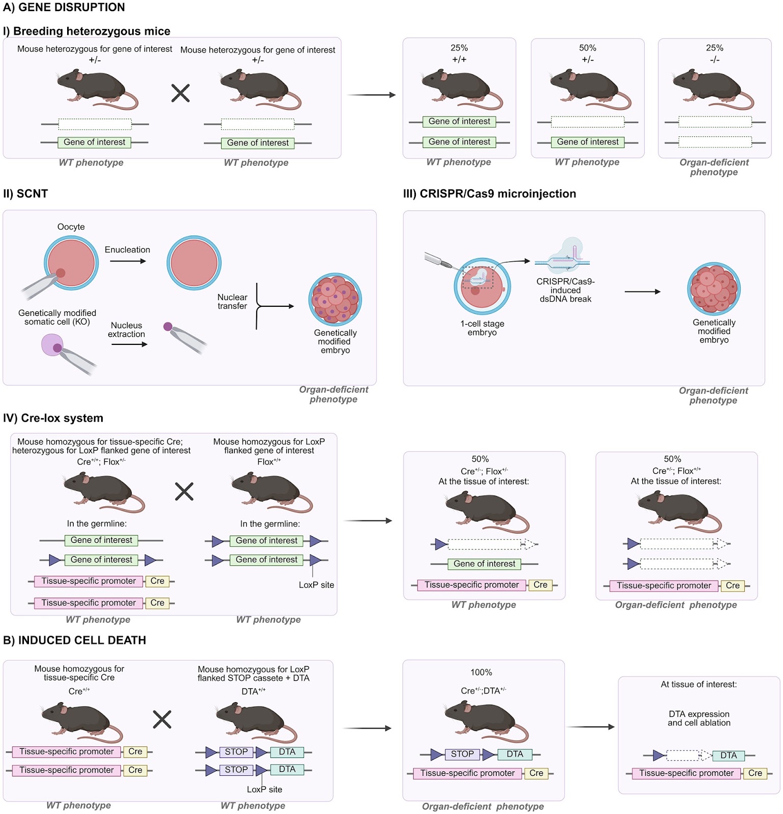
The generation of organs derived from pluripotent stem cells can be achieved in vivo through the blastocyst complementation technique. This method is based on the introduction of pluripotent stem cells into organogenesis-disabled pre-implantation embryos, where environmental signals instruct donor cells to colonize the vacant niche and to develop into the missing organ. When applied interspecies, this approach has the potential to produce human organs in genetically engineered livestock, offering a promising solution to the global transplants’ shortage crisis. In this review, we summarize the current progress in blastocyst complementation research and highlight the key challenges that must be addressed to advance this field.

Read Full Abstract10.1186/s13287-025-04426-y
CD73-expressing endometrial regenerative cell-derived exosomes mitigate acute cardiac allograft rejection through regulating adenosine metabolism in miceGraphical AbstractVerified
Stem Cell Research & Therapy

CD73-expressing endometrial regenerative cell-derived exosomes mitigate acute cardiac allograft rejection through regulating adenosine metabolism in mice

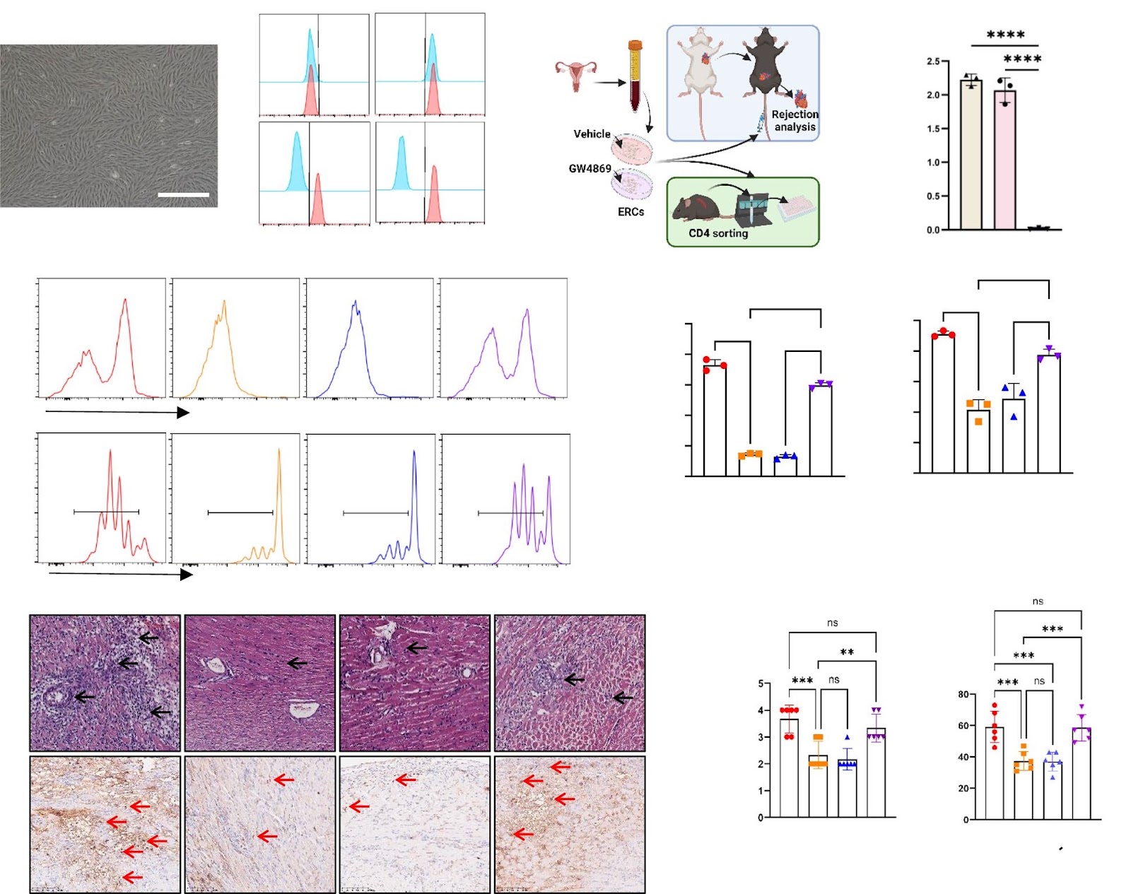
Background Organ transplantation is a life-saving option for end-stage organ dysfunction, but long-term graft survival is limited by unavoidable allograft rejection. While endometrial regenerative cells (ERCs) have been shown to alleviate acute rejection, the underlying mechanisms are not fully understood. This study explored whether ERC-derived exosomes contribute to this effect through CD73-mediated immunoregulation. Methods ERCs were pretreated with GW4869, an exosome inhibitor, to block exosome secretion, and CRISPR-Cas9-based CD73 knockout was performed to validate the role of CD73 in the ERC and ERC-exos. CD73 enzyme activity was measured using an AMP assay in vitro, whereas ATP, AMP, and adenosine levels were quantified using mass spectrometry in vivo. A murine allogeneic heart transplantation model (BALB/c to C57BL/6) was established to evaluate the immunoregulatory effects of ERC-exos in vivo. Graft tissues were analyzed by H&E staining, and immunohistochemistry and flow cytometry analysis of the spleens were performed to assess graft rejection. In vitro, flow cytometry was used to examine CD4+ T-cell activation, proliferation, differentiation, and subsets. Adenosine receptor inhibitors were used to identify receptor-mediated CD73-exosome signaling, and the potential of combining CD73-expressing exosomes with rapamycin to promote long-term graft survival was explored. Results GW4869 reduces the ability of ERCs to inhibit CD4+ T-cell activation and proliferation in vitro and attenuates the ERC-mediated suppression of acute allograft rejection in vivo. ATP, AMP and ADO increase adenosine 2a receptor (A2aR) but not A2bR expression on CD4+ T cells. CD73-expressing ERC-derived exosomes (ERC-exos) metabolize AMP into adenosine, leading to the inhibition of CD4+ T-cell activation, proliferation, and Th1 differentiation in vitro. This regulatory effect is reversed by the A2a receptor inhibitor CPI444. Furthermore, CD73 depletion blocks ERC-derived exosome-mediated adenosine production and impairs the ability of these cells to inhibit CD4+ T-cell activation and proliferation in vitro, as well as attenuate acute cardiac allograft rejection in vivo. Finally, the combination of ERC-exos with rapamycin significantly prolonged allograft survival from 15 days with rapamycin monotherapy to 38 days. Conclusion CD73 expression is crucial for the ability of ERC-exos to generate adenosine to mitigate acute cardiac allograft rejection in mice. ERC-exos combined with rapamycin can prolong allograft survival.

Read Full Abstract10.1186/s13287-025-04398-z
A phase I, open-label study of intravenous human dental pulp stem cells (NestaCellĀ®) at two dose levels in patients with Huntington’s diseaseGraphical AbstractVerified
Stem Cell Research & Therapy

A phase I, open-label study of intravenous human dental pulp stem cells (NestaCellĀ®) at two dose levels in patients with Huntington’s disease

Background Huntington’s disease (HD) is a progressive neurodegenerative disorder with no approved disease-modifying therapies. Human dental pulp stem cells (hDPSCs) offer potential therapeutic benefits due to their neurogenic, neurotrophic, and immunomodulatory properties. This prospective, open-label, single-centre, first-in-human clinical trial evaluated the safety, tolerability, and preliminary efficacy of intravenous hDPSC in patients with HD. Methods Six male patients with HD received intravenous infusions of hDPSCs in two dosage cohorts: three patients received 1 million cells/kg, and three received 2 million cells/kg. The treatment protocol consisted of cycles of three infusions at monthly intervals followed by subsequent administration cycles every six months, as per a protocol amendment based on the initial favourable safety outcomes. The total number of infusions ranged from 4 to 26 over the five years. During the first year, all patients underwent intensive multiparametric monitoring in an intensive care unit (ICU) for 48 h after each infusion. Results No adverse events occurred during the 48-h ICU monitoring or within 15 days post-infusion. Of 41 treatment-emergent adverse events (TEAEs) reported during follow-up, 35 were judged unrelated to the hDPSCs, mainly reflecting disease progression or incidental findings. Six treatment-emergent adverse events (TEAEs) were considered treatment-related, involving transient changes in hair pigmentation or regrowth. One patient discontinued due to a serious adverse event—lung cancer arising from a pre-existing pulmonary nodule identified at enrolment. Genetic analysis of the excised tumour showed no evidence of investigational product engraftment, supporting its non-tumorigenic nature. The same patient experienced a severe depressive episode

Read Full Abstract10.1186/s13287-025-04703-w
Mesenchymal stem cells inhibit mitochondrial fission by upregulating armadillo repeat containing 1, ameliorating oxidative stress in renal fibrosisGraphical AbstractVerified
Stem Cell Research & Therapy

Mesenchymal stem cells inhibit mitochondrial fission by upregulating armadillo repeat containing 1, ameliorating oxidative stress in renal fibrosis

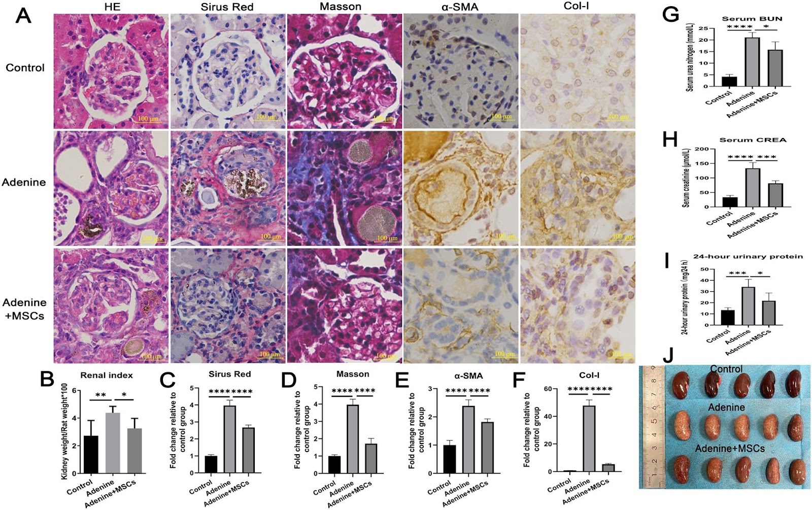
Background Oxidative stress damage is the important mechanism that promotes the process of fibrosis. Whether mesenchymal stem cells (MSCs) regulate mitochondrial dynamics and oxidative stress via armadillo repeat containing 1 (ARMC1) in renal fibrosis? Methods Using proteomics analysis, compare the significant differences in renal tissue proteins before and after MSCs intervention in adenine-induced nephropathy. Using a lentiviral vector to overexpress the ARMC1 gene in HK-2 cells, with the empty vector as a control. MSCs conditioned media (MSCs-CM) was applied to TGF-β1 treated cells, and MSCs were used in a cisplatin-induced nephropathy mouse model to assess mitochondrial dynamics, ROS generation, antioxidant stress, and fibrosis indicators, with Mdivi-1 (a Drp1 inhibitor) and Apocynin (a selective NADPH oxidase inhibitor) as positive controls. Results Renal proteomics showed that MSCs increased ARMC1 protein in the renal tissue of adenine nephropathy (3.521 times). In vitro, MSCs-CM increased ARMC1, reduced DRP1, and enhanced OPA1 and MFN2, lowering ROS, boosting mitochondrial bioactivity, and increasing antioxidant proteins NRF2, SOD1, SOD2, and CAT while decreasing fibrosis markers α-SMA, FN, COL-I, and KIM-1, and raising E-cadherin. The indicator variations in ARMC1-OE cells and OE-Con cells were similar between subgroups; Notably, under identical treatment conditions, the shifts in indicators within ARMC1-OE cells were more significant than those observed in OE-Con cells. In cisplatin-induced nephropathy mice, MSCs, Apocynin, and Mdivi-1 improved renal function and reduced interstitial collagen deposition, inhibited mitochondrial fission, enhanced antioxidant capacity, and reduced fibrosis. However, individual interventions were found to be less effective than their combined counterparts, with the synergistic impact of MSCs and Mdivi-1 achieving the most remarkable outcomes. Conclusion MSCs have the potential to improve renal fibrosis by influencing mitochondrial dynamics and oxidative stress through the upregulation of ARMC1 expression. ARMC1 may be an effective target for anti-fibrosis.

Read Full Abstract10.1186/s13287-025-04706-7
Engineering biomimetic bone marrow niche with gene modified mesenchymal stromal cells for ex vivo culture of human hematopoietic stem and progenitor cellsGraphical AbstractVerified
Stem Cell Research & Therapy

Engineering biomimetic bone marrow niche with gene modified mesenchymal stromal cells for ex vivo culture of human hematopoietic stem and progenitor cells

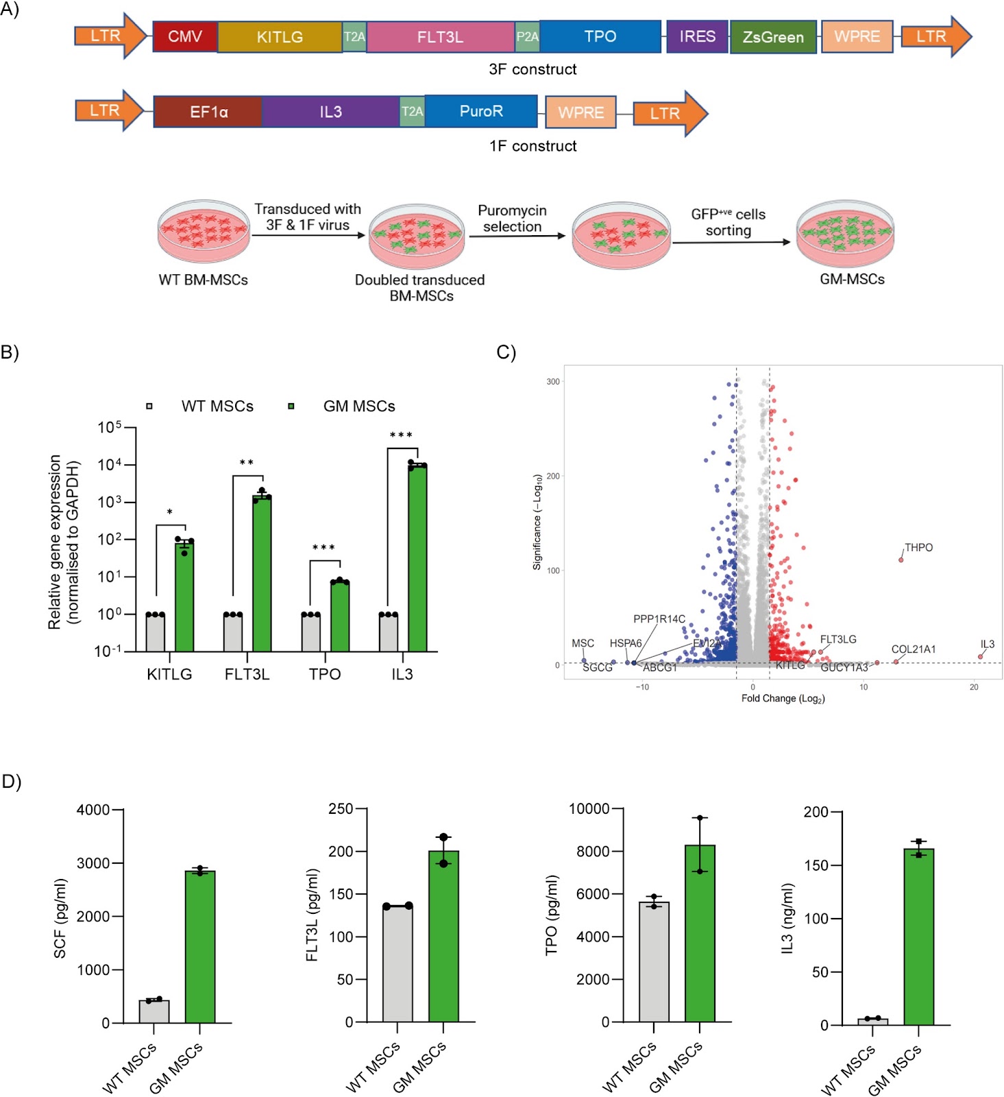
Background: Hematopoietic Stem and Progenitor Cells (HSPCs) gene therapy has shown significant progress, with commercial approval for at least four distinct haematological disorders, and poised for a rapid expansion in the upcoming years. Despite these advancements, the ex vivo culture of HSPCs continues to present significant challenges. The stress induced by ex vivo culture can negatively impact transplantation outcomes, while the need for exogenous cytokine supplementation contributes to the high costs associated with gene therapy products. Methods: We developed genetically modified human bone marrow MSCs (GM-MSCs) secreting cytokines such as Stem cell factor (SCF), Thrombopoietin (TPO), FMS-like tyrosine kinase-3-ligand (FLT3L), and Interleukin-3 (IL3), closely resembling bone marrow cellular niche to augment HSPCs culture. Results: HSPCs proliferate on GM-MSCs akin to standard conditions, devoid of external cytokine supplementation and these HSPCs retain their stem cell characteristics, colony-forming potential, stemness gene signatures, and capacity for long-term multilineage reconstitution in NBSGW mice. We demonstrate that our biomimetic feeder layer supports and alleviates stress associated with Homology Directed Repair (HDR) mediated gene-editing of HSPCs for fetal haemoglobin reactivation for a potential application in β-hemoglobinopathies gene therapy. Conclusion: Our GM-MSCs offer a compelling alternative to traditional cytokine supplementation by establishing a biomimetic bone marrow niche that fosters HSPC expansion while maintaining their stemness. These findings underscore the potential of engineered MSCs to revolutionize ex vivo HSPCs culture, ultimately enhancing their therapeutic value for gene therapy applications.

Read Full Abstract10.1186/s13287-025-04474-4
Melatonin-pretreated mesenchymal stem cell-derived exosomes alleviate cavernous fibrosis in a rat model of nerve injury-induced erectile dysfunction via miR-145-5p/TGF-β/Smad axisGraphical AbstractVerified
Stem Cell Research & Therapy

Melatonin-pretreated mesenchymal stem cell-derived exosomes alleviate cavernous fibrosis in a rat model of nerve injury-induced erectile dysfunction via miR-145-5p/TGF-β/Smad axis

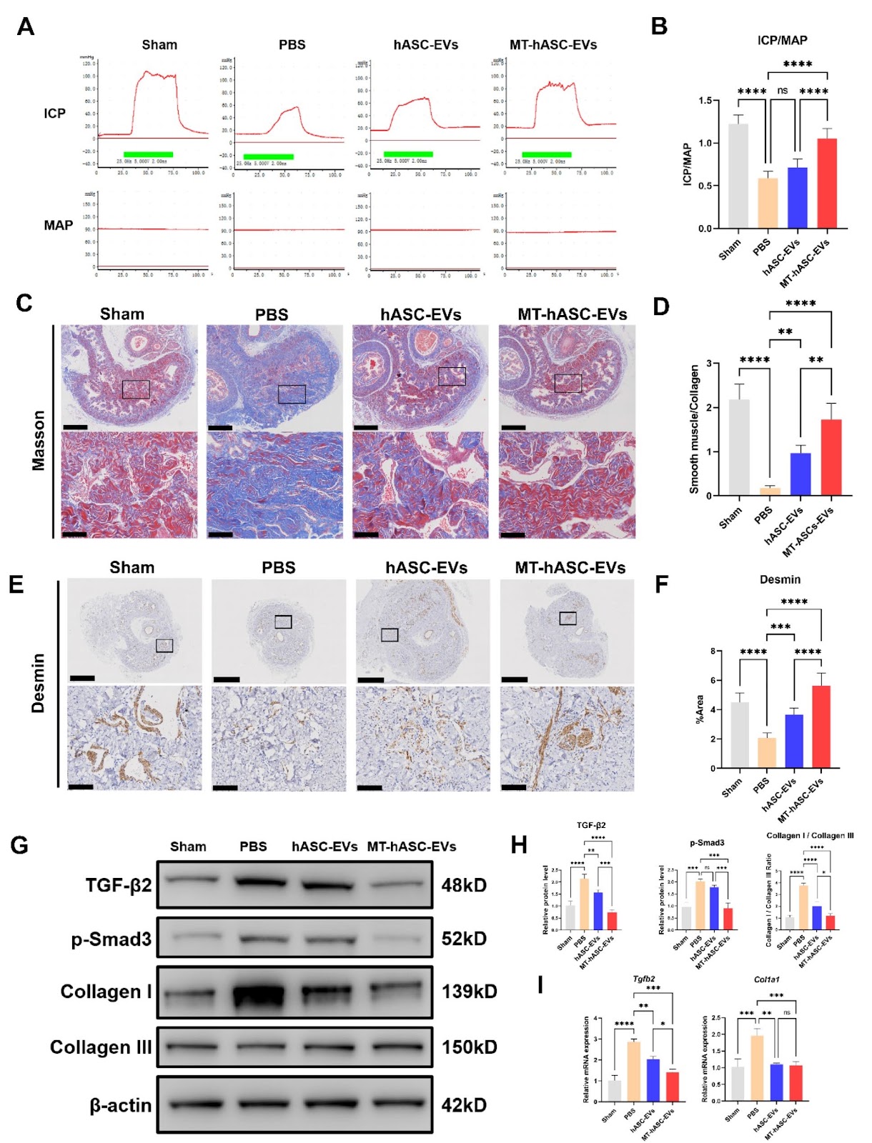
Background Cavernous nerve injury-induced erectile dysfunction (CNI-ED) is a common complication after radical prostatectomy. Conventional treatment approaches have had little success in treating the severe cavernous fibrosis which is a consequence of CNI-ED. Methods Pre-treatment of adipose-derived stem cells with melatonin allows for the extraction of active exosomes (MT-hASC-EVs) from the conditioned medium. The therapeutic effects of MT-hASC-EVs were assessed in a rat model of CNI-ED, and the anti-fibrotic properties were evaluated. MicroRNA sequencing was used to identify specific microRNAs highly expressed in MT-hASC-EVs, and differential microRNAs were screened for regulatory pathways through target gene enrichment analysis. Finally, the conclusions from bioinformatics analysis were validated through in vitro experiments. Results Intracavernous injection of MT-hASC-EVs significantly restored erectile function and reduced the extent of corpus cavernosum fibrosis in the CNI-ED rat model. MT-hASC-EVs promoted the proliferation and anti-apoptotic effects of corpus cavernosum smooth muscle cells (CCSMCs) in vitro. Mechanistically, MT-hASC-EVs inhibit fibrosis by delivering miR-145-5p, which targets TGF-β2/Smad3 axis. Conclusions MT-hASCs-EVs can inhibit cavernous fibrosis and improve erectile function in a rat model of CNI-ED by targeting the miR-145-5p/TGF-β/Smad axis.

Read Full Abstract10.1186/s13287-025-04173-0
The influence of femtosecond laser intrastromal lenticules on the characteristics and maturity in tissue-engineered stem cell-derived retinal pigment epithelium sheetsGraphical AbstractVerified
Stem Cell Research & Therapy

The influence of femtosecond laser intrastromal lenticules on the characteristics and maturity in tissue-engineered stem cell-derived retinal pigment epithelium sheets

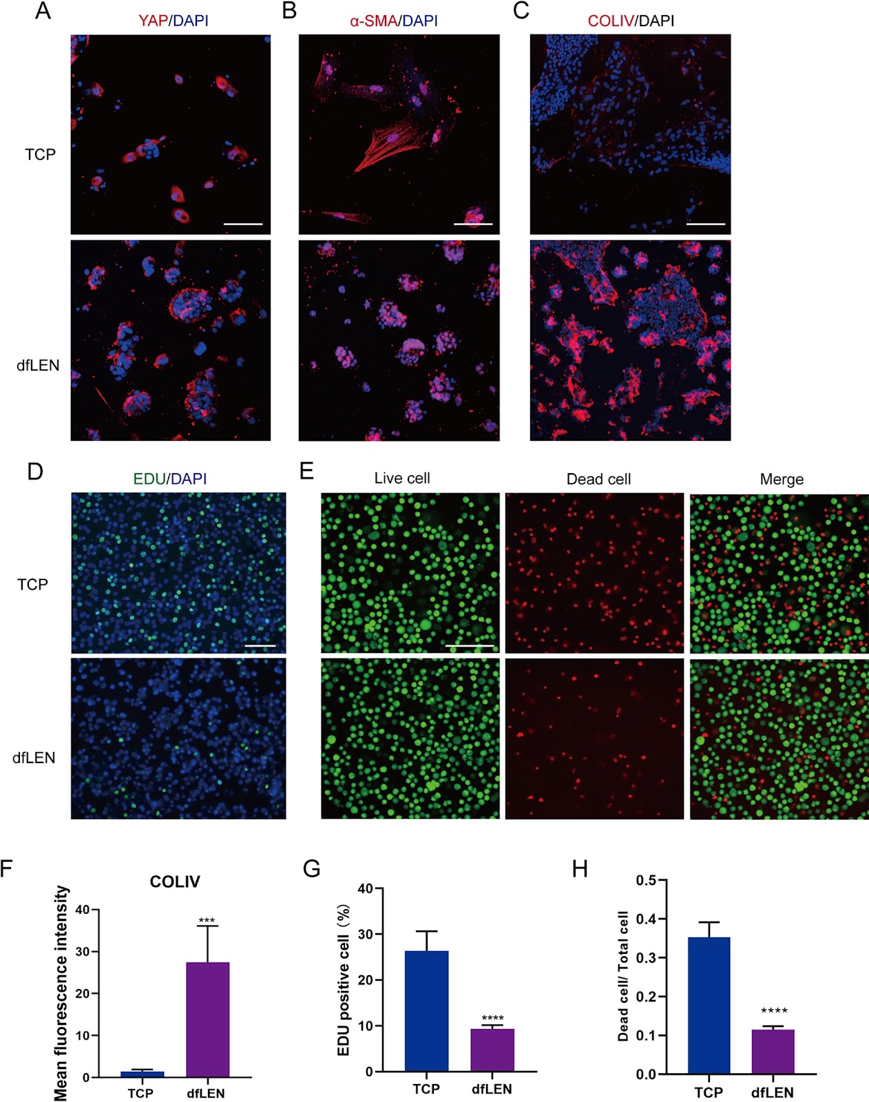
Background: Recent advances in clinical trials have involved the transplantation of induced retinal pigment epithelium (iRPE) cells from stem cells in creating a functional monolayer that mimics the characteristics of natural adult RPE cells. One method of achieving this goal is through the use of tissue engineering. In this research, decellularised femtosecond laser intrastromal lenticules (dfLEN) were employed as a scaffold for cultivating a bioengineered iRPE monolayer sheet. Methods: iRPE cells were obtained by differentiating induced pluripotent stem cells (iPSC). These cells were then seeded on decellularized FLI-lenticules (dfLEN). The functionality, characterization, and oxidative stress of iRPE cultured on dfLEN were compared with those cultured on plates (TCP) using various assays such as immunofluorescence (IF), Edu, CCK8, ELISA, DFCH-DA, and JC-1. Additionally, RNA-seq assays and electron microscope (SEM and TEM) were used to test the iRPE characteristic on engineered dfLEN. Finally, we evaluated the biocompatibility of iRPE-dfLEN sheets by transplanting them into the subretinal space of New Zealand white rabbits. Results: The iRPE cells cultured on dfLEN exhibited morphology and physiology similar to that of native RPE tissue. The dfLEN not only increased the resistance capacity of iRPE cells but also improved their functional properties compared to TCP. In addition, our results indicate that dfLEN enhances the expression of genes associated with cilium assembly, resulting in notable improvements in ciliogenesis in iRPE cells. Finally, the dfLEN-iRPE sheets demonstrated favorable biocompatibility and some viability when transplanted into the subretinal space of rabbits for a period of 14 days.

Read Full Abstract10.1186/s13287-025-04463-7
Transcriptomic insights and feeder-free culturing of porcine expanded potential stem cells from cloned embryosGraphical AbstractVerified
Stem Cell Research & Therapy

Transcriptomic insights and feeder-free culturing of porcine expanded potential stem cells from cloned embryos

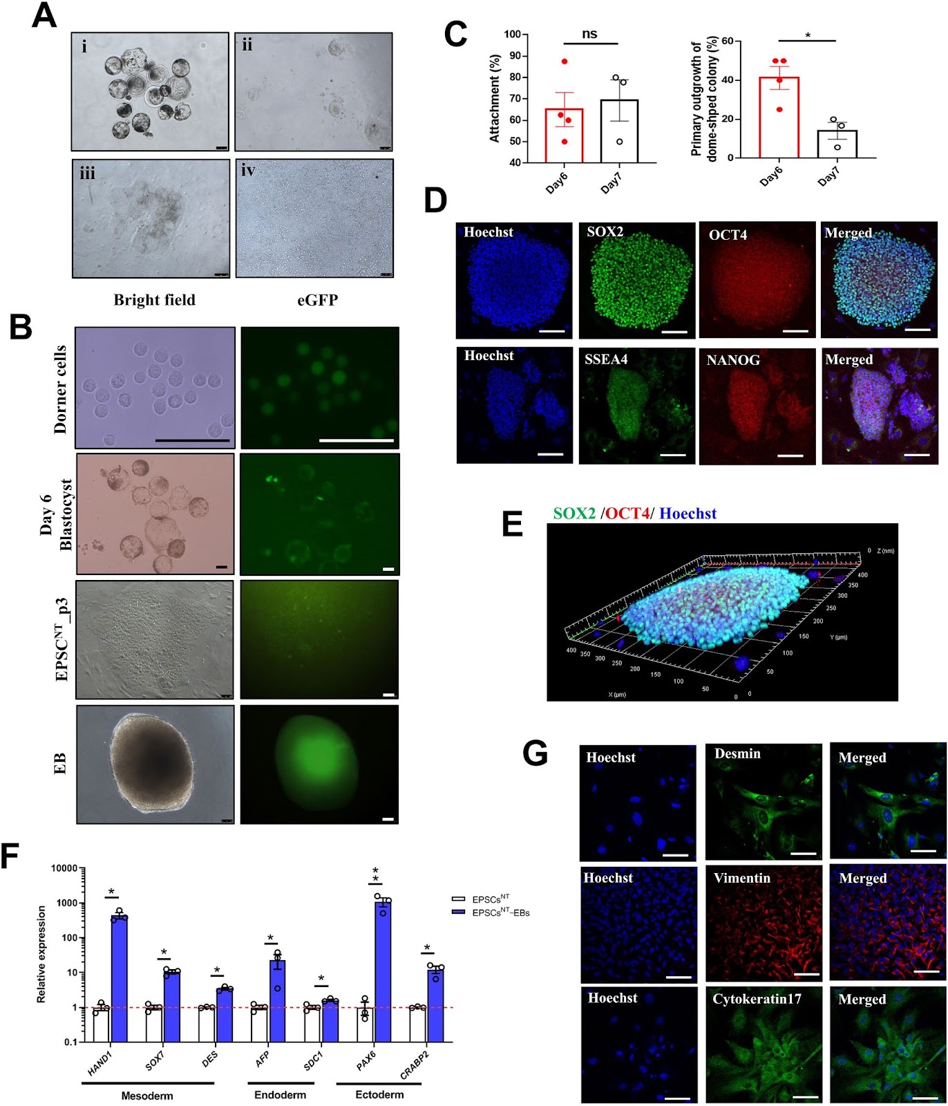
Background Generating expanded potential stem cells from cloned porcine embryos (pEPSCsNT) represents a notable advancement in regenerative medicine and agricultural biotechnology. However, challenges, including low derivation efficiency, limited understanding of transcriptomic features, and unknown feasibility of culturing under feeder-free conditions, remain. This study aimed to generate pEPSCs using blastocysts derived from parthenogenetic activation, in vitro fertilization, and somatic cell nuclear transfer (SCNT) using a modified culture system. Methods We derived pEPSCNT lines using an optimized culture system. We characterized the pEPSC lines from all three origins by analyzing pluripotent marker expression, performing karyotyping, and assessing their differentiation potential into the three germ layers. Furthermore, we performed a comparative transcriptomic analysis using in vivo and cloned embryo data, with a major focus on cell lines derived from SCNT (pEPSCsNT). We optimized feeder-free culture conditions for the pEPSCNT line and derived the pEPSCNT lines using an optimized culture system with an efficiency of ~14%. Results The cells were closely correlated with 8-cell to morula-stage embryos and exhibited significant enrichment of EPSC signature genes, suggesting a unique pluripotent state relatively close to the naĆÆve state, specifically within a formative state. The pEPSCsNT possessed broad differentiation capacity, indicative of Hippo signaling pathway enrichment, blastocyst-like structure formation ability, and potential differentiation into trophoblast lineage cells. Conclusions Our modified culture medium combined with the 2Ɨ Matrigel coating system facilitated the transition to feeder-independent culture conditions. These findings facilitate the establishment of a feeder-free culture system while preserving pluripotency and differentiation potential.

Read Full Abstract10.1186/s13287-025-04627-5
Apremilast improves cardiomyocyte cohesion and arrhythmia in different models for arrhythmogenic cardiomyopathyGraphical AbstractVerified
Stem Cell Research & Therapy

Apremilast improves cardiomyocyte cohesion and arrhythmia in different models for arrhythmogenic cardiomyopathy

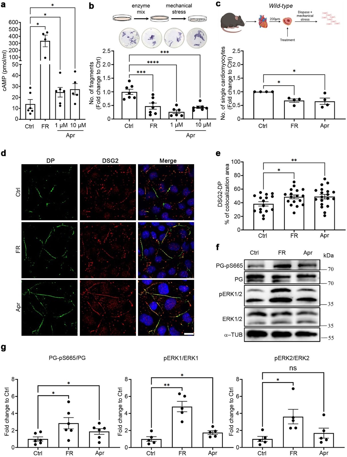
Background Arrhythmogenic cardiomyopathy (ACM) is a genetically inherited desmosome heart disease leading to life-threatening arrhythmias and sudden cardiac death. Currently, ACM treatment paradigms are merely symptom targeting. Recently, apremilast was shown to stabilize keratinocyte adhesion in the desmosomal disease pemphigus vulgaris. Therefore, this study investigated whether apremilast can be a therapeutic option for ACM. Methods Human induced pluripotent stem cells from a healthy control (hiPSC) and an ACM index patient (ACM-hiPSC) carrying a heterozygous desmoplakin (DSP) gene mutation (c.2854G > T, p.Glu952Ter), confirmed by whole exome sequencing (WES), were established. Cyclic-AMP ELISA, dissociation assay, immunostaining, and Western blotting analyses were performed in human iPSC-derived cardiomyocytes (hiPSC-CMs), murine HL-1 cardiomyocytes, and cardiac slices derived from wild-type (WT) mice, plakoglobin (PG, Jup) knockout (Jupāˆ’/āˆ’) (murine ACM model) or PG Serine 665 phosphodeficient (JUP-S665A) mice. Microelectrode array (MEA) analyses in ventricular cardiac slices and Langendorff heart perfusion were performed to analyze heart rate variability and arrhythmia. Results ACM-hiPSC derived cardiomyocytes (ACM-hiPSC-CMs) revealed a significant loss of cohesion, which was rescued by apremilast. Further, treatment with apremilast strengthened basal cardiomyocyte cohesion in HL-1 cells and WT murine cardiac slices, paralleled by phosphorylation of PG at Serine 665 in human and murine models. In HL-1 cells, apremilast in addition activated ERK1/2, inhibition of which abolished apremilast-enhanced cardiomyocyte cohesion. Further, dissociation assays in slice cultures from JUP-S665A and Jupāˆ’/āˆ’ mice revealed that PG is crucial for apremilast's effects. Additionally, apremilast reduced arrhythmic events in ventricular cardiac slices and Langendorff-perfused hearts. Conclusion Apremilast improves cardiomyocyte cohesion and reduces arrhythmia in different models of ACM, suggesting a novel therapeutic strategy for this disease.

Read Full Abstract10.1186/s13287-025-04755-y