🧬 SinoBioData Academic Portal
📚 Peer-Reviewed Translated Literature

All Biomedical & Clinical Articles (Page 37)

Browse complete peer-reviewed translations from top Chinese biomedical, oncology, and genomics journals. Read verified previews and download full authentic clinical reports.

Published Research Papers

Showing 24 of 1542 peer-reviewed translated articles (Page 37 of 65)

Reviving hope: unlocking pancreatic islet immortality by optimizing a trehalose-based cryopreservation media and cell-penetrating peptideGraphical AbstractVerified
Stem Cell Research & Therapy

Reviving hope: unlocking pancreatic islet immortality by optimizing a trehalose-based cryopreservation media and cell-penetrating peptide

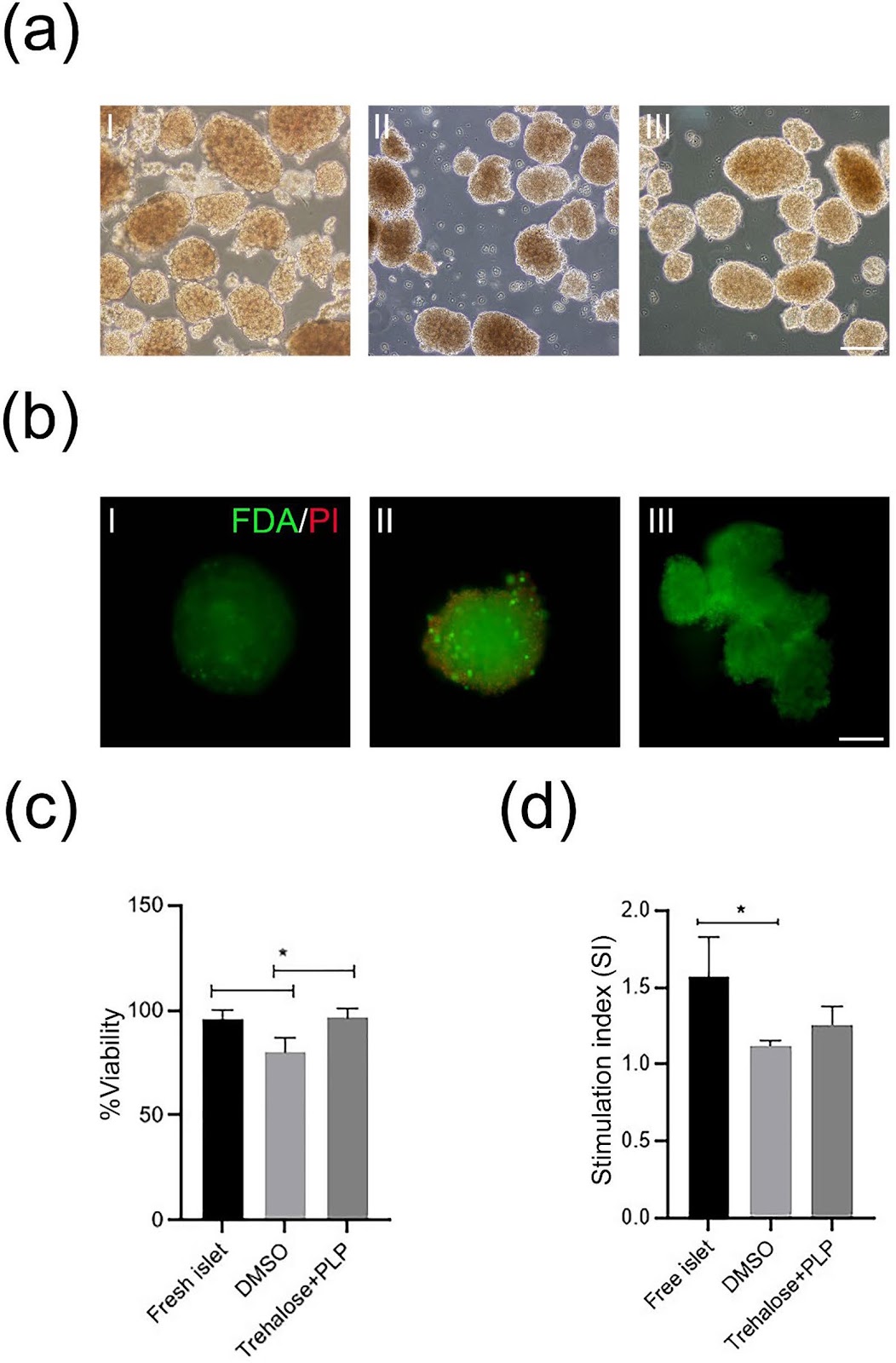
Background Diabetes mellitus remains a pervasive global health concern, urging a deeper exploration of islet transplantation as a potential enduring solution. The efficacy of this therapeutic approach pivots on the precision of cryopreservation techniques, ensuring both the viability and accessibility of pancreatic islets. This study delves into the merits of cryopreserving these islets using the disaccharide trehalose, accompanied by an inventive strategy involving poly L proline (PLP) as a cell-penetrating peptide to overcome the cryoprotectant limitations inherent to trehalose. Methods In our experiments with rat islets, we conducted meticulous viability assessments for fresh and frozen samples. We employed a spectrum of methods, including live/dead staining, insulin/glucagon staining, and measurement of reactive oxygen species (ROS) levels. To gauge functional integrity, we executed glucose-stimulated insulin secretion tests. Subsequently, we transplanted thawed islets into diabetic mice to scrutinize their performance in clinically relevant conditions. Results Our study yielded compelling results, affirming the successful cryopreservation of pancreatic islets using trehalose and PLP. Viability, as corroborated through live/dead and insulin/glucagon staining, underscored the sustained preservation of frozen islets. Moreover, these preserved islets exhibited functional integrity by releasing insulin responsively to glucose stimulation. Significantly, upon transplantation into diabetic mice, the thawed islets proficiently restored euglycemia, evidenced by a substantial reduction in fasting blood glucose and an enhanced glucose tolerance. Conclusion Our findings accentuate the potential of trehalose and PLP as sophisticated cryoprotectants for preserving pancreatic islets. Beyond highlighting viability and functionality, the preserved islets demonstrated a remarkable capacity to restore euglycemia post-transplantation. This research holds promise in addressing the inherent limitations of islet transplantation, particularly in the realm of Type 1 diabetes treatment.
Read Full Abstract10.1186/s13287-025-04168-x
A GelMA/polydopamine hydrogel with PTH and osteogenically stimulated alveolar mucosa-derived stem cells promotes bone regeneration in MRONJ-affected woundsGraphical AbstractVerified
Stem Cell Research & Therapy

A GelMA/polydopamine hydrogel with PTH and osteogenically stimulated alveolar mucosa-derived stem cells promotes bone regeneration in MRONJ-affected wounds

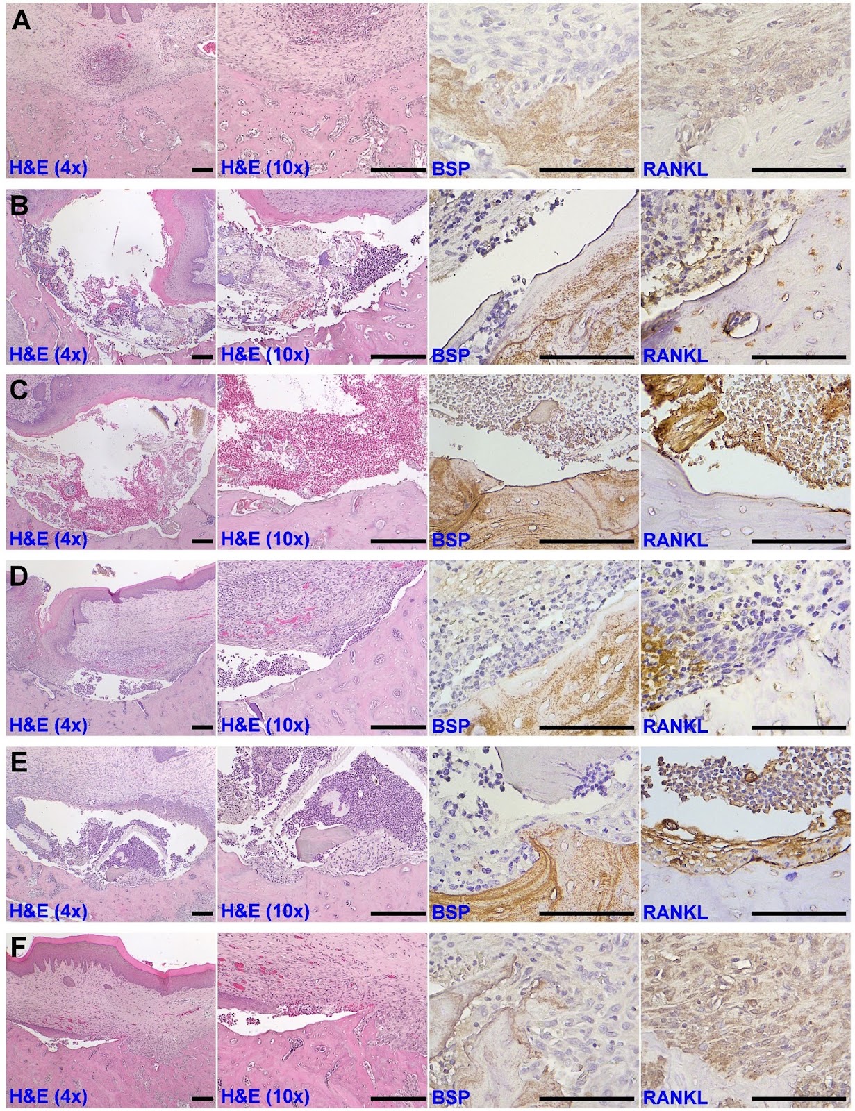
Background Medication-related osteonecrosis of the jaw (MRONJ) is a serious complication in patients taking bisphosphonates. This study aimed at developing a mesenchymal stem cell-based strategy to reduce the incidence of MRONJ and recover regeneration capability of MRONJ-affected wounds by using a gelatin methacryloyl/polydopamine hydrogel (GelMA/PD) to adhere alveolar mucosa-derived stem cells (AMCs) on the bone surface, with the osteogenically stimulated AMCs modulated by microRNA (miR) transfection, and the osteoanabolic environment activated by parathyroid hormone (PTH). Methods GelMA/PD was synthesized by photo-crosslinking, and the incorporation of PD onto GelMA as well as mechanical properties were assessed. Rat AMCs were isolated, and the stemness was characterized. AMCs were osteogenically stimulated by miR transfection. Maxillary osteotomy was created in rats administrated with zoledronic acid and dexamethasone to simulate MRONJ-affected wounds, and osteotomy in rats without ZA served as healthy controls. Wounds were unfilled or filled with GelMA/PD alone, GelMA/PD with AMCs (GA), GelMA/PD with OAMCs (GO), or GelMA/PD with OAMCs and PTH (PO), and were assessed by gross observation, micro-CT imaging, histology, and immunohistochemistry for osteoblast-osteoclast coupling. Results GelMA/PD exhibited modestly decreased compressive strength and superior adhesion strength compared with GelMA. AMCs were double positive for CD73 and CD90, showed trilineage differentiation capability, and were osteogenically stimulated by miR-218 transfection. Among MRONJ-affected wounds, soft tissue coverage was accelerated, with reduced sequestra and significantly greater bone volume in PO group (38.46 ± 10.02%) relative to unfilled group (21.81 ± 6.18%), and osteoblast-osteoclast coupling was evident in GO and PO groups. Soft tissue recovery, inflammation reduction, and matrix deposition on defect surfaces were more prominent in PO group.

Read Full Abstract10.1186/s13287-025-04655-1
Extracellular vesicle bioactivity and potential for clinical development are determined by mesenchymal stromal cell clonal subtypeGraphical AbstractVerified
Stem Cell Research & Therapy

Extracellular vesicle bioactivity and potential for clinical development are determined by mesenchymal stromal cell clonal subtype

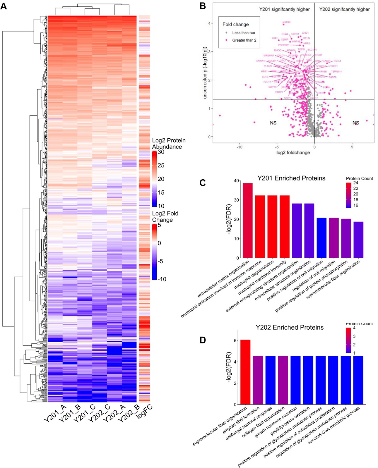
Background Mesenchymal stromal cells (MSCs) have been used in numerous clinical trials but very few reach phase 3 or market authorisation. Progress is often hampered by the use of non-clonal, heterogeneous and uncharacterised MSC cultures and lack of mechanistic understanding. There is limited evidence of MSC engraftment in vivo and disease resolution may be the result of the paracrine effects of the MSC secretome, rather than the cells per se. Extracellular vesicles (EVs) are key components of the MSC secretome and there is growing interest in the use of EVs as cell-free therapies. However, like MSCs, heterogeneity can exist within any therapeutic EV pool. Here we used immortalised clonal MSC lines, termed Y201 and Y202, to examine how MSC phenotype influences EV character and function. Methods EVs were isolated by ultracentrifugation and characterised by nano-sizing, ultrastructural morphometric analysis, western blotting, mass spectrometry and miRNA screening. Bioactivity was determined by phosphorylation of ERK1/2, proliferation and T cell polarisation assays and using two in vivo models of inflammatory disease. Results EVs from Y201 and Y202 MSCs were morphologically similar, however, Y201 EVs were more abundant in EV biomarkers versus Y202 EVs, with an enhanced miRNA and proteomic content. Computational analysis of the Y201 EV proteome identified significant enrichment in matrix-associated proteins, predicted to contribute to an elaborate EV corona particularly abundant in RGD-containing proteins fibronectin and MFG-E8, which was confirmed by western blotting. Y201 EVs, but not Y202 EVs, significantly increased the proliferation of articular chondrocytes in a dose-dependent manner, and the proliferative effect of Y201 EVs was mediated at least in part via an RGD (integrin)-FAK-ERK1/2 axis. Both Y201 and Y202 EV subsets significantly reduced proliferative index scores of activated T cells. However, only Y201 EVs, not Y202 EVs, suppressed disease activity compared to controls in different in vivo models of inflammatory peritonitis and arthritis.

Read Full Abstract10.1186/s13287-025-04665-z
Intranasal delivery of DPSC-derived small extracellular vesicles-encased phloroglucinol attenuates non-motor and motor deficits and promotes neurogenesis in an in vivo rat model of Parkinson’s diseaseGraphical AbstractVerified
Stem Cell Research & Therapy

Intranasal delivery of DPSC-derived small extracellular vesicles-encased phloroglucinol attenuates non-motor and motor deficits and promotes neurogenesis in an in vivo rat model of Parkinson’s disease

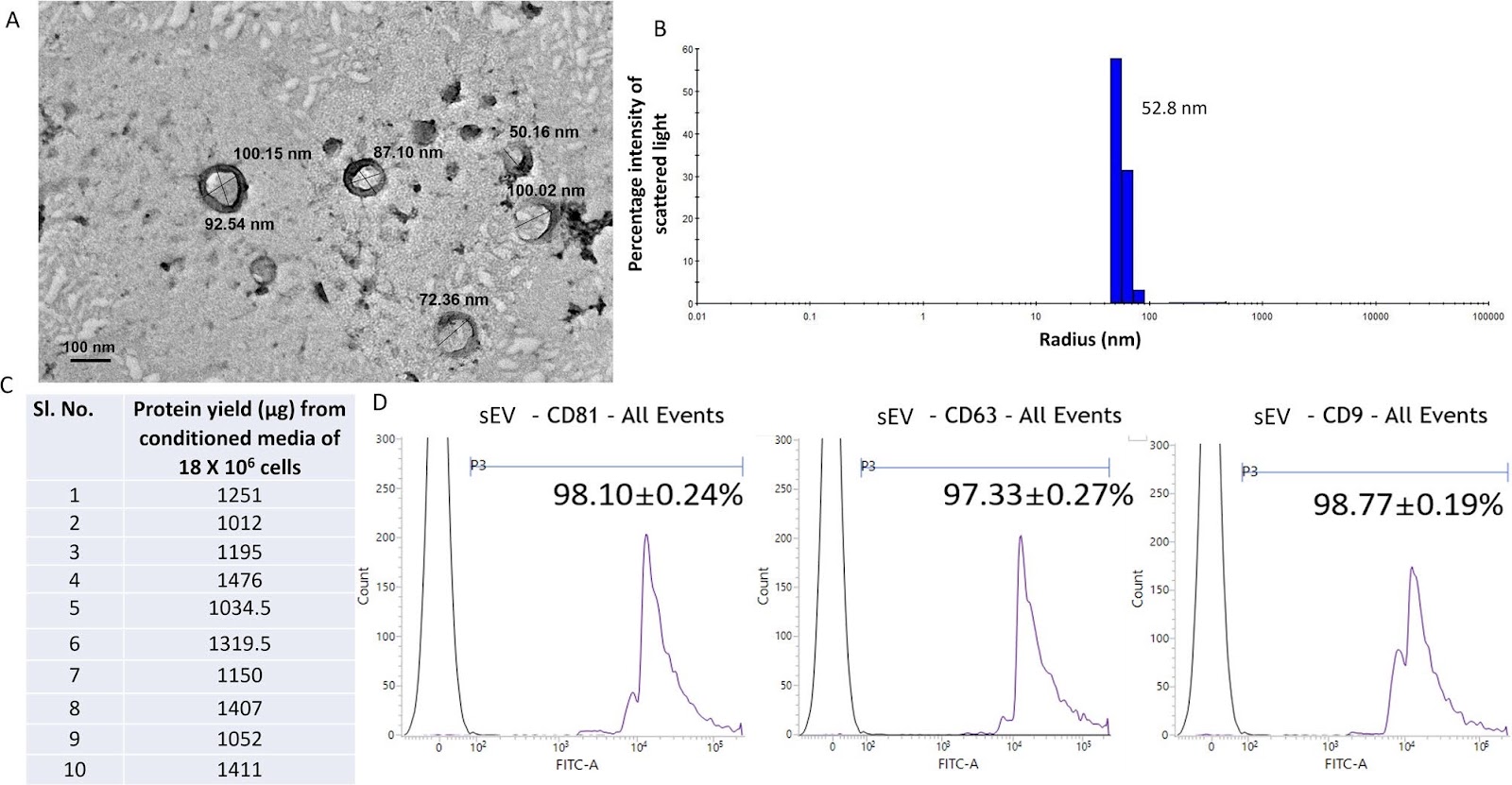
Background  Parkinson’s disease (PD) is characterized by dopaminergic (DA) neuron degeneration in the substantia nigra pars compacta (SNpc) driven by oxidative stress, inflammation, and impaired neurogenesis. Phloroglucinol, a polyphenolic antioxidant, has demonstrated neuroprotective effects in PD models but suffers from limited clinical applicability due to poor blood-brain barrier (BBB) permeability. Small extracellular vesicles (sEV) derived from dental pulp stem cells (DPSCs) exhibit neuroprotective and immunomodulatory properties and serve as promising vehicles for targeted drug delivery across the BBB. This study aimed to evaluate the therapeutic efficacy of intranasally administered sEV-encased phloroglucinol (sEV-Phl) in a chronic MPTP rat model of PD. Methods  DPSC-derived sEV were isolated via density gradient ultracentrifugation and characterized using Transmission Electron Microscopy (TEM), Dynamic-Light-Scattering (DLS), and CD marker expression. Phloroglucinol was encased in sEV (sEV-Phl) using sonication. Antioxidant properties were tested in vitro using an H2DCF.DA assay in SH-SY5Y cells exposed to 6-OHDA. Chronic MPTP-treated male Wistar rats received intranasal sEV-Phl, with motor and non-motor behaviours evaluated up to 4-weeks post-MPTP treatment. TH-positive neurons, neurogenesis (Ki67, BrdU and FOXA2), lipid-peroxidation, and neurotransmitter-levels were analyzed. sEV biodistribution was tracked via near-infrared imaging and localization in neuronal and glial cells was confirmed with PKH-26 labelling, with confocal-imaging further verifying localization in neuronal and glial cells. TNF-α expression was assessed as a marker of neuroinflammation. Results  sEV displayed high purity and homogeneity. sEV-Phl significantly reduced oxidative stress both in vitro and in vivo, as indicated by decreased ROS and lipid peroxidation levels. sEV-Phl treated MPTP rats demonstrated marked improvement in motor and non-motor behaviours compared to MPTP rats. Immunohistochemical analysis revealed

Read Full Abstract10.1186/s13287-025-04573-2
Nano-zinc oxide (nZnO) targets the AMPK-ULK1 pathway to promote bone regenerationGraphical AbstractVerified
Stem Cell Research & Therapy

Nano-zinc oxide (nZnO) targets the AMPK-ULK1 pathway to promote bone regeneration

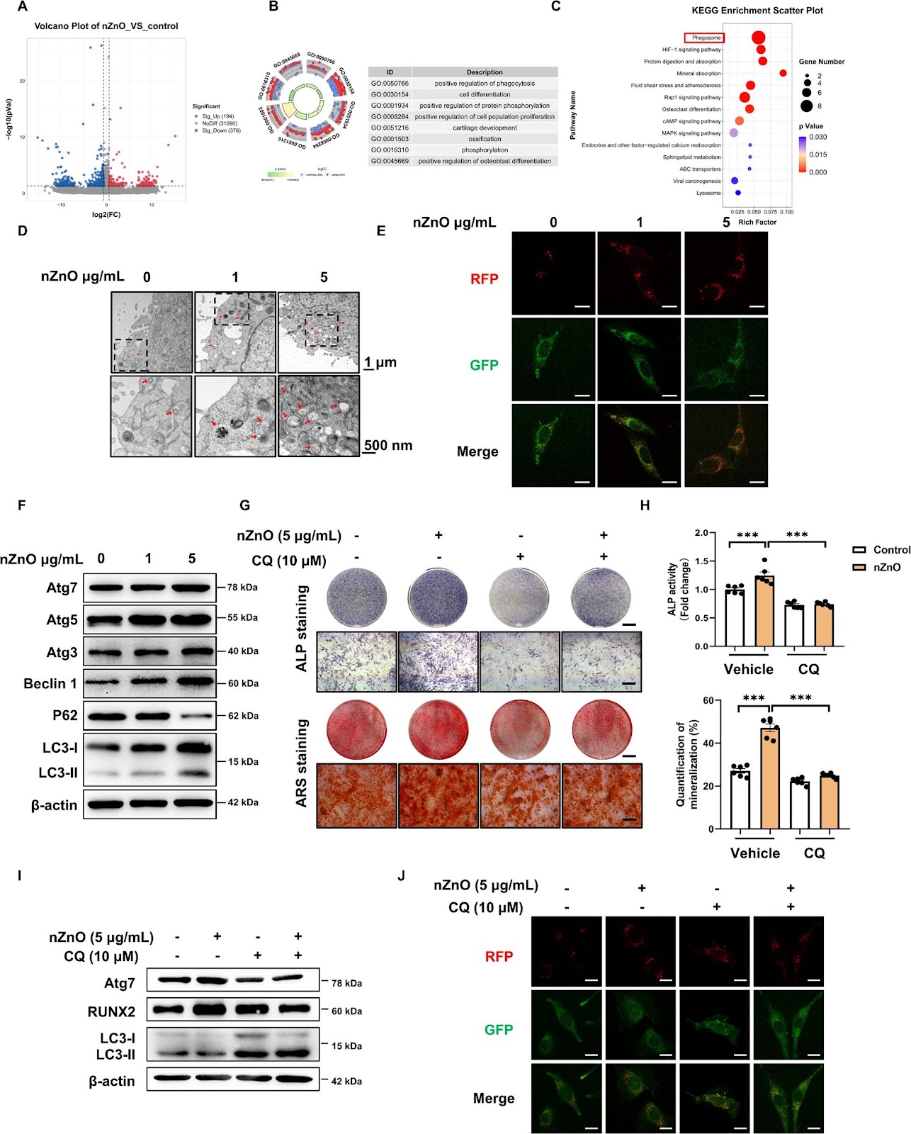
Background Nano-zinc oxide (nZnO) has attracted significant attention in bone tissue engineering due to its antibacterial properties, anti-inflammatory effects, biocompatibility, and chemical stability. Although numerous studies have demonstrated the enhancement of osteogenic differentiation by nZnO-modified tissue engineering materials, the underlying mechanisms remain poorly characterized. Methods This study aimed to identify the molecular mechanisms how nZnO promoted osteogenic differentiation and bone regeneration using transcriptome analysis, drug intervention, and shRNA knockdown techniques, etc. First, the study evaluated the in vivo effects of gelatin methacryloyl (GelMA) containing nZnO on bone regeneration using a mouse calvarial defect model. The impact of nZnO exposure on the osteogenic differentiation of mesenchymal stem cells (MSCs) was then assessed. The combined treatment of nZnO and MSCs in GelMA for bone regeneration was assessed in the mouse calvarial defect model thereafter. Results nZnO induced osteoblastic differentiation to promote bone regeneration. nZnO activated the AMP-dependent protein kinase (AMPK)-ULK1 signals to stimulate autophagosomes formation and facilitate autophagy flow, which was the essential pathway to induce osteogenic differentiation. The combined treatment of MSCs and nZnO significantly enhanced bone regeneration in calvarial defect mice. Conversely, AMPK inhibitor Compound C (C.C) reversed the effects on autophagy flow and osteogenic potentiality induced by nZnO. Conclusions These results highlight that nZnO can regulate bone regeneration by activating autophagy through the AMPK/ULK1 signaling pathway, which may provide a novel therapeutic strategy for addressing bone defects using nZnO.

Read Full Abstract10.1186/s13287-025-04322-5
BACH1 recruits STAT3 to enhance leukemia inhibitory factor receptor activity and augments the self-renewal capacity of mouse embryonic stem cellsGraphical AbstractVerified
Stem Cell Research & Therapy

BACH1 recruits STAT3 to enhance leukemia inhibitory factor receptor activity and augments the self-renewal capacity of mouse embryonic stem cells

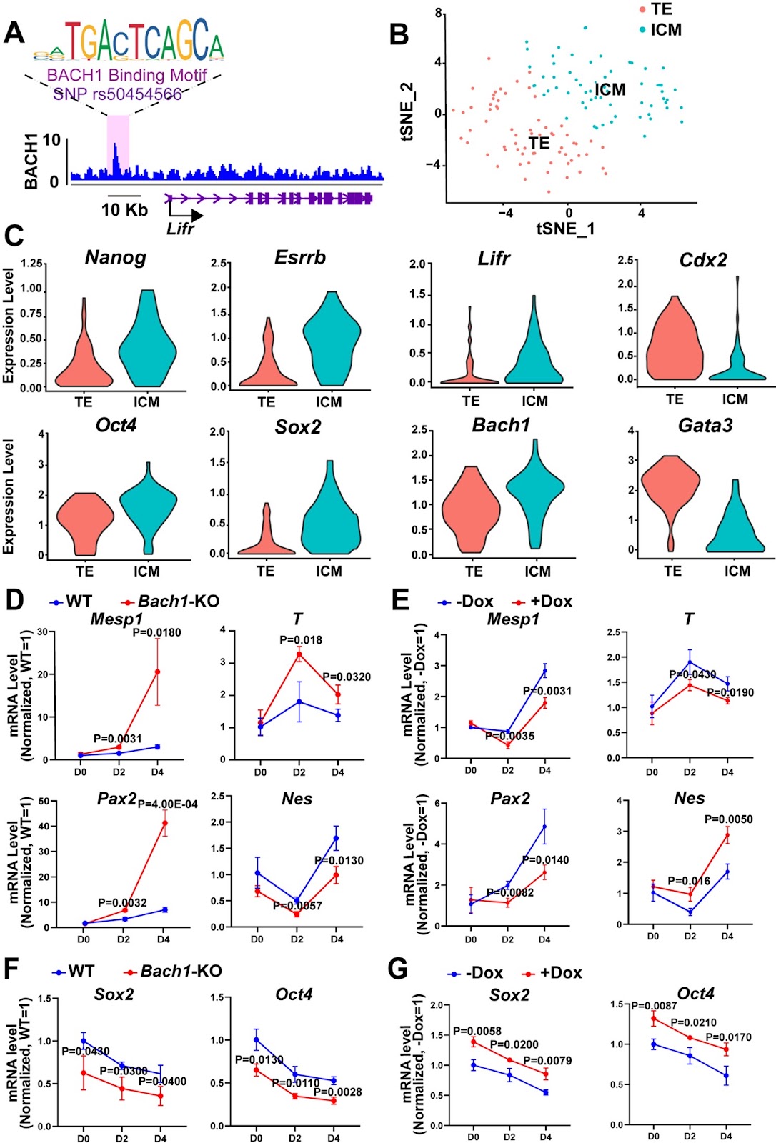
Background Genomic studies have linked single nucleotide variants in the enhancer region of the leukemia inhibitory factor receptor (Lifr) gene to chromatin accessibility and the regulation of self-renewal in mouse embryonic stem cells (mESCs). However, the underlying mechanisms remain unclear. This study investigates the role of the transcription factor BTB and CNC homology 1 (BACH1) in regulating the Lifr enhancer and its impact on mESC pluripotency. Methods We performed RNA-sequencing (RNA-seq) to assess the impact of Bach1 knockout on gene expression in mESCs. Additionally, chromatin immunoprecipitation (ChIP), co-immunoprecipitation (co-IP), and luciferase reporter gene analysis were employed to investigate the mechanism by which BACH1 regulates Lifr expression. Results Genomic analyses identified BACH1 binding at the Lifr enhancer proximal to rs50454566 in mESCs. Integrated single-cell RNA sequencing (scRNA-seq) data revealed co-upregulation of Bach1 and Lifr in inner cell mass (ICM) cells. RNA-seq analyses demonstrated that Bach1 depletion attenuated Lifr expression and impeded LIFR-signal transducer and activator of transcription 3 (STAT3) signaling. Mechanistically, BACH1 recruited STAT3 to the Lifr enhancer, driving Lifr transcription and facilitating the LIFR-STAT3 signaling pathway, thereby enhancing mESC self-renewal. Conclusion Our findings demonstrate that BACH1 enhances Lifr enhancer activity by recruiting STAT3 and activates the LIFR-STAT3 signaling pathway by promoting the LIFR expression, thereby maintaining mESC self-renewal.

Read Full Abstract10.1186/s13287-025-04578-x
From inflammation to healing: the crucial role of GPR91 activation and SDH inhibition in chronic diabetic wound recoveryGraphical AbstractVerified
Stem Cell Research & Therapy

From inflammation to healing: the crucial role of GPR91 activation and SDH inhibition in chronic diabetic wound recovery

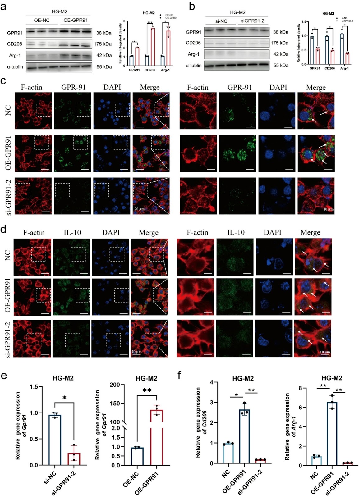
Background Diabetic foot ulcers (DFU) typically exhibit impaired healing due to dysregulated re-epithelialization and excessive inflammation. Succinate, a key metabolic intermediate, is now understood to regulate inflammation through G Protein-Coupled Receptor 91 (GPR91) and succinate dehydrogenase (SDH), although its role in DFU remains unclear. Methods Co-cultures of M2 macrophages and epithelial cells, along with clinical samples, were used to analyze the expression of GPR91 and SDH. Functional assays were performed using high glucose (HG)-treated M2 macrophages (HG-M2) and an in vivo model. Cytokine and growth factor levels in cell supernatant were measured, and molecular mechanisms were explored via qRT-PCR, flow cytometry, and western blot analysis. Results Elevated glucose concentrations increased succinate levels and disrupted M2 macrophage–epidermal stem cells (EpSCs) interactions. GPR91 knockdown worsened HG-M2 dysfunction, while GPR91 overexpression (OE-GPR91) enhanced anti-inflammatory responses and reduced succinate. OE-GPR91-conditioned medium preserved EpSCs stemness and promoted migration mediated by hepatocyte growth factor (HGF). SDH inhibition (via Dimethyl malonate, DMM) boosted M2 macrophage activity by reducing reactive oxygen species (ROS) and upregulating Gpr91 expression. Mechanistically, GPR91 activated the pAkt/pGSK3β/β-catenin pathway, while DMM enhanced M2 macrophage function via the PI3K-Akt/pERK1/2 pathway. Conclusions GPR91 upregulation and SDH inhibition improve HG-M2 macrophage function, reduce inflammation, and enhance HGF-mediated EpSCs repair. Targeting both pathways may represent a promising approach to promote DFU healing.

Read Full Abstract10.1186/s13287-025-04480-6
MSCs with upregulated lipid metabolism block hematopoietic stem cell differentiation via exosomal CTP-1A in MDSGraphical AbstractVerified
Stem Cell Research & Therapy

MSCs with upregulated lipid metabolism block hematopoietic stem cell differentiation via exosomal CTP-1A in MDS

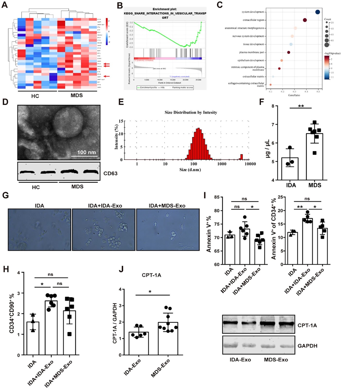
Background Myelodysplastic syndrome (MDS) is a clonal disorder of hematopoietic stem cells (HSCs), characterized by ineffective hematopoiesis and a high risk of progression to acute myeloid leukemia. Elucidating the mechanism underlying the dysfunction of MDS-HSCs is crucial for exploring the pathogenesis of the syndrome. While previous studies have implicated mesenchymal stem cells (MSCs), a principal component of the bone marrow (BM) microenvironment, in the inhibition of normal hematopoiesis, the precise molecular mechanisms have not been fully elucidated. In this study, we investigated the effects of MSCs from MDS patients on hematopoietic functions of HSCs from a metabolic perspective. Methods MSCs were isolated from BM of MDS patients. The proliferation, apoptosis, differentiation and support for hematopoiesis of these cells were analyzed using CCK-8 assay, FC and induction medium and CFU (colony forming units) assay, respectively. Expression levels of metabolic molecules were used as indicators to screen MSCs with different metabolic pathways and were detected by RT-PCR and Western blotting. Exosome derived from MSCs were isolated from the culture supernatant and confirmed by Transmission Electron Microscope, Dynamic Light Scattering and Western blotting. The effects of these exosomes on HSCs were analyzed using the same methods as those used to assess MSCs function. Results Our findings demonstrated that MDS-MSCs exhibited significant functional impairments, including reduced proliferation, impaired differentiation, diminished support for hematopoiesis, and increased apoptosis. Notably, we observed an upregulation of lipid metabolism in these MSCs, which appears to contribute to their dysfunction. Intriguingly, the aberrant lipid metabolic profile can be effectively reversed by the administration of etomoxir (ETO), an inhibitor of carnitine palmitoyltransferase 1A (CPT-1A). Furthermore, MSCs with enhanced lipid metabolism could transmit this dysfunction to HSCs through the secretion of exosomes that are enriched in CPT-1A. Conclusions We suggest that the MDS BM microenvironment disrupts MSCs metabolism by increasing the expression of CPT-1A, which impairs the ability to support normal HSCs. Interestingly, the suppressive effect is mediated by exosomes rich in CPT-1A, which derived from MSCs. These findings provide novel insights into MDS MSCs-metabolism-Exosome axis in ineffective hematopoiesis and offer new strategies for the treatment of MDS.

Read Full Abstract10.1186/s13287-025-04154-3
Small extracellular vesicles secreted from TGF-β1-licensed mesenchymal stromal cells reduce inflammation-associated injury following corneal alkali burnGraphical AbstractVerified
Stem Cell Research & Therapy

Small extracellular vesicles secreted from TGF-β1-licensed mesenchymal stromal cells reduce inflammation-associated injury following corneal alkali burn

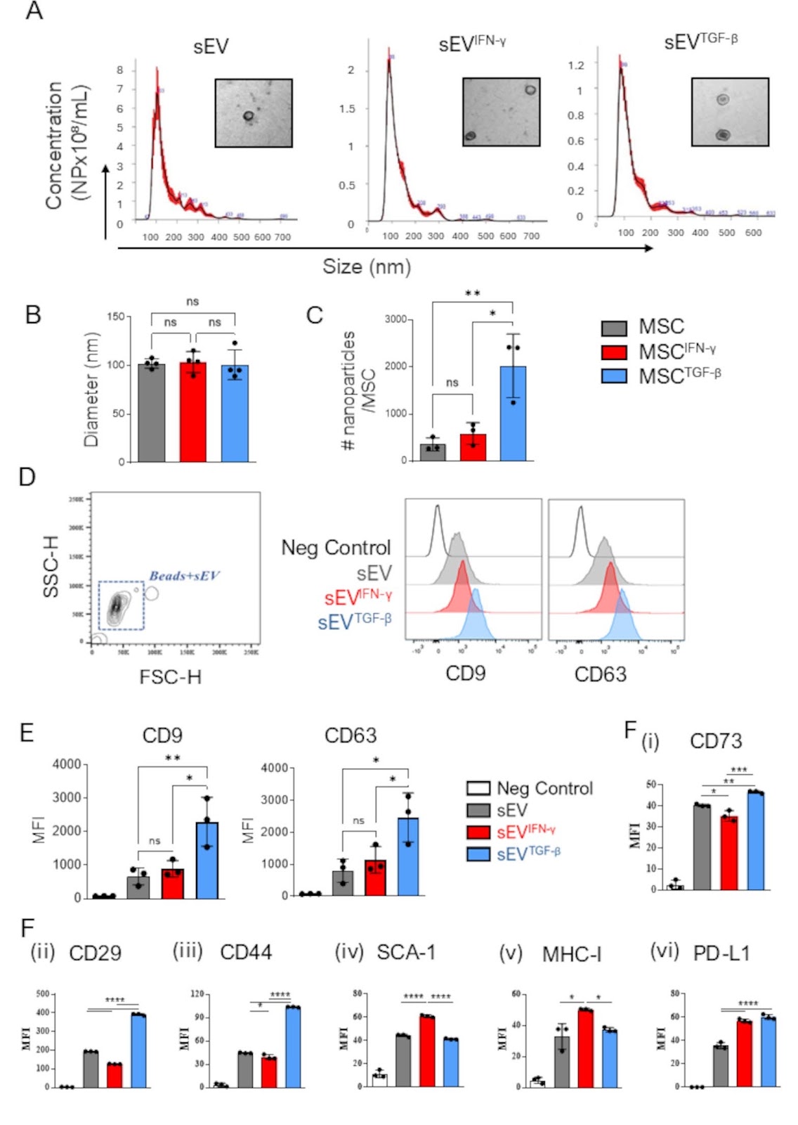
Background It is well established that the mesenchymal stromal cell (MSC) therapeutic potency can be enhanced by cytokine pre-activation or licensing. However, its effects on therapeutic efficacy of small extracellular vesicles (MSC-sEV) have not yet been well established. Here we report on two different cytokine licensing strategies, using either a pro-inflammatory or anti-inflammatory cytokine and evaluate their therapeutic potency in vitro and in a preclinical model of corneal chemical burn. Methods BALB/c MSCs were cultured with no supplement, recombinant IFNγ, or recombinant TGFβ1 for 72 h. sEV, sEVIFNγ, and sEVTGFβ were then isolated from conditioned medium of parental cells by a combination of ultrafiltration and size exclusion chromatography. Following isolation MSC-sEV were thoroughly characterized for size, marker expression and therapeutic efficacy. To evaluate their immunomodulatory capacity, both naïve and licensed MSC-sEV were tested in in vitro macrophage and T cell assays and in a preclinical corneal injury model. Results Relative to unlicensed sEV, sEVIFNγ exhibited increased expression of MHC I and PD-L1 on their surface, whereas sEVTGFβ expressed higher levels of CD44, CD29, and CD73. For immunomodulatory capacity, only sEVTGFβ was found to reduce macrophage expression of MHC II and CD80 and induced the secretion of anti-inflammatory macrophage cytokines. sEVTGFβ were also found to increase Treg expansion and FOXP3 expression. Given the superior efficacy observed of sEVTGFβ in vitro, this product was evaluated in a preclinical mouse model of corneal chemical burn. sEVTGFβ were applied either topically (day 0, 1, and 3) or subconjunctivally (day 0, and 3), and mice were monitored for 14 days. sEVTGFβ ameliorated burn-induced structural damage and accelerated restoration of normal corneal thickness, compared to PBS-treated controls. sEVTGFβ also resulted in reduced inflammatory mediators (IL-1β, iNOS) and minimised levels of fibrosis-associated collagen in the cornea. Mice that received subconjunctival, but not topical, administration of sEVTGFβ exhibited regulatory immune cell profiles with reduced pro-inflammatory-

Read Full Abstract10.1186/s13287-025-04504-1
Apoptotic vesicles derived from bone marrow mesenchymal stem cells increase angiogenesis in a hind limb ischemia model via the NAMPT/SIRT1/FOXO1 axisGraphical AbstractVerified
Stem Cell Research & Therapy

Apoptotic vesicles derived from bone marrow mesenchymal stem cells increase angiogenesis in a hind limb ischemia model via the NAMPT/SIRT1/FOXO1 axis

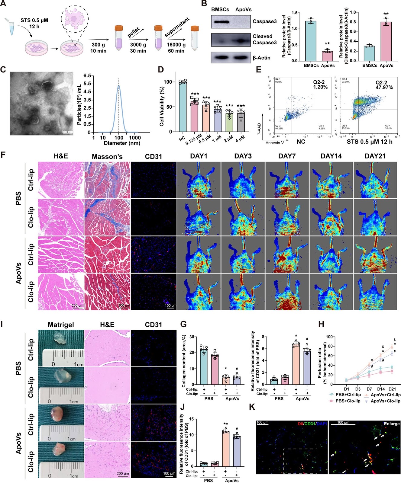
Background Chronic limb-threatening ischemia (CLTI) is the most severe form of peripheral arterial disease (PAD). Mesenchymal stem cell (MSC) transplantation holds promise as a treatment for CLTI; however, the harsh local environment poses challenges to its effectiveness. Apoptotic vesicles (ApoVs) are extracellular vesicles produced by cells undergoing apoptosis, and they can carry various biomolecules from their parent cells, including proteins, RNA, DNA, lipids, ions, and gas neurotransmitters. ApoVs play significant roles in anti-inflammatory responses, anti-tumor activities, and tissue regeneration through intercellular communication, and they have demonstrated potential as drug carriers. In this study, we investigated the potential of bone marrow stem cell (BMSC)-derived ApoVs for treating CLTI. Methods In vivo, we explored the therapeutic effect of ApoVs on a hindlimb ischemia model through Laser Doppler, matrigel plug assay, and histological analysis. In vitro, we analyzed the effects of ApoVs on the proliferation, migration, and angiogenesis of HUVECs and explored the uptake process of ApoVs. In addition, Proteomic analysis, western blotting, quantitative real-time PCR, shRNA, and siRNA were used to analyze ApoVs-induced HUVECs activation and downstream signaling pathways. Results BMSCs transplantation showed improvement in a hind limb ischemia model, and this effect still exists after apoptosis of BMSCs. Subsequently, ApoVs of BMSCs were isolated and found to improve mouse hind limb ischemia in vivo. In vitro, ApoVs can be ingested by HUVECs through dynamin-, clathrin-, and caveolin-mediated endocytosis and promote its proliferation, migration, and angiogenesis. Mechanistically, ApoVs transferred NAMPT to HUVECs, therefore activating the NAMPT/SIRT1/FOXO1 axis, influencing the transcriptional activity of FOXO1, and promoting angiogenesis. Conclusions Our results demonstrate that the transplanted BMSCs can ameliorate hindlimb ischemia by releasing ApoVs during apoptosis. The main mechanism of this effect is promoting the proliferation, migration, and angiogenesis of endothelial cells via the NAMPT/SIRT1/FOXO1 axis.

Read Full Abstract10.1186/s13287-025-04245-1
Integrin signaling pathways in mesenchymal stem cellsGraphical AbstractVerified
Stem Cell Research & Therapy

Integrin signaling pathways in mesenchymal stem cells

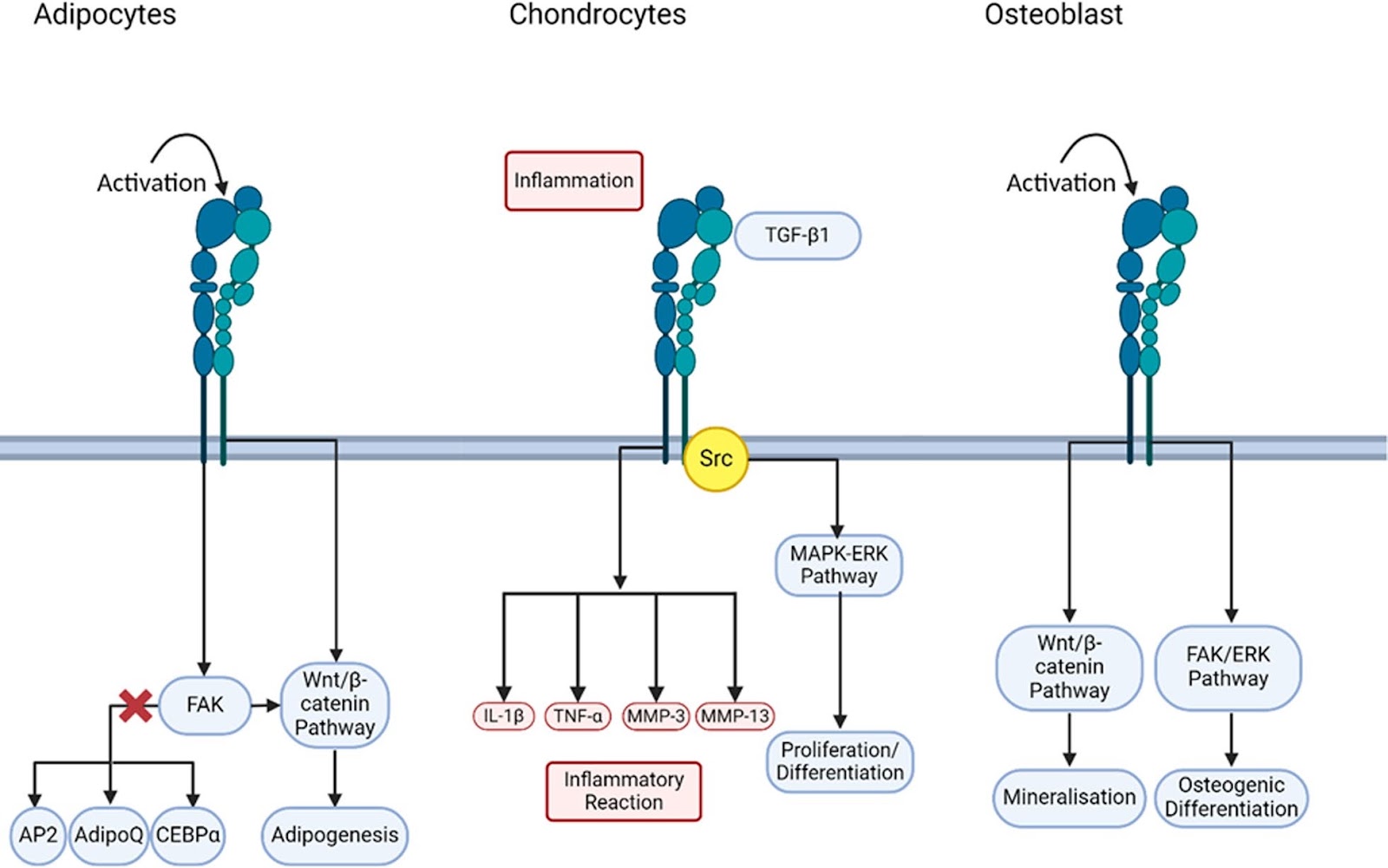
This review provides an overview of the integrin signaling pathways and their roles in mesenchymal stem cell differentiation into adipocytes, chondrocytes, and osteoblasts. In these three differentiated cells, the cell extracellular matrix plays an important role in regulating the integrin signaling pathway, as the presence of growth factors and other molecules in the extracellular matrix will affect the cell differentiation. The focus of this review is to elucidate the role of the integrin signaling pathway in adipogenesis, chondrogenesis, and osteogenesis, highlighting its diverse contributions to tissue homeostasis and repair. By synthesizing current knowledge, this paper aims to inspire further research into the therapeutic potential of targeting integrin pathways in stem cell-based tissue engineering.

Read Full Abstract10.1186/s13287-025-04608-8
Human embryonic stem cell-derived Sertoli cells as an immune modulator of cell transplantation therapy in a diabetic mice modelGraphical AbstractVerified
Stem Cell Research & Therapy

Human embryonic stem cell-derived Sertoli cells as an immune modulator of cell transplantation therapy in a diabetic mice model

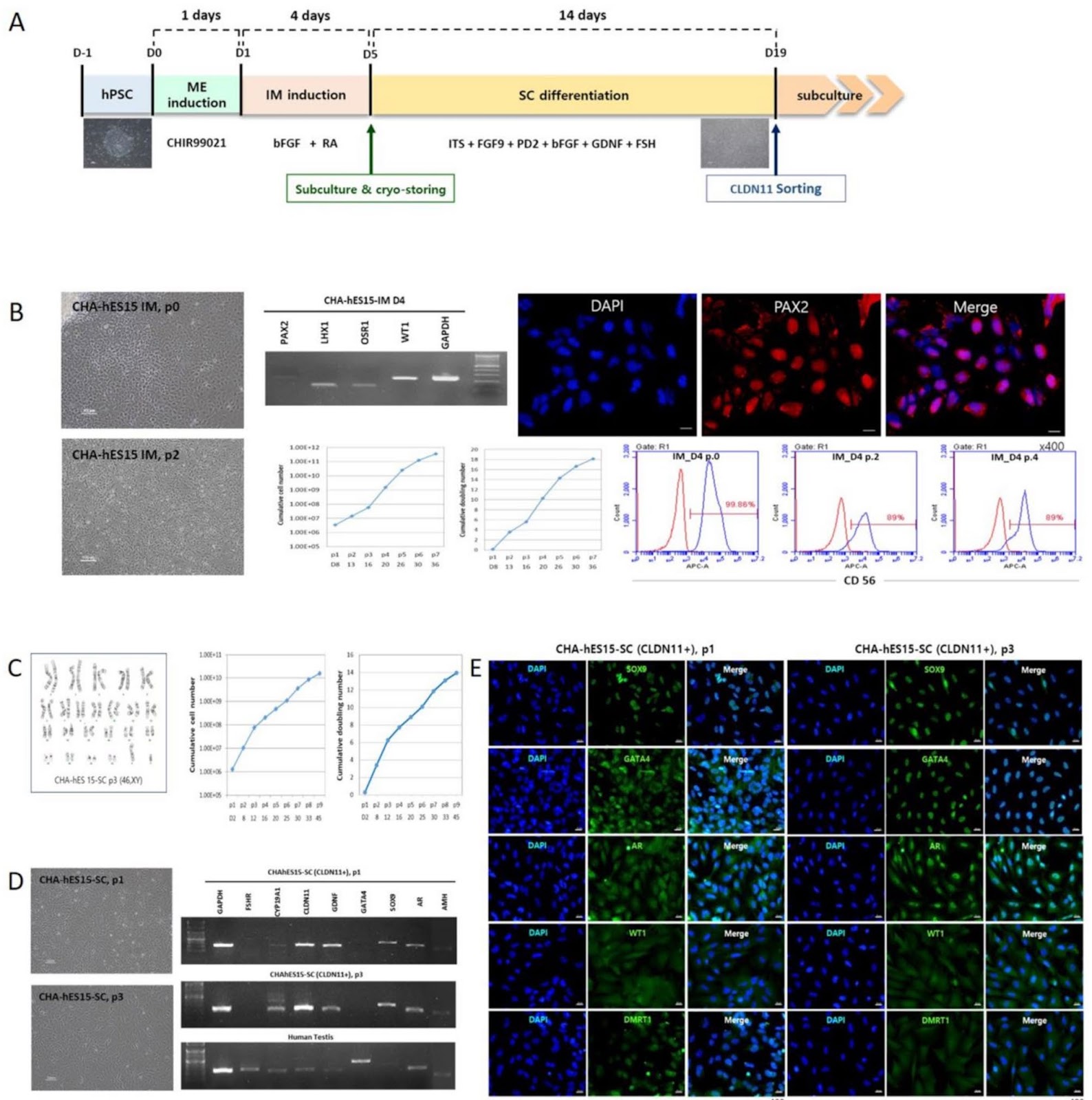
Objective Sertoli cells (SCs) are somatic cells that are a part of the seminiferous tubules in the testes and support germ cell development and maturation. Additionally, SCs play another role in protecting male germ cells from immune destruction via the formation of the blood-testis barrier and the secretion of several immunoregulatory factors. Based on these characteristics, SCs have been suggested to create a tolerogenic environment to protect co-transplanted cells as immune modulators. Because mature SCs are quiescent somatic cells and show lower proliferation activity in vitro, it is difficult to obtain the number of human cells needed for clinical applications. Materials and methods We established a protocol for mass production of SCs from human ESCs (hESC-SCs) and their functional properties were analyzed in vitro and in diabetic-induced mice after their co-transplantation with human insulin-secreting cells. Results hESC-SCs were successfully produced via a stepwise differentiation protocol. In addition, a mass culture method was established to secure the number of hESC-SCs available for cell therapy. hESC-SCs obtained from in vitro derivation highly express marker genes of SCs, such as GATA4, SOX9, CLDN11, and AR, and have shown immune-modulation activity similar to that of human bone marrow-mesenchymal stem cells. In diabetic-induced mice subcutaneously co-transplanted with EndoC-βH1 cells (insulin-secreting cells) and hESC-SCs, lower blood glucose levels were maintained for 6 months than in those transplanted with EndoC-βH1 cells alone. Conclusions We believe that hESC-SCs could be useful tool for securing cell therapy to treat human diseases in the future.

Read Full Abstract10.1186/s13287-025-04532-x
Synergistic potential of bone marrow mesenchymal stem cells and miR181-a combinational therapy against multiple sclerosisGraphical AbstractVerified
Stem Cell Research & Therapy

Synergistic potential of bone marrow mesenchymal stem cells and miR181-a combinational therapy against multiple sclerosis

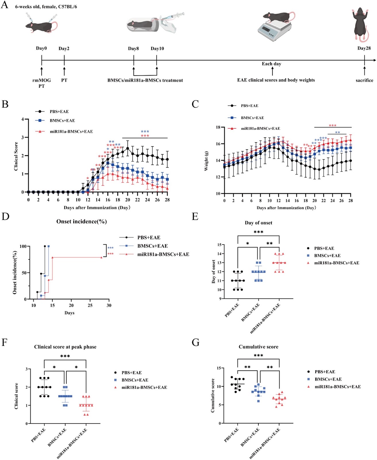
Background: Multiple sclerosis (MS) is a progressive autoimmune disease characterized by massive inflammatory infiltration, demyelination, and subsequent axonal injury and neuronal damage in the central nervous system (CNS). The etiology of MS remains unclear and there is not yet a definitive therapeutic schedule for the disease. Bone marrow mesenchymal stem cells (BMSCs), exhibiting neuroimmune-modulatory functions to alleviate various autoimmune diseases, show great potential in the treatment of MS. However, the instability of BMSCs-mediated immunosuppression in vivo has limited their application. MiR181-a, a positive regulator of immune balance, which has a preference for T cells and B cells differentiation, but degrade rapidly upon entering systemic circulation due to their unstable molecular structure. Methods: We propose a synergistic therapy approach that combines the penetrative targeting capability of BMSCs with the immuno-modulatory effects of miR181-a by overexpressing miR181-a to BMSCs through lentivirus packaging system. With this strategy, on the basis of the establishment of the experimental autoimmune encephalomyelitis (EAE) model, miR181-a overexpressing BMSCs (miR181a-BMSCs) would have a stronger immuno-modulatory treatment benefit, in terms of attenuating MS development. Results: Indicate that this method prolongs the modulatory effects of BMSCs and resulted in significantly enhancements of the proliferation of regulatory B cells (Bregs), regulatory T cells (Tregs) and the inhibition of Th17 cells compared to the traditional BMSCs group. Moreover, 10-fold miRNA’s concentration in the exosome of miR181a-BMSCs, leading to an increased duration of miRNAs to exert their biological effects. By immunotherapy and synergistic treatment, the effectiveness of the treatment is significantly enhanced, showing consistent results in different groups of the animal model.

Read Full Abstract10.1186/s13287-025-04401-7
OCT4 translationally promotes AKT signaling as an RNA-binding protein in stressed pluripotent stem cellsGraphical AbstractVerified
Stem Cell Research & Therapy

OCT4 translationally promotes AKT signaling as an RNA-binding protein in stressed pluripotent stem cells

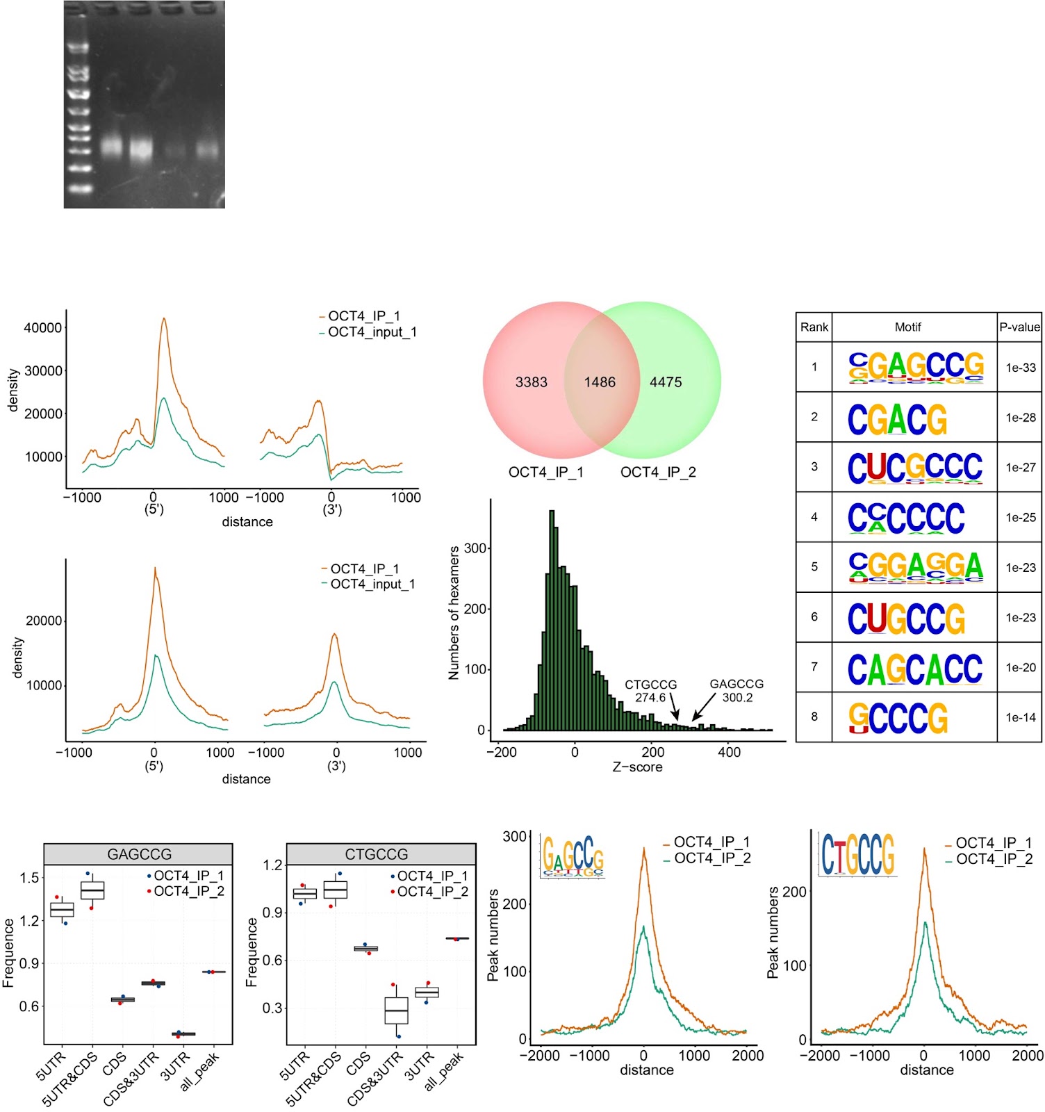
Background Despite numerous studies addressing the molecular mechanisms by which pluripotent stem cells (PSCs) maintain self-renewal and pluripotency under normal culture conditions, the fundamental question of how PSCs manage to survive stressful conditions remains largely unresolved. Post-transcriptional/translational regulation emerges to be vital for PSCs, but how PSCs coordinate and balance their survival and differentiation at translational level under extrinsic and intrinsic stress conditions is unclear. Methods The high-throughput sequencing of cross-linking immunoprecipitation cDNA library (HITS-CLIP) was employed to decipher the genome-wide OCT4-RNA interactome in human PSCs, a combined RNC-seq/RNA-seq analysis to assess the role of OCT4 in translational regulation of hypoxic PSCs, and an OCT4-protein interactome to search for OCT4 binding partners that regulate cap-independent translation initiation. By taking the Heterozygous Knocking In N-terminal Tags (HKINT) approach that specifically disrupts the 5'-UTR secondary structure and tagging its protein product of the mRNA from one allele while leaving that from the other allele intact, we examined the effect of disrupting the OCT4/5'-UTR interaction on translation of AKT1 mRNA. Results We revealed OCT4 as a bona fide RNA-binding protein (RBP) in human PSCs that bound to the 5'-UTR, 3'-UTR and CDS regions of mRNAs. Multiple known proteins participating in IRES-mediated translation initiation were detected in the OCT4-protein interactome, and a combined RNC-seq/RNA-seq analysis further confirmed a crucial role of OCT4 in translational regulation of PSCs in response to hypoxic stress. Remarkably, OCT4 bound to the GC-rich elements in the 5'-UTR of AKT1 and multiple PI3K/AKT-pathway-gene mRNAs, and promoted their translation initiation via IRES-mediated pathways under stress conditions. Specifically disrupting the AKT1 mRNA 5'-UTR structure and the OCT4/5'-UTR interaction by the HKINT approach significantly reduced the translation level of AKT1 that led to a higher susceptibility of PSCs to oxidative stress-induced apoptotic death and prioritized differentiation toward ectoderm and endoderm. Conclusions Our results reveal OCT4 as an anti-stress RBP for translational regulation that critically coordinates the survival and differentiation of PSCs in response to various stressors.

Read Full Abstract10.1186/s13287-025-04229-1
A traditional herbal decoction regulates skeletal muscle satellite cell osteogenesis and myogenesis for repairing osteosarcopenic fractures via β-cateninGraphical AbstractVerified
Stem Cell Research & Therapy

A traditional herbal decoction regulates skeletal muscle satellite cell osteogenesis and myogenesis for repairing osteosarcopenic fractures via β-catenin

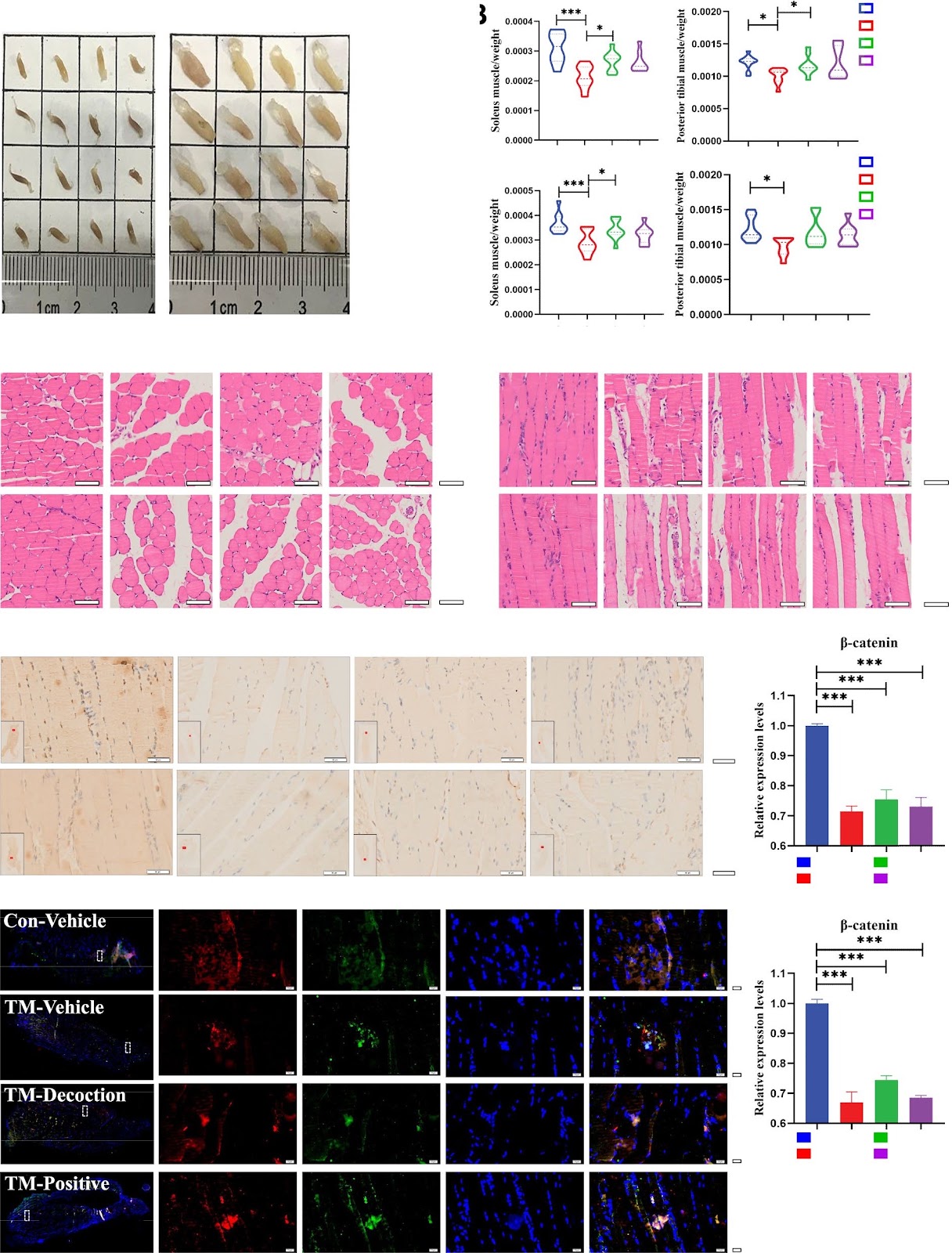
Introduction: Osteosarcopenic fractures, an emerging geriatric syndrome characterized by sarcopenia-osteoporotic fractures coexistence, delayed fracture healing, and elevated risk of re-fracture. Limited research has investigated the mechanisms by which skeletal muscle satellite cells (SMSCs) promote muscle regeneration and osteoporotic fracture healing. The aim of this study was to investigate the impact of a traditional herbal decoction (HD), the Invigorate the Spleen and Tonify the Kidney Formula, on SMSC regulation, muscle regeneration, and fracture healing. Method: Using conditional knockout mice, the role of SMSCs in promoting fracture healing and mitigating sarcopenia was evaluated by visualizing the fracture area and surrounding muscle tissue. The signaling pathways involved were comprehensively analyzed using a combination of Western blotting, real-time PCR analysis, immunohistochemical staining, and immunofluorescent staining. And the key elements and compounds facilitating osteogenesis and myogenesis were identified using HPLC and network pharmacology analysis. Results: This study demonstrated that the herbal decoction mediates the β-catenin signaling pathway, mobilizes SMSCs to migrate to the fracture area, facilitates their osteogenic and myogenic differentiation, and enhances osteoporotic fracture healing. Knockdown of β-catenin in SMSCs in Pax7-CreERT2/+;β-cateninfx/fx conditional knockout mice led to sarcopenia and osteoporosis. Additionally, the herbal decoction significantly increased bone mass, repaired bone microstructure, and promoted muscle fiber remodeling around fractures in mice. Conclusions: These findings provide the first evidence that the HD, as a β-catenin agonist, not only promotes fracture healing by modulating the osteogenic and myogenic effects of SMSCs but also ameliorates sarcopenia.

Read Full Abstract10.1186/s13287-025-04642-6
Human induced pluripotent stem cells for in vitro modeling of impaired mucociliary clearance in cystic fibrosis lung diseaseGraphical AbstractVerified
Stem Cell Research & Therapy

Human induced pluripotent stem cells for in vitro modeling of impaired mucociliary clearance in cystic fibrosis lung disease

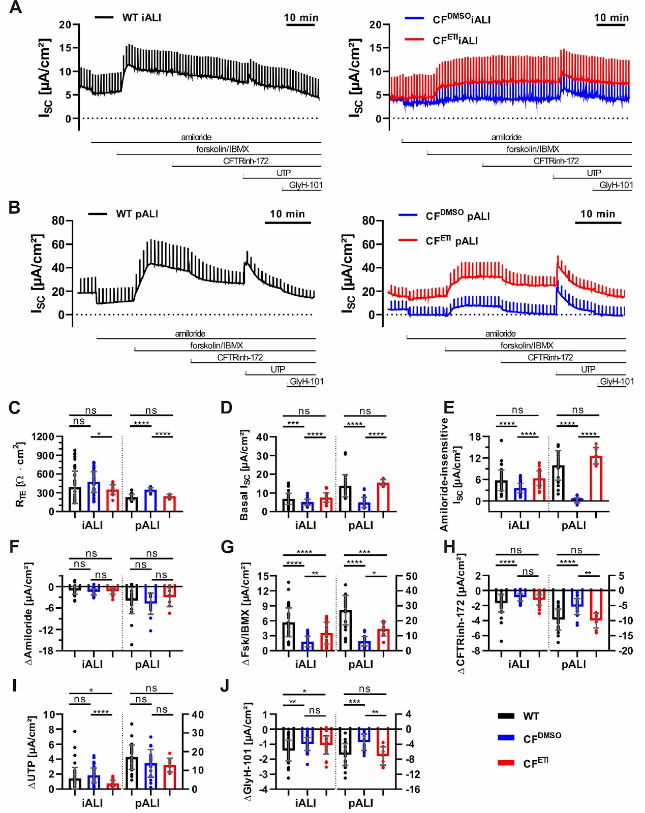
Severely impaired mucociliary airway function is the primary pathomechanism in Cystic Fibrosis (CF) lung disease. Despite significant advances in CF therapy, there is still a critical need for alternative, individualized treatment options, especially for patients with untreatable CFTR mutations. Although intestinal organoids and primary airway cells are widely used as preclinical models of CF, both systems exhibit limitations with regard to the proper modelling of mucociliary clearance or the availability of sufficient cell quantities. Patient-specific human induced pluripotent stem cells (hiPSCs) are a promising alternative due to their unlimited expansion potential and capacity to differentiate into airway epithelia. However, cellular inhomogeneities in iPSC-derived airway cultures complicated conventional assays that determine CFTR function such as Ussing chamber measurements, and a comprehensive demonstration of CF pathophysiology in hiPSC-derived airway models has been largely lacking. This study provides comprehensive data demonstrating very similar gene expression, (ultra)structure and CFTR function in CF iPSC-derived airway (iALI) and primary airway (pALI) cultures. Addressing current limitations, we have implemented a sensitive, straightforward, and automatable ciliary beat frequency (CBF) assay, which is largely unaffected by inhomogeneities and directly reflects disturbed mucus viscosity and mucociliary transport in CF lung disease. Electron microscopy images confirmed the disease phenotype showing a highly dense and dehydrated mucus layer on top of CF iALI cultures. Furthermore, established CFTR modulator drugs partially rescued the disease phenotype in CF iALI cultures, which validated the utility of iALI cultures as a scalable, patient-specific platform for CF research and personalized drug development.

Read Full Abstract10.1186/s13287-025-04737-0
Modeling pathogenesis and progression of metabolic dysfunction-associated steatotic liver disease and therapeutic drug screening using hESC-derived mature polarized hepatocyte organoidsGraphical AbstractVerified
Stem Cell Research & Therapy

Modeling pathogenesis and progression of metabolic dysfunction-associated steatotic liver disease and therapeutic drug screening using hESC-derived mature polarized hepatocyte organoids

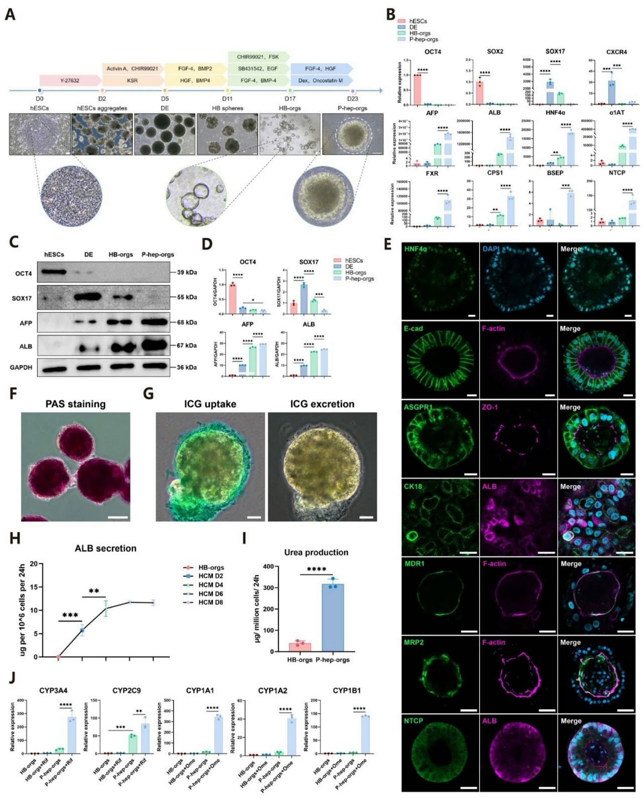
Background: Metabolic dysfunction-associated steatotic liver disease (MASLD), the most prevalent chronic liver disorder worldwide, exhibits complex pathogenesis and lacks effective targeted therapeutics. Existing animal models are limited by prolonged induction periods and interspecies discrepancies, while conventional monolayer hepatocyte cultures fail to recapitulate disease pathology due to inadequate polarization and functional immaturity. Methods: To overcome these limitations, we established an in vitro MASLD model by treating human embryonic stem cell (hESC)-derived mature polarized hepatocyte organoids (P-hep-orgs) with free fatty acids (FFAs). Pathogenesis and progression of MASLD in this model were characterized using multiple assays, and its utility for drug screening was validated with three known antioxidant or lipid-lowering agents. Results: P-hep-orgs derived from hESCs expressed mature hepatocyte markers (e.g., ALB), exhibited polarized architecture (e.g., MRP2) and demonstrated functionalities of mature hepatocytes (e.g., urea production). Moreover, we developed an in vitro MASLD model by treating P-hep-orgs with FFAs. This model recapitulated key pathological progression hallmarks, including disrupted glucose/lipid metabolism, oxidative stress, apoptosis, loss of polarization, impaired liver function, and ductular reaction. Furthermore, transcriptomic analysis revealed that P-hep-orgs treated with FFAs for 10 days shared similar molecular signatures with human MASH liver tissues (581 overlap DEGs). Finally, this model was used to assess the potential efficacy of established antioxidant or lipid-lowering agents (e.g., Vitamin E) in alleviating pathological phenotypes, including lipid accumulation and oxidative stress.

Read Full Abstract10.1186/s13287-025-04865-7
hUMSC-derived exosomes alleviate follicular interstitial cell autophagy by let-7a-5p/AMPK/mTOR axis in POI ratsGraphical AbstractVerified
Stem Cell Research & Therapy

hUMSC-derived exosomes alleviate follicular interstitial cell autophagy by let-7a-5p/AMPK/mTOR axis in POI rats

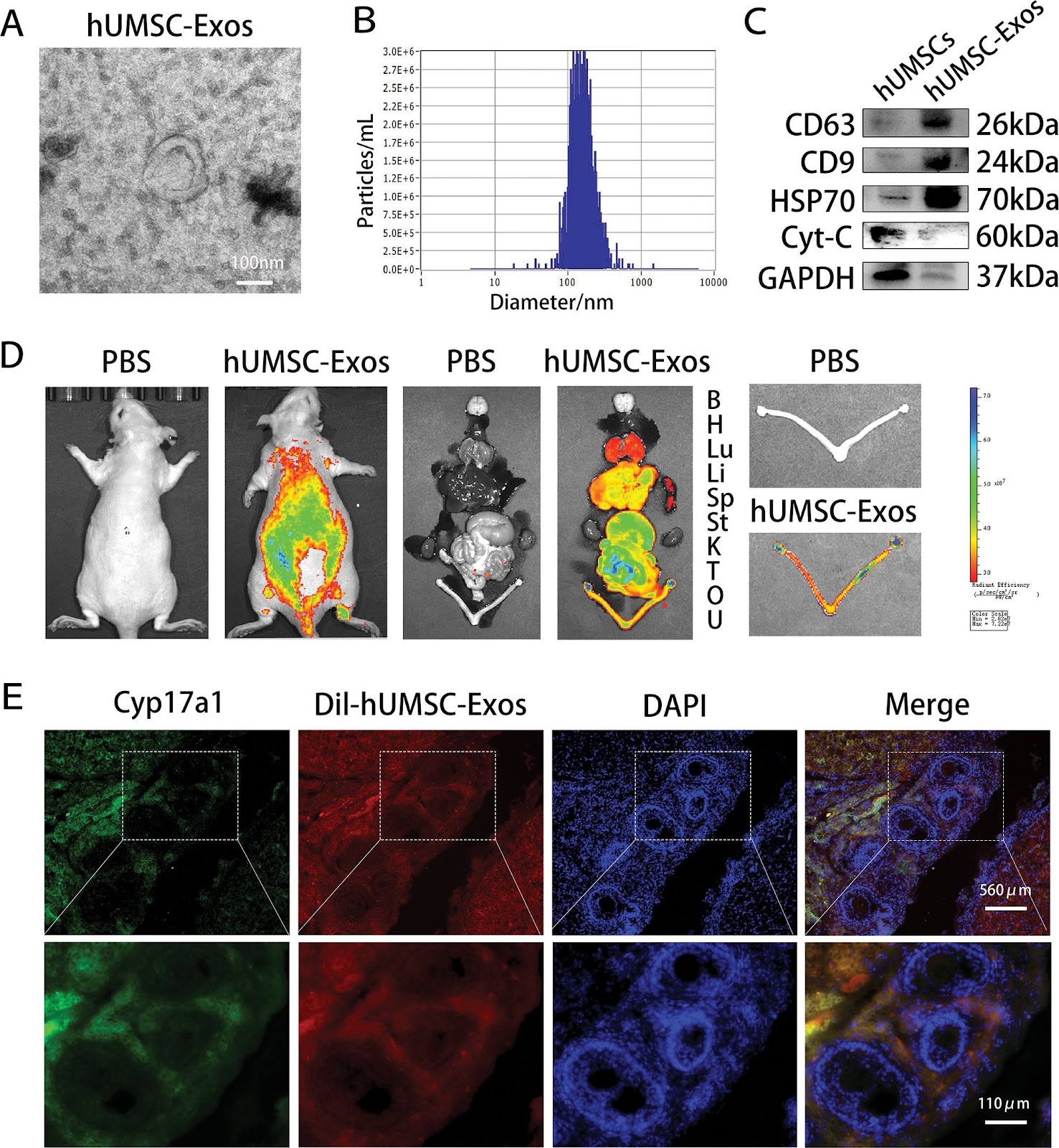
Background  One major factor contributing to infertility in women of childbearing age is premature ovarian insufficiency (POI). Exosomes produced from human umbilical cord mesenchymal stem cells (hUMSC-Exos) have drawn a lot of attention lately as a potential treatment for ovarian dysfunction brought on by POI. However, its therapeutic mechanism is still unclear and needs further exploration. Methods  POI model was established by intraperitoneal injection of cyclophosphamide (CTX) in female Wistar rats. These POI rats were treated with hUMSC-Exos for one week. In addition to in vivo experiments, in vitro POI models were also established. In vitro experiments, theca interstitial cells (TICs) treated with CTX were exposed to normal as well as let-7a-5p inhibitory hUMSC-Exos. The ovary structure, morphology, endocrine function, and reproductive ability of POI rats were observed by H&E staining and ELISA. Western blot, immunofluorescence staining (IF), and quantitative real-time polymerase chain reaction (qRT-PCR) were used to evaluate the autophagy-related indexes in ovary and TICs of POI rats in each group. Results  CTX induced abnormalities of ovarian morphology, structure, endocrine, and reproductive function in rats, and accompanied by autophagy of TICs. Notably, hUMSC-Exos diminishes ovarian structural and functional damage in POI rats and TICs autophagy via targeting the AMPK/mTOR pathway. Furthermore, downregulating let-7a-5p in hUMSC-Exos weakened their ability to prevent TICs autophagy. Conclusions  Overall, the findings suggested that hUMSC-Exos improves ovarian function in POI rats by inhibiting TICs autophagy via the let-7a-5p/AMPK/mTOR pathway. Our study provided further evidence that POI patients can benefit from hUMSC-Exos-mediated therapy.

Read Full Abstract10.1186/s13287-025-04396-1
Human spindle-shaped urine-derived stem cell exosomes alleviate severe fatty liver ischemia–reperfusion injury by inhibiting ferroptosis via GPX4Graphical AbstractVerified
Stem Cell Research & Therapy

Human spindle-shaped urine-derived stem cell exosomes alleviate severe fatty liver ischemia–reperfusion injury by inhibiting ferroptosis via GPX4

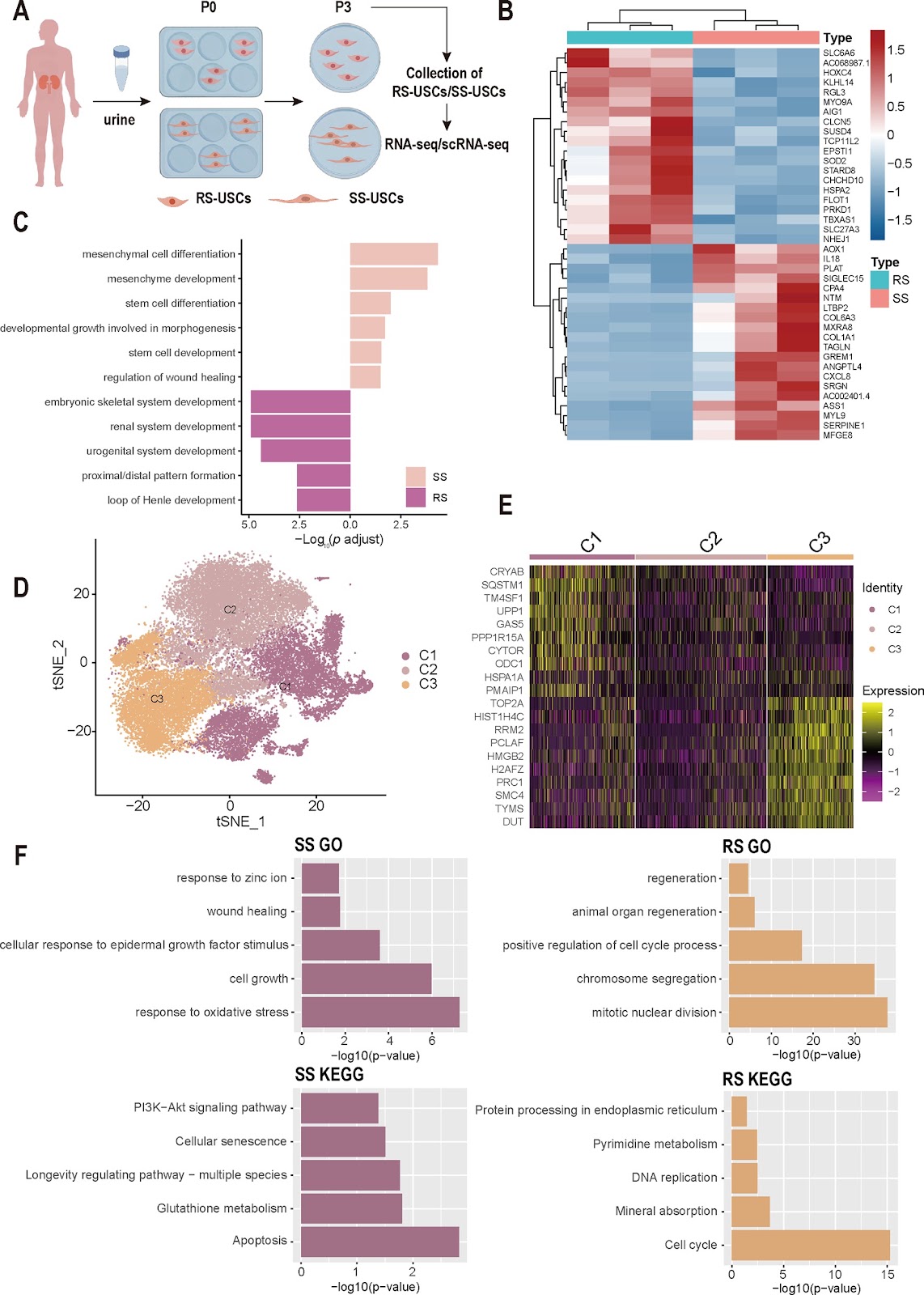
Background Severe hepatic steatosis can exacerbate Ischemia–reperfusion injury (IRI), potentially leading to early graft dysfunction and primary non-function. In this study, we investigated the heterogeneity of different subpopulations of Urine-derived stem cells (USCs) to explore the most suitable cell subtype for treating severe steatotic liver IRI. Methods This study utilized scRNA-seq and Bulk RNA-seq to investigate the transcriptional heterogeneity between Spindle-shaped USCs (SS-USCs) and Rice-shaped USCs (RS-USCs). Additionally, rat fatty Liver transplantation (LT) model, mouse fatty liver IRI model, and Steatotic Hepatocyte Hypoxia-Reoxygenation (SHP-HR) model were constructed. Extracellular vesicles derived from SS-USCs and RS-USCs were isolated and subjected to mass spectrometry analysis. The therapeutic effects of Spindle-shaped USCs Exosomes (SS-USCs-Exo) and Rice-shaped USCs Exosomes (RS-USCs-Exo) were explored, elucidating their potential mechanisms in inhibiting ferroptosis and alleviating IRI. Results Multiple omics analyses confirmed that SS-USCs possess strong tissue repair and antioxidant capabilities, while RS-USCs have the potential to differentiate towards specific directions such as the kidney, nervous system, and skeletal system, particularly showing great application potential in renal system reconstruction. Further experiments demonstrated in vivo and in vitro models confirming that SS-USCs and SS-USCs-Exo significantly inhibit ferroptosis and alleviate severe fatty liver IRI, whereas the effects of RS-USCs/RS-USCs-Exo are less pronounced. Analysis comparing the proteomic differences between SS-USCs-Exo and RS-USCs-Exo revealed that SS-USCs-Exo primarily inhibit ferroptosis and improve cellular viability by secreting exosomes containing Glutathione Peroxidase 4 (GPX4) protein. This highlights the most suitable cell subtype for treating severe fatty liver IRI. Conclusions SS-USCs possess strong tissue repair and antioxidant capabilities, primarily alleviating ferroptosis in the donor liver of fatty liver through the presence of GPX4 protein in their exosomes. This highlights SS-USCs as the most appropriate cell subtype for treating severe fatty liver IRI.

Read Full Abstract10.1186/s13287-025-04202-y
KRT5high TP63-expressing urothelial basal cells act as a driver to bladder urothelium regeneration in rabbitGraphical AbstractVerified
Stem Cell Research & Therapy

KRT5high TP63-expressing urothelial basal cells act as a driver to bladder urothelium regeneration in rabbit

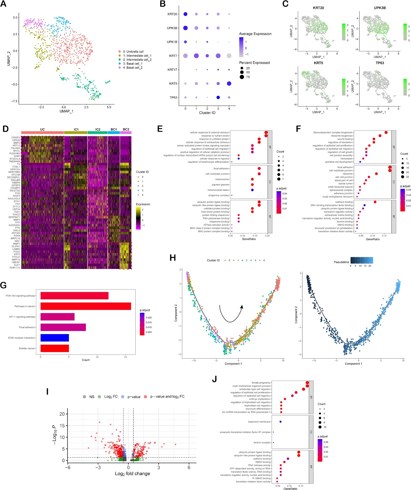
Background  Urothelial regeneration is a crucial part of bladder tissue engineering. However, there is a lack of ideal “seed cells” in current practices. Here, we demonstrated that a sub-population of p63 positive basal cells could be activated and differentiate into intermediate and superficial umbrella cells after full-thickness mucosal resection in rabbit. Methods  A focal mucosal resection model was used to characterize the role of different urothelial cells during regeneration. Urothelial basal cells were isolated from rabbit bladder mucosa and cultured in vitro. The basal cells were then transplanted in vivo in a manner of cell sheet for reconstruction. Results  Via single-cell RNA sequencing (scRNA-seq), it has been confirmed that the cluster of KRT5high TP63-expressing cells possesses a ‘stemness’ signature which can give rise to lineage cell types sequentially. With a strong support from the underneath pre-set capsule vascular bed, the transplanted cell sheet could develop into a physio-morphology resembled to the native mucosa in vivo. Importantly, we validated that the bioengineered urothelium implemented perfect barrier function after implanted to bladder. Conclusions  In summary, bioengineering urothelium with KRT5high TP63-expressing basal cells on a capsule vascular bed offers a promising strategy for bladder tissue engineering and provides a model for drug screening and bladder disease research.

Read Full Abstract10.1186/s13287-025-04417-z
Intratracheal administration of mesenchymal stem cells ameliorates hyperoxia-induced bronchopulmonary dysplasia by inhibiting NLRP3 inflammasome activation: the critical role of Aldh1a2Graphical AbstractVerified
Stem Cell Research & Therapy

Intratracheal administration of mesenchymal stem cells ameliorates hyperoxia-induced bronchopulmonary dysplasia by inhibiting NLRP3 inflammasome activation: the critical role of Aldh1a2

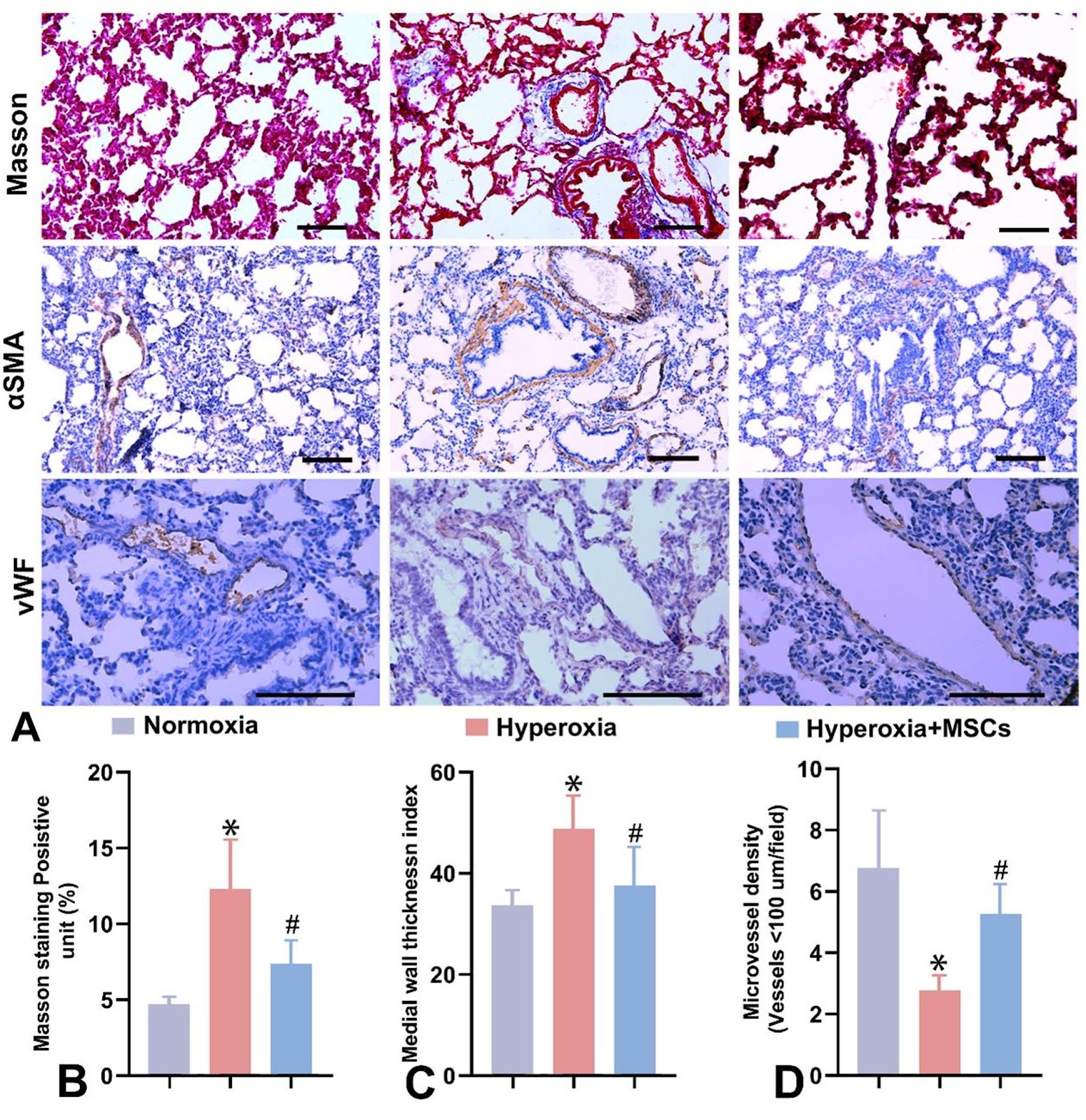
Background Bronchopulmonary dysplasia (BPD) is a chronic lung disease driven by inflammation and oxidative stress. Mesenchymal stem cells (MSCs) have shown protective effects against hyperoxic lung injury. However, few studies have thoroughly examined the significantly differentially expressed genes (DEGs) in the lungs before and after MSC treatment. In this study, we analyzed the significant DEGs in lung tissues during both in vivo and vitro umbilical cord-derived mesenchymal stem cells (UCMSCs)-mediated repair of hyperoxic lung injury and investigated their potential mechanisms of action. Methods Neonatal rats were exposed to hyperoxia and subsequently treated with UCMSCs. Inflammatory responses were quantified via ELISA and RT‒qPCR, while Western blotting (WB) and immunohistochemistry (IHC) were used to examine NLRP3 inflammasome and IL-1β expression. Transcriptomic analysis of UCMSC-mediated lung repair revealed 46 DEGs, which were validated by RT‒qPCR, and WB verified the significant differential expression of ALDH1A2. In RLE-6TN cells, Aldh1a2 expression was reduced during MSC-mediated repair of H2O2-induced oxidative stress injury. Functional evaluations were performed. WB further analyzed NLRP3 inflammasome and IL-1β expression in these processes. A recombinant adenoviral overexpression vector was intratracheally administered to hyperoxia-exposed neonatal rats. Arterial blood gas and RT‒qPCR were performed, and ELISA, WB, and IHC were used to evaluate the impact of Aldh1a2 overexpression on lung inflammation and oxidative stress, focusing on the NLRP3 inflammasome. Results UCMSCs ameliorated hyperoxia-induced alveolar simplification and microvessel loss, reduced inflammation and oxidative stress injury, and inhibited the expression of the NLRP3 inflammasome. RT‒qPCR and WB analyses revealed significant differential expression of Aldh1a2 in UCMSC-treated hyperoxia-induced lung injury. UCMSCs also mitigated H2O2-induced oxidative stress injury in RLE-6TN cells. Inhibition of Aldh1a2 expression exacerbated oxidative stress, upregulated NLRP3 inflammasome and IL-1β expression, and impaired the reparative effects of UCMSCs. Conversely, Aldh1a2 overexpression or UCMSC intervention ameliorated hyperoxia-induced alveolar simplification and microvascular abnormalities, suppressed inflammation, and enhanced lung ventilation and angiogenesis. These findings indicated that Aldh1a2 overexpression inhibits NLRP3 inflammasome activation and IL-1β release. Conclusions Aldh1a2 was significantly differentially expressed in UCMSC-mediated repair of hyperoxic lung injury, and its overexpression ameliorates BPD by inhibiting NLRP3 inflammasome activation, suggesting a novel therapeutic target for BPD.

Read Full Abstract10.1186/s13287-025-04851-z
The human umbilical cord–mesenchymal stem cell secretome regulates hair growth and cycle transition by promoting methylthioadenosine synthesis via the PI3K/AKT/mTOR pathwayGraphical AbstractVerified
Stem Cell Research & Therapy

The human umbilical cord–mesenchymal stem cell secretome regulates hair growth and cycle transition by promoting methylthioadenosine synthesis via the PI3K/AKT/mTOR pathway

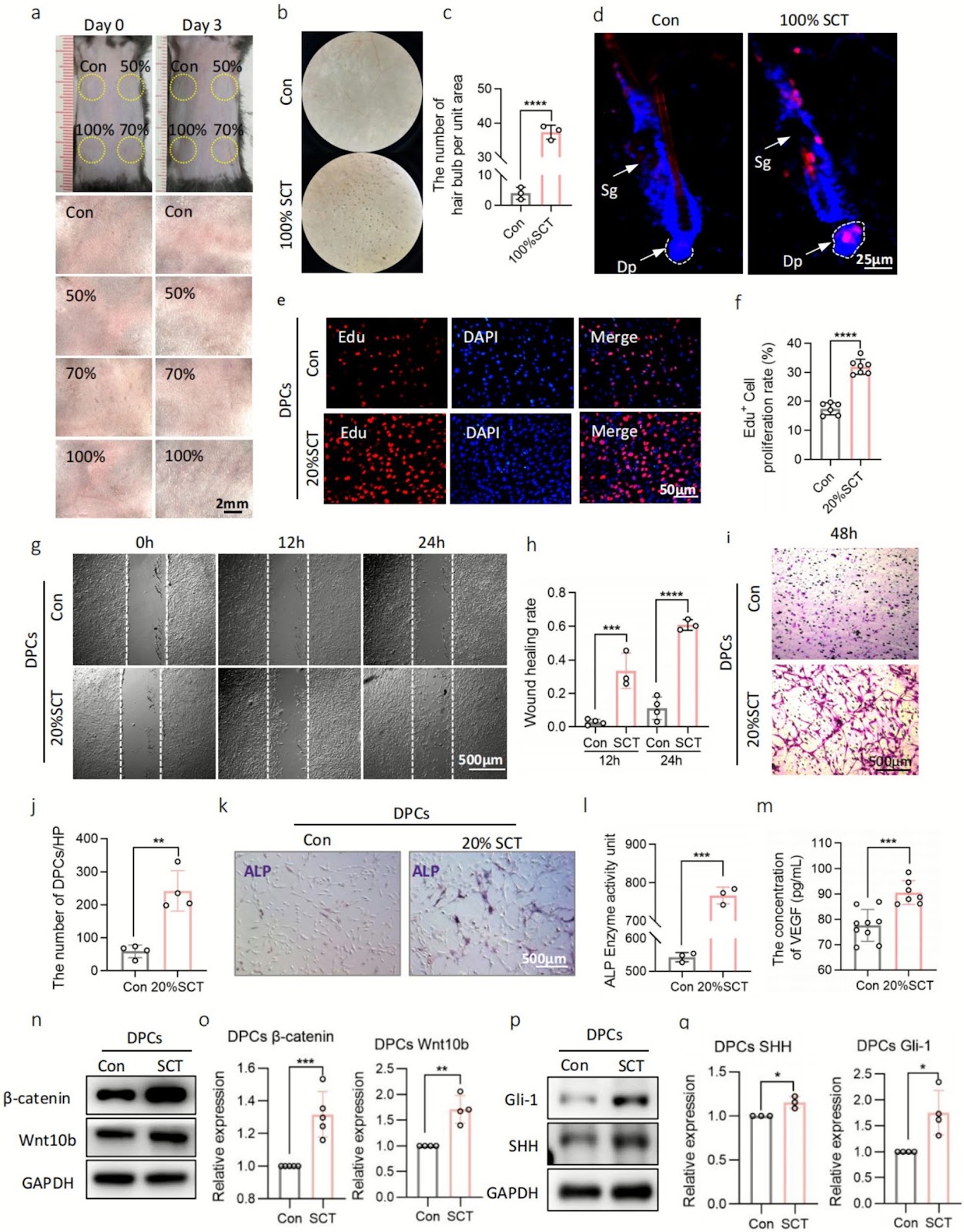
Background The human umbilical cord (hUC)–mesenchymal stem cell (MSC) secretome (SCT) is a cell-free therapy that may emerge as a novel therapeutic strategy for hair loss prevention. Here, we aimed to elucidate the underlying mechanisms through which SCT regulates hair growth and cycle transition. Methods Using C57BL/6 mice, ex vivo follicles, and cell experiments, we studied the effects and mechanisms of SCT on hair growth and cycling using untargeted metabolomics and phosphoproteomics. A three-month double-blind clinical study was conducted to validate the effects of SCT on human hair. Results SCT promotes the telogen-to-anagen transition, hair thickening, and elongation of the vibrissae in mice; regulates dermal papilla cells and hair matrix cells through cysteine and methionine metabolism; and stimulates methylthioadenosine synthesis in hair matrix cells by activating the PI3K/AKT/mTOR signaling pathway. Clinical studies demonstrated that SCT increased human hair density and average hair diameter. Scalp physiological tests and subjective feedback indicated no related adverse reactions on the scalp or hair. Conclusions SCT promoted hair growth, thickening, and the hair follicle cycle via the PI3K/AKT/mTOR signaling pathway. This research provides a basis for the application of cell-free alternatives in hair care and hair loss prevention.

Read Full Abstract10.1186/s13287-025-04806-4
A systematic review of preclinical studies on therapeutic potential of mesenchymal stem/stromal cells and their secretome in bacterial infectionsGraphical AbstractVerified
Stem Cell Research & Therapy

A systematic review of preclinical studies on therapeutic potential of mesenchymal stem/stromal cells and their secretome in bacterial infections

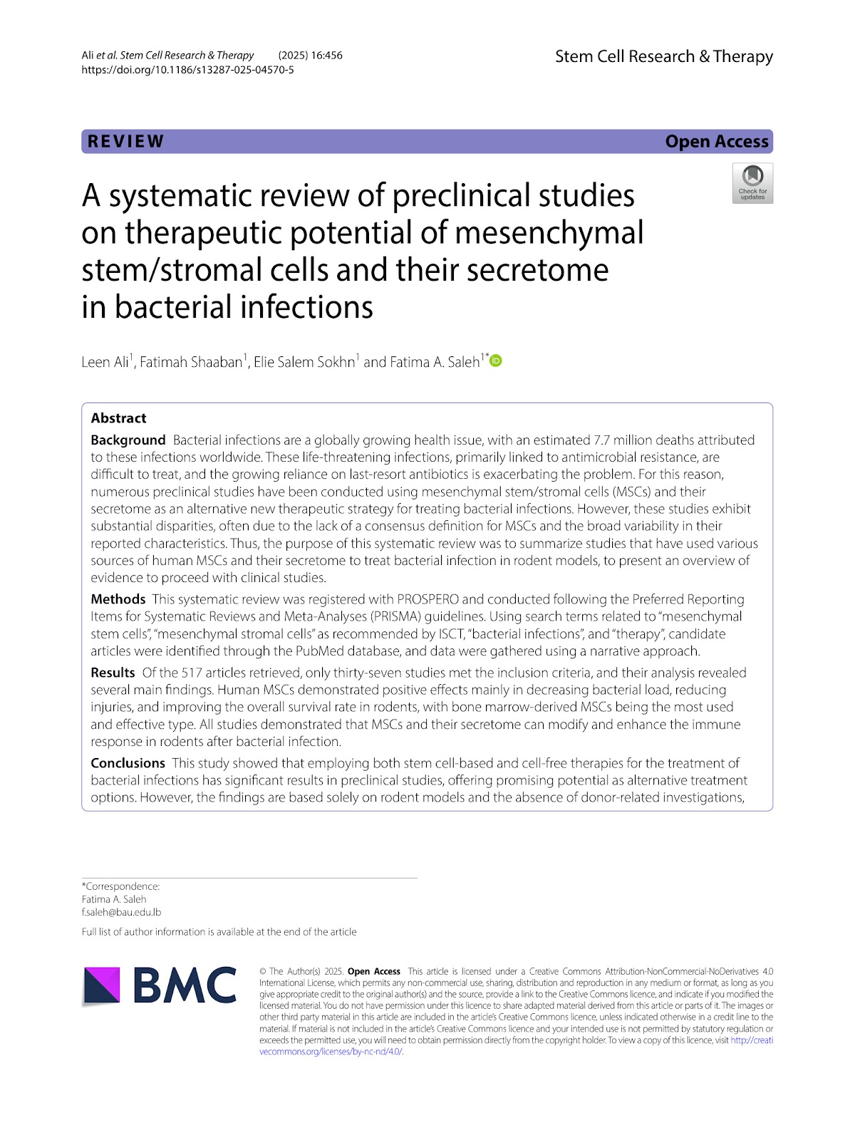
Background  Bacterial infections are a globally growing health issue, with an estimated 7.7 million deaths attributed to these infections worldwide. These life-threatening infections, primarily linked to antimicrobial resistance, are difficult to treat, and the growing reliance on last-resort antibiotics is exacerbating the problem. For this reason, numerous preclinical studies have been conducted using mesenchymal stem/stromal cells (MSCs) and their secretome as an alternative new therapeutic strategy for treating bacterial infections. However, these studies exhibit substantial disparities, often due to the lack of a consensus definition for MSCs and the broad variability in their reported characteristics. Thus, the purpose of this systematic review was to summarize studies that have used various sources of human MSCs and their secretome to treat bacterial infection in rodent models, to present an overview of evidence to proceed with clinical studies. Methods  This systematic review was registered with PROSPERO and conducted following the Preferred Reporting Items for Systematic Reviews and Meta-Analyses (PRISMA) guidelines. Using search terms related to “mesenchymal stem cells”, “mesenchymal stromal cells” as recommended by ISCT, “bacterial infections”, and “therapy”, candidate articles were identified through the PubMed database, and data were gathered using a narrative approach. Results  Of the 517 articles retrieved, only thirty-seven studies met the inclusion criteria, and their analysis revealed several main findings. Human MSCs demonstrated positive effects mainly in decreasing bacterial load, reducing injuries, and improving the overall survival rate in rodents, with bone marrow-derived MSCs being the most used and effective type. All studies demonstrated that MSCs and their secretome can modify and enhance the immune response in rodents after bacterial infection. Conclusions  This study showed that employing both stem cell-based and cell-free therapies for the treatment of bacterial infections has significant results in preclinical studies, offering promising potential as alternative treatment options. However, the findings are based solely on rodent models and the absence of donor-related investigations, necessitating further research to translate these findings into clinical applications.

Read Full Abstract10.1186/s13287-025-04570-5
Editorial Expression of Concern: Any closer to successful therapy of multiple myeloma? CAR-T cell is a good reason for optimismGraphical AbstractVerified
Stem Cell Research & Therapy

Editorial Expression of Concern: Any closer to successful therapy of multiple myeloma? CAR-T cell is a good reason for optimism

The Editor-in-Chief of Stem Cell Research & Therapy is issuing an editorial expression of concern to alert readers that this article shows indication of irregularities in authorship during the publication process. The substantial authorship change that took place at revision does not appear to match the extent of the revision itself, and no sufficient rationale for the change has been provided by the authors. Navid Shomali, Roza Motavali did not state explicitly whether he agrees to this Expression of Concern. Faroogh Marofi, Safa Tahmasebi, Heshu Sulaiman Rahman, Max Stanley Chartrand, Rebar N. Mohammed, Yashwant Pathak and Roza Motavalli do not agree to this Expression of concern. Denis Kaigorodov, Alexander Markov, Alexei Valerievich Yumashev, Mostafa Jarahian and Farhad Motavalli Khiavi did not reply to correspondence from the Editor about this Expression of Concern.

Read Full Abstract10.1186/s13287-025-04260-2