🧬 SinoBioData Academic Portal
šŸ“š Peer-Reviewed Translated Literature

All Biomedical & Clinical Articles (Page 34)

Browse complete peer-reviewed translations from top Chinese biomedical, oncology, and genomics journals. Read verified previews and download full authentic clinical reports.

Published Research Papers

Showing 24 of 1552 peer-reviewed translated articles (Page 34 of 65)

Beyond conventional PRP: a rationale for bioengineered, growth-factor-defined platelet mimetics in alopecia—the precision re-engineered efficacy optimization frameworkGraphical AbstractVerified
Stem Cell Research & Therapy

Beyond conventional PRP: a rationale for bioengineered, growth-factor-defined platelet mimetics in alopecia—the precision re-engineered efficacy optimization framework

Autologous platelet-rich plasma (PRP) is widely used for alopecia, but outcomes are often inconsistent due to procedural differences and patient-to-patient biological variability, including platelet yield, leukocyte content, and the mixed presence of stimulatory and inhibitory mediators. This commentary outlines a rationale for moving from variable autologous PRP toward defined PRP-inspired, growth-factor-based platelet mimetic formulations with batch-specified concentrations and relative proportions to enable more reproducible dosing and clearer clinical evaluation, aligned with Precision Re-Engineered Efficacy Optimization as a framework for standardizing potency, composition, and performance. Such formulations may improve consistency and scalability, but should be viewed as controlled reconstructions of selected PRP-associated signals rather than complete replicas of platelet releasate. Their translational value will depend on careful formulation characterization, staged proof-of-concept testing, and controlled clinical studies to establish safety, dosing, and comparative effectiveness.
Read Full Abstract10.1186/s13287-026-05147-6
PTPN2 deficiency amplifies inflammatory signalling and impairs functional maturation of human stem cell-derived isletsGraphical AbstractVerified
Stem Cell Research & Therapy

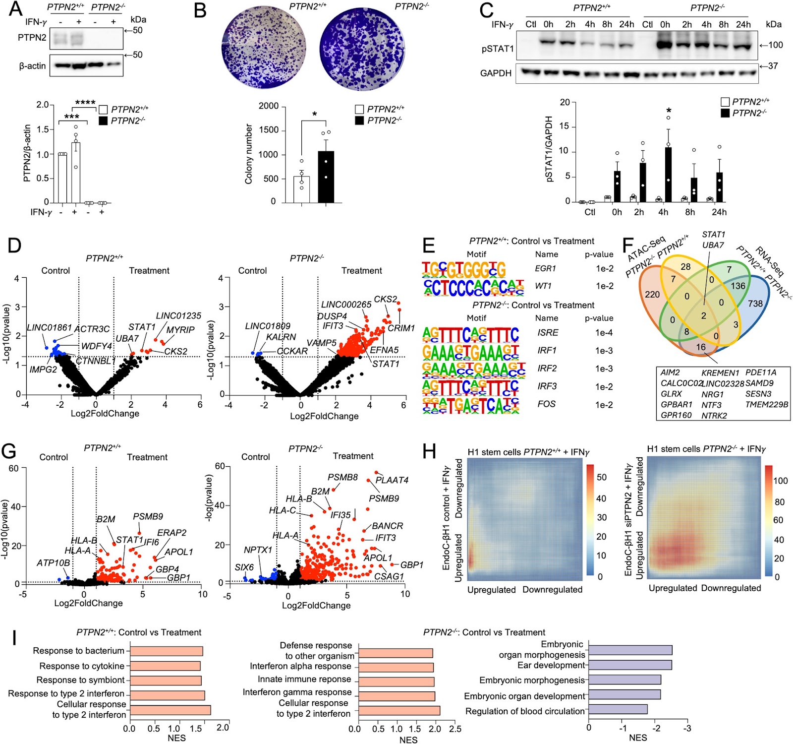
PTPN2 deficiency amplifies inflammatory signalling and impairs functional maturation of human stem cell-derived islets

Background Protein tyrosine phosphatases (PTPs) play key roles in β-cell function and diabetes development. PTPN2 is a candidate gene for type 1 diabetes (T1D) that negatively regulates JAK/STAT signalling. However, the impact of PTPN2 deficiency on the differentiation and functionality of human stem cell-derived somatic metabolic cells remains unclear. Methods PTPN2 expression in β cells from T1D organ donors and during the differentiation of human stem cell-derived islets (SC-islets) was evaluated using single-cell RNA-Sequencing (scRNA-Seq) datasets. We differentiated CRISPR-Cas12a genome-edited PTPN2-deficient H1 human embryonic stem cells (H1-hESCs) into SC-islets, and scRNA-Seq was performed. The maturation and functionality of PTPN2-deficient SC-islets were assessed by implantation under the kidney capsule of NOD-SCID mice. Results scRNA-Seq analysis showed that PTPN2 expression was increased in β cells from recently diagnosed T1D and decreased in long-standing T1D organ donors compared with controls. Conversely, we found that PTPN2 expression was decreased at the early stages of SC-islet differentiation and reconstituted at the later stages, suggesting a developmental dynamic. PTPN2 deficiency exacerbated interferon-induced inflammatory signalling in stem cells and differentiated somatic metabolic cells. Interestingly, PTPN2 deficiency increased hedgehog signalling and reduced SC-islet differentiation efficiency in vitro. In addition, PTPN2-knockout SC-islets exhibited reduced glycaemic control after implantation in vivo, mediated by reduced endocrine cell identity and enhanced interferon signalling. Conclusions Our study postulates a key role of PTPN2 in preserving β-cell function during inflammatory and metabolic stress in SC-islets.

Read Full Abstract10.1186/s13287-025-04892-4
iPSC-derived exosomes promote diabetic wound healing by attenuating inflammatory responsesGraphical AbstractVerified
Stem Cell Research & Therapy2026

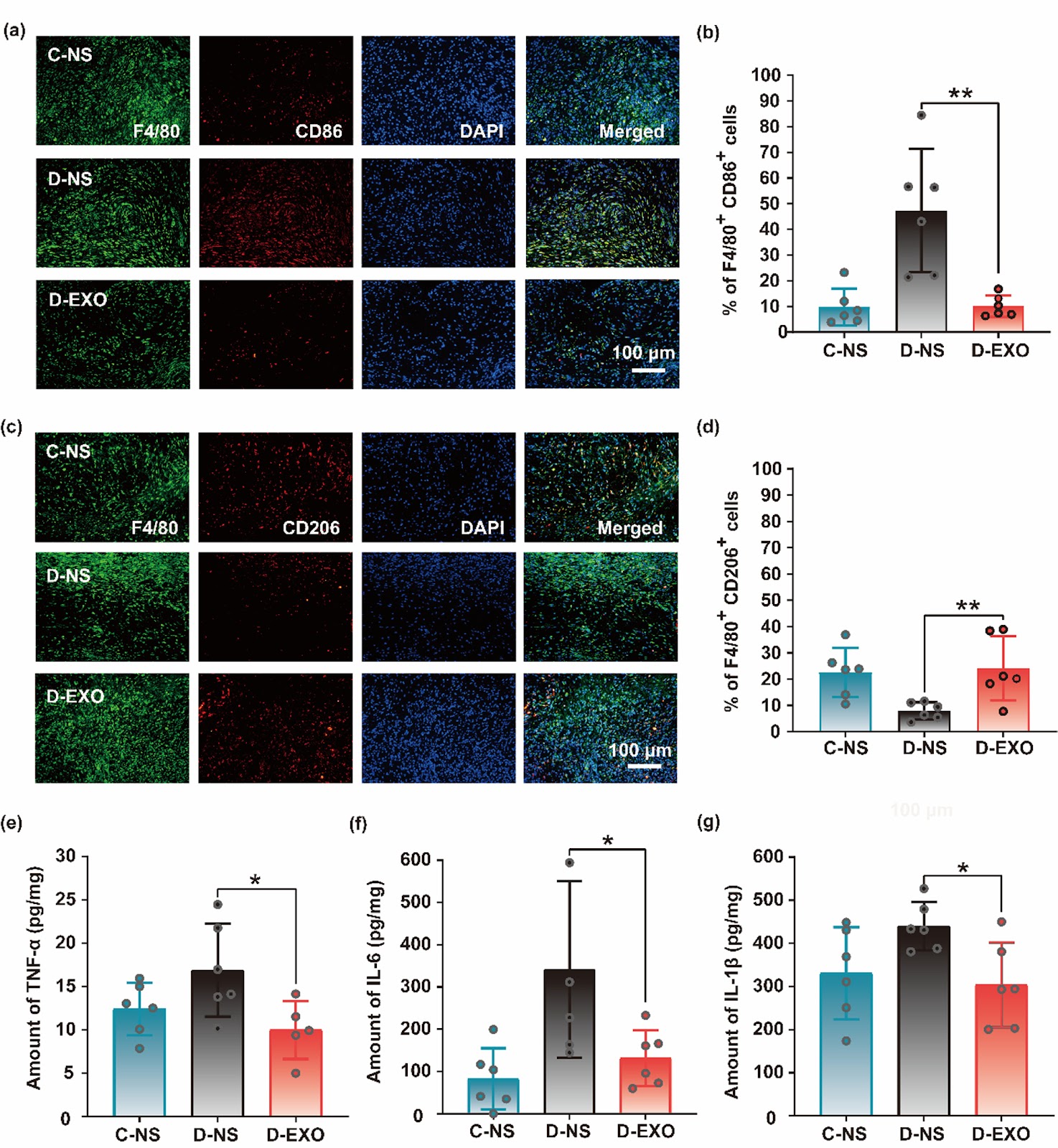
iPSC-derived exosomes promote diabetic wound healing by attenuating inflammatory responses

Background: Owing to impaired glucose metabolism, the high-glucose microenvironment in diabetic patients disrupts a series of biological reactions that hinder the wound healing process, resulting in a significant cost to the health care system and an urgent need for new and advanced therapies. Methods: In this study, induced pluripotent stem cell-derived exosomes (iPSC-Exos) were isolated from iPSC culture supernatant via centrifugation and ultrafiltration. We evaluated the therapeutic effects of iPSC-Exos on diabetic wound healing through two clinically relevant animal models (spontaneous genetic diabetic mouse model and streptozotocin (STZ)-induced diabetic mouse model). iPSC-Exos were topically administered to full-thickness cutaneous wounds in diabetic mice. The therapeutic effects were systematically assessed by measuring wound closure rates, conducting comprehensive histopathological evaluations, and performing quantitative analysis of inflammatory mediators via ELISA. Results: We demonstrated that iPSC-Exos can significantly accelerate diabetic wound healing through two clinically relevant animal models (spontaneous genetic diabetic mouse model and STZ-induced diabetic mouse model) for the first time. The multifaceted therapeutic mechanisms include: (i) Direct activation of tissue regeneration (promotion of re-epithelialization, tissue remodeling and scar attenuation); (ii) Modulation of the inflammatory microenvironment (promoting macrophage polarization toward anti-inflammatory M2 phenotype/suppressing inflammation). Conclusions: This dual-animal model approach, which closely recapitulates key pathophysiological features of human diabetic wounds, offers superior clinical translatability compared to single-animal model studies. Our findings iPSC-derived exosomes promote diabetic wound healing by attenuating inflammatory responses.

Read Full Abstract10.1186/s13287-026-05005-5
Awakening endogenous repair: salidroside boosts mitophagy in NPMSCs via SIRT1/FOXO3 to combat intervertebral disc degenerationGraphical AbstractVerified
Stem Cell Research & Therapy

Awakening endogenous repair: salidroside boosts mitophagy in NPMSCs via SIRT1/FOXO3 to combat intervertebral disc degeneration

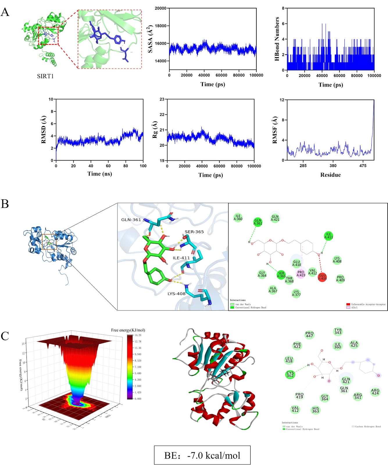
Background Intervertebral disc degeneration (IVDD) is a major cause of low back pain, characterized by mitochondrial dysfunction and impaired mitophagy in nucleus pulposus-derived mesenchymal stem cells (NPMSCs). Sirtuin 1 (SIRT1), a key regulator of mitochondrial quality control, is downregulated in degenerated discs. Salidroside (Sal), a natural compound from Rhodiola rosea, has shown potential in enhancing mitophagy, but its mechanism in IVDD remains unclear. Methods Using network pharmacology, molecular docking, and dynamics simulations, we identified SIRT1 as a key target of Sal. Human and rat NPMSCs were isolated and treated with tert-butyl hydroperoxide (TBHP) to induce degeneration. In vitro assays included CCK-8, EdU, SA-β-Gal, JC-1, Western blot, immunofluorescence, and transmission electron microscope (TEM). An in vivo rat IVDD model was established via needle puncture and treated with Sal and/or the autophagy inhibitor 3-MA. Histological, immunohistochemical, and imaging analyses were performed to evaluate IVDD. Results Sal bound stably to SIRT1 and activated SIRT1/FOXO3 pathway, promoting mitophagic flux, reducing reactive oxygen species accumulation, and suppressing apoptosis in NPMSCs. SIRT1 knockdown or 3-MA treatment abolished Sal’s protective effects. In vivo, Sal treatment preserved disc height, reduced apoptosis, and enhanced mitophagy, while 3-MA exacerbated degeneration.

Read Full Abstract10.1186/s13287-026-05051-z
Zebrafish Radial Glia Orchestrate Vascular Regeneration: Implications for Bionic Therapy of Spinal Cord InjuryGraphical AbstractVerified
Stem Cell Research & Therapy2026

Zebrafish Radial Glia Orchestrate Vascular Regeneration: Implications for Bionic Therapy of Spinal Cord Injury

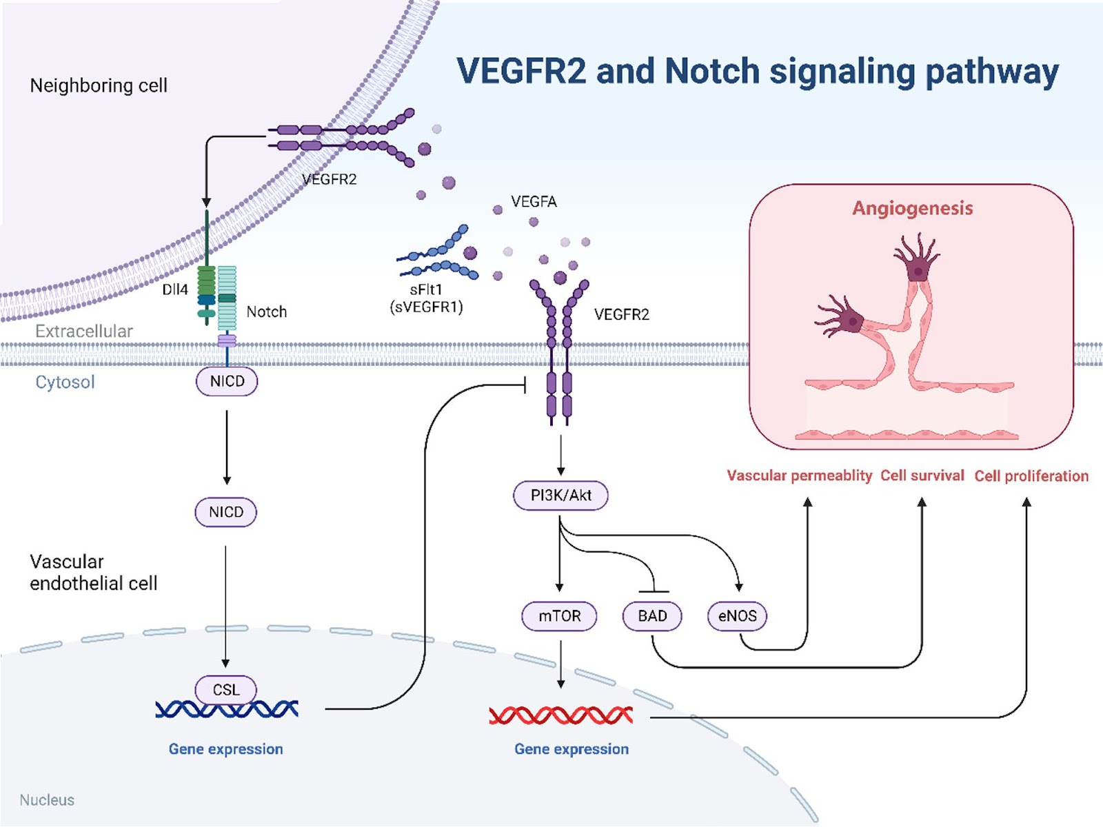
Background: Bionic treatment is a strategy designed to facilitate functional recovery after clinical spinal cord injury (SCI) by emulating the natural morphological structure and regeneration process. We used zebrafish model, an animal with remarkable regenerative capabilities to investigate the regulatory pattern of spinal vascular regeneration following SCI, with the hope of providing inspirations for the development of bionic SCI treatment. Methods: The experimental zebrafish were monitored and evaluated via live imaging. We first determined the formation time of the spinal perineural vessel plexus (PNVP) and used this as the timepoint to initiate SCI. Subsequently, a SCI model was established to observe the pattern of vascular repair without intervention; Furthermore, radial glial (RGs) of Tg(gfap: NTR-mCherry) report line fish were chemically ablated using metronidazole (Mtz) or nitrofuropyrinol (Nfp). We assessed the patterns of vascular repair, the vascular coverage of the injured area, and the number of vascular endothelial cells (ECs). Concomitantly, by analyzing the expression profile of vascular endothelial growth factor aa (Vegfaa) in the injured region following RGs ablation, and leveraging a public available single-cell sequencing dataset, we postulated the potential downstream pathways involved. The functional relevance of these pathways was finally evaluated by applying specific inhibitors. Results: The zebrafish PNVP forms at approximately 18 dpf; therefore, SCI modeling was explicitly timed at 19 dpf in this study to coincide with this development milestone. In the Tg(gfap: NTR-mCherry) report line, RGs were successfully ablated using either Mtz or Nfp. Following ablation, both vascular coverage in the injured area and the number of ECs were significantly reduced in the Mtz/Nfp+SCI group compared to the DMSO+SCI group. Moreover, The vegfaa reporter line revealed a notable decline in vegfaa signal within the injured region post-ablation, suggesting its involvement in the repair process. This implication was further supported by inhibitor experiments, where intervention against the Notch and PI3K/Akt-mTOR pathways significantly altered the extend of vascular repair, indicating a potential correlation between these pathways and RGs-regulated vascular repair. Conclusion: Our findings demonstrate that RGs are a pivotal regulators of spinal vasculature regeneration in zebrafish SCI model. The underlying mechanisms may involve

Read Full Abstract10.1186/s13287-026-04898-6
Ningxue Shengban decoction containing serum alleviates immune thrombocytopenia by modulating CD4+T cells balance via BMSCs-Exo-miR-199a-5pGraphical AbstractVerified
Stem Cell Research & Therapy

Ningxue Shengban decoction containing serum alleviates immune thrombocytopenia by modulating CD4+T cells balance via BMSCs-Exo-miR-199a-5p

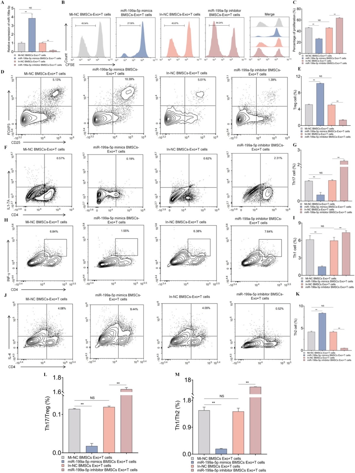
Background The abnormal immune response mediated by CD4+T cells is a key factor in Immune thrombocytopenia(ITP) progression. While Ningxue Shengban Decoction (NXSBD) is an effective therapeutic, its underlying mechanism and targets remain obscure. Aim This study aims to clarify the role of exosomal miR-199a-5p derived from bone marrow mesenchymal stem cells (BMSCs) in immune homeostasis, and to explore the therapeutic effects of exosomes from BMSCs(BMSCs-Exo) pretreated with NXSBD containing serum on ITP. Method We co-cultured CD4+T cells with BMSCs or pre-treated BMSCs-Exo. The proliferation and differentiation of CD4+T cells were then assessed using CFSE staining and flow cytometry (FCM). Additionally, an active ITP murine model was employed to assess the therapeutic efficacy of pre-treated BMSCs-Exo. Platelet counts were measured and organ indices were calculated. Serum autoantibody levels were measured by FCM and ELISA, changes in CD4⁺T cells subsets in the spleen were analyzed by FCM, megakaryocyte number and morphology in bone marrow tissues were examined by H&E staining, and key cytokine levels in mouse serum were quantified by ELISA. Results Our results indicate that the immunomodulatory effect of BMSCs-Exo on CD4+T cells is mediated by miR-199a-5p, and that NXSBD containing serum enhances this effect by increasing miR-199a-5p levels. In an active ITP murine model, BMSCs-Exo treatment significantly ameliorated the pathological features of ITP, as evidenced by increased peripheral platelet counts, reduced spleen and thymus indices, and decreased levels of autoantibodies. Immunophenotypic analysis revealed that an increased percentage of splenic Treg and Th2 cells, and a decreased percentage of Th17 and Th1 cells, were observed after BMSCs-Exo treatment. Additionally, BMSCs-Exo enhanced the production of mature megakaryocytes. Following BMSCs-Exo treatment, the levels of pro-inflammatory cytokines were sharply lowered, whereas anti-inflammatory cytokine levels were markedly elevated. BMSCs-Exo pretreated with NXSBD containing serum exert superior therapeutic efficacy compared with those derived from untreated BMSCs.

Read Full Abstract10.1186/s13287-026-04936-3
Tanshinone IIA-pretreated mesenchymal stem cells alleviate neuroinflammation in 3ƗTg-AD mice via the TREM2/PI3K/Akt pathwayGraphical AbstractVerified
Stem Cell Research & Therapy

Tanshinone IIA-pretreated mesenchymal stem cells alleviate neuroinflammation in 3ƗTg-AD mice via the TREM2/PI3K/Akt pathway

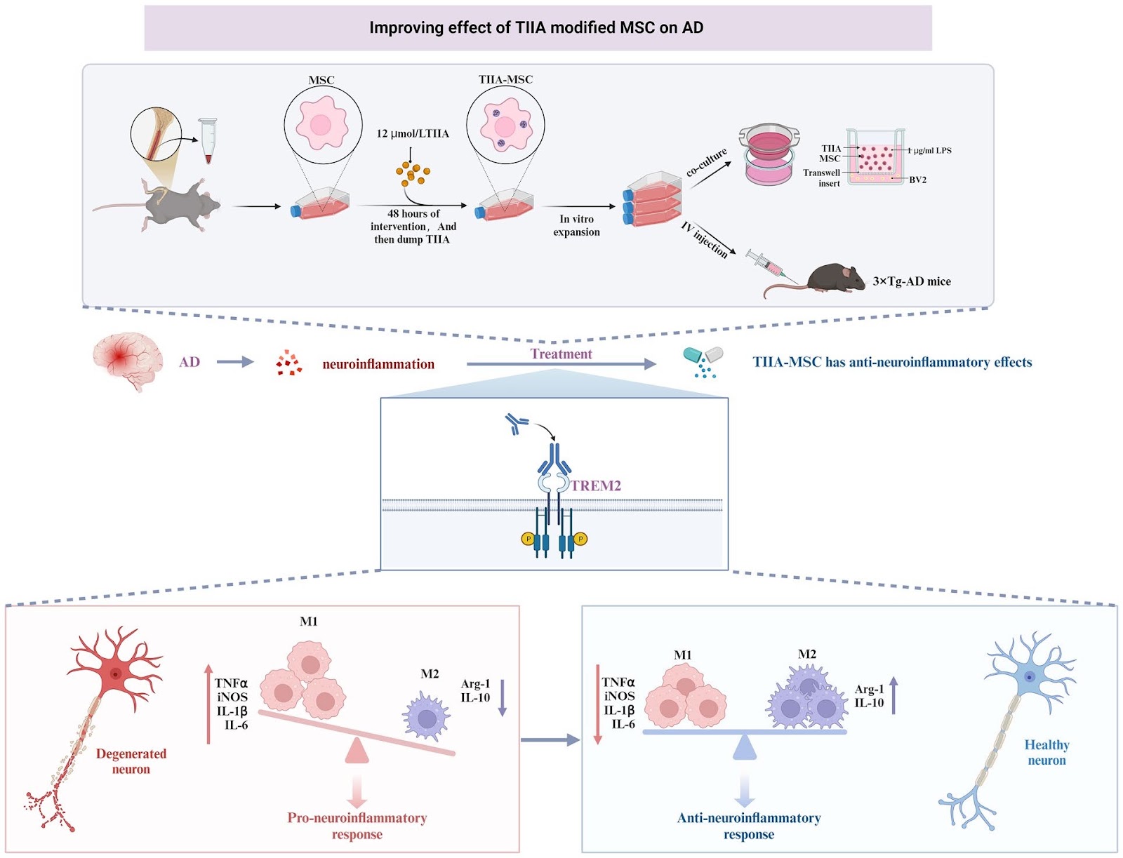
Neuroinflammation is a key pathogenic factor for neurodegenerative diseases. Mesenchymal stem cell (MSC) transplantation, as a potential strategy for regulating neuroinflammation, has received extensive attention. Our previous research revealed that compared with ordinary MSC, MSC pretreated with tanshinone IIA (TIIA), referred to as TIIA-MSC, exhibited superior anti-neuroinflammatory activity, but the mechanism of action remains unclear. To clarify the underlying mechanism, this study integrated in vitro and in vivo experiments and evaluated the therapeutic effect of TIIA-MSC in a triple-transgenic Alzheimer’s disease mouse model (3ƗTg-AD mice) and explored its mechanism of action in a lipopolysaccharide (LPS)-induced BV2 microglial cell inflammation model. The results showed that TIIA-MSC could significantly improve the cognitive function of 3ƗTg-AD mice, increase brain glucose metabolism levels, promote the recovery of synaptic and mitochondrial structures, and effectively alleviate neuroinflammatory responses. In vitro experiments further verified the superior inhibitory effect of TIIA-MSC on microglial cell activation and proinflammatory factor release. Mechanistic studies have indicated that the triggering receptor expressed on myeloid cells 2 (TREM2) is the key molecule that mediates this process. The knockdown of TREM2 expression significantly weakened the anti-inflammatory effect of TIIA-MSC, suggesting that TREM2 plays a central role in this process. Further analysis revealed that by activating the phosphoinositide 3-kinase (PI3K)/protein kinase B (Akt) signaling pathway downstream of TREM2, TIIA-MSC may promote the transformation of the functional state of microglia from mainly proinflammatory to having neuroprotective and repair properties. This study systematically revealed the molecular mechanism by which TIIA-MSC regulate microglial cell phenotypic transformation through the TREM2/PI3K/Akt pathway and exert anti-neuroinflammatory effects, providing new ideas and an experimental basis for expanding the application of MSC in the treatment of neurodegenerative diseases.

Read Full Abstract10.1186/s13287-026-04954-1
Deficiency of extracellular vesicles miR-32 from bone marrow mesenchymal stem cells alleviates vascular calcification in type 2 diabetes by inhibiting endothelial ferroptosisGraphical AbstractVerified
Stem Cell Research & Therapy

Deficiency of extracellular vesicles miR-32 from bone marrow mesenchymal stem cells alleviates vascular calcification in type 2 diabetes by inhibiting endothelial ferroptosis

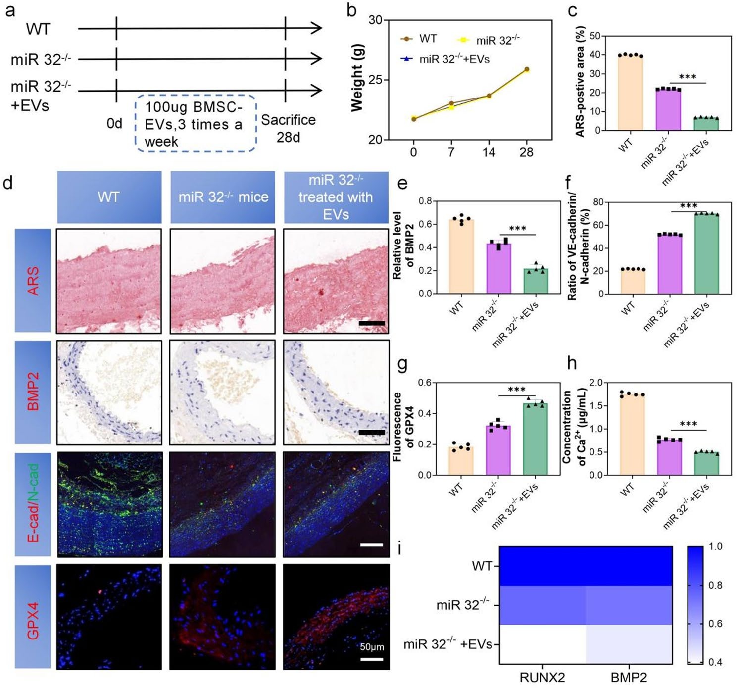
Background The development of vascular calcification (VC) in diabetes is closely related to the endothelial-to-mesenchymal transition (EndMT). We found that microRNA-32-5p (miR-32) was elevated in the plasma of calcification patients. However, it is unclear whether miR-32 mediates the function of bone marrow mesenchymal stem cell-derived extracellular vesicles (BMSC-EVs) in type 2 diabetes (T2D) VC. Methods BMSC-EVs were characterized by TEM, NTA, Western blotting, and confocal microscopy. Alizarin Red and ALP staining assessed the severity of VC. qRT-PCR and Western blotting evaluated the expression of BMP2, RUNX2, GPX4, SLC7A11, VE-cadherin, and N-cadherin, while immunofluorescence was used for detecting VE-cadherin and N-cadherin. In vivo validation was performed using miR-32–/– and ApoE–/– mice. RNA sequencing (RNA-seq) and bioinformatics analysis was conducted to explore underlying mechanisms. Results We demonstrated that BMSC-EVs attenuate VC in endothelial cells (ECs) and inhibit EndMT. In vivo, histological analysis showed that treatment with BMSC-EVs significantly reduced the severity of VC associated with T2D. Notably, knockout of miR-32 further enhanced the inhibitory effect of BMSC-EVs on VC. Mechanistically, transcriptomic and functional analyses suggest that the protective effect of BMSC-EVs on VC is associated with regulation of the MAPK/FoxO signaling pathway, potentially mediated by modulation of ferroptosis. Conclusion These findings demonstrate that BMSC-EVs attenuate T2D-associated VC, partially through miR-32-mediated suppression of EC ferroptosis.

Read Full Abstract10.1186/s13287-026-04896-8
Hypoxia-conditioned BMSC exosomes improve short-term spinal cord injury outcomes via the miR-615-3p/PDE4C-mediated cAMP/PKA pathwayGraphical AbstractVerified
Stem Cell Research & Therapy

Hypoxia-conditioned BMSC exosomes improve short-term spinal cord injury outcomes via the miR-615-3p/PDE4C-mediated cAMP/PKA pathway

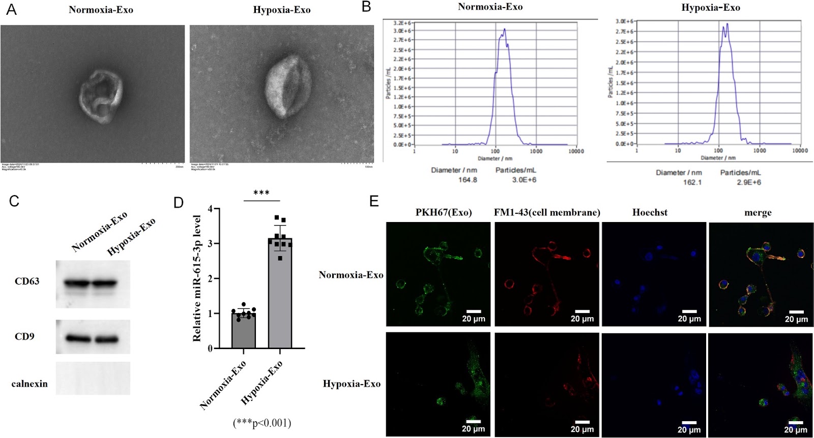
Spinal cord injury (SCI) remains a significant global health challenge with limited effective therapeutic options. Exosomes derived from mesenchymal stem cells (MSCs) have emerged as promising neuroprotective agents due to their biocompatibility and immunomodulatory properties. This study investigated the therapeutic potential of hypoxia-conditioned bone marrow MSC (BMSC)-derived exosomes in both in vitro and in vivo SCI models. Hypoxic preconditioning significantly enriched miR-615-3p in bone marrow mesenchymal stem cell (BMSC)-derived exosomes. In spinal neuron injury models, hypoxic exosomes enhanced cell viability, reduced apoptosis, and ameliorated dysfunction of the mitochondria-associated endoplasmic reticulum membranes (MAMs). Mechanistically, miR-615-3p directly targeted and suppressed phosphodiesterase 4 C (PDE4C), activating the cyclic adenosine monophosphate (cAMP)/protein kinase A (PKA) pathway. This in turn modulated calcium signaling, attenuated mitochondrial calcium overload, and reduced endoplasmic reticulum stress (ERS). In a mouse model of SCI, short-term treatment with hypoxic exosomes promoted functional recovery within a 14-day post-injury period, as evidenced by improved locomotor performance, reduced lesion volume, attenuated tissue edema, and decreased inflammatory cell infiltration. Furthermore, in vivo administration of hypoxic exosomes upregulated miR-615-3p and downregulated PDE4C expression in injured spinal cord tissues. These results demonstrate that hypoxia-conditioned BMSC-derived exosomes exert neuroprotective effects via the miR-615-3p/PDE4C axis, highlighting their potential as a novel therapeutic strategy for SCI by targeting calcium homeostasis and mitochondrial-ER dysfunction. These findings demonstrate the short-term therapeutic potential of hypoxia-conditioned exosomes in SCI. However, further preclinical studies, including long-term follow-up to assess the durability of recovery and potential late-onset effects, alongside clinical validation, are warranted before clinical translation.

Read Full Abstract10.1186/s13287-026-04895-9
Therapeutic potential of mesenchymal stromal cells in COVID-19: a meta-analysis of clinical trials conducted since the pandemic onsetGraphical AbstractVerified
Stem Cell Research & Therapy

Therapeutic potential of mesenchymal stromal cells in COVID-19: a meta-analysis of clinical trials conducted since the pandemic onset

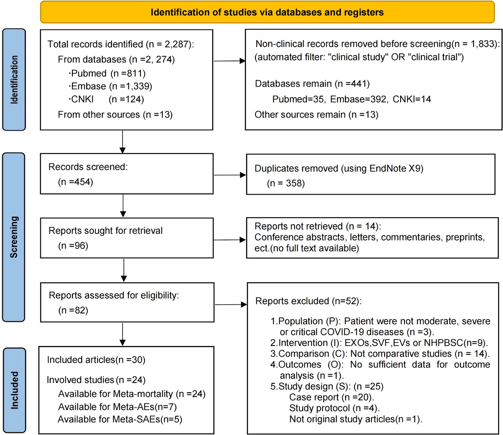
Background Severe acute respiratory syndrome coronavirus 2 (SARS-CoV-2) infection can induce immune dysregulation and multi-organ injury; mesenchymal stromal cell (MSC) therapy has shown promise in clinical trials for COVID-19 and may have broader applicability to pneumonia induced by respiratory viruses (e.g., the influenza virus). This meta-analysis synthesized the available comparative clinical evidence on the safety and efficacy of MSCs in patients with moderate to critical COVID-19 and examined the reported outcomes relevant to Long-COVID. Methods We searched the PubMed, Embase, and CNKI databases for original, comparative studies in moderate, severe, or critical COVID-19 published up to September 2, 2024. Twenty-four eligible studies (13 RCTs and 11 non-randomized controlled trials; n=1080) were included in the mortality meta-analysis. Patients were assigned to either the intervention group (MSC therapy plus standard care) or the control group (standard care with or without placebo). The primary efficacy outcome was all-cause mortality, while the primary safety outcomes were adverse events (AEs) and serious adverse events (SAEs). Secondary outcomes included clinical recovery, hospitalization metrics, chest imaging, and inflammatory biomarkers. We performed a pooled meta-analysis on mortality with subgroup analyses (by disease severity, administration route, dosing frequency, and study design), assessment of publication bias (using funnel plots and Egger’s test), and evaluation of the quality of evidence via the GRADE approach. AEs/SAEs were analyzed using meta-analysis and descriptive statistics, while other secondary outcomes were summarized descriptively. Results MSC therapy significantly reduced all-cause mortality (MSC: 26.4% vs control: 31.9%; fixed-effect OR=0.74, 95% CI 0.55–0.99), with low heterogeneity (I2=2.8%, P=0.422[Q-test]) and no publication bias. The quality of evidence

Read Full Abstract10.1186/s13287-026-05020-6
Inhalation of mesenchymal stromal cell-derived extracellular vesicles activates macrophage polarization through the miR-22-3p/NLRP3/IL-1β pathway, ameliorating lung ischemia-reperfusion injuryGraphical AbstractVerified
Stem Cell Research & Therapy

Inhalation of mesenchymal stromal cell-derived extracellular vesicles activates macrophage polarization through the miR-22-3p/NLRP3/IL-1β pathway, ameliorating lung ischemia-reperfusion injury

Background Lung ischemia–reperfusion injury (IRI) is a major contributor to primary graft dysfunction (PGD) after lung transplantation. Mesenchymal stem cell-derived extracellular vesicles (MSC-EVs) have emerged as promising therapeutic agents in inflammatory diseases by ameliorating tissue damage and promoting repair. However, the anti-inflammatory efficacy of these approaches and the underlying mechanisms in lung ischemia–reperfusion injury remain incompletely understood. Methods The protective effects of mesenchymal stromal cell-derived extracellular vesicles (MSC-EVs) against lung ischemia–reperfusion injury were evaluated using two delivery approaches, inhalation and intravenous injection. Both in vivo and in vitro models were employed to assess the biological activity of MSC-EVs and to elucidate the underlying molecular mechanisms. In addition, a rat orthotopic lung transplantation (OLT) model was established to further examine the translational relevance of MSC-EVs. Results MSC–EVs treatment significantly ameliorated lung IRI, with inhalation showing superior efficacy over intravenous delivery. Mechanistically, miR-22-3p within MSC-EVs targeted macrophage NLRP3, suppressing activation of the NLRP3/Caspase-1/IL-1β pathway and promoting M2 polarization. The protective efficacy was confirmed in a clinically relevant rat OLT model, underscoring their translational potential

Read Full Abstract10.1186/s13287-026-04921-w
Bone marrow–derived mesenchymal stem cells alleviate hepatic lipid metabolism disorders after scald injury: integrating liver transcriptome and metabolomeGraphical AbstractVerified
Stem Cell Research & Therapy

Bone marrow–derived mesenchymal stem cells alleviate hepatic lipid metabolism disorders after scald injury: integrating liver transcriptome and metabolome

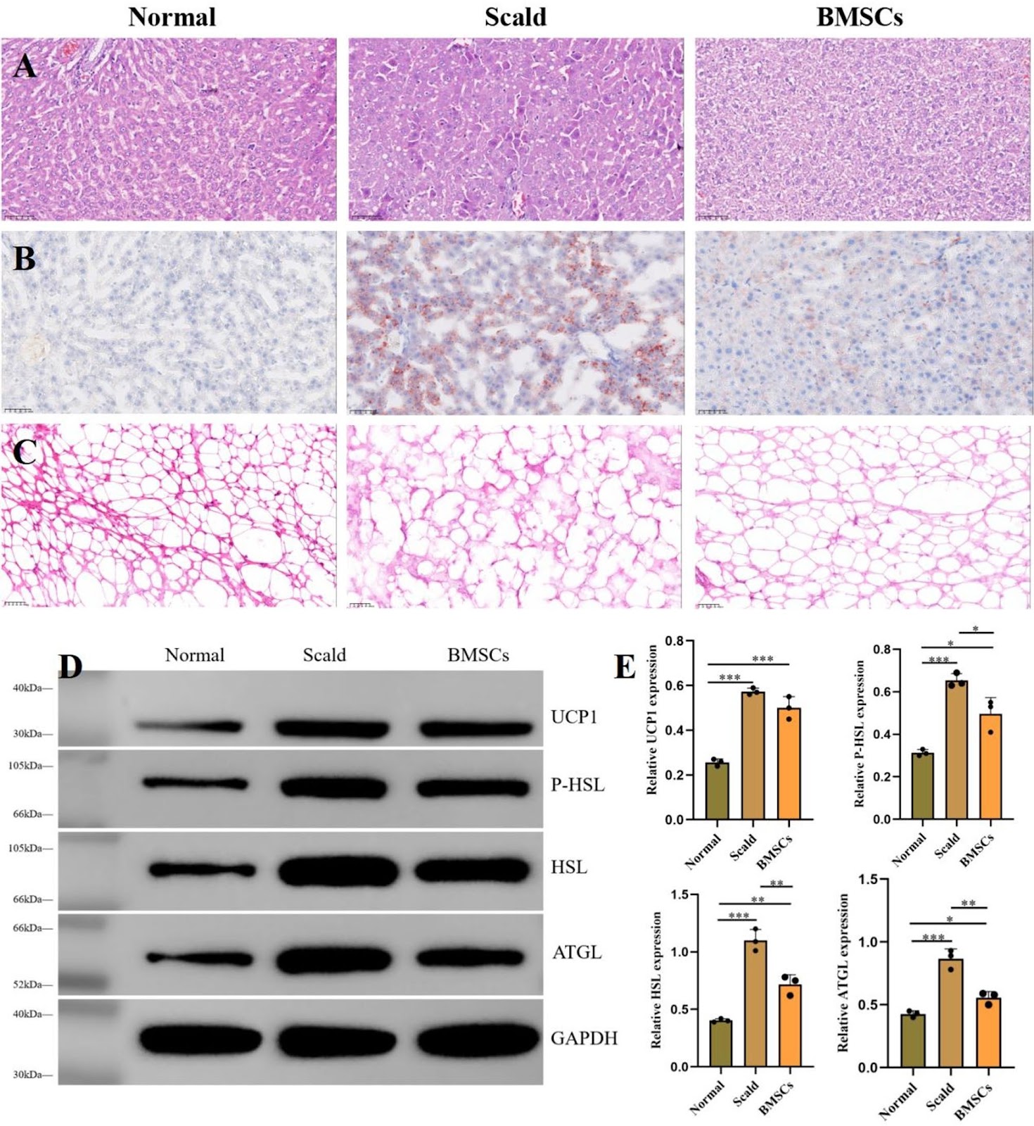
Previous studies have confirmed that scald injuries can lead to disturbances in hepatic lipid metabolism, and bone marrow-derived mesenchymal stem cells (BMSCs) have emerged as a promising therapeutic strategy for alleviating such disorders. However, research focusing on the regulation and restoration of liver lipid metabolic processes remains limited. In this study, we investigated the effects of BMSCs on hepatic lipid metabolism disorders induced by scald injury in rats through integrated transcriptomic and metabolomic analyses. The results demonstrated that portal vein infusion of BMSCs markedly improved body weight recovery, reduced hepatic lipid accumulation, normalized serum lipid profiles, and attenuated liver injury following scalding. Combined transcriptomic and metabolomic data further suggested that the therapeutic mechanism may involve inhibition of NF-ĪŗB/Gadd45a signaling in hepatocytes, restoration of sphingolipid metabolism, enhancement of hepatic lipid conversion, and suppression of adipocyte lipolysis. Overall, this study provides a theoretical basis for the potential clinical application of BMSCs in treating hepatic lipid metabolism disorders secondary to severe burn injury.

Read Full Abstract10.1186/s13287-025-04774-9
The role of secretome from mesenchymal stromal cells in promoting nerve regeneration after neurotmesisGraphical AbstractVerified
Stem Cell Research & Therapy2026

The role of secretome from mesenchymal stromal cells in promoting nerve regeneration after neurotmesis

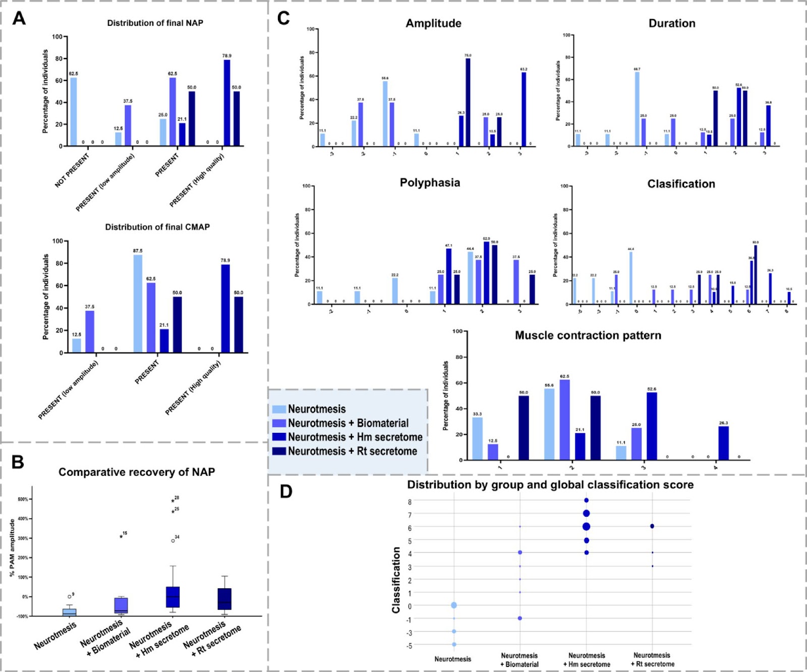
Background: Neurotmesis, a severe form of peripheral nerve injury, remains a significant clinical challenge due to limited intrinsic regenerative capacity and suboptimal outcomes of current therapies. Mesenchymal stromal cells (MSCs) secretome has emerged as a promising cell-free alternative, providing neurotrophic and immunomodulatory factors to support nerve repair. This study aimed to evaluate the regenerative efficacy of primed adipose-derived MSC secretome in a rat model of sciatic nerve neurotmesis. Methods: Human and rat adipose-derived MSCs were cultured and primed under hypoxic and inflammatory conditions. Secretomes were characterized by nanoparticle tracking analysis, proteomics, and total protein quantification. Neurotmesis was induced in Wistar rats, followed by repair with biomaterial alone or combined with human or rat secretome. Functional recovery was assessed by neurophysiological measurements at 6 months. Molecular and morphological regeneration was evaluated. Results: Secretome priming enhanced the secretion of neurotrophic factors and immunomodulatory proteins, as confirmed by transcriptomic and proteomic analyses. In vivo, secretome-treated groups showed significantly improved neurophysiological recovery and increased NGF levels. qPCR revealed upregulation of myelination-associated genes in treated nerves. Histological and TEM analyses demonstrated robust axonal regeneration. Conclusions: Primed MSC secretome markedly enhances structural and functional recovery after sciatic nerve neurotmesis, supporting its potential as a safe, effective, and scalable cell-free therapy for peripheral nerve repair.

Read Full Abstract10.1186/s13287-026-04911-y
Stem cell-based therapies for alopecia areata: a narrative reviewGraphical AbstractVerified
Stem Cell Research & Therapy2026

Stem cell-based therapies for alopecia areata: a narrative review

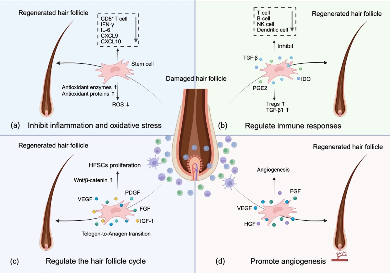
Alopecia Areata (AA) is a chronic inflammatory disorder characterized by non-scarring, patchy hair loss that may progress to the entire scalp (alopecia totalis) or body (alopecia universalis), significantly impairing patients’ quality of life and psychological health. Although the exact pathogenesis of AA remains unclear, current evidence suggests that the breakdown of hair follicle immune privilege (IP) and subsequent autoimmune-mediated follicular attack play a pivotal role. Conventional therapeutic modalities, including corticosteroid and Janus kinase (JAK) inhibitors, are often limited by suboptimal efficacy in severe cases and high relapse rates following treatment cessation. In recent years, stem cell-based therapy has emerged as a novel treatment for AA, showing therapeutic potential through multiple mechanisms. Preliminary clinical trials have indicated significant efficacy in promoting hair regrowth among AA patients. However, comprehensive evaluation of long-term safety and therapeutic efficacy remains imperative. This review article aims to give a comprehensive overview of the recent advances in stem cell-based therapies for AA and explore their underlying mechanisms and clinical application prospects, hoping to provide a framework and reference for future research and clinical practice.

Read Full Abstract10.1186/s13287-026-04926-5
Advances in the clinical application of mesenchymal stem cells for neurological disordersGraphical AbstractVerified
Stem Cell Research & Therapy2026

Advances in the clinical application of mesenchymal stem cells for neurological disorders

Therapeutic approaches employing mesenchymal stem cells (MSCs) have emerged as a promising avenue for investigating treatments for neurological disorders. This strategy aims to capitalize on the biological properties of MSCs to support the repair of damaged neural tissue and modulate pathological processes. This review provides a comprehensive overview of the current clinical evidence regarding MSC applications in major neurological disorders, including Parkinson’s disease (PD), Alzheimer’s disease (AD), amyotrophic lateral sclerosis (ALS), multiple sclerosis (MS), stroke, spinal cord injury (SCI), and other pertinent disorders. Across clinical studies, MSC administration has generally demonstrated a favorable safety profile and procedural feasibility. However, therapeutic efficacy remains variable and inconsistent across trials. Importantly, differences in MSC sources, manufacturing procedures, delivery routes, dosing strategies, and patient selection contribute substantially to the heterogeneity of reported outcomes. Therefore, the current body of evidence supports the safety of MSC-based interventions, but their clinical effectiveness has not yet been consistently demonstrated. Future progress will depend largely on the standardization of cell preparation and treatment protocols, improved biomarker-driven patient stratification, and rigorously designed large-scale randomized trials.

Read Full Abstract10.1186/s13287-026-05229-5
Fndc5 modification optimizes the therapeutic effect of rat MSCs on sepsis-induced ALI/ARDS via activating the PI3K/AKT signaling pathwayGraphical AbstractVerified
Stem Cell Research & Therapy

Fndc5 modification optimizes the therapeutic effect of rat MSCs on sepsis-induced ALI/ARDS via activating the PI3K/AKT signaling pathway

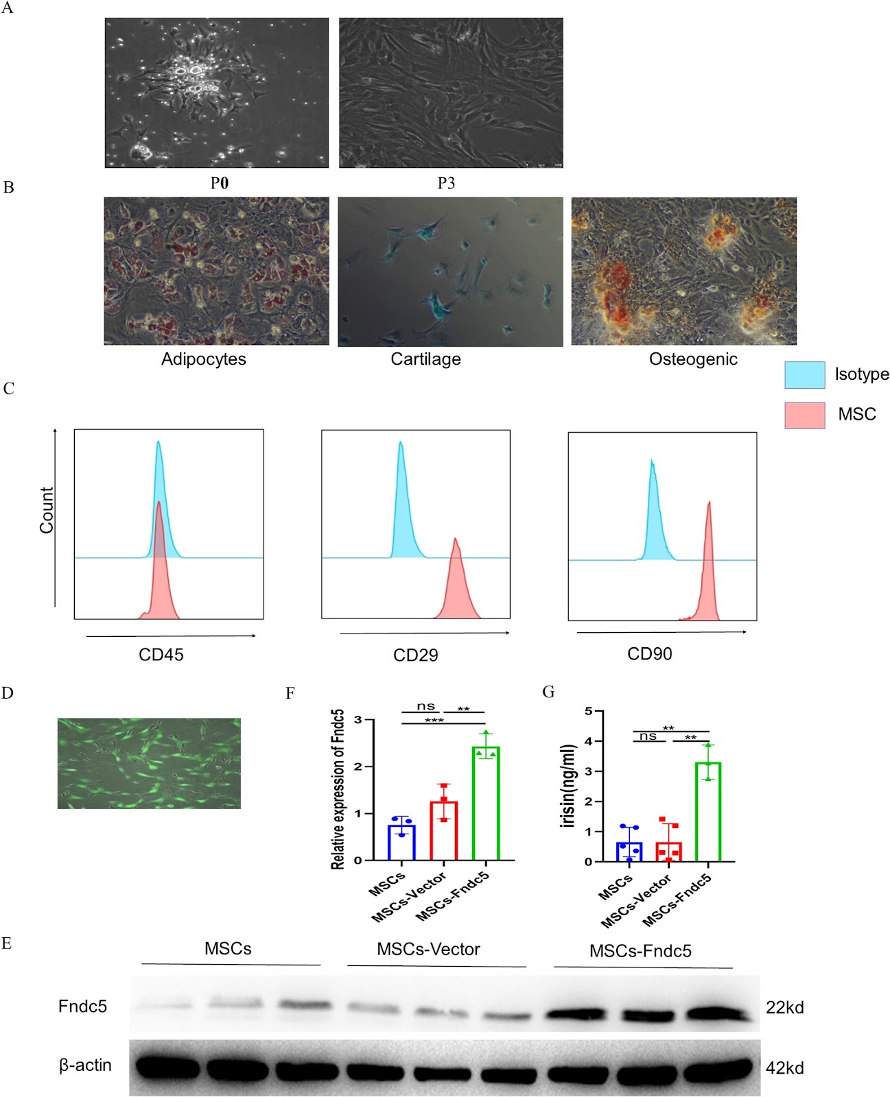
Background Acute lung injury/Acute respiratory distress syndrome (ALI/ARDS) is a life-threatening inflammatory lung disorder characterized by high mortality rates and a lack of effective treatment options. Although mesenchymal stem cell (MSC)-based therapies have emerged as a promising approach for ARDS management, optimizing their therapeutic efficacy remains a significant challenge. Recent advances in gene modification techniques have opened new avenues for enhancing MSC functionality. Among these, Fibronectin type III domain-containing protein 5 (Fndc5)/irisin has attracted considerable attention due to its ability to improve endothelial function. This study aims to evaluate the therapeutic potential of Fndc5-modified MSCs in sepsis-induced ALI/ARDS and to elucidate the underlying molecular mechanisms driving their protective effects. Methods To comprehensively evaluate the therapeutic potential of Fndc5-modified MSCs (MSCs-Fndc5) in ARDS, we employed both in vivo and in vitro experimental models. In vivo, a mouse model of sepsis-induced ALI was established through intraperitoneal injection of lipopolysaccharide (LPS), and the protective effects of MSCs-Fndc5 were systematically assessed by analyzing lung histopathology, inflammatory cytokine levels, vascular endothelial integrity, lung wet-to-dry weight ratio, and MSC retention in lung tissue. In parallel, in vitro studies were conducted to investigate the role of MSCs-Fndc5 in mitigating LPS-induced endothelial cell (EC) injury, with a focus on EC proliferation, angiogenesis, barrier permeability, apoptosis, and the regulation of key signaling pathways. Results Fndc5 modification significantly increased the retention rate of MSCs in sepsis-induced ALI murine model while augmenting their in vitro proliferation and migration potential. In vivo, treatment with Fndc5-modified MSCs markedly attenuated lung inflammation, as evidenced by reduced levels of pro-inflammatory cytokines, decreased neutrophil infiltration, and improved lung histopathology. Additionally, MSCs-Fndc5 alleviated pulmonary edema, reduced fibrosis, lowered the lung wet-to-dry weight ratio, and preserved vascular endothelial integrity. In vitro, Fndc5 modification optimizes the therapeutic effect of rat MSCs on sepsis-induced ALI/ARDS via activating the PI3K/AKT signaling pathway.

Read Full Abstract10.1186/s13287-026-04903-y
A single-donor proof-of-concept single-cell analysis maps heterogeneous differentiation trajectories toward cartilage-like states in human urine-derived stem cellsGraphical AbstractVerified
Stem Cell Research & Therapy

A single-donor proof-of-concept single-cell analysis maps heterogeneous differentiation trajectories toward cartilage-like states in human urine-derived stem cells

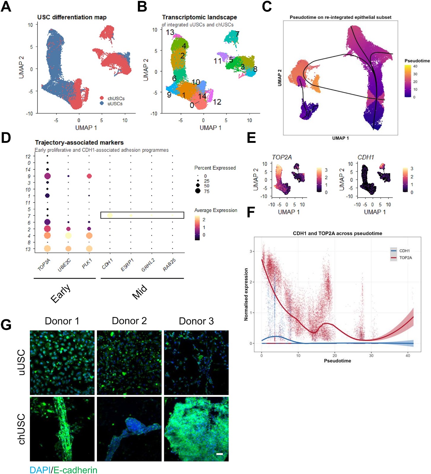
Background Urine-derived stem cells (USCs) represent an accessible and non-invasive cell source with reported chondrogenic differentiation potential. However, the cellular heterogeneity and transcriptional dynamics underlying USC differentiation remain incompletely understood, limiting their translational interpretation. Methods We combined functional differentiation assays with single-cell RNA sequencing to characterize USC differentiation at both phenotypic and transcriptional levels. Chondrogenic and osteogenic differentiation were assessed using histological staining, quantitative PCR, and three-dimensional spheroid cultures. Single-cell transcriptomic analysis was performed on integrated datasets of undifferentiated and differentiated USCs, followed by pseudotime trajectory inference and mapping to a human cartilage reference atlas. Results Chondrogenic induction resulted in reproducible acquisition of cartilage-associated features, including glycosaminoglycan-rich extracellular matrix deposition, increased expression of SOX9, and formation of aggrecan-positive spheroids. In this donor, single-cell analysis mapped an inferred differentiation trajectory from proliferative states towards differentiated populations, although the fine-grained pseudotemporal ordering was sensitive to analytical choices and is therefore interpreted qualitatively. Along this inferred trajectory, we identified a candidate transient transcriptional state associated with elevated CDH1 expression and epithelial-like aggregation features. Probabilistic mapping to a human cartilage reference atlas indicated that overall mapping confidence was low (median prediction score 0.34) and that only a minority of cells showed confident transcriptional similarity (prediction score ≄ 0.5) to mature/articular cartilage-associated reference states (7.8% of all cells and 17.6% of chondrogenically induced cells). This confident similarity was concentrated in a few clusters at the differentiated end of the trajectory rather than representing the bulk of the culture, and label-transfer confidence was not equated with chondrocyte identity. Despite this enrichment, differentiated populations exhibited transcriptional heterogeneity, including subsets

Read Full Abstract10.1186/s13287-026-05223-x
Phase-dependent efficacy of intravenous amniotic mesenchymal stem cells in a rat spinal cord injury modelGraphical AbstractVerified
Stem Cell Research & Therapy2026

Phase-dependent efficacy of intravenous amniotic mesenchymal stem cells in a rat spinal cord injury model

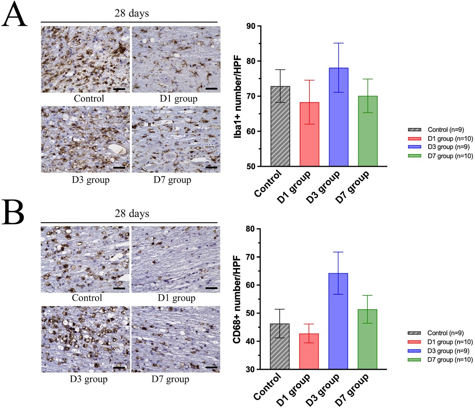
Background: Spinal cord injury results in profound neurological disability driven initially by primary mechanical damage and subsequently by secondary injury processes characterized by progressive neuroinflammation. Intravenous administration of human amniotic mesenchymal stem cells (MSC) has emerged as a promising therapeutic approach; however, the optimal timing of administration and its relationship to dynamic immune responses remain unclear. Methods: A rat contusion model of spinal cord injury was used to evaluate the effects of intravenous MSC administration at three post-injury time points: days 1, 3, and 7. Functional and histological assessments were performed for each group. Systemic inflammatory responses were evaluated through blood analysis of neutrophil and macrophage counts, systemic inflammation index (SII), and plasma proteomics. Local immune responses were assessed by quantifying infiltrating immune cells within the injured spinal cord. Results: The most substantial improvement in locomotor function was observed in the day-1 treatment group, followed by the day-7 group, whereas the day-3 group showed minimal benefit. The day-3 group also demonstrated a trend toward greater lesion length and increased macrophage infiltration 28 days after injury. MSC administration reduced SII in the day-1 and day-7 groups but not in the day-3 group, which instead showed an increased systemic inflammatory response. Analysis of spinal cord tissue demonstrated that MSC treatment on day-1 effectively reduced neutrophil infiltration, which peaks at this time point, while day-7 administration reduced macrophage infiltration during its peak phase. In contrast, MSC administration on day-3 failed to attenuate either neutrophil or macrophage accumulation. Plasma proteomic profiling revealed enhanced complement and coagulation pathway activation specifically on day-3. Conclusions: The therapeutic efficacy of intravenously administered MSC is highly dependent on the timing of intervention. Optimal benefit is achieved when treatment coincides with peak activation of a dominant target immune cell population and avoids the peak of complement and coagulation signaling. These findings support a phase-matched therapeutic strategy to maximize MSC effectiveness following spinal cord injury.

Read Full Abstract10.1186/s13287-026-05018-0
Mesenchymal stem cell-derived extracellular vesicles in the treatment of type 2 diabetes and its complications: current progress and future directionsGraphical AbstractVerified
Stem Cell Research & Therapy

Mesenchymal stem cell-derived extracellular vesicles in the treatment of type 2 diabetes and its complications: current progress and future directions

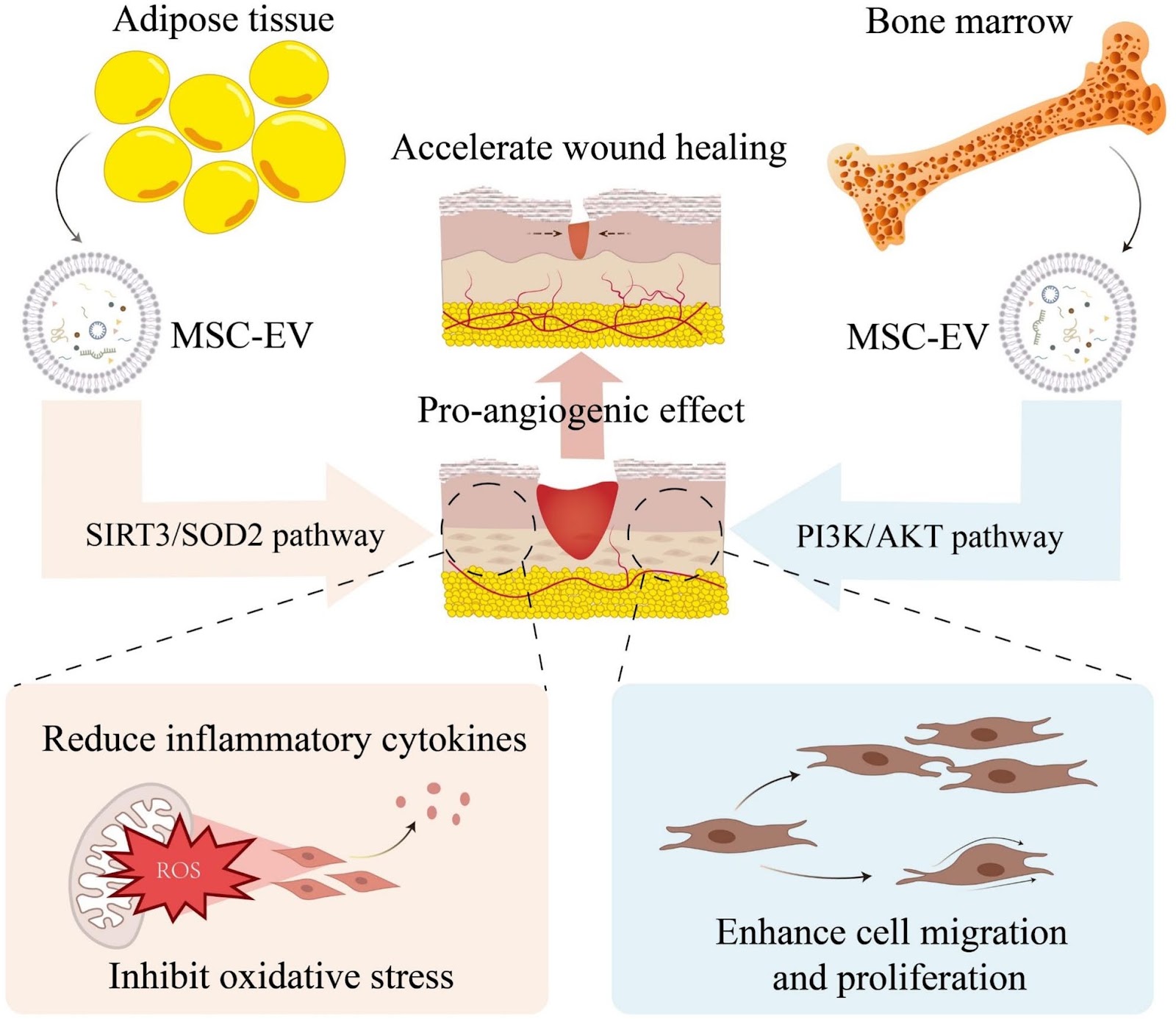
Type 2 diabetes (T2D) and its complications represent a complex disorder involving multiple pathophysiological processes. Although conventional therapeutic approaches partially regulate blood glucose, they fail to fundamentally reverse disease progression or effectively prevent complications. This review summarizes the current research advance and challenges of using different forms of mesenchymal stem cell-derived extracellular vesicles (MSC-EVs) in treating T2D and complications. It begins with an introduction to the characteristics of MSC-EVs. Subsequently, the mechanisms and therapeutic prospects of natural MSC-EVs are analyzed, with a focus on their roles in inflammatory modulation, tissue regeneration, and improving insulin resistance. Engineering MSC-EVs, covering strategies including optimizing MSC culture conditions, modifying EV contents, and establishing MSC-EV delivery systems based on bioactive materials are then discussed, which boost EV yield and quality while enhancing therapeutic efficacy. Current challenges, including the limited yield and high heterogeneity of natural MSC-EVs, as well as issues related to long-term safety, immunocompatibility, and large-scale production of engineered MSC-EVs are finally overviewed, with emphasizing artificial intelligence in guiding future research directions. These summaries are crucial for clinical translation of MSC-EVs and will ultimately provide T2D patients with an effective and safe treatment option.

Read Full Abstract10.1186/s13287-026-04991-w
DMOG pretreatment restores osteogenic–adipogenic balance and mitochondrial function in ONFH BMSCs through the HIF-1α/Homer3 pathwayGraphical AbstractVerified
Stem Cell Research & Therapy

DMOG pretreatment restores osteogenic–adipogenic balance and mitochondrial function in ONFH BMSCs through the HIF-1α/Homer3 pathway

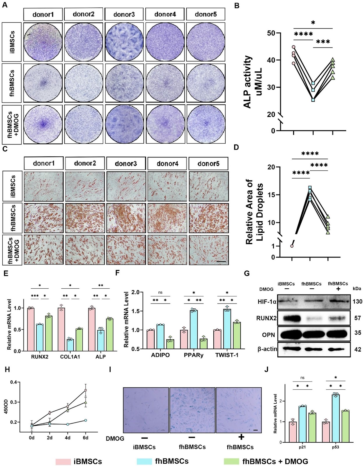
Background  Osteonecrosis of the femoral head (ONFH) is a progressive orthopedic disorder that often culminates in femoral head collapse and joint failure. Dysfunction of bone marrow mesenchymal stem cells (BMSCs), including impaired osteogenesis, enhanced adipogenesis, and mitochondrial dysfunction, has been increasingly recognized as a central driver of ONFH pathogenesis. However, the molecular mechanisms linking metabolic stress to lineage imbalance remain poorly defined. Methods  Paired BMSCs were isolated from necrotic femoral head regions (fhBMSCs) and the iliac crest (iBMSCs) of ONFH patients. Functional assays, RNA sequencing, and molecular analyses were performed to evaluate the effects of the hypoxia mimetic dimethyloxalylglycine (DMOG) on osteogenic–adipogenic balance, mitochondrial function, and senescence. Loss-of-function experiments targeting hypoxia-inducible factor-1α (HIF-1α) and Homer3 were conducted to elucidate mechanistic pathways. Results  Compared with iBMSCs, fhBMSCs exhibited impaired osteogenesis, enhanced adipogenesis, mitochondrial dysfunction, and increased senescence. DMOG pretreatment restored osteogenic differentiation, suppressed adipogenesis, improved mitochondrial dynamics, reduced oxidative stress, and enhanced bioenergetic metabolism. These protective effects were dependent on HIF-1α stabilization. Transcriptomic profiling identified Homer3 as a downstream negative regulator of HIF-1α. Homer3 was aberrantly upregulated in fhBMSCs but suppressed by DMOG, and its knockdown mimicked the effects of DMOG by promoting osteogenesis, inhibiting adipogenesis, enhancing mitophagy, and restoring mitochondrial function. Conversely, silencing HIF-1α abolished DMOG-mediated benefits and reinstated Homer3 expression.

Read Full Abstract10.1186/s13287-026-05026-0
OSBPL2-Mediated Lipid Metabolism Alteration Governs Lung Cancer Stem Cells PropertiesGraphical AbstractVerified
Stem Cell Research & Therapy2026

OSBPL2-Mediated Lipid Metabolism Alteration Governs Lung Cancer Stem Cells Properties

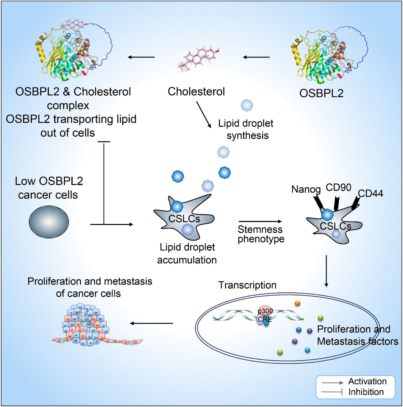
Lung cancer is the first leading cause of cancer death worldwide. Oxysterol-binding protein-like 2 (OSBPL2) is a lipid transport protein regulating cholesterol homeostasis. Here, we clarified the previously unreported role of OSBPL2 in lung cancer stemness properties. We observed that OSBPL2 reduced cholesterol content by HPLC-MS. It inhibited the accumulation of lipid droplets (LDs) in lung cancer. OSBPL2-mediated lipid transportation significantly suppressed tumor sphere formation, stemness markers expression and in vivo tumorigenesis and tumor metastasis. In clinical specimens, we also demonstrated that OSBPL2 repressed the expression of Lung cancer stem-like cells (LCSCs) markers-ALDH1A1, CD133 and Nanog. The level of OSBPL2 was negatively correlated with malignant of lung cancer, such as tumor stage progression and lymph node metastasis. Taken together, these findings illustrated that OSBPL2-mediated lipid transportation inhibited the stemness and aggressiveness of lung cancer cells. OSBPL2 was a potential therapeutic target to develop novel cancer-preventive compound.

Read Full Abstract10.1186/s13287-026-04919-4
Extracorporeal cardiac shock wave stimulation enhances the therapeutic efficacy of intravenously delivered endothelial colony-forming cells via PI3K/AKT signaling in a rat myocardial infarction modelGraphical AbstractVerified
Stem Cell Research & Therapy

Extracorporeal cardiac shock wave stimulation enhances the therapeutic efficacy of intravenously delivered endothelial colony-forming cells via PI3K/AKT signaling in a rat myocardial infarction model

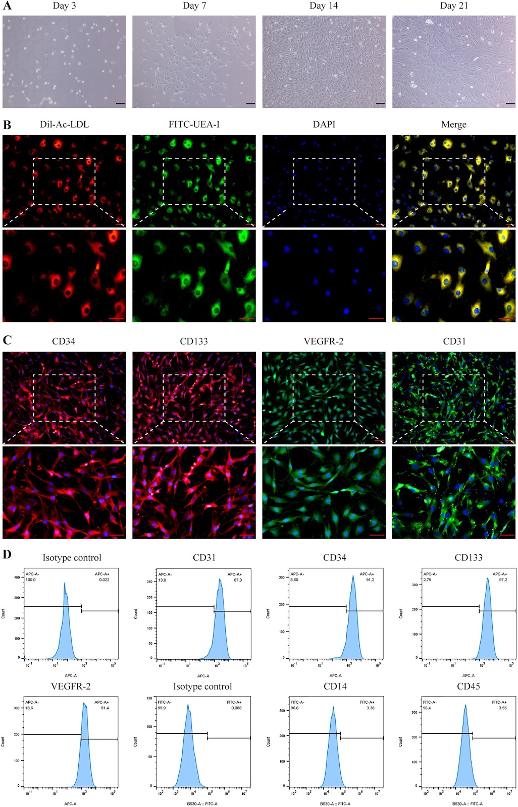
Background Extracorporeal cardiac shock wave (ECSW) therapy enhances the function of endothelial colony-forming cells (ECFCs), but whether it can serve as a preconditioning strategy to enhance myocardial infarction (MI) therapy remains unclear. This study investigated the efficacy and mechanism of intravenously delivered ECSW-preconditioned ECFCs (SW-ECFCs) in a rat MI model. Methods ECFCs were isolated from the bone marrow of ApoE-/- rats and fully characterized. RNA sequencing of control ECFCs versus SW-ECFCs revealed significant enrichment of the PI3K/AKT pathway. We therefore performed a series of in vitro functional assays on these cells, including Transwell migration, Matrigel tube formation, CCK-8 proliferation, flow cytometric apoptosis analysis, and VEGF-A ELISA. The role of the PI3K/AKT pathway was interrogated using the inhibitor LY294002. Subsequently, an acute MI model was established in ApoE-/- rats via left anterior descending coronary artery ligation. Rats were randomized into four groups: MI+PBS, MI+ECFCs, MI+SW-ECFCs, and MI+LY294002-pretreated SW-ECFCs (LY-SW-ECFCs), with sham-operated rats as controls. Comprehensive evaluations included echocardiography, serum injury biomarkers, TTC, and histopathological (H&E, Masson) staining, immunohistochemical detection of cardiomyocyte apoptosis and p-eNOS, immunofluorescence assessment of ECFC homing and vascular markers (CD31, α-SMA, VEGF-A), tissue/plasma nitric oxide measurement, and Western blot analysis of PI3K/AKT signaling proteins. Results Transcriptomic analysis revealed significant enrichment of the PI3K/AKT pathway in SW-ECFCs. Functionally, ECSW enhanced ECFCs migration, tube formation, proliferation, and VEGF-A secretion, while reducing apoptosis;

Read Full Abstract10.1186/s13287-026-04913-w
An open phase I/IIa study evaluating safety, patient-reported outcomes and voice function after surgery, local administration of mesenchymal stromal cells and voice training in patients with vocal fold scarring and dysphoniaGraphical AbstractVerified
Stem Cell Research & Therapy

An open phase I/IIa study evaluating safety, patient-reported outcomes and voice function after surgery, local administration of mesenchymal stromal cells and voice training in patients with vocal fold scarring and dysphonia

Background Damage to the vocal folds can result in scarring, leading to chronic, severe voice impairments for which lasting and effective treatments are currently lacking. The aim of this clinical trial was to evaluate the safety and effectiveness of autologous bone marrow-derived Mesenchymal Stromal Cell (MSC) therapy for patients with vocal fold scarring and severe dysphonia. Additionally, the study sought to propose a post-operative voice training protocol and explore its potential role in facilitating voice improvement. Methods Eight patients with vocal fold scarring and chronic dysphonia underwent surgical scar resection and autologous MSC injection, followed by voice training. Safety was continuously monitored for up to 36 months postoperatively. Data to evaluate therapeutic efficacy was collected pre-treatment, 3 and 12 months post-treatment. Assessments included analysis of vocal fold vibrations, Phonation Threshold Pressure, and Maximum Phonation Time. Patient-reported measures were collected using the Voice Handicap Index, the Vocal Fatigue Index, and ratings of major symptoms and their impact on daily life. Treatment effectiveness was analyzed at both group and individual levels, with clinically relevant changes predefined.

Read Full Abstract10.1186/s13287-026-05022-4
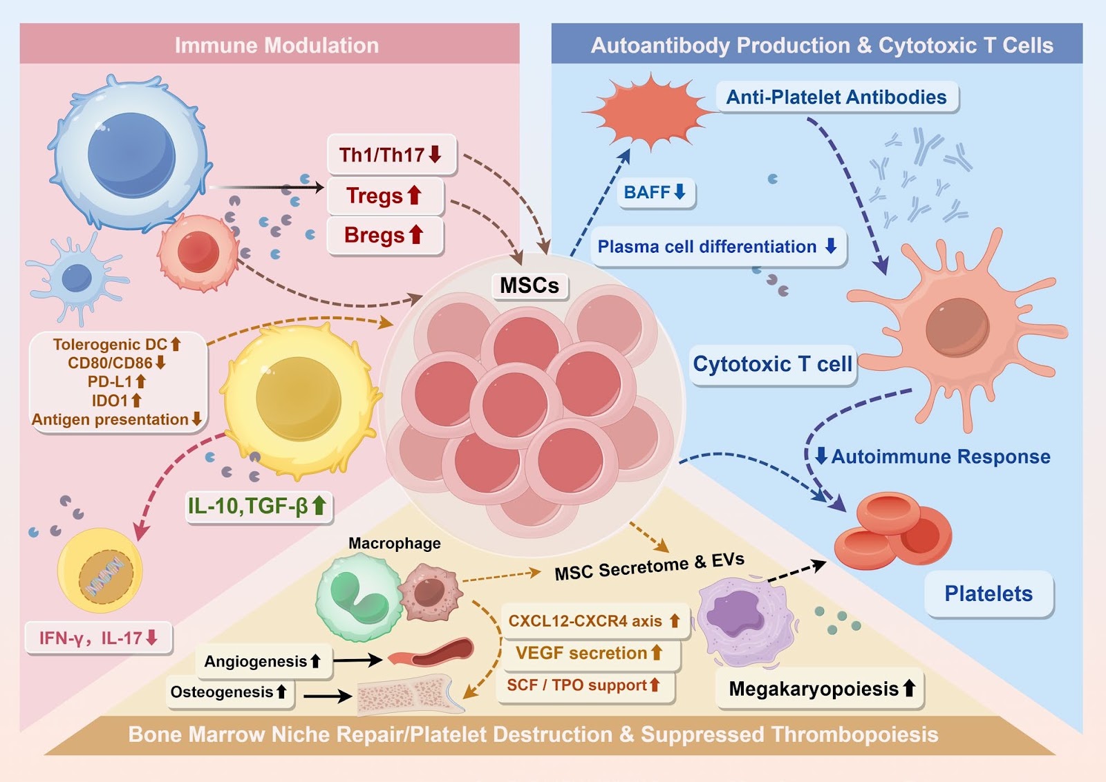
Innovative strategies for immune thrombocytopenia treatment: immunomodulatory mechanisms and clinical potential of mesenchymal stem cellsGraphical AbstractVerified
Stem Cell Research & Therapy

Innovative strategies for immune thrombocytopenia treatment: immunomodulatory mechanisms and clinical potential of mesenchymal stem cells

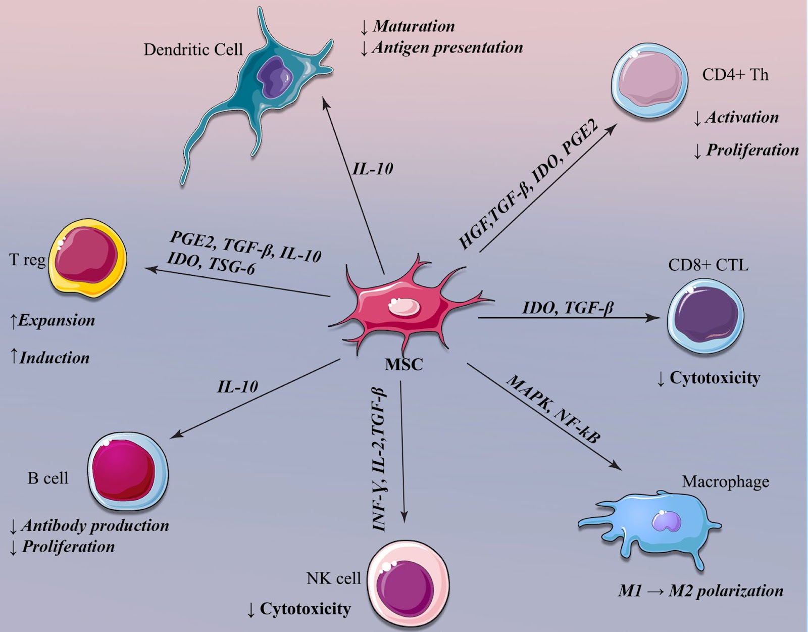
Immune thrombocytopenia (ITP) is a heterogeneous autoimmune disorder characterized by increased platelet destruction and impaired megakaryopoiesis within a dysregulated bone marrow niche. Conventional therapies often achieve only transient platelet recovery, failing to restore immune tolerance, thereby underscoring the need for mechanism-based therapeutic strategies. Mesenchymal stem cells (MSCs) have emerged as promising candidates due to their ability to modulate immune responses and repair the hematopoietic microenvironment. This review synthesizes current evidence regarding the biological properties, immunomodulatory mechanisms, and therapeutic applications of MSCs in ITP, emphasizing intrinsic abnormalities of patient-derived MSCs and the corrective potential of exogenous MSCs from distinct tissue sources. It further integrates emerging insights into MSC functional heterogeneity, optimization of culture conditions, priming strategies, and cellular engineering approaches that may enhance therapeutic efficacy and safety. By highlighting the interplay between immune tolerance restoration and bone marrow niche remodeling, this review provides a translational framework that links mechanistic understanding to the future clinical development of MSC-based therapies for ITP.

Read Full Abstract10.1186/s13287-026-05000-w