🧬 SinoBioData Academic Portal
šŸ“š Peer-Reviewed Translated Literature

All Biomedical & Clinical Articles (Page 35)

Browse complete peer-reviewed translations from top Chinese biomedical, oncology, and genomics journals. Read verified previews and download full authentic clinical reports.

Published Research Papers

Showing 24 of 1542 peer-reviewed translated articles (Page 35 of 65)

FHOD3 deficiency disrupts sarcomere organization and activates CaMKII signaling in human stem cell-derived cardiomyocytesGraphical AbstractVerified
Stem Cell Research & Therapy

FHOD3 deficiency disrupts sarcomere organization and activates CaMKII signaling in human stem cell-derived cardiomyocytes

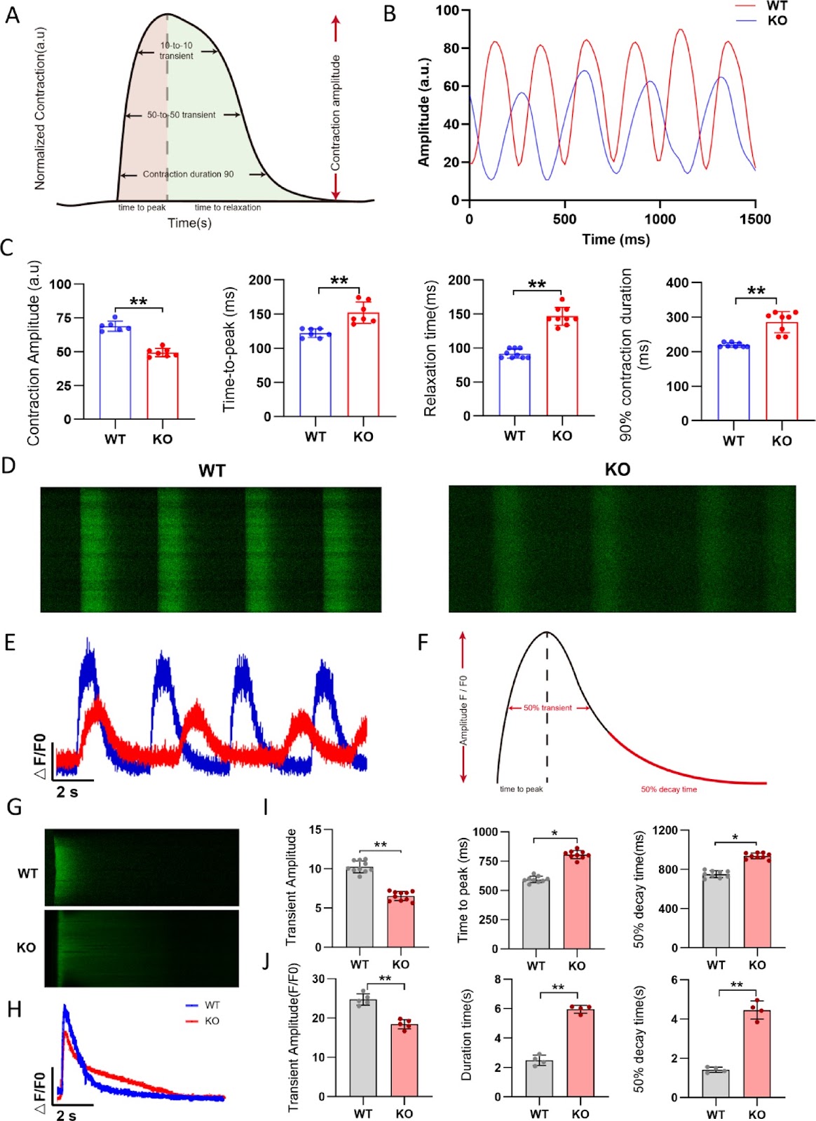
Background Inherited cardiomyopathy (ICM) is a genetic disorder characterized by abnormal myocardial structure and function, often progressing to heart failure. FHOD3, a member of the Formin gene family, plays a crucial role in cardiomyocyte cytoskeletal organization. Mutations in FHOD3 have been associated with various cardiomyopathies, including hypertrophic cardiomyopathy (HCM), dilated cardiomyopathy (DCM) and left ventricular noncompaction (LVNC). However, the molecular mechanisms underlying FHOD3 deficiency-induced cardiomyopathy remain elusive. Methods A FHOD3 knockout (FHOD3-/-) human embryonic stem cell (hESC) line was generated using the CRISPR/Cas9 system and subsequently differentiated into cardiomyocytes (hESC-CMs). Sarcomere structure, calcium handling, mitochondrial function, and contractility were evaluated via immunofluorescence, electron microscopy, Seahorse metabolic analysis, and high-definition video analysis, respectively. Transcriptomic sequencing was performed to identify differentially expressed genes and enriched pathways. Results FHOD3-deficient hESC-CMs exhibited marked sarcomere disorganization and degradation, impaired calcium handling and compromised mitochondrial function, ultimately leading to reduced contractility. Transcriptomic analysis revealed significant downregulation of sarcomere-related genes and calcium-handling genes, with enrichment in pathways associated with cardiomyopathy and calcium signaling. Furthermore, FHOD3 deficiency triggered the phosphorylation of CaMKII (Thr286), a key regulator of cardiac hypertrophy and remodeling, contributing to the progression of heart failure. Treatment with the myosin activator Omecamtiv mecarbil (OM) partially restored contractility without affecting calcium handling, highlighting its potential as a therapeutic strategy.
Read Full Abstract10.1186/s13287-026-04902-z
A review of the circadian regulation of stem cells: harnessing the internal body clock for enhanced regenerative therapiesGraphical AbstractVerified
Stem Cell Research & Therapy

A review of the circadian regulation of stem cells: harnessing the internal body clock for enhanced regenerative therapies

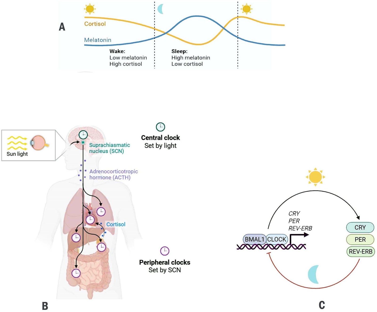
Background Circadian rhythms are endogenous, transcription-translation feedback loops that align cellular activities with the 24-h light–dark cycle. Stem-cell populations across tissues exhibit circadian oscillations that influence their self-renewal, proliferation, and differentiation. Key developmental pathways (Wnt/β-catenin, Notch, and Hedgehog) are increasingly recognized as both regulators and targets of circadian machinery. Objectives This review synthesizes current knowledge on the bidirectional crosstalk between circadian clock components and major stem-cell regulatory pathways, and evaluates how this interplay shapes tissue homeostasis, regenerative capacity, and therapeutic potential. Methods Literature examining molecular interfaces between circadian clock genes and Wnt, Notch, and Hedgehog signaling was surveyed, with emphasis on transcriptional regulation, chromatin dynamics, post-translational control, and functional outcomes for stem-cell behavior and regeneration. Results Evidence indicates that core clock components modulate stem-cell pathways through direct transcriptional control, shared enhancer architecture, altered chromatin accessibility, and rhythmic protein modification. In turn, Wnt, Notch, and Hedgehog signals feed back onto clock genes, influencing circadian amplitude and phase within stem-cell niches. Perturbation of this reciprocal regulation disrupts tissue maintenance, diminishes regenerative responses, alters metabolic equilibrium, and may promote tumorigenesis. Conclusions Circadian oscillators act as temporal gatekeepers of stem-cell function. Mapping the molecular interfaces between clock genes and developmental signaling pathways reveals new opportunities to refine regenerative therapies. Chronotherapeutic strategies, i.e. timing interventions to intrinsic circadian phases may enhance the efficacy, precision, and safety of stem-cell–based treatments.

Read Full Abstract10.1186/s13287-026-04979-6
Phenotypic alterations and PI3K-AKT pathway regulation in senescence of human tonsil mesenchymal stem cellsGraphical AbstractVerified
Stem Cell Research & Therapy2026

Phenotypic alterations and PI3K-AKT pathway regulation in senescence of human tonsil mesenchymal stem cells

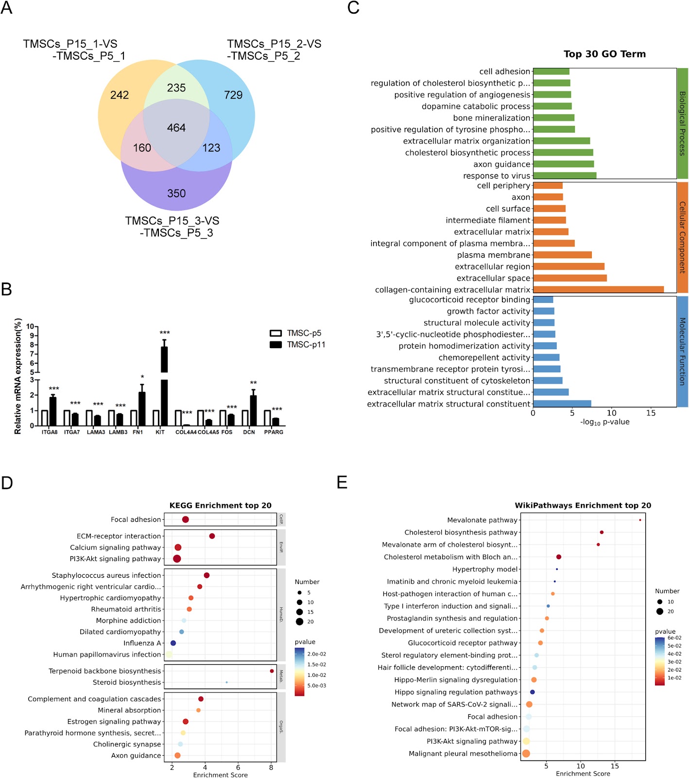
Background: Tonsil mesenchymal stem cells (TMSCs) are a promising regenerative medicine source but require continuous subculturing for expansion. Long-term expansion in vitro induces cellular senescence, impairing their function. This study aimed to elucidate senescence-related phenotypic alterations and regulatory mechanisms in human tonsil-derived mesenchymal stem cells. Methods: Human-derived TMSCs were isolated from palatine tonsils, cultured under standard conditions, and characterized for mesenchymal markers. Senescence-associated changes were evaluated across early (P1–P5) and late passages (beyond P10). Proliferation capacity was assessed via CCK-8 assays, while senescence-associated β-galactosidase (SA-β-gal) activity and protein levels of p16, p53, and p21 were quantified. RNA sequencing identified differentially expressed genes (DEGs) between young and senescent TMSCs, followed by KEGG pathway enrichment analysis. Key findings were validated by measuring the p-Akt/Akt ratio via Western blot. Results: TMSCs showed a progressive decline in proliferative capacity with increasing passages. SA-β-gal staining revealed a significantly higher percentage of positive cells in late-passage TMSCs compared to early-passage cells. Expression levels of P16, P53, and P21 proteins were markedly upregulated in aged TMSCs. KEGG analysis of DEGs indicated significant enrichment in the PI3K-Akt signaling pathway, ECM-receptor interaction, and calcium signaling. Consistent with this, Western blot confirmed a significantly increased p-Akt/Akt ratio in senescent TMSCs. Conclusion: Our research proved that replicative senescence in TMSCs is associated with PI3K-Akt pathway activation, which likely orchestrates senescence via p16 and p53-p21 cascades. These findings provide new insights into the mechanisms of stem cell aging and suggest potential molecular targets for developing strategies to delay senescence in TMSCs for regenerative medicine.

Read Full Abstract10.1186/s13287-026-04986-7
Efficacy of multi-layered human iPS cell-derived cardiovascular cell sheets in a pacing-induced canine dilated cardiomyopathy modelGraphical AbstractVerified
Stem Cell Research & Therapy

Efficacy of multi-layered human iPS cell-derived cardiovascular cell sheets in a pacing-induced canine dilated cardiomyopathy model

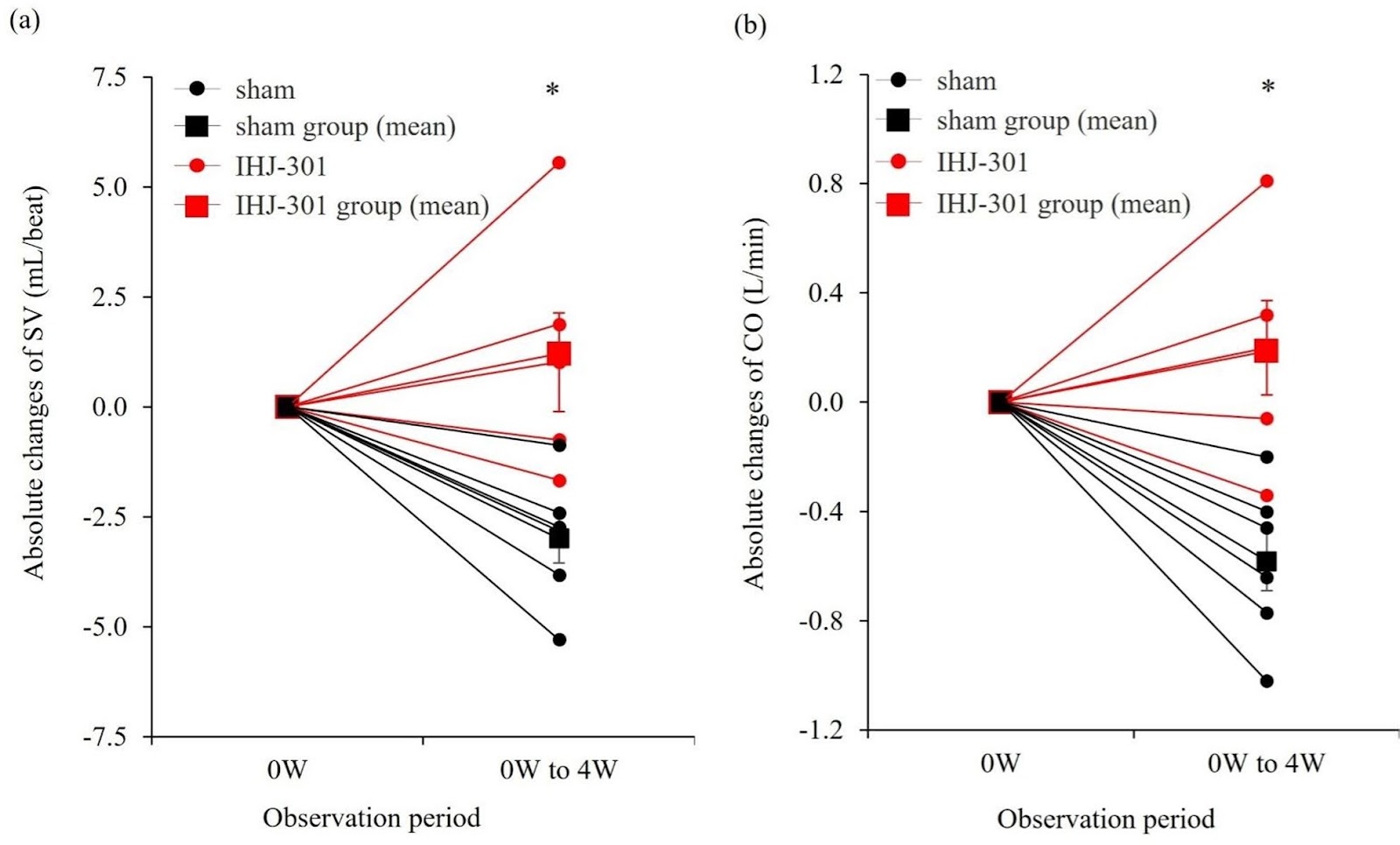
Background Dilated cardiomyopathy (DCM) is a progressive, intractable disease that leads to heart failure. Heart transplantation is the only curative treatment; however, access is limited by donor scarcity. Induced pluripotent stem cell (iPSC)-based therapies are attracting attention for DCM, but suitable large-animal models and robust preclinical data have been limited. Methods We generated multi-layered cardiovascular cell sheets from human iPSCs by combining cardiomyocytes with endothelial and stromal cells and overcoming stacking limits using interleaved gelatin hydrogel microspheres, yielding a thicker cardiac tissue-like construct (product code: IHJ-301). To enable rigorous testing in non-ischemic heart failure, we established a modified canine rapid-pacing heart failure model that maintains depressed function without mortality by continuing pacing at a slightly reduced rate after induction (Step-Down Pacing Heart Failure model). IHJ-301 was implanted epicardially onto the left ventricular surface via thoracotomy, and cardiac function was assessed by echocardiography and right-heart catheterization. Results After 4 weeks of rapid pacing (230±10 bpm), left ventricular ejection fraction (LVEF) was reduced from 77.8±1.1% (pre-pacing) to 44.9±1.9% (n=11) (0 W). Continued pacing at 210±10 bpm for additional 4 weeks resulted in no mortality and maintained depressed function (4 W LVEF 47.3±2.6%). IHJ-301 was implanted at 0 W. At 4 weeks post-implantation (4 W), all animals in the IHJ-301 group (n=5) showed greater functional improvement than sham (n=6). Absolute changes from 0 W to 4 W were: Ī”LVEF (%) 9.38±1.47 vs. 1.90±0.34; Ī”fractional shortening (%) 4.84±0.75 vs. 0.97±0.18; stroke volume (mL/beat) 1.21±1.26 vs. āˆ’2.99±0.60; cardiac output (L/min) 0.19±0.19 vs. āˆ’0.58±0.12 (all p<0.05). Conclusions We established a non-ischemic large-animal heart failure model that sustains depressed function for one month, enabling clear therapeutic readouts. IHJ-301 significantly improved multiple parameters of cardiac function, providing preclinical evidence that IHJ-301 could offer a promising therapeutic option for DCM.

Read Full Abstract10.1186/s13287-026-05207-x
Research based on serine metabolism indicates mesenchymal stem cells alleviate psoriasis by regulating the PSPH-PINK1-Parkin-NLRP3 pathway in HaCaTGraphical AbstractVerified
Stem Cell Research & Therapy

Research based on serine metabolism indicates mesenchymal stem cells alleviate psoriasis by regulating the PSPH-PINK1-Parkin-NLRP3 pathway in HaCaT

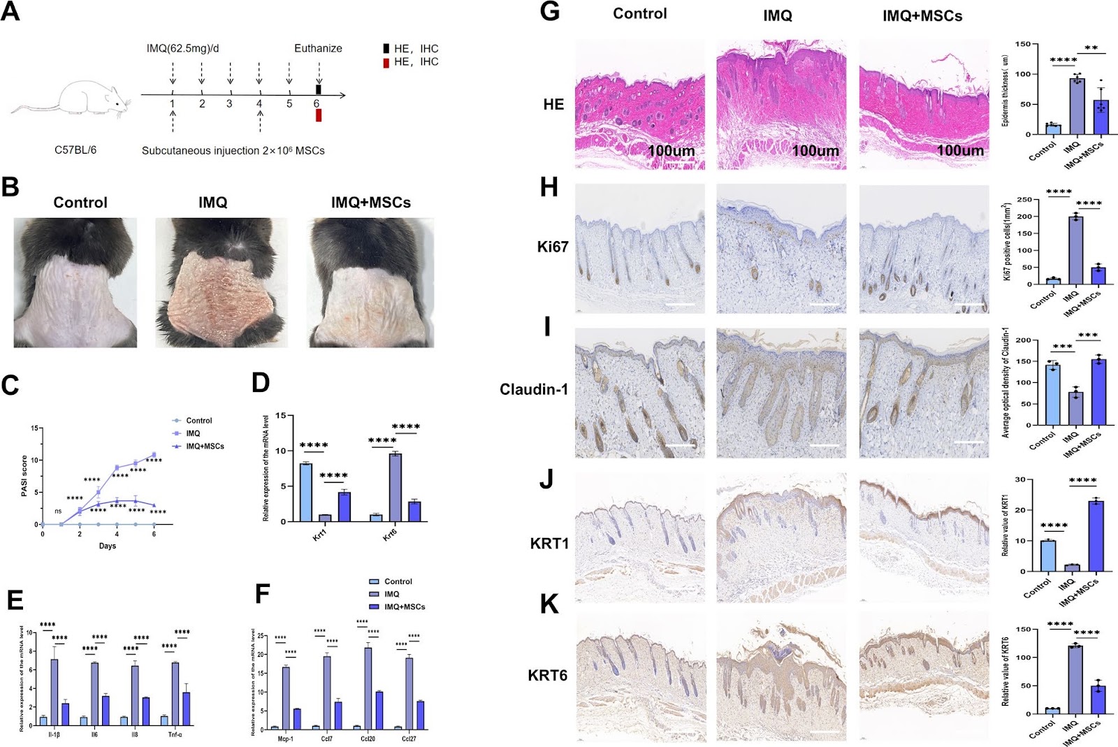
Background Psoriasis is a refractory immune-related disease. In recent years, it has been discovered that mesenchymal stem cells (MSCs) can be used as a new therapeutic approach for psoriasis, but their potential therapeutic mechanism remains unclear. This study aims to explore the role of MSCs in the treatment of psoriasis. Methods We employed a mouse psoriasis model induced by imiquimod (IMQ) in vivo and a co-culture system of MSCs and HaCaT keratinocytes (KCs) cell line in vitro. These approaches allowed us to investigate the effect of MSCs on the levels of inflammatory factors and the activation of inflammasomes in both contexts. Mouse-targeted amino acid sequencing, transmission electron microscopy for in vitro observation, immunofluorescence for both in vivo and in vitro analyses, and siRNA transfection in vitro were employed in this study. Results Our results showed that MSCs significantly improved the skin lesion of mice with psoriasis, and reduced the levels of inflammatory factors and chemokines including IL-1β, IL-6, IL-8, TNF-α, MCP-1, CCL7, CCL20 and CCL27 in the mouse skin lesion areas and M5- induced psoriatic KCs models in vitro. Likewise, MSCs repaired the skin barrier by enhancing claudin-1 expression in vivo. In addition, MSCs increased KRT1 and decreased KRT6 levels in vivo and in vitro. Amino acid metabolism analysis showed that MSCs could improve the serine metabolism level in the mouse skins and upregulated the key enzyme phosphoserine phosphatase (PSPH) in serine metabolism. In vitro experiments demonstrated that knockdown of PSPH could reverse the therapeutic effects of MSCs on psoriasis. Furthermore, studies in vitro and in vivo revealed that MSCs can activate the PINK1-Parkin pathway. It was specifically manifested by elevated levels of PINK1, Parkin, p-Parkin, Beclin-1, and LC3B-II/I, coupled with a reduction in P62 protein. Subsequently, the activation of PINK1-Parkin led to decreased expressions of IL-1β, IL-6, IL-8, TNF-α, CCL7, CCL20, CCL27, and MCP-1. In vitro and in vivo experiments indicated that MSCs can reduce the levels of IL-1β, IL-6, IL-8, TNF-α, CCL7, CCL20, CCL27, and MCP-1 by inhibiting the activation of NLRP3 inflammasomes. Meanwhile, PSPH knockdown in vitro can reverse the activating effects of MSCs on the PINK1-Parkin, as shown by decreased levels of PINK, Parkin, p-Parkin, Beclin-1, and LC3B-II/I, concurrently with an elevation in P62. Conclusions The results of this study indicated that MSCs can alleviate IMQ-induced psoriasiform dermatitis in mice by upregulating serine metabolism. The key serine metabolism enzyme PSPH may enhance PINK1/Parkin-mediated mitochondrial autophagy in psoriatic HaCaT and inhibit NLRP3 inflammasome activation in HaCaT cells, thereby alleviating skin inflammatory responses and suppressing skin proliferation in psoriatic mice.

Read Full Abstract10.1186/s13287-026-04964-z
Correction: Development of a robust induced pluripotent stem cell atrial cardiomyocyte differentiation protocol to model atrial arrhythmiaGraphical AbstractVerified
Stem Cell Research & Therapy

Correction: Development of a robust induced pluripotent stem cell atrial cardiomyocyte differentiation protocol to model atrial arrhythmia

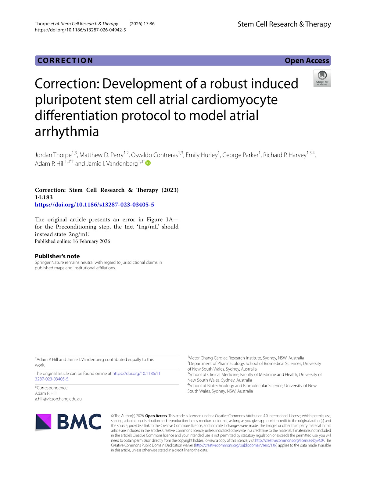
The original article presents an error in Figure 1A—for the Preconditioning step, the text ā€˜1ng/mL’ should instead state ā€˜2ng/mL’.

Read Full Abstract10.1186/s13287-026-04942-5
Mesenchymal stromal cells alleviate pulmonary arterial hypertension by suppressing pulmonary arterial adventitial fibroblast activation and extracellular matrix remodeling via the SOCS3/STAT3 pathwayGraphical AbstractVerified
Stem Cell Research & Therapy

Mesenchymal stromal cells alleviate pulmonary arterial hypertension by suppressing pulmonary arterial adventitial fibroblast activation and extracellular matrix remodeling via the SOCS3/STAT3 pathway

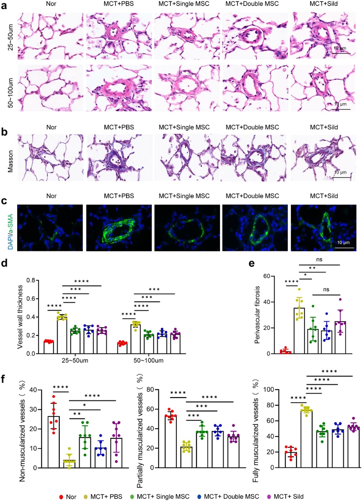
Background Pulmonary arterial hypertension (PAH) is a fatal condition characterized by progressive vascular remodeling in the pulmonary arteries, eventually leading to right heart failure and death. Dysregulated extracellular matrix (ECM) remodeling is central to PAH pathogenesis and represents a potential therapeutic target. Mesenchymal stromal cells (MSCs) have shown promise in preclinical studies; however, the optimal therapeutic window, dosing frequency, and mechanistic basis for their regulation of vascular ECM remain unclear. Methods We employed a monocrotaline (MCT)-induced rat model of PAH to evaluate different MSC treatment regimens, including early administration (day 1 post-MCT), delayed administration (days 7 and 14), and repeated dosing (days 1 and 11). Additionally, we combined in vivo and in vitro approaches to investigate how MSCs modulate the activation of pulmonary arterial adventitial fibroblasts (PAAFs) and influence ECM remodeling. Results Biodistribution studies indicated that MSC retention in lung tissue peaked within 24 h and gradually declined by day 21. A single early dose of MSCs (on day 1) significantly ameliorated PAH progression, increasing the 28-day survival rate, reducing right ventricular systolic pressure (RVSP), improving right ventricular function, and attenuating small pulmonary vascular remodeling, including reductions in medial thickening, excessive muscularization, and collagen deposition. Repeated MSC administration did not provide additional therapeutic benefit. Both in animal models and cell cultures, MSCs effectively suppressed PAAF activation and reduced ECM protein production. This anti-fibrotic effect was mediated via the SOCS3/STAT3 signaling pathway. Conclusions Early single-dose MSC therapy effectively alleviates PAH by suppressing PAAF activation and ECM remodeling through the SOCS3/STAT3 pathway, offering a potential therapeutic strategy for PAH.

Read Full Abstract10.1186/s13287-025-04883-5
Autologous bone marrow mesenchymal stem cell mitochondrial transplantation in recurrent assisted reproductive technology failure: a randomized controlled trialGraphical AbstractVerified
Stem Cell Research & Therapy

Autologous bone marrow mesenchymal stem cell mitochondrial transplantation in recurrent assisted reproductive technology failure: a randomized controlled trial

Background Mitochondrial dysfunction contributes to poor embryo quality and recurrent assisted reproductive technology (ART) failure. Mitochondrial transplantation (MIT), which involves supplementing oocytes with exogenous mitochondria, has been proposed as a novel strategy to improve ART outcomes. However, both its clinical efficacy and safety remain unclear. Methods In this single-center trial, 151 patients with a history of ≄ 2 failed ART cycles provided 1178 metaphase II (MII) oocytes. Sibling oocytes were randomized 1:1 to receive autologous bone marrow mesenchymal stem cells (BMSCs) mitochondria co-injection during intracytoplasmic sperm injection (ICSI) or standard ICSI. The primary outcome was the rate of day-3 good-quality embryos. Results MIT significantly accelerated early embryonic cleavage at the 3-cell stage and 5-cell stage, but this morphokinetic alteration did not translate into improvements in good-quality embryo rate, clinical pregnancy rate, or live birth rate. Long-term follow-up of 23 live births revealed no adverse effects, with all offspring exhibiting normal growth and development. Exploratory analysis revealed that oocytes yielding ≄ 70% transferable embryos after MIT harbored an elevated higher burden of medium frequency (0.05–0.5) mtDNA point mutations. Conclusions While autologous BMSCs-MIT transiently alters early cleavage kinetics, it does not demonstrate a clinical advantage in unselected patients with recurrent ART failure. Nevertheless, its observed safety profile and the identification of mtDNA mutation burden as a potential predictive biomarker provide a foundation for shifting future MIT research from a universal approach toward precision application in molecularly stratified populations.

Read Full Abstract10.1186/s13287-026-05059-5
Spatiotemporal single-cell atlas of suture stem cell dynamics in craniosynostosisGraphical AbstractVerified
Stem Cell Research & Therapy2026

Spatiotemporal single-cell atlas of suture stem cell dynamics in craniosynostosis

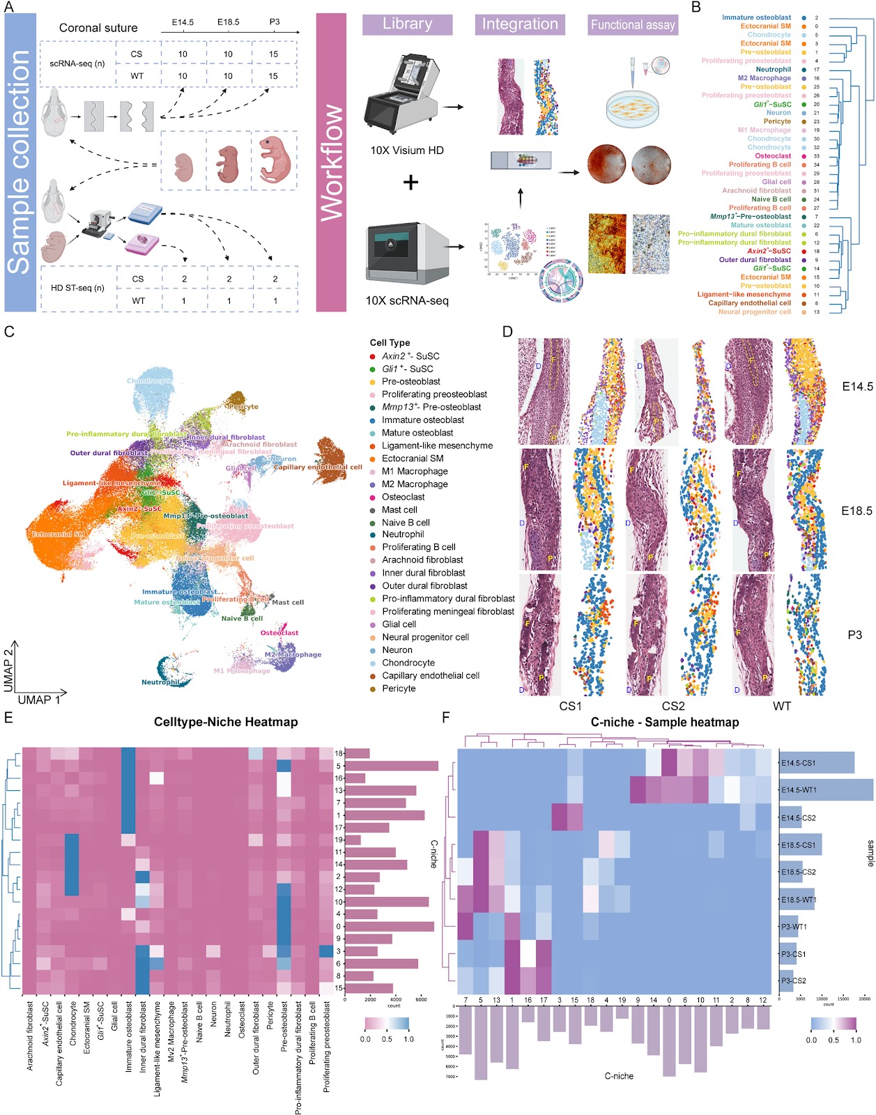
Background: Craniosynostosis is a congenital disorder characterized by premature suture fusion and aberrant skull morphogenesis. The cellular dynamics and regulatory mechanisms of suture mesenchymal stem cells (SuSCs) in this disease remain poorly defined. Methods: We integrated single-cell RNA sequencing and 2-μm-resolution Visium HD spatial transcriptomics to build a spatiotemporal atlas of coronal suture cells in Fgfr2C342Y/+ mice, a murine model recapitulating human Crouzon syndrome, alongside wild-type controls across three key developmental stages (E14.5, E18.5, and P3). To obtain near single-cell spatial resolution, we created SpatialCell, which combines morphology-based segmentation and machine-learning classification using a reference trained on our single-cell datasets. Results: The atlas reveals stage-specific remodeling of SuSC niches and a shift of SuSC spatial associations toward osteogenic mesenchyme in craniosynostosis. Along the SuSC-to-osteoblast trajectory, pre-osteoblasts were depleted earlier than upstream SuSCs, and SuSCs displayed premature acquisition of osteogenic programs near the suture midline. Temporal Gene Ontology patterns indicated early extracellular-matrix disruption, mid-gestation chondrogenic activation, and postnatal mineralization. Network analysis nominated Foxa3 as a candidate regulator in SuSC subsets; siRNA knockdown of Foxa3 reduced ex vivo mineralization in the craniosynostosis background. Spatial communication analyses implicated signals from suture meningeal fibroblasts and immune cells that converge on SuSC fate. Conclusions: Our results support a model where craniosynostosis may involve disrupted temporal coordination of developmental programs, not merely accelerated bone formation. The atlas and analytic framework pinpoint when and where SuSC fate diverges, propose Foxa3 as an intervention target, and provide a high-resolution resource for mechanistic and therapeutic exploration.

Read Full Abstract10.1186/s13287-026-04987-6
Intervertebral disc progenitor cells: roles in regeneration and diseaseGraphical AbstractVerified
Stem Cell Research & Therapy2026

Intervertebral disc progenitor cells: roles in regeneration and disease

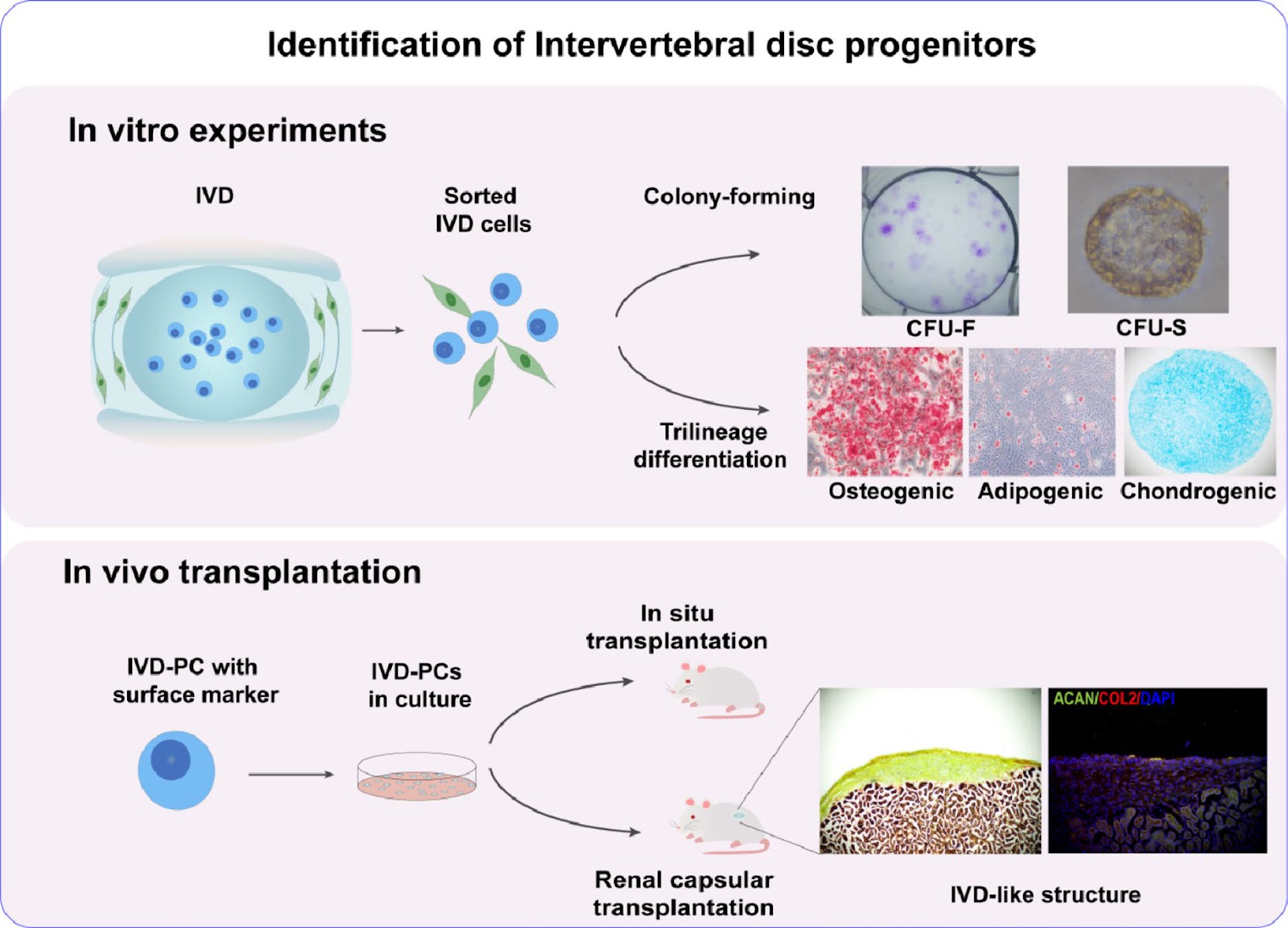
Intervertebral disc (IVD) degenerative disease is a prevalent and debilitating spinal disease. Current treatments only focus on symptomatic relief but fail to halt disease progression or restore the native biomechanical function of the spine. Regenerative medicine strategies, particularly those harnessing endogenous progenitor cells, offer a promising avenue for achieving biological repair and functional homeostasis. The identification of intervertebral disc progenitor cells (IVD-PCs) has unveiled a potential cellular reservoir for self-repair, given their demonstrated stemness attributes, including clonogenicity and multipotent differentiation. However, the clinical translation of IVD-PCs is significantly hampered by an incomplete understanding of their inherent heterogeneity, hierarchical organization, and, most critically, the dynamic interplay with their unique microenvironment, which dictates their fate decisions. This review synthesizes recent advances in deciphering the molecular signatures and functional plasticity of IVD-PCs. We place a particular emphasis on how key physicochemical, mechanical, and cellular cues within the IVD niche orchestrate progenitor cell behavior—ranging from maintenance and activation to aberrant differentiation—during both homeostasis and degeneration. Furthermore, we propose forward-looking insights to bridge critical knowledge gaps, aiming to propel the development of novel progenitor cell-based therapeutics for IVD degeneration.

Read Full Abstract10.1186/s13287-026-04918-5
ATG5 overexpression enhances the therapeutic efficacy of mesenchymal stem cells in a mouse colitis model by augmenting anti-inflammatory and antioxidative mechanismsGraphical AbstractVerified
Stem Cell Research & Therapy

ATG5 overexpression enhances the therapeutic efficacy of mesenchymal stem cells in a mouse colitis model by augmenting anti-inflammatory and antioxidative mechanisms

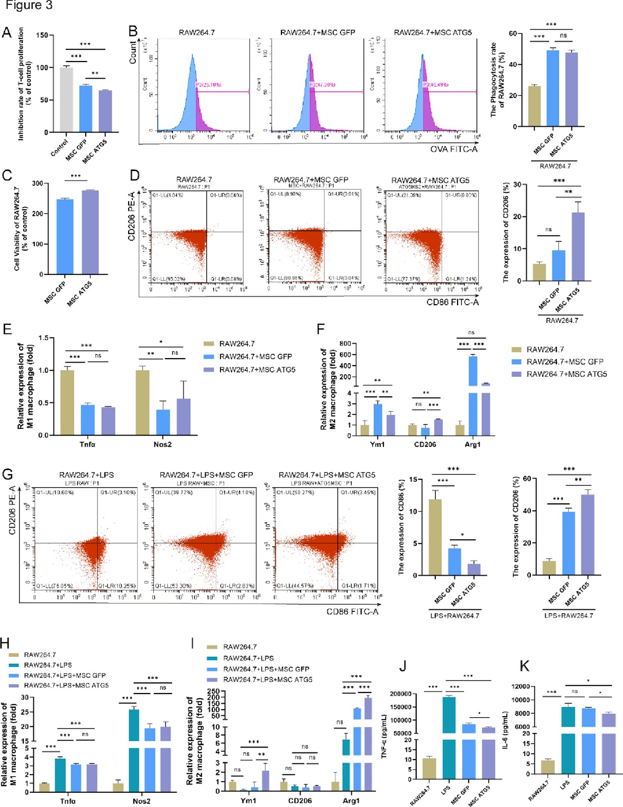
Background The therapeutic efficacy of mesenchymal stem cells (MSCs) can be improved by enhancing their adaptation to the inflammatory microenvironment. Autophagy maintains MSCs functionality, and autophagy-related gene 5 (ATG5) mediates autophagy and regulates the biological functions and therapeutic efficacy of these cells. The aim of this study was to investigate the role of ATG5 in the antioxidant capacity and evaluate the therapeutic effect of ATG5-engineered MSCs for colitis treatment. Methods Cell viability was assessed using a Cell Counting Kit-8. The mRNA expression of autophagy-, antioxidant-, and polarization-related genes was determined through real-time quantitative polymerase chain reaction, and protein expression was analyzed via western blotting. Macrophage polarization markers were analyzed using flow cytometry. Multiomics approaches, including RNA transcriptome sequencing, untargeted metabolomics, and 16S ribosomal RNA microbiota analysis, were also used. Mice with dextran sulfate sodium-induced colitis were used to evaluate the therapeutic efficacy of MSCs. Results Preconditioning MSCs with hypoxia (1% Oā‚‚) and serum deprivation significantly enhanced autophagy and upregulated ATG5 expression. Adenovirus-mediated ATG5 overexpression in MSCs (MSCs-ATG5) enhanced their autophagic activity and antioxidant capacity, upregulated HMOX-1, SOD2, and CAT expression, and increased glutathione peroxidase and catalase enzymatic activity, while enhancing cell proliferation, without altering surface marker expression. Further, MSCs-ATG5 significantly promoted M2 macrophage polarization and regulated oxidative stress-related signaling pathways. Additionally, MSCs-ATG5-based therapy markedly ameliorated colitis disease signs in mice. Transcriptome analysis revealed that MSCs-ATG5 suppressed the IL-17/NF-ĪŗB inflammatory signaling pathway. This treatment also regulated levels of the anti-inflammatory metabolite prostaglandin D2 (PGD2) in colon tissues.

Read Full Abstract10.1186/s13287-026-05008-2
Research progress on the effects of M1/M2 macrophages on the differentiation and maturation of stem cell-derived cardiomyocytes: a reviewGraphical AbstractVerified
Stem Cell Research & Therapy

Research progress on the effects of M1/M2 macrophages on the differentiation and maturation of stem cell-derived cardiomyocytes: a review

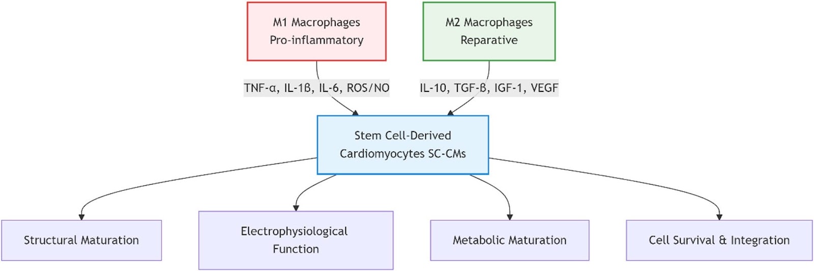
Stem cell-derived cardiomyocytes (SC-CMs) represent a promising cell source for cardiac regenerative medicine, disease modeling, and drug screening. However, their clinical translation faces significant challenges, including functional immaturity, poor long-term survival, and inadequate integration with host tissue following transplantation. The immune microenvironment, particularly the dynamic polarization of macrophages into pro-inflammatory (M1) or reparative (M2) phenotypes, is increasingly recognized as a critical regulator of cardiac repair, yet a systematic understanding of its specific effects on SC-CM fate remains incomplete. This review aims to comprehensively evaluate the dual regulatory roles of M1 and M2 macrophages on the differentiation efficiency, structural and functional maturation, and in vivo transplantation efficacy of SC-CMs. A systematic literature search was conducted in PubMed, Web of Science, Nature, and CNKI for relevant studies published from database inception to July 2025. After screening, 92 articles were included for analysis. The synthesized evidence demonstrates that M1 macrophages and their secreted factors (e.g., TNF-α, IL-1β) impede cardiac differentiation by inhibiting the Wnt/β-catenin pathway, disrupt sarcomeric organization and calcium handling, and maintain SC-CMs in a glycolytic, immature state. In contrast, M2 macrophages enhance SC-CM maturation by providing trophic support (e.g., IGF-1, HGF), promoting electrophysiological maturation and metabolic reprogramming towards oxidative phosphorylation, and facilitating angiogenesis via VEGF. The novelty of this review lies in its integrated perspective on macrophage-driven immunomodulation as a central axis for SC-CM maturation. Furthermore, it discusses emerging therapeutic strategies—such as optimized transplantation timing, co-transplantation with immunomodulatory cells, engineered exosomes, and smart biomaterials—that leverage macrophage polarization to create a favorable microenvironment for SC-CMs. Ultimately, harnessing macrophage-SC-CM crosstalk is a crucial step toward advancing clinically effective and immunologically informed cardiac regeneration therapies.

Read Full Abstract10.1186/s13287-026-04938-1
A novel OTUD5 variant disrupts neural progenitor cell homeostasis: mechanistic insights from HEK293T cell-based analysesGraphical AbstractVerified
Stem Cell Research & Therapy

A novel OTUD5 variant disrupts neural progenitor cell homeostasis: mechanistic insights from HEK293T cell-based analyses

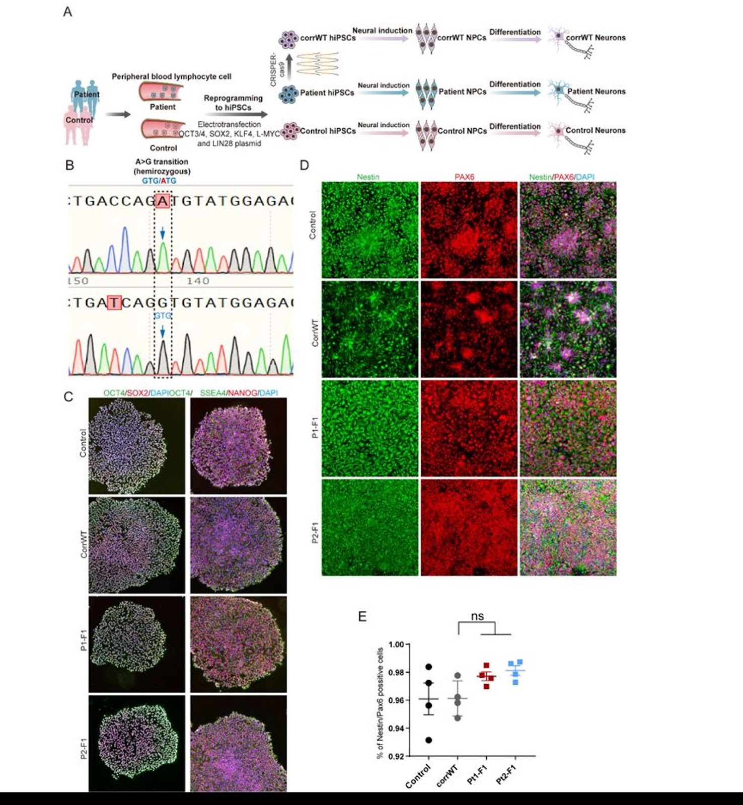
Background Variants in OTUD5 are associated with neurodevelopmental disorders (NDDs), yet the underlying molecular mechanisms remain unclear. This study aimed to investigate the pathogenicity of a novel OTUD5 variant (c.697G > A, p.Val233Met) and elucidate its regulatory role in neural progenitor cell (NPC) proliferation and differentiation, thereby uncovering the function of OTUD5 in neurodevelopment. Methods The OTUD5 variant was identified in two NDD patients via exome sequencing. Patient-derived induced pluripotent stem cells (iPSCs) and CRISPR/Cas9-corrected isogenic controls were generated. NPC proliferative activity was assessed by Ki67 immunofluorescence staining, cell-cycle distribution was analyzed by flow cytometry, and neuronal differentiation was evaluated by Tuj1/MAP2 immunofluorescence staining. Substrate screening was conducted in HEK293T cells using co-immunoprecipitation (Co-IP) and mass spectrometry. Deubiquitination capacity and protein stability were validated through ubiquitination assays and cycloheximide (CHX) chase experiments. Results The p.Val233Met variant, located within the catalytic OTU domain, induced a marked conformational alteration in the OTUD5 protein. Functionally, the variant caused aberrant NPC proliferation (1.8-fold increase in Ki67+ cells, accompanied by release of G1 arrest) and impaired neuronal differentiation (60% reduction in Tuj+ cells). Mechanistically, wild-type OTUD5 stabilized GSK3β by removing K48-linked ubiquitin chains, whereas the mutant isoform exhibited diminished deubiquitinase activity, accelerating GSK3β degradation and shortening its half-life by 40%. Conclusion This study establishes a novel disease mechanism whereby OTUD5 mutations disrupt NPC homeostasis through GSK3β destabilization, highlighting the critical role of ubiquitination regulation in neurodevelopment. Our iPSC model provides a platform for testing GSK3β-targeted therapies in OTUD5-related NDDs.

Read Full Abstract10.1186/s13287-026-04974-x
Construction of liver organoid models by hepatobiliary differentiation from human induced pluripotent stem cells: state of the art, challenges and improving strategiesGraphical AbstractVerified
Stem Cell Research & Therapy

Construction of liver organoid models by hepatobiliary differentiation from human induced pluripotent stem cells: state of the art, challenges and improving strategies

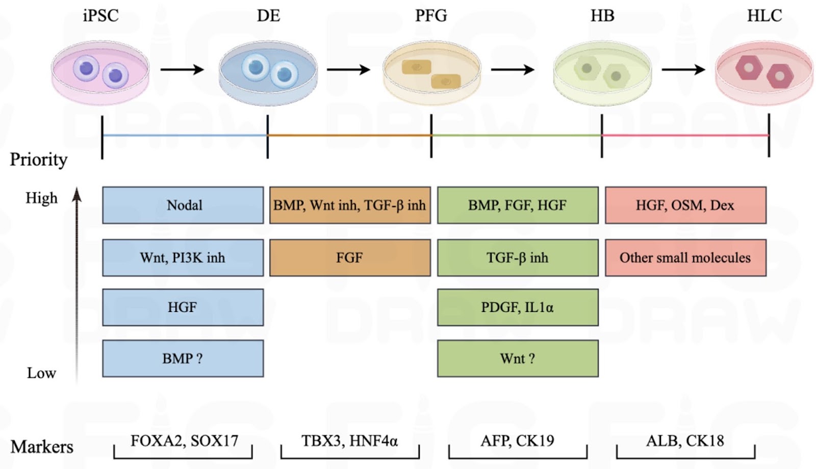
Physiologically relevant liver models are essential for advancing hepatic disorder research, especially for disease modeling and drug development, yet current in vitro systems fail to adequately recapitulate the architecture and function of the liver. Owing to the accessibility, robust proliferation and multilineage differentiation potential of human induced pluripotent stem cells (iPSCs), liver organoids derived from iPSCs have emerged as a promising resource in hepatology. Despite this promise, the field still faces persistent bottlenecks including incomplete hepatic maturation, insufficient incorporation of non-parenchymal cells (notably immune and stromal populations), phenotypic instability, and a lack of consensus on standardized differentiation protocols. Therefore, this review systematically analyzes the challenges and strategies of iPSC differentiation into liver organoids and the related influencing factors by focusing on multidimensional regulation of hepatobiliary development as well as the effects of cellular origin, culture system and liver microenvironment on hepatic differentiation of iPSCs. Moving forward, priority should be given to the following directions: (1) Elucidating the self-assembly mechanism of liver organoids to enable precise control of hepatobiliary differentiation, thereby better governing organoid morphology and improving reproducibility; (2) Replacing exogenous cytokines with small-molecule compounds at different stages of iPSC differentiation to simplify and standardize differentiation protocols; (3) Advancing liver organoid transplantation as a means to validate physiological functionality and shift cell therapy from passive replacement toward active tissue reconstruction; (4) Integrating artificial intelligence to achieve intelligent and precise regulation of hepatic differentiation.

Read Full Abstract10.1186/s13287-026-05080-8
A Noncanonical Neuroligin 3-Centered Complex Promotes Functional Recovery of Spinal Cord InjuryGraphical AbstractVerified
Stem Cell Research & Therapy2026

A Noncanonical Neuroligin 3-Centered Complex Promotes Functional Recovery of Spinal Cord Injury

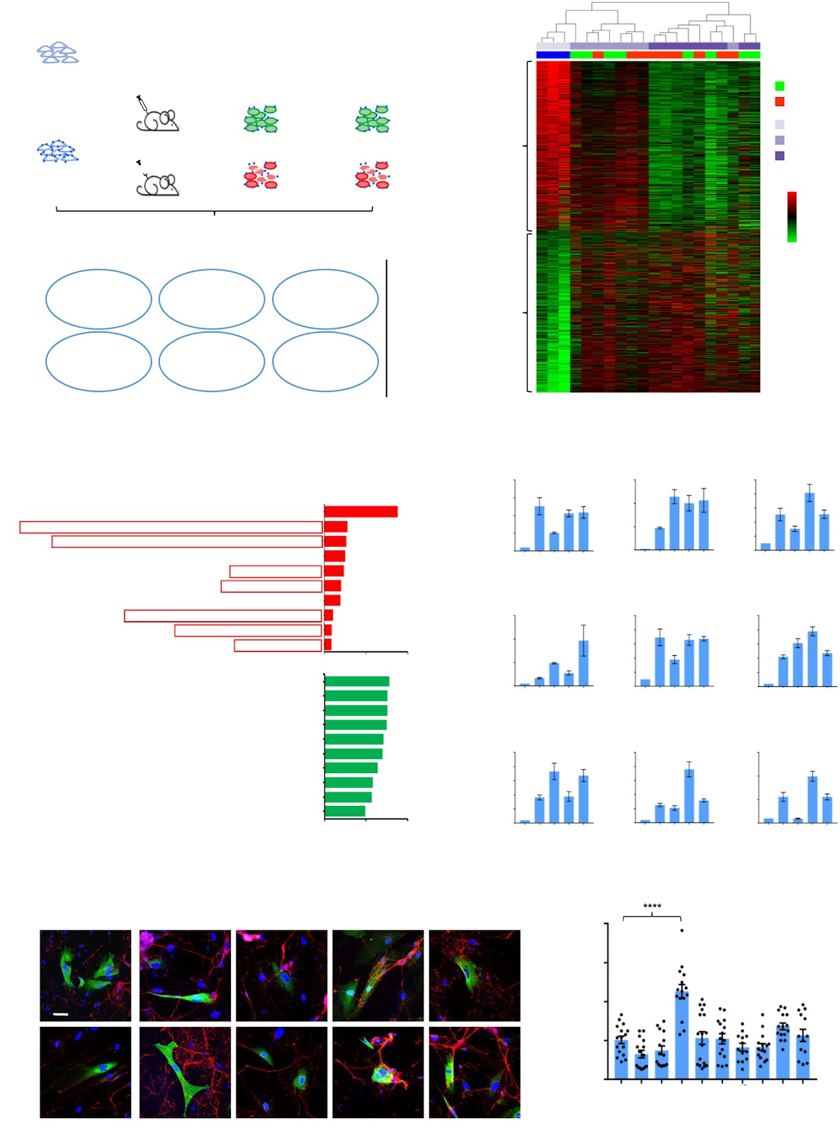
Background: While human umbilical cord-derived mesenchymal stem cells (hUC-MSCs) hold great potential for spinal cord injury (SCI) treatment, their intrinsic mechanisms are not fully understood. Given their multipotency, especially their neuronal transdifferentiation potential, we hypothesized that investigating the in situ transcriptional reprogramming of transplanted hUC-MSCs within the spinal cord microenvironment (SCE) could shed light on crucial genes for SCI repair. Methods: DiD-labeled hUC-MSCs were intrathecally transplanted in rats with or without sub-acute spinal cord bilateral hemisection injury and subsequently retrieved for RNA-seq. Comparative analysis of the transcriptomes of hUC-MSCs and functional screenings in vitro and in vivo, including heterologous synapse formation assay, transplantation of MSCs with gene overexpression or knockdown, AAV-mediated neuron-specific gene expression in SCI rats, behavioral tests, and motor evoked potentials (MEPs) were performed to identify the novel target gene Neuroligin 3 (Nlgn3). Immunoprecipitation followed by mass spectrometry (IP-Mass spec), cell aggregation assay, and immuno-electron microscopy were used to reveal the functional interacting partners of Nlgn3. Moreover, RT-qPCR, western blotting, immunofluorescence staining, and co-IP were used to elucidate the underlying mechanism. Results: Using RNA-seq and functional screening, we identified NLGN3 as a neuronal cell adhesion molecule (CAM) activated by the SCE in transplanted hUC-MSCs to promote therapeutic efficacy. Critically, the neuron-specific restoration of Nlgn3 in the injured spinal cord alone was sufficient to achieve a comparable therapeutic effect. Mechanistically, Nlgn3 recruits the synaptic vesicle proteins Sar1a and Hspa8 to modulate synaptic strength. The combinatorial restoration of Nlgn3 with either Sar1a or Hspa8 synergistically enhanced SCI repair, highlighting the functional importance of this noncanonical Nlgn3-Sar1a-Hspa8 axis. Conclusions: This work unveils a novel therapeutic role for Nlgn3 in SCI treatment, demonstrating its ability to both enhance MSC transplantation efficacy and directly promote neural circuit reconstruction. We also propose a combinatorial strategy of targeting the noncanonical Nlgn3-centered complex for SCI repair.

Read Full Abstract10.1186/s13287-026-05100-7
Stem cell-driven biomedical technologies for tooth regeneration: engineering scaffolds, organoid models, and molecular targeted strategiesGraphical AbstractVerified
Stem Cell Research & Therapy

Stem cell-driven biomedical technologies for tooth regeneration: engineering scaffolds, organoid models, and molecular targeted strategies

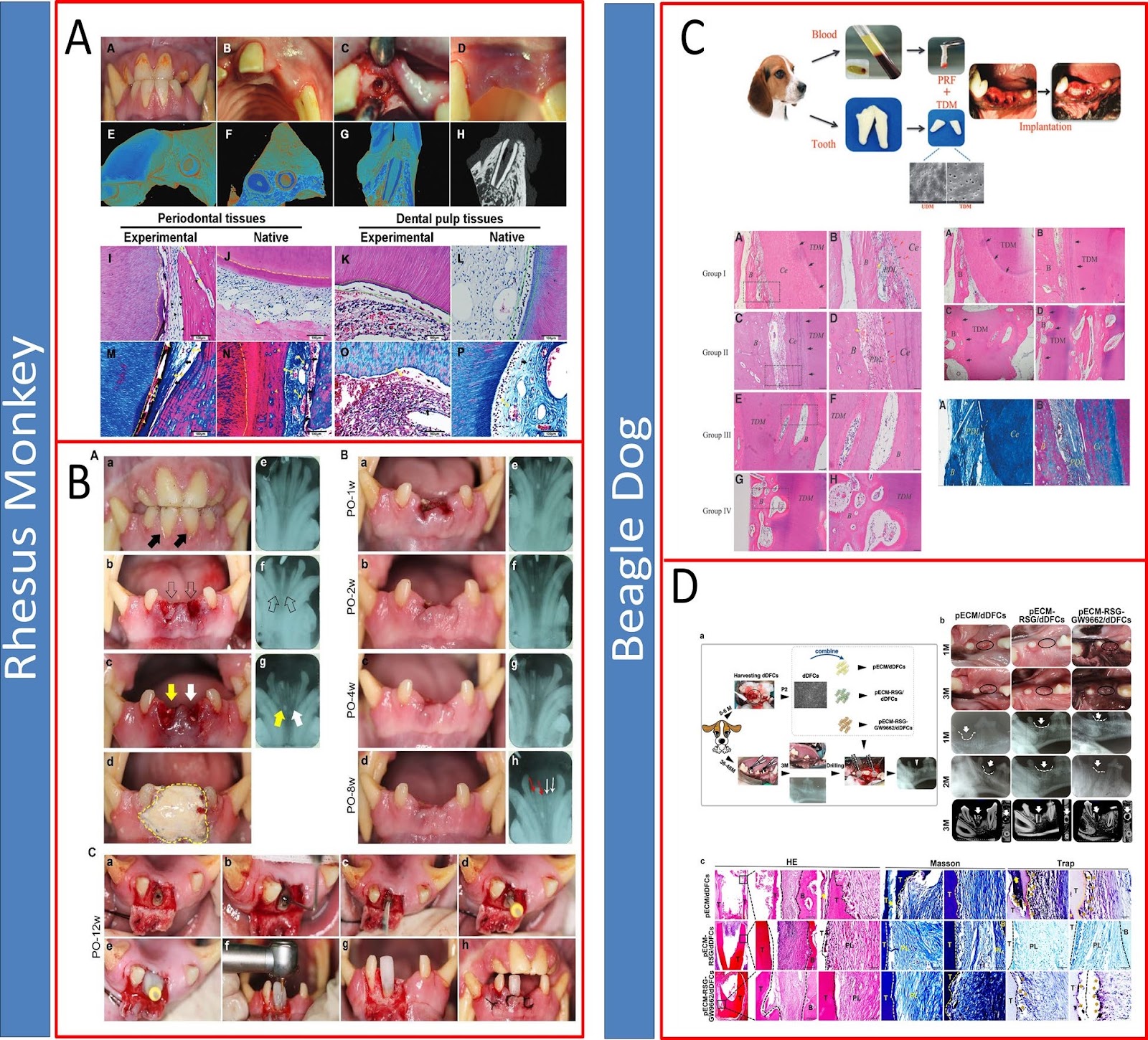
Tooth loss remains a major unmet clinical challenge, and current prosthetic approaches cannot restore the biological complexity, sensory function, or regenerative capacity of natural teeth. Recent progress in stem cell biology, developmental engineering, and regenerative biomaterials has opened new possibilities for biological tooth regeneration. This review integrates advances across three major research domains that together define the current landscape of translational regenerative dentistry. First, we discuss stem cell-based, scaffold-guided strategies for tooth regeneration. These approaches combine dental and nondental stem cells, including DPSCs, SCAPs, PDLSCs, SHED, and iPSC-derived lineages, with bioactive materials such as HA/TCP ceramics, dentin-derived extracellular matrix scaffolds, and natural or synthetic polymers to promote odontogenic differentiation, vascularization, and periodontal attachment. Second, we summarize emerging tooth organoid and bioengineered tooth germ technologies that recapitulate epithelial–mesenchymal interactions and enable controlled reconstruction of dentin–pulp and periodontal compartments for modeling human odontogenesis. Third, we highlight molecular regulation-driven therapeutic strategies, focusing on the modulation of Wnt, BMP, FGF, TGF-β, and USAG-1 pathways to stimulate endogenous tooth regeneration and correct developmental defects. Despite marked progress, challenges remain, including stable neurovascular integration, optimization of stem cell–material crosstalk, precise control of spatiotemporal signaling, and long-term functional stability in vivo. Finally, we outline future directions involving smart biomaterials, gene- and protein-based molecular targeting, organoid-guided regeneration, and iPSC-enabled personalized therapies, which may further accelerate the clinical translation of stem cell-based tooth regeneration.

Read Full Abstract10.1186/s13287-026-05044-y
Functional Development of Photoreceptors in Human Retinal OrganoidsGraphical AbstractVerified
Stem Cell Research & Therapy2026

Functional Development of Photoreceptors in Human Retinal Organoids

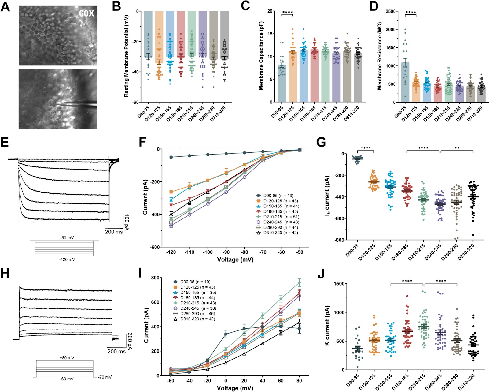
Background: Retinal organoids (ROs) derived from human pluripotent stem cells are crucial for modeling retinal development and disease. However, the functional electrophysiological maturation of photoreceptors within ROs remains poorly characterized. This study aimed to define the functional maturation timeline of photoreceptors in human embryonic stem cell (hESC)-derived ROs. Methods: H9 hESC-derived ROs which included a CRX-tdTomato reporter line for specific photoreceptor identification were utilized. An integrated approach of RNA-sequencing analysis, immunofluorescence staining, and whole-cell patch-clamp recordings was employed to systematically assess photoreceptor maturation over 300 days of differentiation. Results: Transcriptional and protein analysis revealed progressive upregulation of key ion channels. Patch-clamp recordings demonstrated stage-dependent maturation of membrane properties, which stabilized by D120–125. Hyperpolarization-activated cyclic nucleotide-gated (HCN) channel-mediated currents (Ih) increased progressively, peaking at D240, with amplitudes comparable to mature primate photoreceptors. Voltage-gated sodium (Nav) currents also showed significant developmental upregulation, reaching a maximum, stable plateau from D210–215 onward. Pharmacological blockade confirmed the identity of HCN and Nav currents. Critically, the capacity for action potential (AP) generation increased developmentally, with the proportion of photoreceptors capable of firing APs rising from 16.7% at D90–95 to a peak of 90.2% by D240–245. Conclusions: This study defines a comprehensive electrophysiological maturation timeline for photoreceptors in human ROs and establishes D240 as a key benchmark for functional maturity, characterized by peak Ih currents and AP generation capacity equivalent to mature native photoreceptors. These findings provide essential physiological criteria for standardizing RO quality control, enhancing their utility for modeling retinal degenerative diseases and developing cell replacement therapies.

Read Full Abstract10.1186/s13287-026-05027-z
Endothelial progenitor cell susceptibility to DNA damaging and DDR-modulating compounds determines endothelial differentiation accuracyGraphical AbstractVerified
Stem Cell Research & Therapy

Endothelial progenitor cell susceptibility to DNA damaging and DDR-modulating compounds determines endothelial differentiation accuracy

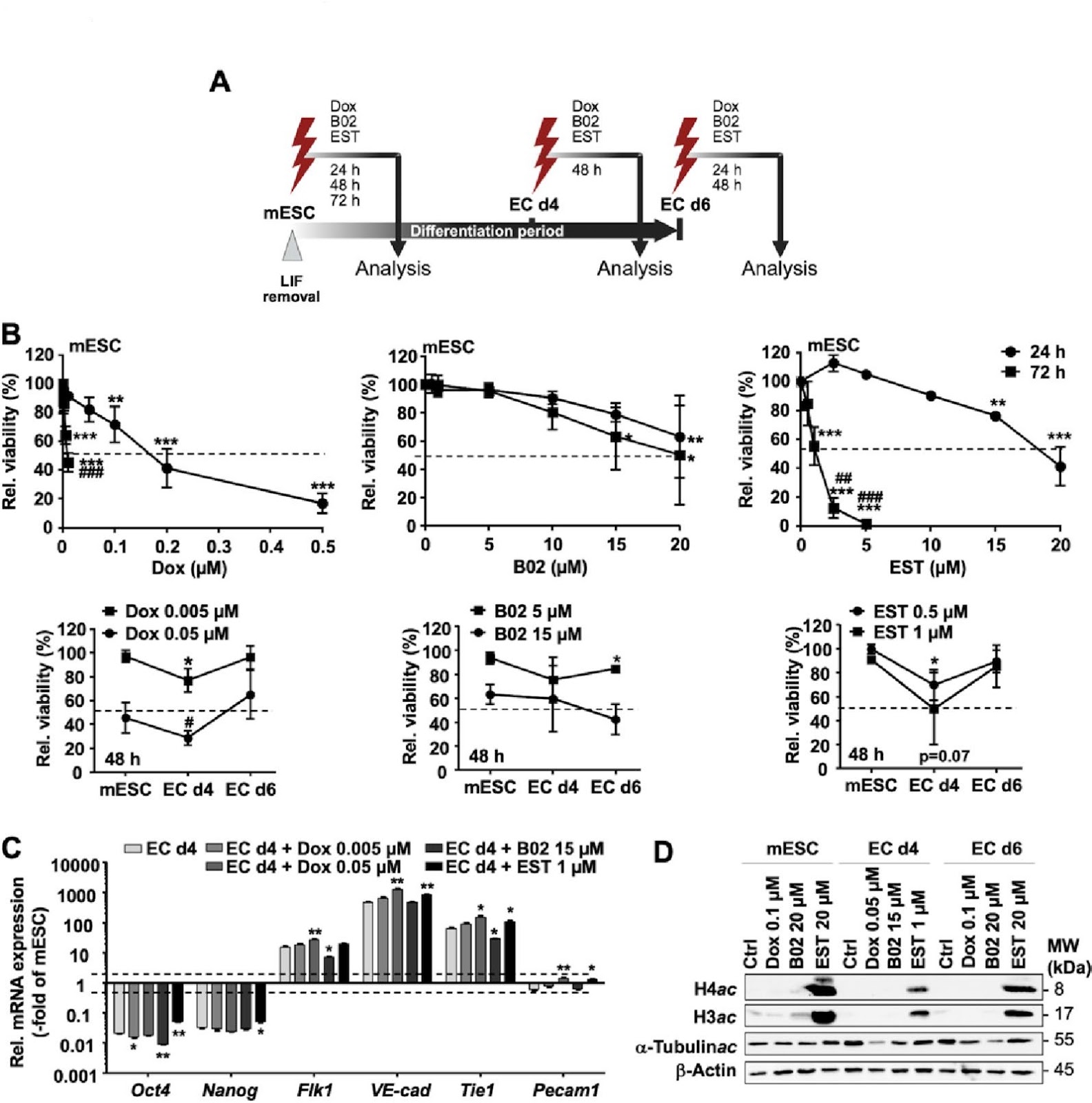
The clinical use of the anticancer drug doxorubicin (Dox) is limited by irreversible cardiotoxicity. The pathophysiological relevance of different cardiac cell types, including endothelial progenitor cells (EPC), in this process is unclear. Since progenitor cells are particularly relevant for tissue regeneration, we hypothesize that residual damage resulting from Dox-based therapeutic regimen may influence their endothelial differentiation accuracy. Therefore, we comparatively investigated the response of murine embryonic stem cells (mESC), endothelial progenitor cells (EC d4) and terminally differentiated endothelial-like cells (EC d6) following exposure to Dox and selected pharmacological inhibitors of DNA repair/DNA damage response (DDR) (RAD51i B02; HDACi entinostat (EST)). We show that EC d4 exhibit enhanced Dox sensitivity as compared to mESC and EC d6. EdU incorporation and replication fork progression analyses revealed pronounced agent-specific differences between mESC, EC d4 and EC d6. Furthermore, DNA damage formation varied in a drug-dependent manner, with mESC showing enhanced residual levels of DNA single-strand breaks (SSB) as compared to EC d4 and EC d6 while EC d6 revealed highest levels of DNA double-strand breaks (DSB). Dox treatment of EC d4 did not prevent their further differentiation into EC d6. However, it caused several functional impairments in the surviving EC d6 progeny, including defects in mitochondrial homeostasis, endothelial barrier function related to cell-cell adhesion factors (ZO1, VE-cadherin), cytokine response and low-density lipoprotein (LDL) uptake. This is accompanied by increased senescence. Summarizing, we demonstrate both overlapping and agent-specific responses of mESC, EC d4 and EC d6 to Dox and DNA repair/DDR inhibitors. Notably, drug treatment of EPC (EC d4) causes multiple dysfunctions in differentiated EC d6. Hence, pharmacological measures aiming to specifically protect EPC from Dox-induced damage are suggested to foster the maintenance of healthy endothelial functionality during regeneration, thereby lowering the risk of detrimental late cardiotoxicity resulting from Dox-based anticancer regimen.

Read Full Abstract10.1186/s13287-026-05087-1
Mesenchymal stem cell derived exosomes mitigate COVID-19 cytokine storm via Annexin A1 and TGF-β mediated MAPK pathway inhibitionGraphical AbstractVerified
Stem Cell Research & Therapy

Mesenchymal stem cell derived exosomes mitigate COVID-19 cytokine storm via Annexin A1 and TGF-β mediated MAPK pathway inhibition

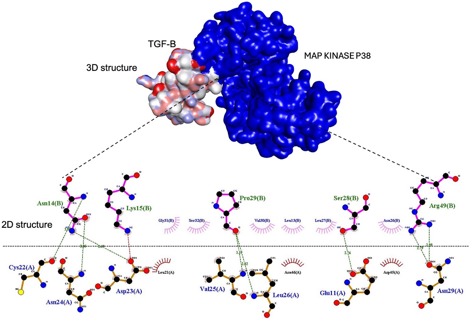
Background Severe COVID-19 is marked by a dysregulated inflammatory response, known as a cytokine storm, resulting in acute respiratory distress syndrome (ARDS) and multiple organ failure. Mesenchymal stem cell-derived exosomes (MSC-Exos) have demonstrated potential as immunomodulatory agents. This work investigates the possibility of MSC-Exos to mitigate excessive inflammation in COVID-19 by targeting the mitogen-activated protein kinase (MAPK) signalling pathway. Methodology We integrated molecular docking analysis between TGF-β and Annexin A1 as exosomal proteins and key component proteins of the MAPK pathway (p38, ERK1/2, JNK1). The in-silico results were then validated in vivo using a Syrian hamster model of SARS-CoV-2 infection. Quantitative PCR (qPCR), western blotting, and histological examination were employed to evaluate the effects of MSC-Exos therapy on MAPK pathway activation, cytokine production, and lung tissue pathology. Results The in-silico study revealed extensive hydrogen bonding and hydrophobic interactions at the protein–protein interfaces between exosomal proteins and MAPK components. These interactions suggest that exosomal proteins may modulate MAPK signaling pathways. In vivo, MSC-Exos administration led to marked downregulation of pivotal genes in the MAPK signaling pathway (MEKK1, MEKK2, MEKK3), diminished phosphorylation of JNK1, p38, and ERK1/2, and lowered production of pro-inflammatory cytokines (IL-1β, IL-6, TNF-α). Histopathological examination demonstrated ameliorated lung tissue structure, characterized by diminished alveolar wall thickness and decreased immune cell infiltration.

Read Full Abstract10.1186/s13287-026-04980-z
Prevascularization of electrospun PCL/PLA scaffolds using human adipose-derived stem and endothelial cells enhances vascular integration and host angiogenesis in vivoGraphical AbstractVerified
Stem Cell Research & Therapy

Prevascularization of electrospun PCL/PLA scaffolds using human adipose-derived stem and endothelial cells enhances vascular integration and host angiogenesis in vivo

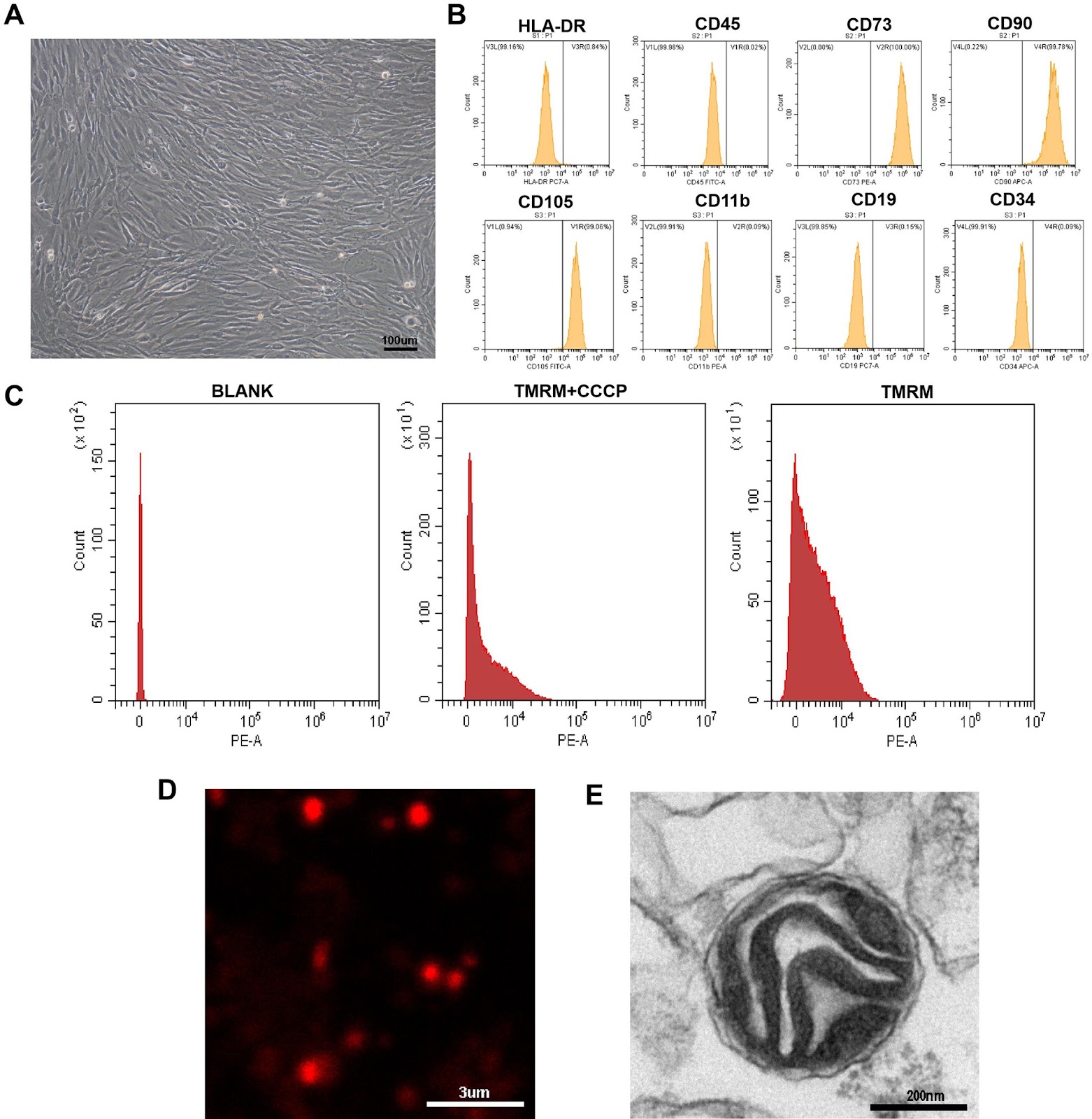
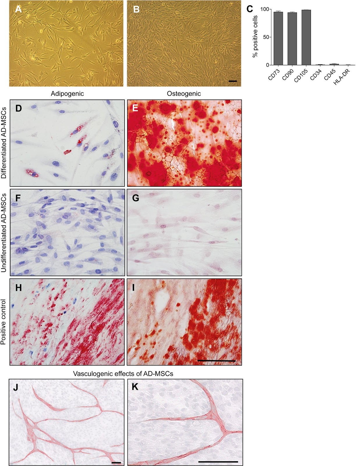
Background Inadequate vascularization remains a major limitation in tissue engineering, often leading to graft failure due to limited oxygen and nutrient supply. Prevascularization, the formation of microvascular networks within scaffolds before implantation, aims to accelerate perfusion and improve graft integration. We developed bilayer electrospun poly(ε-caprolactone)/poly(l-lactide) (PCL/PLA) scaffolds prevascularized by co-culture of human adipose-derived mesenchymal stem cells (AD-MSCs) and human placental arterial endothelial cells (HPAECs). Methods AD-MSCs were isolated from lipoaspirates and characterized by flow cytometry and functional assays. Bilayered PCL/PLA scaffolds were engineered with a wide-meshed layer for cell infiltration and a fine-meshed layer for mechanical stability. Scaffolds were seeded with AD-MSCs, HPAECs, or both (co-culture). Cell viability, adhesion, and apoptosis were analyzed histologically. Angiogenic and vasculogenic potential was evaluated in vitro and in vivo using the chick chorioallantoic membrane (CAM) assay. Results AD-MSCs expressed characteristic markers, demonstrated adipogenic and osteogenic differentiation, and promoted angiogenesis in 2D co-culture. ELISA analyses indicated dynamic secretion of VEGF, HGF, and bFGF, reflecting both paracrine and contact-dependent AD-MSC–HPAEC interactions. On scaffolds, cells primarily adhered to the wide-meshed layer. Co-culture induced vessel-like structures within a multicellular stromal environment; monocultures did not support prevascularization. Five days post-implantation, prevascularized scaffolds exhibited human microvessels at the scaffold–CAM interface and in adjacent tissue, closely associated with AD-MSCs and containing chicken erythrocytes—indicating successful anastomosis and functional perfusion. Quantitative analysis showed a significant increase in vessel branching points in the host CAM tissue in response to AD-MSC-only (2.8-fold)

Read Full Abstract10.1186/s13287-026-05066-6
Extracellular vesicles from Yiguanjian-primed bone-marrow mesenchymal stem cells ameliorate chronic liver fibrosis via miR-7045-5pGraphical AbstractVerified
Stem Cell Research & Therapy

Extracellular vesicles from Yiguanjian-primed bone-marrow mesenchymal stem cells ameliorate chronic liver fibrosis via miR-7045-5p

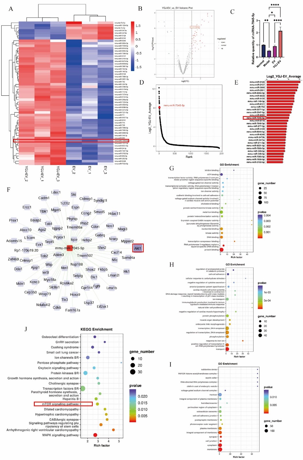
Background Liver fibrosis is a crucial pathological stage in the progression of chronic liver diseases. Yiguanjian (YGJ), a Chinese herbal formula, exhibits anti-inflammatory, anti-fibrotic, and hepatoprotective effects. Extracellular vesicles from bone-marrow mesenchymal stem cells (BMSC-EVs) have shown potential in treating various disorders, including liver fibrosis. This study investigated the regulatory effects of EVs from YGJ-preconditioned BMSCs (YGJ-EVs) on TGF-β1-stimulated hepatic stellate cells (HSCs) and their therapeutic potential in a mouse model of liver fibrosis, with a focus on identifying the causative microRNA cargo. Methods YGJ-EVs and control EVs were isolated from BMSC culture supernatants and characterized via western blotting, transmission electron microscopy, and nanoparticle tracking analysis. Their cellular uptake in vitro and in vivo was evaluated using DIR labeling. To identify candidate miRNAs mediating YGJ-EV bioactivity, miRNA microarray analysis was conducted. To assess the effect of YGJ-EVs on liver fibrosis, TGF-β1-activated HSC cells were treated with YGJ-EVs or control-EVs for 24 h, and then the expression of proteins related to fibrotic activation (COL1-A1 and α-SMA), lysosomal biogenesis (LAMP1, TPP1, CTSD, and CTSB) mitophagy (p62, LC3, PINK1, and Parkin), and the Akt/AMPK/TFEB pathway was assessed. To determine whether miR-7045-5p is the causative factor, HSC cells transfected with miR-7045-5p were similarly analyzed. Results miRNA microarray analysis revealed miR-7045-5p upregulation in YGJ-EVs versus control EVs. In CCl4-treated mice, YGJ-EV-derived miR-7045-5p ameliorated the liver fibrosis, improved the hepatic function, and suppressed the HSC activation by inhibiting the Akt/AMPK/TFEB pathway. In vitro, miR-7045-5p overexpression attenuated TGF-β1-induced HSC activation. Conclusion YGJ increases miR-7045-5p abundance in BMSC-EVs. YGJ-EVs alleviate liver fibrosis by delivering the anti-fibrotic miRNA miR-7045-5p, which inhibits the Akt/AMPK/TFEB pathway, thereby promoting lysosomal biogenesis and mitophagy in HSCs.

Read Full Abstract10.1186/s13287-025-04780-x
In vitro assays for investigating the immunomodulatory properties of human mesenchymal stromal cellsGraphical AbstractVerified
Stem Cell Research & Therapy2026

In vitro assays for investigating the immunomodulatory properties of human mesenchymal stromal cells

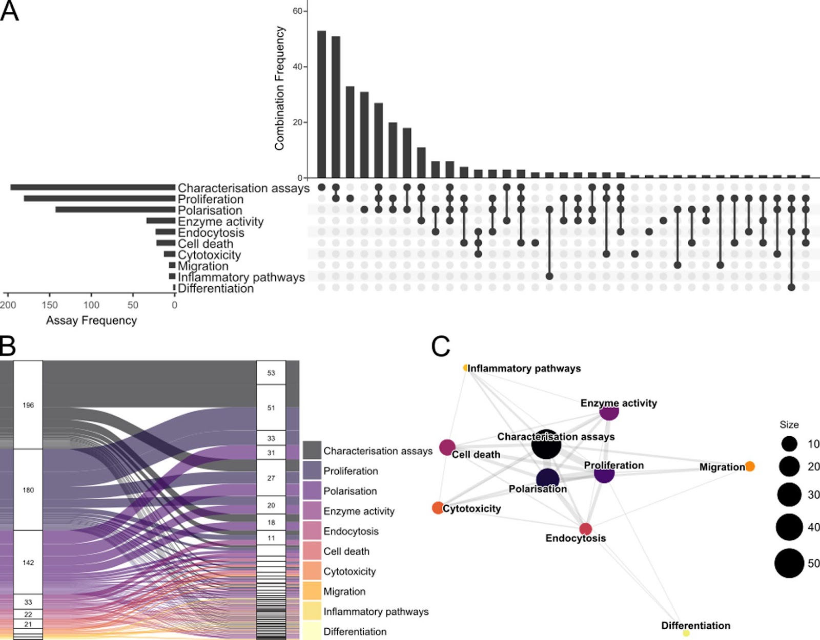
Mesenchymal stromal cells (MSCs) are widely recognized for their immunomodulatory properties, which underpin their therapeutic potential in inflammatory and immune-mediated diseases. Although MSC therapies have consistently proven safe, clinical efficacy remains inconclusive, maybe due to incomplete understanding of MSC interactions with the immune environment. This review evaluates current trends in MSC immunomodulation research, based on 318 studies published since 2019 until medio 2024. The most frequently used assays included characterization, proliferation, and polarization, employing methods such as flow cytometry, enzyme-linked immunosorbent assays and colorimetric assays, and polymerase chain reaction. Many studies incorporated strategies for priming of MSCs or included immune cells, most commonly peripheral blood mononuclear cells, T cells, and macrophages. We identify key sources of variability and propose a minimum reporting checklist including MSC source, priming conditions, assay design, and immune cell characteristics. We further recommend implementation of multi-assay workflows combining phenotypic characterization with at least one functional assay. These measures may improve transparency, comparability across studies, and guide robust assay design.

Read Full Abstract10.1186/s13287-026-04920-x
The metabolic profiles of cancer stem cellsGraphical AbstractVerified
Stem Cell Research & Therapy2026

The metabolic profiles of cancer stem cells

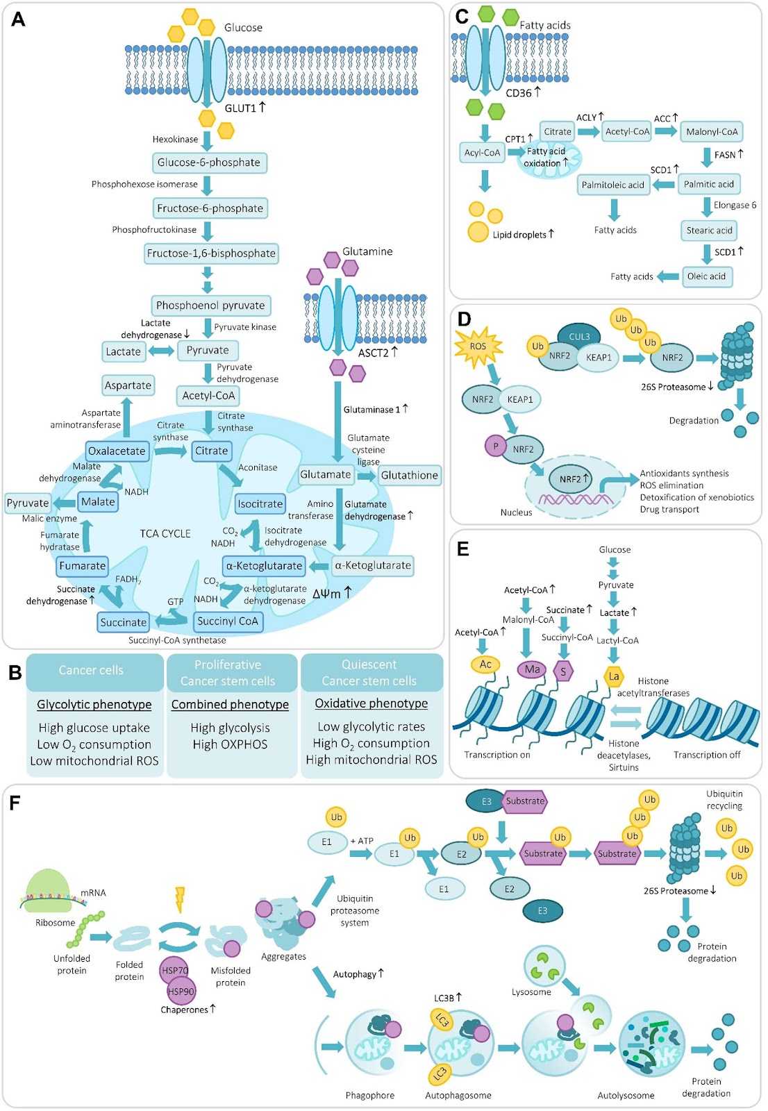
Cancer stem cells (CSCs) represent a minor but highly adaptable subpopulation within tumors that drives long-term growth, metastasis, and therapy resistance. Their ability to survive and regenerate under metabolic and therapeutic stress relies on a unique integration of energy flexibility, redox balance, and proteostatic programs. While bulk tumor cells typically favor aerobic glycolysis and high protein turnover, CSCs often exhibit elevated mitochondrial activity, fatty acid oxidation, and selective suppression of proteasome function. These metabolic features support quiescence, stress tolerance, and self-renewal. Beyond energy production, metabolic intermediates such as acetyl-CoA, succinate, and lactate serve as epigenetic cofactors, linking nutrient availability to chromatin remodeling and transcriptional plasticity. Reactive oxygen species and antioxidant responses further tune this balance, shaping the transition between glycolytic and oxidative CSC states. These intrinsic programs are continuously influenced by the tumor microenvironment, where hypoxia, cytokine-driven signaling, and metabolic coupling with stromal and immune cells modulate CSC metabolism and reinforce stemness. Despite rapid progress, major conceptual and methodological gaps still limit our understanding of CSC metabolism and this review highlights these unresolved issues and further outline key contextual factors—including tumor-intrinsic, microenvironmental, systemic, and metastatic cues—that shape CSC metabolism and help explain the divergent observations reported across studies. Understanding this network will be essential for designing combinatorial therapies that target CSC metabolism while accounting for their heterogeneity and plasticity.

Read Full Abstract10.1186/s13287-026-05014-4
Targeting cellular senescence in progenitor cells as a strategy to enhance bone regeneration by cell therapies: a systematic review of pre-clinical investigationsGraphical AbstractVerified
Stem Cell Research & Therapy

Targeting cellular senescence in progenitor cells as a strategy to enhance bone regeneration by cell therapies: a systematic review of pre-clinical investigations

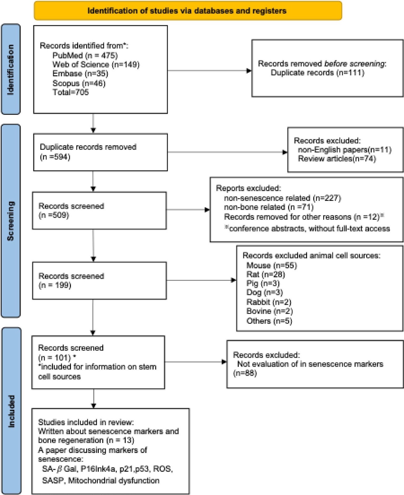
Background With the global population aging, optimizing bone regeneration is becoming increasingly important for enhancing the quality of life among elderly individuals. Progenitor cell-based therapies, such as mesenchymal stromal cells and induced pluripotent stem cells for bone regeneration have shown challenges due to cellular senescence and the control of the differentiation processes remain significant hurdles. In particular, elevated expression of senescence markers may play a pivotal role in limiting bone regeneration. This systematic review examines how these senescence markers influence the efficacy of progenitor cell therapies and whether targeting them could improve outcomes. Methods We conducted a systematic literature review following the PRISMA guidelines, using the PubMed, Web of Science, Embase and Scopus with the algorithm of ā€œbone regeneration AND senescence AND markerā€. Data synthesis focused on human cell sources and specifically examined senescence markers related to bone regeneration. Results Studies using human cells were discussed in 101 papers. Based on our inclusion and exclusion criteria, 13 papers remained for our review on senescence markers in human cells within the context of bone regeneration and senescence, with and without interventional strategies. More than half of the cell sources in current aging-related studies are derived from bone marrow. Markers of aging relevant to bone regeneration include changes in cell size and morphology, increased levels of β-galactosidase (β-Gal) and Reactive Oxygen Species (ROS), and the presence of a senescence-associated secretory phenotype (SASP). Additionally, distinct senescence markers such as p16Ink4a, p21, and p53, and mitochondrial dysfunction were associated with reduced osteogenic potential and impaired regenerative capacity. Conclusion Bone marrow is the most common source of cells for studies of senescence. Cellular senescence characterized by elevated expression of specific markers was consistently shown to be negatively associated with osteogenic capacity and regenerative outcomes. The most common strategies to rejuvenate senescent cells include

Read Full Abstract10.1186/s13287-025-04767-8