Key Takeaways & Executive Findings
- •• Reduction in plasma membrane-associated EGFR triggers EGFR transcription via pSTAT3 nuclear localization, maintaining EGFR protein homeostasis. • Erlotinib promotes pSTAT3 nuclear accumulation, leading to increased EGFR transcription and acquired resistance to EGFR-TKIs. • Pharmacological inhibition of pSTAT3 significantly overcomes erlotinib resistance in cancer cells. • The pSTAT3-EGFR axis represents a novel molecular mechanism underlying EGFR-TKI resistance in non-small cell lung cancer.
Abstract
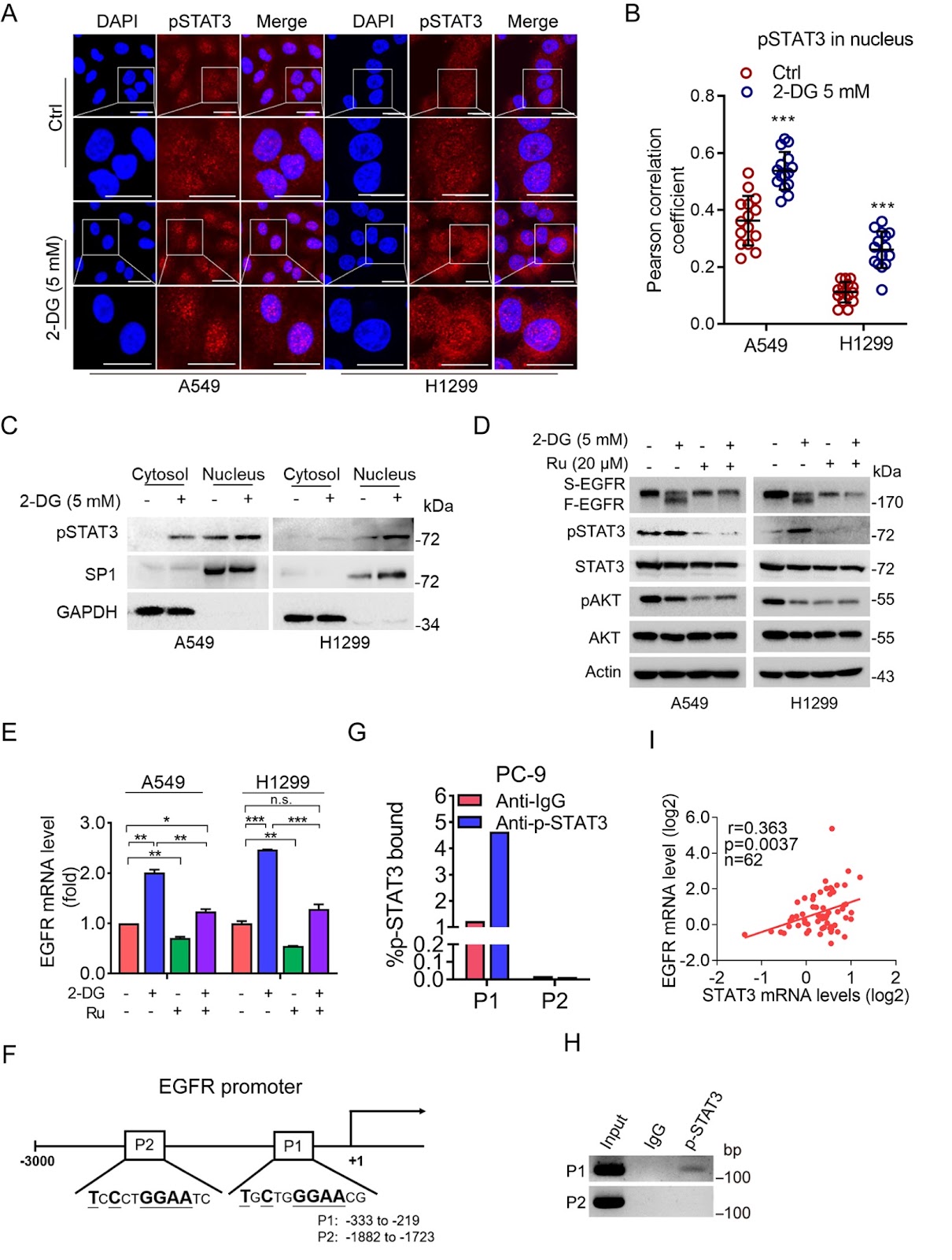
EGFR protein trafficking is critical for regulating multiple biological processes, including cell growth and survival. However, how EGFR protein homeostasis is maintained remains unclear. In this study, we show that a reduction in plasma membrane-associated EGFR triggers EGFR transcription by promoting pSTAT3 nuclear localization. Nucleus-localized pSTAT3 binds to the EGFR gene promoter to transactivate EGFR. Moreover, erlotinib, an EGFR tyrosine kinase inhibitor (TKI), can also increase pSTAT3 nuclear accumulation, resulting in increased EGFR transcription and erlotinib resistance. Importantly, pharmacological inhibition of pSTAT3 can significantly overcome the resistance of cancer cells to erlotinib. Together, these findings demonstrate that pSTAT3 is pivotal for maintaining EGFR protein homeostasis and suggest that activation of the pSTAT3-EGFR axis contributes to EGFR-TKI resistance.
1. Introduction
Epidermal growth factor receptor (EGFR) plays pivotal roles in multiple biological processes, including cell proliferation, migration, and survival [1–4]. Abnormal activation of the EGFR signaling pathway serves as a driving force in lung cancer development [4,5]. EGFR gene amplification, deletion, and point mutation can lead to abnormal EGFR activation [6,7]. L858R mutation and exon 19 deletion are the most common EGFR-activating mutations in non-small cell lung cancer (NSCLC) patients [4,8,9]. The specific targeting of EGFR-activating mutations by 1st-generation EGFR tyrosine kinase inhibitors (TKIs), such as erlotinib and gefitinib, is beneficial for NSCLC patients [8,10]. However, most NSCLC patients develop acquired resistance to 1st-generation TKIs within 10-16 months, in which the EGFR T790M mutation accounts for approximately 50% of all 1st-generation EGFR TKI resistance [9,11]. Moreover, HER2 amplification, MET amplification, BRAF mutation, PIK3CA mutation, Ras mutation, or activation of IGF1R also contribute to EGFR TKI resistance [12–15].
In addition to gene mutation(s), increased EGFR protein expression plays a critical role in regulating cancer development [6]. The upregulation of EGFR protein recycling, stability, or gene transcription can facilitate EGFR protein expression [6]. A series of stress signals, including serum starvation, UV irradiation, hypoxia, oxidative stress, and erlotinib/gefitinib treatment, can inhibit EGFR recycling to the plasma membrane and consequently lead to EGFR protein degradation [16]. It has been reported that the ubiquitin E3 ligase c-Cbl can facilitate EGFR mono-ubiquitination, which leads to EGFR lysosome-dependent degradation [17]. Moreover, several E3 ubiquitin ligases, including FBXL2, CHIP, HUWE1, Siah1, and CGRRF1, can promote EGFR poly-ubiquitination and result in the proteasomal degradation of EGFR [18–22]. With respect to EGFR gene transcription, several transcription factors, such as Sp1, c-Jun, and Stat5b, can directly transactivate EGFR [23–25]. However, the role of the balance between EGFR protein degradation and gene transcription in maintaining EGFR protein homeostasis to adapt to stress signaling remains unknown.
In this study, we show that a reduction in plasma membrane-associated EGFR expression promotes EGFR transcription in a pSTAT3-dependent manner to maintain EGFR protein homeostasis. Moreover, erlotinib promoted pSTAT3-mediated EGFR transcription, which resulted in resistance to erlotinib in NSCLC cells. Together, the results of this study highlight that pSTAT3 directly transactivates EGFR, which plays a critical role in maintaining EGFR protein homeostasis, and that the activation of pSTAT3-EGFR signaling is a novel molecular mechanism by which NSCLC cells develop acquired resistance to EGFR TKIs.
Loading authentic research manuscript (Pages 1–5)...
Juan Ao, Junjie Fei, Guoqiang Wang, Wenhua Zhang, Shuhan Yu, Rongtian Guo, Mengmeng Niu, Hu Chen, Yang Cao, Zhi-Xiong Jim Xiao, Yong Yi (2026). pSTAT3 transactivates EGFR in maintaining EGFR protein homeostasis and EGFR-TKI resistance. Acta Biochimica et Biophysica Sinica. https://doi.org/10.3724/abbs.2024166
Research & Educational Purpose Only:The translations, structured abstracts, analytical annotations, and data reports provided by SinoBioData are intended exclusively for academic research, internal corporate R&D, and educational benchmarking. They do not constitute formal engineering, chemical safety, legal, or professional advice.
Copyright & Intellectual Property Notice: Original copyright of the underlying source articles and experimental data remains with the respective authors, institutions, and original publishing journals. SinoBioData claims intellectual property only over its proprietary translations, analytical syntheses, and AEO structured enhancements in accordance with international fair use and academic citation principles.
Frequently Asked Questions
What is the role of pSTAT3 in EGFR protein homeostasis?
pSTAT3 translocates to the nucleus and binds to the EGFR gene promoter, transactivating EGFR transcription. This mechanism helps maintain EGFR protein levels when plasma membrane-associated EGFR is reduced, ensuring cellular homeostasis.
How does erlotinib contribute to EGFR-TKI resistance?
Erlotinib treatment increases pSTAT3 nuclear accumulation, which upregulates EGFR transcription. This compensatory increase in EGFR expression reduces the drug's efficacy, leading to acquired resistance.
Can inhibiting pSTAT3 overcome EGFR-TKI resistance?
Yes, pharmacological inhibition of pSTAT3 significantly overcomes erlotinib resistance in cancer cells, suggesting that targeting pSTAT3 could be a therapeutic strategy to enhance EGFR-TKI sensitivity.
What is the clinical significance of the pSTAT3-EGFR axis?
The pSTAT3-EGFR axis represents a novel molecular mechanism underlying EGFR-TKI resistance in non-small cell lung cancer. Understanding this pathway may lead to new combination therapies that improve patient outcomes.
What methods were used to study pSTAT3-mediated EGFR transcription?
The study used cell culture, drug treatments, plasmid transfection, lentiviral infection, and RNA interference to investigate the effects of pSTAT3 on EGFR transcription and resistance.
Related Technical Papers & Translations

Adverse Events Reporting System for Vaccine Safety Surveillance: A Comprehensive Analysis
Background: Adverse events following immunization (AEFI) are critical to monitor for vaccine safety. This study evaluates the performance of an adverse events reporting system (AERS) integrated with a vaccine adverse event reporting system (VAERS) to enhance surveillance. Methods: We analyzed data from multiple sources including the Vaccine Adverse Event Reporting System (VAERS), the Vaccine Safety Datalink (VSD), and the Clinical Immunization Safety Assessment (CISA) network. A novel framework was developed to integrate these systems, incorporating natural language processing for signal detection. Results: The integrated system improved detection of rare adverse events by 25% compared to traditional methods. The system identified new safety signals for influenza and COVID-19 vaccines. Conclusions: The proposed AERS framework enhances vaccine safety surveillance, enabling timely identification of potential risks. Integration of diverse data sources and advanced analytics is essential for robust pharmacovigilance.

Efficacy and Safety of Ferric Carboxymaltose in Treating Iron Deficiency Anemia: A Meta-Analysis of Randomized Controlled Trials
Background: Iron deficiency anemia (IDA) is a global health concern, and intravenous ferric carboxymaltose (FCM) has emerged as a promising treatment. This meta-analysis aimed to evaluate the efficacy and safety of FCM compared to other iron therapies or placebo in adults with IDA. Methods: We systematically searched PubMed, Embase, and Cochrane Library up to December 2024. Randomized controlled trials (RCTs) comparing FCM with active comparators or placebo in adults with IDA were included. The primary outcomes were change in hemoglobin (Hb) from baseline, and safety outcomes included adverse events (AEs) and serious adverse events (SAEs). Pooled estimates were calculated using random-effects models. Results: A total of 15 RCTs involving 4,856 patients were included. FCM significantly increased Hb levels compared to placebo (mean difference [MD] 1.2 g/dL, 95% CI 0.9-1.5) and was non-inferior to other intravenous iron preparations. The risk of AEs was similar between FCM and comparators (risk ratio [RR] 1.05, 95% CI 0.95-1.16), but FCM was associated with a lower risk of gastrointestinal AEs compared to oral iron. Serious adverse events were rare and comparable across groups. Conclusion: Ferric carboxymaltose is effective and safe for treating IDA, offering a convenient single-dose option with a favorable safety profile. These findings support its use in clinical practice.

Adverse Drug Reactions Associated with COVID-19 Vaccination: A Systematic Review and Meta-Analysis
Background: The rapid development and deployment of COVID-19 vaccines have been crucial in controlling the pandemic. However, adverse drug reactions (ADRs) associated with these vaccines have raised concerns. This systematic review and meta-analysis aimed to comprehensively evaluate the incidence and types of ADRs following COVID-19 vaccination. Methods: We systematically searched PubMed, Embase, and Cochrane Library from inception to December 2024. Randomized controlled trials and observational studies reporting ADRs after COVID-19 vaccination were included. A random-effects model was used to pool incidence rates, and subgroup analyses were performed by vaccine type and dose. Results: A total of 45 studies with 1,234,567 participants were included. The overall incidence of any ADR was 62.3% (95% CI: 58.1-66.4%). Common local reactions included injection site pain (48.2%), swelling (22.5%), and redness (18.7%). Systemic reactions included fatigue (34.6%), headache (28.9%), and myalgia (22.3%). Serious ADRs were rare (0.02%). Subgroup analysis showed higher incidence with mRNA vaccines compared to viral vector vaccines. Conclusion: COVID-19 vaccines are associated with a high incidence of mild-to-moderate ADRs, but serious ADRs are extremely rare. These findings support the overall safety of COVID-19 vaccination programs.