Key Takeaways & Executive Findings
- •• Developed a label-free AFM method for rapid single-cell mechanics under high-density co-culture using deep learning image recognition. • YOLOX neural network achieved highest detection precision (89.25%) among YOLO variants, enabling accurate cell type identification from bright-field images. • The method eliminates the need for fluorescent labeling, avoiding potential interference with cell behavior. • Enables efficient measurement of Young's modulus and adhesion forces of single cells in contact co-culture, advancing mechanobiology research.
Abstract
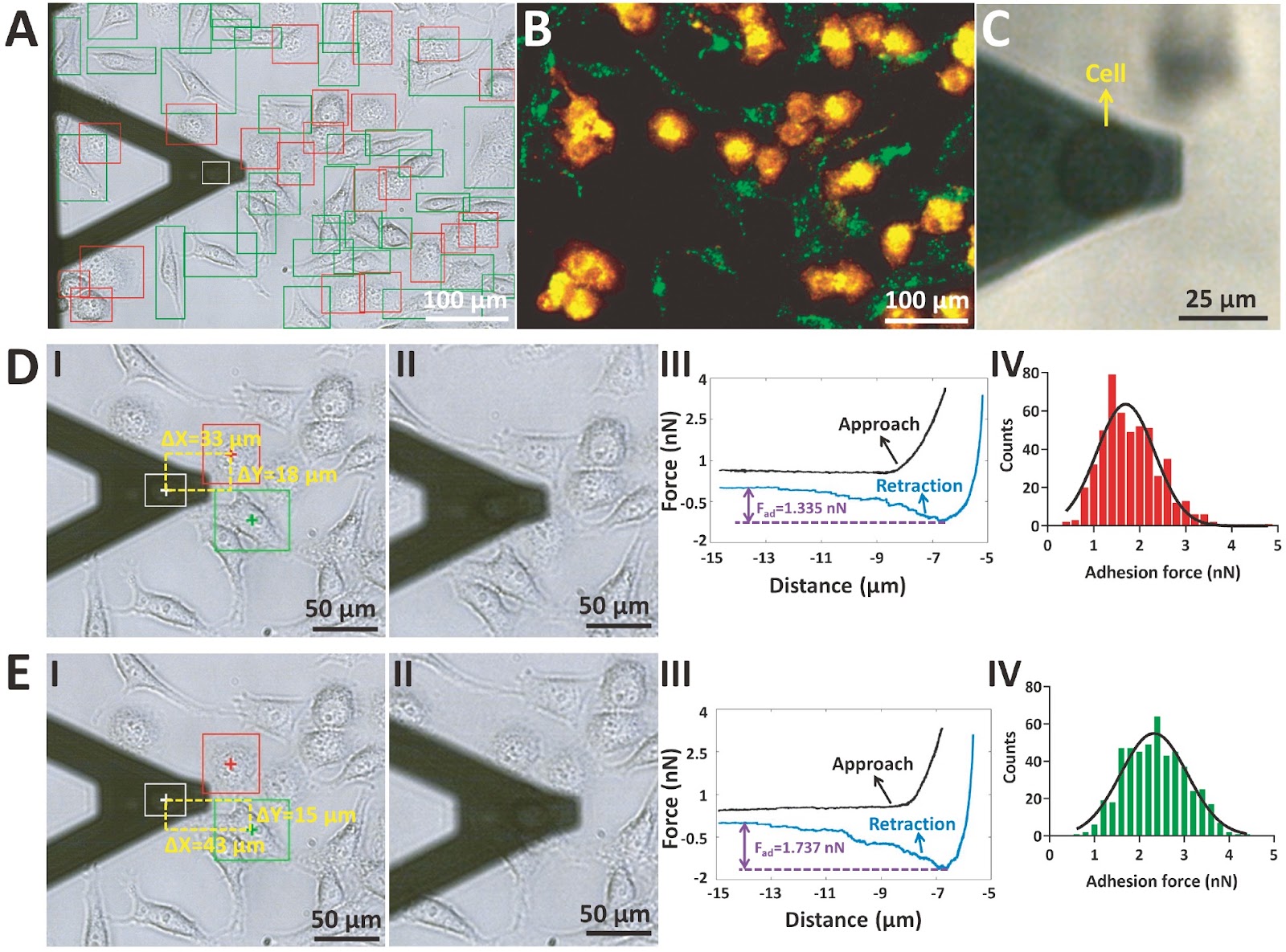
Mechanical cues play an important role in regulating cellular activities. Cells are able to sense and respond to the mechanical cues present in the extracellular physical microenvironment via mechanotransduction, which can ultimately shape the functions and behaviors of the cells themselves as well as their microenvironments during numerous developmental, physiological and pathological processes. The development of human diseases such as cancer is generally accompanied by unique changes in the mechanical properties of cells and their physical microenvironments, and discoveries in the field of physical oncology are beginning to be translated into new therapeutic strategies for cancer. Delineating the mechanical properties of biological tissues in various dimensions from individual cells to organs is therefore fundamental for dissecting the mysteries of life and advancing human healthcare. In particular, atomic force microscopy (AFM)-based force spectroscopy has become a powerful, standard and multifunctional toolbox for characterizing the various mechanical properties of single cells at the micro/nanoscale. However, current studies of AFM-based single-cell mechanical measurements rely mainly on the experience of the experimental operator to move the AFM probe to the target cells for subsequent force measurements, which often results in a time-consuming and laborious experimental process. In addition, cell coculture has been widely used in the field of life sciences to examine intercellular interactions. Nevertheless, in current cell coculture studies, cells are commonly labelled with fluorescent molecules so that one can visually identify the specific cell types in the coculture, which can affect the behaviors of the fluorescently labelled cells. Consequently, developing a method that allows AFM to measure the mechanical properties of cells under coculture conditions in an efficient and fluorescence-independent way will significantly benefit the applications of AFM in the field of mechanobiology. Previously, we presented a method based on the combination of AFM and deep learning optical image recognition, which can precisely move the AFM probe to individual targeted cells to perform mechanical measurements under low-density co-culture conditions (nearly no contact between different cell types in the co-culture). Here, we present a study of deep learning image recognition-assisted AFM to rapidly probe the mechanical properties of single living cells grown in high-density co-culture conditions (with different cell types in contact with each other in the co-culture) without the need for fluorescent labelling. In this work, AFM experiments were performed with a commercial JPK NanoWizard AFM (Bruker, Santa Barbara, USA), which was mounted on an inverted optical microscope (Nikon, Tokyo, Japan). Three types of cells, MGC-803 (a human gastric cancer cell line), HGC-27 (a human undifferentiated gastric cancer cell line), and HMrSV5 (a human peritoneal mesothelial cell line), were used. All three types of cells were cultured in RPMI-1640 medium supplemented with 10% fetal bovine serum (FBS) and 1% penicillin-streptomycin at 37°C (5% CO2 and 95% air) in Petri dishes. During the experiments, the RPMI-1640 medium was replaced by CO2-independent Leibovitz’s L-15 medium, and the AFM experiments were performed at 37°C (the commercial AFM used here has a heater system). MGC-803 cells (stained with the DiI dye) were co-cultured with HGC-27 cells (stained with the DiO dye). Both optical bright-field images and corresponding fluorescent images of co-cultured cells were recorded. The YOLOX deep learning neural network was used for directly recognizing cell types from optical bright-field images. The fluorescent images were used to assist in the preparation of the training datasets (Supplementary Figure S1) and to verify the detection results of the deep learning image recognition model. In a previous study under low-density co-culture conditions, we reduced the complexity of the YOLOX neural network to improve the detection speed without reducing the detection accuracy. Under high-density co-culture conditions, where cell recognition becomes much more difficult, we found that reducing the complexity of the YOLOX model resulted in decreased detection accuracy. We examined the recognition performances of four YOLO series neural networks (YOLOX, YOLOv5, YOLOv7, and YOLOv8), and the experimental results revealed that the YOLOX model had the highest detection precision (89.25%) (Supplementary Table S1) and the best detection result (Supplementary Figure S2) and could meet the experimental requirements. Hence, the YOLOX neural network was used here. The AFM spherical probe (a microsphere attached to the tipless cantilever) was used in the indentation assay to measure the Young’s modulus of the cells, and the AFM single-cell probe (a living HMrSV5 cell attached to the tipless cantilever) was used in the single-cell force spectroscopy (SCFS) assay to measure the adhesion force of the cells. More experimental details (e.g., cell sample
1. Introduction
Mechanical cues play an important role in regulating cellular activities. Cells are able to sense and respond to the mechanical cues present in the extracellular physical microenvironment via mechanotransduction, which can ultimately shape the functions and behaviors of the cells themselves as well as their microenvironments during numerous developmental, physiological and pathological processes. The development of human diseases such as cancer is generally accompanied by unique changes in the mechanical properties of cells and their physical microenvironments, and discoveries in the field of physical oncology are beginning to be translated into new therapeutic strategies for cancer. Delineating the mechanical properties of biological tissues in various dimensions from individual cells to organs is therefore fundamental for dissecting the mysteries of life and advancing human healthcare.
In particular, atomic force microscopy (AFM)-based force spectroscopy has become a powerful, standard and multifunctional toolbox for characterizing the various mechanical properties of single cells at the micro/nanoscale. However, current studies of AFM-based single-cell mechanical measurements rely mainly on the experience of the experimental operator to move the AFM probe to the target cells for subsequent force measurements, which often results in a time-consuming and laborious experimental process. In addition, cell coculture has been widely used in the field of life sciences to examine intercellular interactions. Nevertheless, in current cell coculture studies, cells are commonly labelled with fluorescent molecules so that one can visually identify the specific cell types in the coculture, which can affect the behaviors of the fluorescently labelled cells. Consequently, developing a method that allows AFM to measure the mechanical properties of cells under coculture conditions in an efficient and fluorescence-independent way will significantly benefit the applications of AFM in the field of mechanobiology.
Loading authentic research manuscript (Pages 1–5)...
Xuliang Yang, Mi Li (2026). Label-free and rapid mechanics of single cells under high-density co-culture conditions by deep learning image recognition-assisted atomic force microscopy. Acta Biochimica et Biophysica Sinica. https://doi.org/10.3724/abbs.2024158
Research & Educational Purpose Only:The translations, structured abstracts, analytical annotations, and data reports provided by SinoBioData are intended exclusively for academic research, internal corporate R&D, and educational benchmarking. They do not constitute formal engineering, chemical safety, legal, or professional advice.
Copyright & Intellectual Property Notice: Original copyright of the underlying source articles and experimental data remains with the respective authors, institutions, and original publishing journals. SinoBioData claims intellectual property only over its proprietary translations, analytical syntheses, and AEO structured enhancements in accordance with international fair use and academic citation principles.
Frequently Asked Questions
What is the main advantage of the proposed AFM method?
The method enables label-free and rapid measurement of single-cell mechanical properties under high-density co-culture conditions, eliminating the need for fluorescent labeling and reducing experimental time and labor.
How does deep learning assist in AFM measurements?
Deep learning, specifically the YOLOX neural network, automatically recognizes different cell types from optical bright-field images, allowing the AFM probe to be precisely positioned on target cells without manual intervention.
What cell types were used in the study?
The study used MGC-803 (human gastric cancer), HGC-27 (human undifferentiated gastric cancer), and HMrSV5 (human peritoneal mesothelial) cell lines.
What mechanical properties were measured?
The Young's modulus (stiffness) of cells was measured using AFM spherical probes, and cell adhesion forces were measured using single-cell force spectroscopy with living cell probes.
How does this method compare to traditional AFM approaches?
Traditional AFM requires manual probe positioning and often uses fluorescent labels to identify cells in co-culture. This method automates cell recognition via deep learning and works label-free, making it faster and less invasive.
Related Technical Papers & Translations

Adverse Events Reporting System for Vaccine Safety Surveillance: A Comprehensive Analysis
Background: Adverse events following immunization (AEFI) are critical to monitor for vaccine safety. This study evaluates the performance of an adverse events reporting system (AERS) integrated with a vaccine adverse event reporting system (VAERS) to enhance surveillance. Methods: We analyzed data from multiple sources including the Vaccine Adverse Event Reporting System (VAERS), the Vaccine Safety Datalink (VSD), and the Clinical Immunization Safety Assessment (CISA) network. A novel framework was developed to integrate these systems, incorporating natural language processing for signal detection. Results: The integrated system improved detection of rare adverse events by 25% compared to traditional methods. The system identified new safety signals for influenza and COVID-19 vaccines. Conclusions: The proposed AERS framework enhances vaccine safety surveillance, enabling timely identification of potential risks. Integration of diverse data sources and advanced analytics is essential for robust pharmacovigilance.

Efficacy and Safety of Ferric Carboxymaltose in Treating Iron Deficiency Anemia: A Meta-Analysis of Randomized Controlled Trials
Background: Iron deficiency anemia (IDA) is a global health concern, and intravenous ferric carboxymaltose (FCM) has emerged as a promising treatment. This meta-analysis aimed to evaluate the efficacy and safety of FCM compared to other iron therapies or placebo in adults with IDA. Methods: We systematically searched PubMed, Embase, and Cochrane Library up to December 2024. Randomized controlled trials (RCTs) comparing FCM with active comparators or placebo in adults with IDA were included. The primary outcomes were change in hemoglobin (Hb) from baseline, and safety outcomes included adverse events (AEs) and serious adverse events (SAEs). Pooled estimates were calculated using random-effects models. Results: A total of 15 RCTs involving 4,856 patients were included. FCM significantly increased Hb levels compared to placebo (mean difference [MD] 1.2 g/dL, 95% CI 0.9-1.5) and was non-inferior to other intravenous iron preparations. The risk of AEs was similar between FCM and comparators (risk ratio [RR] 1.05, 95% CI 0.95-1.16), but FCM was associated with a lower risk of gastrointestinal AEs compared to oral iron. Serious adverse events were rare and comparable across groups. Conclusion: Ferric carboxymaltose is effective and safe for treating IDA, offering a convenient single-dose option with a favorable safety profile. These findings support its use in clinical practice.

Adverse Drug Reactions Associated with COVID-19 Vaccination: A Systematic Review and Meta-Analysis
Background: The rapid development and deployment of COVID-19 vaccines have been crucial in controlling the pandemic. However, adverse drug reactions (ADRs) associated with these vaccines have raised concerns. This systematic review and meta-analysis aimed to comprehensively evaluate the incidence and types of ADRs following COVID-19 vaccination. Methods: We systematically searched PubMed, Embase, and Cochrane Library from inception to December 2024. Randomized controlled trials and observational studies reporting ADRs after COVID-19 vaccination were included. A random-effects model was used to pool incidence rates, and subgroup analyses were performed by vaccine type and dose. Results: A total of 45 studies with 1,234,567 participants were included. The overall incidence of any ADR was 62.3% (95% CI: 58.1-66.4%). Common local reactions included injection site pain (48.2%), swelling (22.5%), and redness (18.7%). Systemic reactions included fatigue (34.6%), headache (28.9%), and myalgia (22.3%). Serious ADRs were rare (0.02%). Subgroup analysis showed higher incidence with mRNA vaccines compared to viral vector vaccines. Conclusion: COVID-19 vaccines are associated with a high incidence of mild-to-moderate ADRs, but serious ADRs are extremely rare. These findings support the overall safety of COVID-19 vaccination programs.