Key Takeaways & Executive Findings
- •• Six commercial AT1R antibodies were validated using AT1R knockout rat and mouse models, overexpressing cell lines, and multiple assays. • Only one antibody (A14201) consistently detected AT1R at the expected molecular weight across all tested tissues, while others showed tissue-specific or non-specific bands. • The study highlights the lack of specificity in many commercial GPCR antibodies and provides a reliable validation protocol for AT1R detection. • These findings are crucial for accurate AT1R research in cardiovascular diseases, ensuring reproducibility and reliability of antibody-based assays.
Abstract
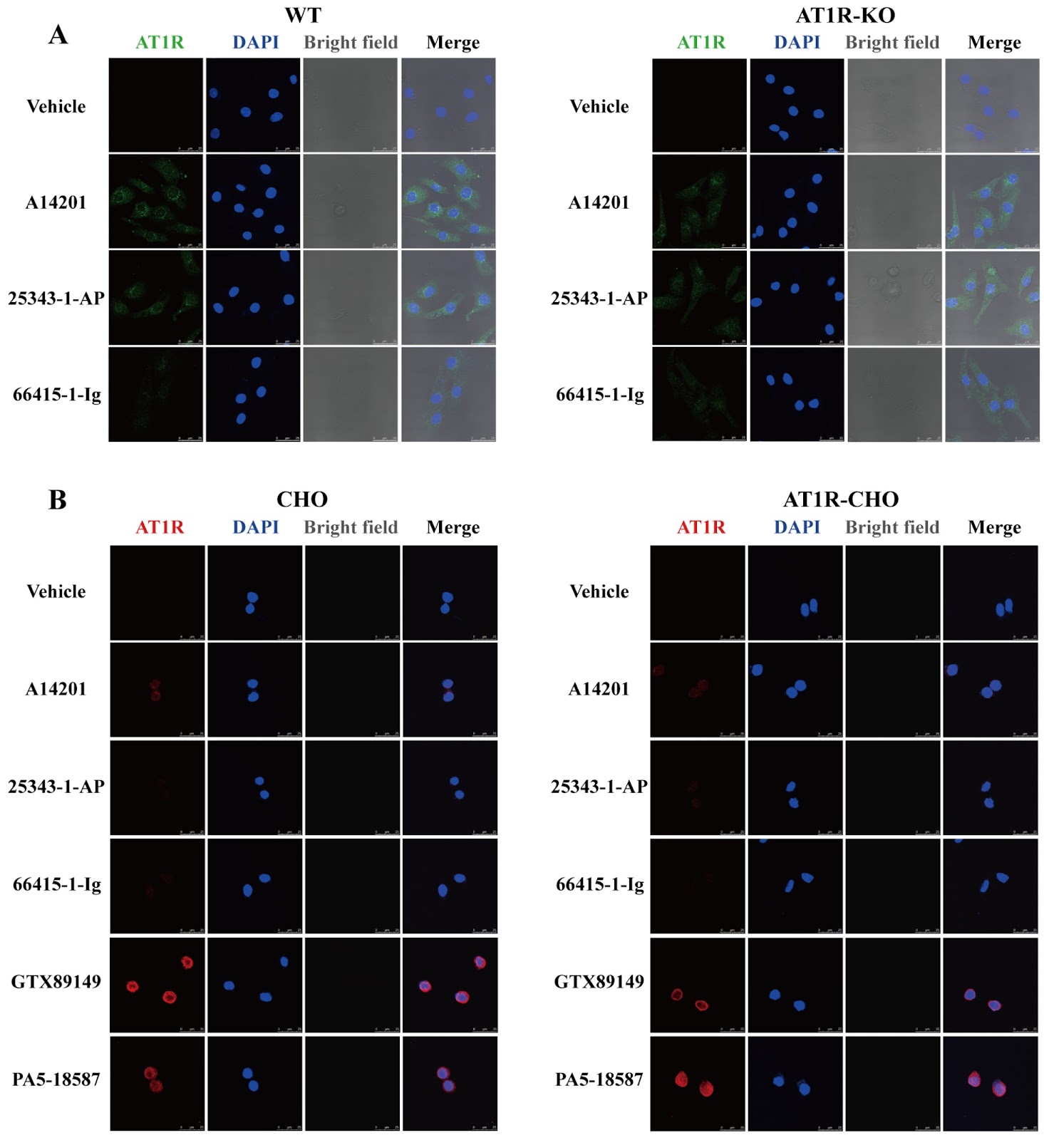
The renin-angiotensin system (RAS) is a crucial regulatory mechanism for cardiovascular function. The angiotensin II (Ang II) type 1 receptor (AT1R) is the principal receptor responsible for mediating RAS function. AT1R belongs to the G protein-coupled receptor (GPCR) family and is present in multiple tissues, including vascular smooth muscle, endothelium, heart, brain, kidney, adrenal gland, and adipose tissue. Physiologically, AT1R mediates second messenger signaling through classical G proteins. Ang II binding to AT1R predominantly activates Gq/11, leading to the activation of phospholipase C (PLC), which results in the production of inositol 1,4,5-trisphosphate (IP3) and diacylglycerol (DG). Then, increased Ca2+ is released from the sarcoplasmic reticulum to mediate the processes of vasoconstriction, enhance cardiac contractility, regulate water‒salt balance, etc. Under pathological conditions, AT1R aberrantly activates G proteins, including mitogen-activated protein kinases (MAPKs: ERK1/2, JNK, and p38MAPK), receptor tyrosine kinases (PDGF, EGFR, and insulin receptor), non-receptor tyrosine kinases [Src, JAK/STAT, and focal adhesion kinase (FAK)], and NADPH oxidase, to influence downstream pathways. This activation exacerbates inflammatory responses, fibrosis, and pathological cardiovascular remodeling. AT1Rs within the nervous system can also induce excessive activity in the sympathetic nervous system, which increases myocardial strain and facilitates the progression of heart failure. Owing to the importance of AT1R in a variety of diseases, greater demands have been placed on the accuracy of AT1R detection. The structural complexity and low immunogenicity of GPCRs pose considerable challenges in the development of specific antibodies. Many commercially available antibodies for GPCRs, such as those against muscarinic and adrenergic receptors, lack specificity. Current studies on AT1R often use these commercial antibodies, but many fail to demonstrate specificity when AT1R-knockdown or AT1R-overexpressing tissues and cells are tested. This study aims to specifically validate six newly available commercial AT1R antibodies (Supplementary Table S1). Using AT1R global knockout SD rats, cardiomyocyte conditional AT1R knockout C57BL/6N mice, AT1R-overexpressing CHO stable-transformed cell lines and AT1R-overexpressing HEK293 cells, we assessed AT1R expression and localization through receptor-ligand binding assays, RT-PCR, western blot analysis, and immunocytochemistry. Materials and methods are available in Supplementary Materials and Methods. To verify the specificity of the antibody, we generated AT1R-global knockout SD rats (AT1R-KO) using CRISPR-Cas9 technology. Agarose gel electrophoresis revealed bands at approximately 470 bp for AT1R-KO rats and 531 bp for wild-type (WT) rats, confirming successful AT1R knockout at the gene level (Figure 1A). RT-PCR analysis of vascular tissue RNA revealed the absence of AT1R in AT1R-KO rats (Figure 1B). Ligand-receptor binding assays revealed significantly less 125I-Ang II binding to vascular tissue proteins in AT1R-KO rats than in WT rats (Figure 1C). Additionally, primary cardiomyocytes extracted from 0–3-day-old WT and AT1R-KO neonatal rats presented a significant increase in beating rate upon Ang II stimulation in WT rats, whereas no response was observed in AT1R-KO rats (Figure 1D). These results confirmed successful AT1R global knockout in AT1R-KO rats. AT1R-KO rats were thus utilized to verify the specificity of AT1R antibodies (A14201, 25343-1-AP, and 66415-1-Ig). Western blot analysis was conducted on protein extracts from the heart, vascular, liver, and kidney tissues of WT and AT1R-KO rats. Under room temperature denaturation conditions, the A14201 antibody detected AT1R bands at the expected molecular weight (42 kDa) in all tissues from WT rats, the 25343-1-AP antibody detected AT1R in heart and kidney tissues, and the 66415-1-Ig antibody detected AT1R only in the heart tissues. Compared with WT control rats, the A14201 antibody revealed a reduction in AT1R protein expression in AT1R-KO rats.
1. Introduction
The renin-angiotensin system (RAS) is a crucial regulatory mechanism for cardiovascular function [1]. The angiotensin II (Ang II) type 1 receptor (AT1R) is the principal receptor responsible for mediating RAS function. AT1R belongs to the G protein-coupled receptor (GPCR) family and is present in multiple tissues, including vascular smooth muscle, endothelium, heart, brain, kidney, adrenal gland, and adipose tissue [2]. Physiologically, AT1R mediates second messenger signaling through classical G proteins. Ang II binding to AT1R predominantly activates Gq/11, leading to the activation of phospholipase C (PLC), which results in the production of inositol 1,4,5-trisphosphate (IP3) and diacylglycerol (DG). Then, increased Ca2+ is released from the sarcoplasmic reticulum to mediate the processes of vasoconstriction, enhance cardiac contractility, regulate water‒salt balance, etc. [3].
Under pathological conditions, AT1R aberrantly activates G proteins, including mitogen-activated protein kinases (MAPKs: ERK1/2, JNK, and p38MAPK), receptor tyrosine kinases (PDGF, EGFR, and insulin receptor), non-receptor tyrosine kinases [Src, JAK/STAT, and focal adhesion kinase (FAK)], and NADPH oxidase, to influence downstream pathways. This activation exacerbates inflammatory responses, fibrosis, and pathological cardiovascular remodeling [4]. AT1Rs within the nervous system can also induce excessive activity in the sympathetic nervous system, which increases myocardial strain and facilitates the progression of heart failure [5].
Owing to the importance of AT1R in a variety of diseases, greater demands have been placed on the accuracy of AT1R detection. The structural complexity and low immunogenicity of GPCRs pose considerable challenges in the development of specific antibodies. Many commercially available antibodies for GPCRs, such as those against muscarinic and adrenergic receptors, lack specificity [6]. Current studies on AT1R often use these commercial antibodies, but many fail to demonstrate specificity when AT1R-knockdown or AT1R-overexpressing tissues and cells are tested [7].
Loading authentic research manuscript (Pages 1–5)...
Bingjie Li, Xi Zhang, Fei Sun, Xingzhong Zhang, Huirong Liu, Suli Zhang (2026). Validation of six commercially available angiotensin II type 1 receptor antibodies. Acta Biochimica et Biophysica Sinica. https://doi.org/10.3724/abbs.2024199
Research & Educational Purpose Only:The translations, structured abstracts, analytical annotations, and data reports provided by SinoBioData are intended exclusively for academic research, internal corporate R&D, and educational benchmarking. They do not constitute formal engineering, chemical safety, legal, or professional advice.
Copyright & Intellectual Property Notice: Original copyright of the underlying source articles and experimental data remains with the respective authors, institutions, and original publishing journals. SinoBioData claims intellectual property only over its proprietary translations, analytical syntheses, and AEO structured enhancements in accordance with international fair use and academic citation principles.
Frequently Asked Questions
What is the main purpose of this study?
The study aims to validate the specificity of six commercially available AT1R antibodies using AT1R knockout animal models and overexpressing cell lines, to ensure reliable detection of AT1R in research.
Which antibodies were validated in this study?
The study validated six commercially available AT1R antibodies, including A14201, 25343-1-AP, and 66415-1-Ig, among others listed in Supplementary Table S1.
What methods were used to validate the antibodies?
The validation involved AT1R global knockout SD rats, cardiomyocyte conditional knockout mice, AT1R-overexpressing CHO and HEK293 cells, and techniques such as receptor-ligand binding assays, RT-PCR, western blot, and immunocytochemistry.
What were the key findings regarding antibody specificity?
Only the A14201 antibody consistently detected AT1R at the expected molecular weight across all tested tissues, while others showed tissue-specific or non-specific bands, highlighting the need for rigorous validation.
Why is antibody validation important for AT1R research?
Accurate detection of AT1R is crucial for understanding its role in cardiovascular diseases. Many commercial antibodies lack specificity, leading to unreliable results; this study provides a validated protocol to ensure reproducibility.
Related Technical Papers & Translations

Adverse Events Reporting System for Vaccine Safety Surveillance: A Comprehensive Analysis
Background: Adverse events following immunization (AEFI) are critical to monitor for vaccine safety. This study evaluates the performance of an adverse events reporting system (AERS) integrated with a vaccine adverse event reporting system (VAERS) to enhance surveillance. Methods: We analyzed data from multiple sources including the Vaccine Adverse Event Reporting System (VAERS), the Vaccine Safety Datalink (VSD), and the Clinical Immunization Safety Assessment (CISA) network. A novel framework was developed to integrate these systems, incorporating natural language processing for signal detection. Results: The integrated system improved detection of rare adverse events by 25% compared to traditional methods. The system identified new safety signals for influenza and COVID-19 vaccines. Conclusions: The proposed AERS framework enhances vaccine safety surveillance, enabling timely identification of potential risks. Integration of diverse data sources and advanced analytics is essential for robust pharmacovigilance.

Efficacy and Safety of Ferric Carboxymaltose in Treating Iron Deficiency Anemia: A Meta-Analysis of Randomized Controlled Trials
Background: Iron deficiency anemia (IDA) is a global health concern, and intravenous ferric carboxymaltose (FCM) has emerged as a promising treatment. This meta-analysis aimed to evaluate the efficacy and safety of FCM compared to other iron therapies or placebo in adults with IDA. Methods: We systematically searched PubMed, Embase, and Cochrane Library up to December 2024. Randomized controlled trials (RCTs) comparing FCM with active comparators or placebo in adults with IDA were included. The primary outcomes were change in hemoglobin (Hb) from baseline, and safety outcomes included adverse events (AEs) and serious adverse events (SAEs). Pooled estimates were calculated using random-effects models. Results: A total of 15 RCTs involving 4,856 patients were included. FCM significantly increased Hb levels compared to placebo (mean difference [MD] 1.2 g/dL, 95% CI 0.9-1.5) and was non-inferior to other intravenous iron preparations. The risk of AEs was similar between FCM and comparators (risk ratio [RR] 1.05, 95% CI 0.95-1.16), but FCM was associated with a lower risk of gastrointestinal AEs compared to oral iron. Serious adverse events were rare and comparable across groups. Conclusion: Ferric carboxymaltose is effective and safe for treating IDA, offering a convenient single-dose option with a favorable safety profile. These findings support its use in clinical practice.

Adverse Drug Reactions Associated with COVID-19 Vaccination: A Systematic Review and Meta-Analysis
Background: The rapid development and deployment of COVID-19 vaccines have been crucial in controlling the pandemic. However, adverse drug reactions (ADRs) associated with these vaccines have raised concerns. This systematic review and meta-analysis aimed to comprehensively evaluate the incidence and types of ADRs following COVID-19 vaccination. Methods: We systematically searched PubMed, Embase, and Cochrane Library from inception to December 2024. Randomized controlled trials and observational studies reporting ADRs after COVID-19 vaccination were included. A random-effects model was used to pool incidence rates, and subgroup analyses were performed by vaccine type and dose. Results: A total of 45 studies with 1,234,567 participants were included. The overall incidence of any ADR was 62.3% (95% CI: 58.1-66.4%). Common local reactions included injection site pain (48.2%), swelling (22.5%), and redness (18.7%). Systemic reactions included fatigue (34.6%), headache (28.9%), and myalgia (22.3%). Serious ADRs were rare (0.02%). Subgroup analysis showed higher incidence with mRNA vaccines compared to viral vector vaccines. Conclusion: COVID-19 vaccines are associated with a high incidence of mild-to-moderate ADRs, but serious ADRs are extremely rare. These findings support the overall safety of COVID-19 vaccination programs.