Key Takeaways & Executive Findings
- •• OGG1 deficiency exacerbates bleomycin-induced cellular senescence in lung cells, characterized by increased SA-β-gal positivity and elevated p21 and p-H2AX levels. • OGG1 overexpression reverses senescence markers, promotes cell cycle progression, and maintains TERT and LaminB1 levels in BLM-treated cells. • OGG1 binds to p53, inhibiting its activation; silencing p53 reverses OGG1's protective effect, implicating the p53-p21 pathway as a key mediator. • In vivo, OGG1-deficient mice show augmented senescence, suggesting OGG1 as a potential therapeutic target for pulmonary fibrosis and aging-related lung diseases.
Abstract
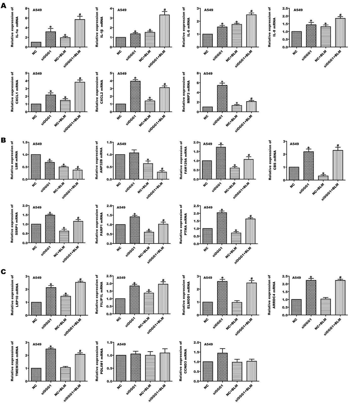
Cellular senescence is an important factor leading to pulmonary fibrosis. Deficiency of 8-oxoguanine DNA glycosylase (OGG1) in mice leads to alleviation of bleomycin (BLM)-induced mouse pulmonary fibrosis, and inhibition of the OGG1 enzyme reduces the epithelial mesenchymal transition (EMT) in lung cells. In the present study, we find decreased expression of OGG1 in aged mice and BLM-induced cell senescence. In addition, a decrease in OGG1 expression results in cell senescence, such as increases in the percentage of SA-β-gal-positive cells, and in the p21 and p-H2AX protein levels in response to BLM in lung cells. Furthermore, OGG1 promotes cell transformation in A549 cells in the presence of BLM. We also find that OGG1 siRNA impedes cell cycle progression and inhibits the levels of telomerase reverse transcriptase (TERT) and LaminB1 in BLM-treated lung cells. The increase in OGG1 expression results in the opposite phenomenon. The mRNA levels of senescence-associated secretory phenotype (SASP) components, including IL-1α, IL-1β, IL-6, IL-8, CXCL1/CXCL2, and MMP-3, in the absence of OGG1 are obviously increased in A549 cells treated with BLM. Interestingly, we demonstrate that OGG1 binds to p53 to inhibit the activation of p53 and that silencing of p53 reverses the inhibition of OGG1 on senescence in lung cells. Additionally, the augmented cell senescence is shown in vivo in OGG1-deficient mice. Overall, we provide direct evidence in vivo and in vitro that OGG1 plays an important role in protecting tissue cells against aging associated with the p53 pathway.
1. Introduction
Senescence is irreversible growth inhibition of cells during aging as well as for different cell stimulation sources, including activated oncogenes, cytokines, reactive oxygen species, DNA damage, and nucleotide depletion [1–3]. Cellular senescence is accelerated in patients and appears to play a role in aging-associated morbidity. Traditionally, cell senescence is considered to be a beneficial physiological mechanism in the process of development, wound healing and tumor inhibition. However, in recent years, several lines of evidence have demonstrated that senescent cells persist or accumulate, which have harmful consequences. Aging of alveolar epithelial cells (AEC) may be detrimental to lung repair [4]. Depletion of senescent epithelial cells in vitro and ex vivo decreases fibrotic markers [5]. Notably, cellular senescence may play a positive or negative role in regulating organ fibrosis [5–7].
Senescent cells are involved in different pathophysiological processes. The senescence process is characterized by a variety of nonunique markers, including constitutive DNA damage response (DDR) signaling, senescence-associated β-galactosidase (SA-β-gal) activity, increased expression of cyclin-dependent kinase (CDK) inhibitors, p16INK4A (CDKN2A) and p21CIP1 (CDKN1A), increased secretion of many bioactive factors, including the senescence-associated secretory phenotype (SASP), and reduced expression of the nuclear lamina protein LaminB1 (LMNB1) [8,9]. The p53 transcription factor plays an important role in cellular responses to stress. Its activation in response to DNA damage results in cell growth arrest, allowing for DNA repair, or induces cellular senescence or apoptosis, thereby leading to the maintenance of genome integrity [10].
8-Oxo-7,8-dihydroguanine (8-oxo-G) is the main product of oxidative DNA damage in cells and one of the most common types of endogenously generated mutagenic base damage [11,12]. The corresponding DNA repair enzyme is 8-oxoguanine DNA glycosylase (OGG1), a DNA repair glycosylase that localizes to both the nucleus and mitochondria [13–16], and it has been shown to activate α-SMA polymerization and to increase the levels of α-SMA exposure to stress fibres [17]. OGG1 inhibition mediated by EGFR is involved in the cell transformation induced by wood dust exposure [18]. Our recent study demonstrated that OGG1 enhanced cell transformation and that OGG1 knockout relieved bleomycin (BLM)-induced pulmonary fibrosis in mice [14]. Numerous studies have shown that cellular senescence is an important factor in the occurrence and development of a variety of lung diseases [19], so what role does OGG1 play in cellular senescence in lung fibrosis?
Loading authentic research manuscript (Pages 1–5)...
Shenglan Gao, Lujun Chen, Ziying Lin, Zhiliang Xu, Yahong Wang, Huayu Ling, Zijun Wu, Yu Yin, Weimin Yao, Keng Wu, Gang Liu (2026). 8-Oxoguanine DNA glycosylase protects cells from senescence via the p53-p21 pathway. Acta Biochimica et Biophysica Sinica. https://doi.org/10.3724/abbs.2023264
Research & Educational Purpose Only:The translations, structured abstracts, analytical annotations, and data reports provided by SinoBioData are intended exclusively for academic research, internal corporate R&D, and educational benchmarking. They do not constitute formal engineering, chemical safety, legal, or professional advice.
Copyright & Intellectual Property Notice: Original copyright of the underlying source articles and experimental data remains with the respective authors, institutions, and original publishing journals. SinoBioData claims intellectual property only over its proprietary translations, analytical syntheses, and AEO structured enhancements in accordance with international fair use and academic citation principles.
Frequently Asked Questions
What is the role of OGG1 in cellular senescence?
OGG1 protects cells from senescence by inhibiting p53 activation via direct binding, thereby reducing senescence markers and SASP components in lung cells.
How does OGG1 deficiency affect bleomycin-induced pulmonary fibrosis?
OGG1 deficiency alleviates bleomycin-induced pulmonary fibrosis in mice, but also leads to increased cellular senescence, suggesting a complex role in fibrosis and aging.
What is the significance of the p53-p21 pathway in OGG1-mediated protection?
OGG1 binds to p53 and inhibits its activation, which in turn reduces p21 expression and prevents senescence. Silencing p53 reverses OGG1's protective effect, confirming the pathway's importance.
Could OGG1 be a therapeutic target for aging-related lung diseases?
Yes, enhancing OGG1 activity may protect against cellular senescence and pulmonary fibrosis, offering a potential therapeutic strategy for aging-related lung diseases.
What are the key senescence markers affected by OGG1?
OGG1 modulates SA-β-gal activity, p21 and p-H2AX levels, TERT and LaminB1 expression, and SASP components such as IL-1α, IL-1β, IL-6, IL-8, CXCL1/CXCL2, and MMP-3.
Related Technical Papers & Translations

Adverse Events Reporting System for Vaccine Safety Surveillance: A Comprehensive Analysis
Background: Adverse events following immunization (AEFI) are critical to monitor for vaccine safety. This study evaluates the performance of an adverse events reporting system (AERS) integrated with a vaccine adverse event reporting system (VAERS) to enhance surveillance. Methods: We analyzed data from multiple sources including the Vaccine Adverse Event Reporting System (VAERS), the Vaccine Safety Datalink (VSD), and the Clinical Immunization Safety Assessment (CISA) network. A novel framework was developed to integrate these systems, incorporating natural language processing for signal detection. Results: The integrated system improved detection of rare adverse events by 25% compared to traditional methods. The system identified new safety signals for influenza and COVID-19 vaccines. Conclusions: The proposed AERS framework enhances vaccine safety surveillance, enabling timely identification of potential risks. Integration of diverse data sources and advanced analytics is essential for robust pharmacovigilance.

Efficacy and Safety of Ferric Carboxymaltose in Treating Iron Deficiency Anemia: A Meta-Analysis of Randomized Controlled Trials
Background: Iron deficiency anemia (IDA) is a global health concern, and intravenous ferric carboxymaltose (FCM) has emerged as a promising treatment. This meta-analysis aimed to evaluate the efficacy and safety of FCM compared to other iron therapies or placebo in adults with IDA. Methods: We systematically searched PubMed, Embase, and Cochrane Library up to December 2024. Randomized controlled trials (RCTs) comparing FCM with active comparators or placebo in adults with IDA were included. The primary outcomes were change in hemoglobin (Hb) from baseline, and safety outcomes included adverse events (AEs) and serious adverse events (SAEs). Pooled estimates were calculated using random-effects models. Results: A total of 15 RCTs involving 4,856 patients were included. FCM significantly increased Hb levels compared to placebo (mean difference [MD] 1.2 g/dL, 95% CI 0.9-1.5) and was non-inferior to other intravenous iron preparations. The risk of AEs was similar between FCM and comparators (risk ratio [RR] 1.05, 95% CI 0.95-1.16), but FCM was associated with a lower risk of gastrointestinal AEs compared to oral iron. Serious adverse events were rare and comparable across groups. Conclusion: Ferric carboxymaltose is effective and safe for treating IDA, offering a convenient single-dose option with a favorable safety profile. These findings support its use in clinical practice.

Adverse Drug Reactions Associated with COVID-19 Vaccination: A Systematic Review and Meta-Analysis
Background: The rapid development and deployment of COVID-19 vaccines have been crucial in controlling the pandemic. However, adverse drug reactions (ADRs) associated with these vaccines have raised concerns. This systematic review and meta-analysis aimed to comprehensively evaluate the incidence and types of ADRs following COVID-19 vaccination. Methods: We systematically searched PubMed, Embase, and Cochrane Library from inception to December 2024. Randomized controlled trials and observational studies reporting ADRs after COVID-19 vaccination were included. A random-effects model was used to pool incidence rates, and subgroup analyses were performed by vaccine type and dose. Results: A total of 45 studies with 1,234,567 participants were included. The overall incidence of any ADR was 62.3% (95% CI: 58.1-66.4%). Common local reactions included injection site pain (48.2%), swelling (22.5%), and redness (18.7%). Systemic reactions included fatigue (34.6%), headache (28.9%), and myalgia (22.3%). Serious ADRs were rare (0.02%). Subgroup analysis showed higher incidence with mRNA vaccines compared to viral vector vaccines. Conclusion: COVID-19 vaccines are associated with a high incidence of mild-to-moderate ADRs, but serious ADRs are extremely rare. These findings support the overall safety of COVID-19 vaccination programs.