🧬 SinoBioData Academic Portal
📚 Peer-Reviewed Translated Literature

All Biomedical & Clinical Articles (Page 48)

Browse complete peer-reviewed translations from top Chinese biomedical, oncology, and genomics journals. Read verified previews and download full authentic clinical reports.

Published Research Papers

Showing 24 of 1542 peer-reviewed translated articles (Page 48 of 65)

Puerarin prevents cadmium-induced endoplasmic reticulum stress via SIRT1-dependent PERK-CHOP pathway in HepG2 cellsGraphical AbstractVerified
Acta Biochimica et Biophysica Sinica

Puerarin prevents cadmium-induced endoplasmic reticulum stress via SIRT1-dependent PERK-CHOP pathway in HepG2 cells

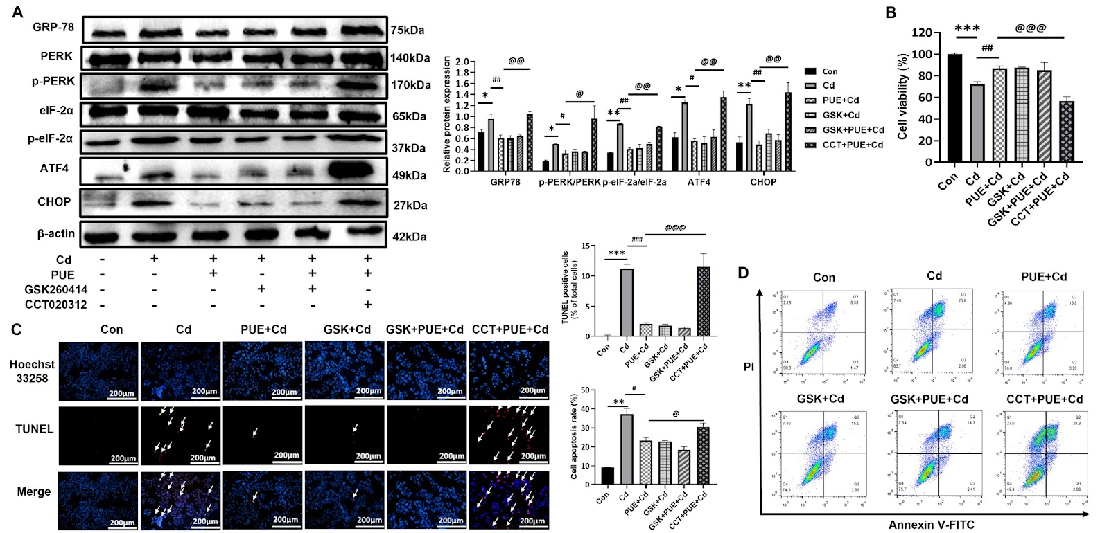
Cadmium (Cd) is a high-risk heavy metal that induces oxidative stress, endoplasmic reticulum (ER) stress and inflammation, damaging organs such as the liver. Puerarin (PUE) has been shown to treat liver injury and especially prevent Cd-induced hepatic damage via its antioxidant activity. Sirtuin 1 (SIRT1), a histone deacetylase, is a key protector against various stress insults. However, its role in the protection of PUE against Cd-induced liver damage has not been clarified. Thus, this study is designed to elucidate the molecular mechanism in the human hepatoma cell line HepG2. The results first reveal that Cd-induced apoptosis is significantly restored by PUE pretreatment, as confirmed by the CCK-8, flow cytometric, Hoechst 33258 and TUNEL assays. Mechanistically, PUE significantly decreases ROS production and increases SOD levels in Cd-treated HepG2 cells. Moreover, PUE pretreatment alleviates ER stress by inhibiting the PERK-eIF2α-ATF4-CHOP axis and subsequently partially restores ER function as revealed by decreased Ca2+ release from the ER. In addition, further study demonstrates that PUE upregulates SIRT1 expression, which suppresses the PERK signaling cascade and reduces CHOP levels. Collectively, our results first demonstrate that PUE protects HepG2 cells from Cd-induced apoptosis at least partially by inhibiting the PERK-eIF2α-ATF4-CHOP pathway in a SIRT1 expression-dependent manner. Puerarin appears to have great potential as a hepatoprotective agent.

Read Full Abstract10.3724/abbs.2025039
Ageing-associated gut dysbiosis deteriorates mouse cognitionGraphical AbstractVerified
Acta Biochimica et Biophysica Sinica

Ageing-associated gut dysbiosis deteriorates mouse cognition

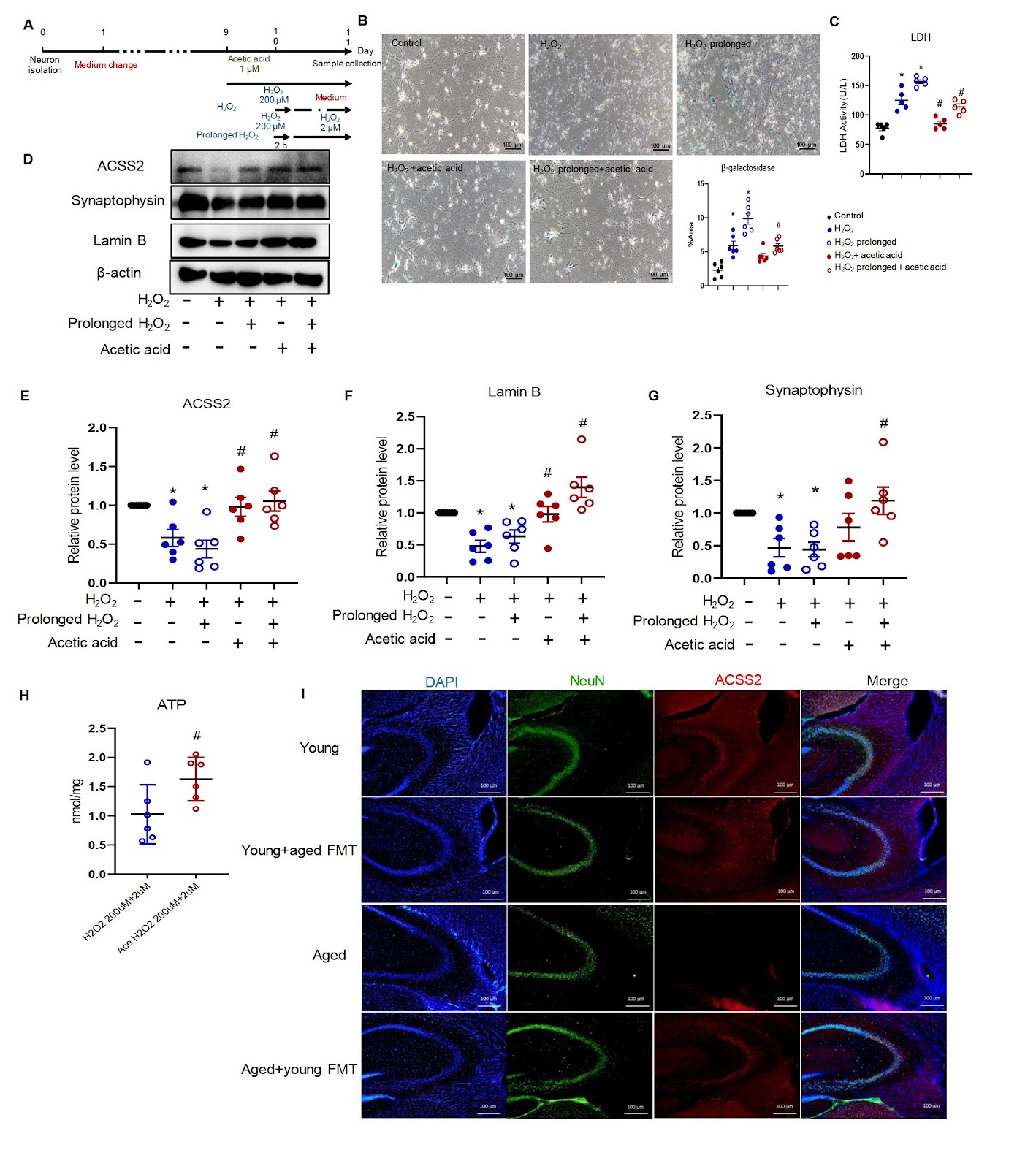
Ageing is an independent factor for cognitive dysfunction. Ageing-associated alterations in the gut microbiota also affect cognition. The present study is designed to investigate changes in the gut microbiota and their participation in ageing-associated cognitive impairment. Both 10-week-old and 18-month-old mice are used. Mouse cognition is examined by novel object recognition and T-maze tests. Mouse feces are collected for sequencing and transplantation. Protein expression in the mouse intestine and hippocampus is studied using immunohistochemistry and immunofluorescence staining. Senescent neurons are induced by hydrogen peroxide in vitro. The cell lysates are used for western blot analysis and adenosine triphosphate (ATP) measurement. Our results show that 18-month-old mice exhibit cognitive dysfunction compared with young mice. In aged mice, transplanting the microbiota of young mice increases the protein presence of synaptophysin in the hippocampus and partially restores cognition. The protein expressions of mucin-2 and E-cadherin in the intestine are reduced in aged mice but are increased by transplantation. Gut microbiota analyses reveal that the reduced abundance of the microbe Bacilli-Lactobacillales-Lactobacillaceae-Lactobacillus in aged mice is restored by transplantation. Fecal microbiota transplantation in young mice increases the serum level of acetic acid in aged mice. Hydrogen peroxide stimulation induces senescence and reduces the protein expression levels of synaptophysin and acetyl-coenzyme A synthetase member 2 (ACSS2) in primary neurons. Incubation with acetic acid upregulates the protein expressions of ACSS2 and synaptophysin and further increases ATP production in senescent neurons. In summary, gut microbiota transplantation increases the abundance of Lactobacillales, elevates serum acetic acid level, and improves cognitive function in aged mice. Gut microbiota transplantation has therapeutic importance for ageing-associated cognitive decline.

Read Full Abstract10.3724/abbs.2024217
Levosimendan ameliorates cardiomyocyte injury and mitochondrial dysfunction in an Nrf2-dependent manner in mice with sepsis-induced cardiomyopathyGraphical AbstractVerified
Acta Biochimica et Biophysica Sinica

Levosimendan ameliorates cardiomyocyte injury and mitochondrial dysfunction in an Nrf2-dependent manner in mice with sepsis-induced cardiomyopathy

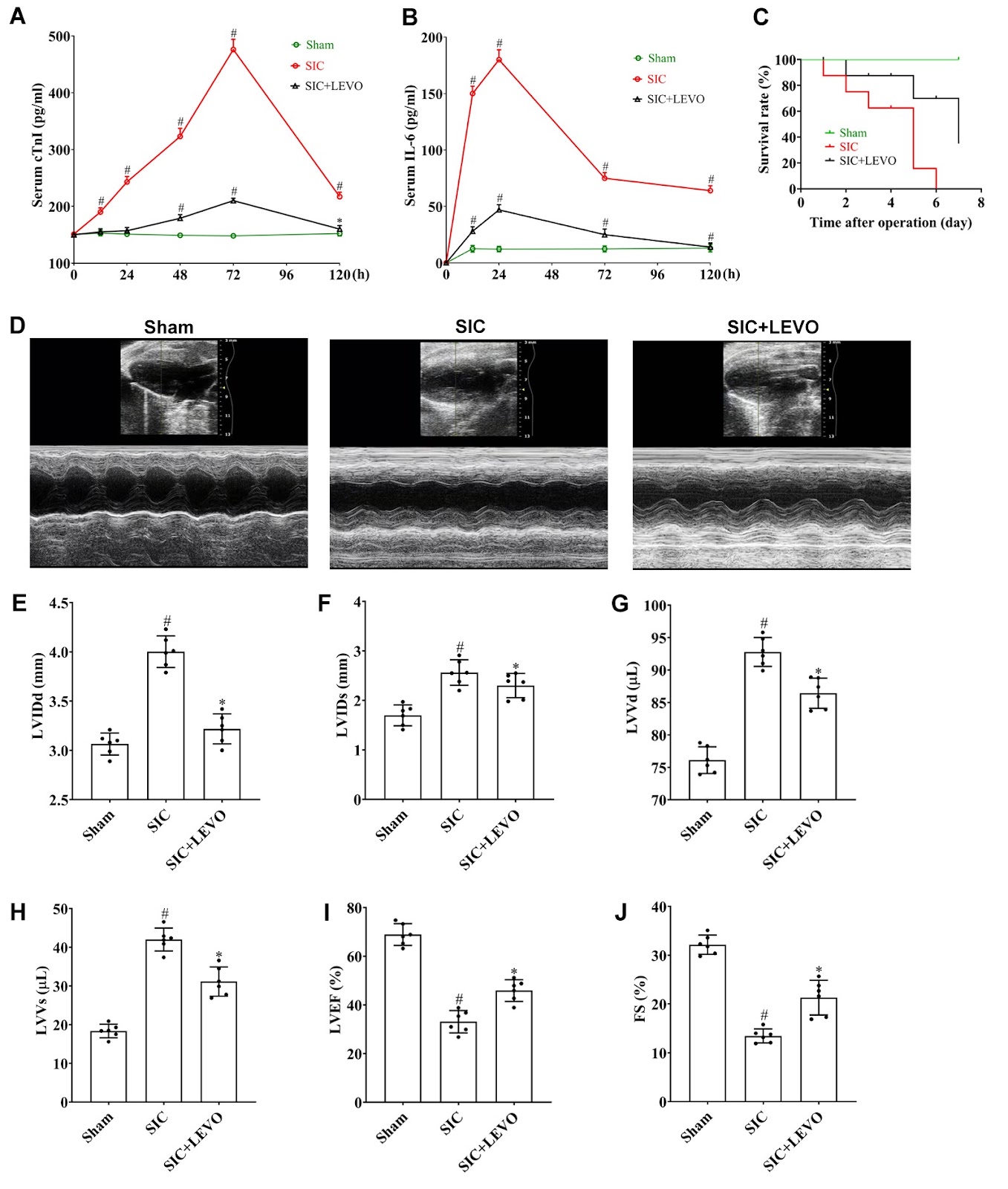
Sepsis-induced cardiomyopathy (SIC) is a severe complication of sepsis and septic shock and is characterized by cardiac dysfunction. Levosimendan (LEVO), a calcium sensitizer, has shown therapeutic potential in SIC, although its underlying mechanism remains unclear. Nrf2, a pivotal regulator of antioxidant and anti-inflammatory responses, may represent a potential target for SIC treatment. In this study, we examine the effects of LEVO on SIC and explore the mechanistic role of Nrf2 in mediating its cardioprotective effects. A murine SIC model is established via cecal ligation and puncture (CLP), and cardiomyocyte injury is induced in vitro via lipopolysaccharide (LPS) exposure in HL-1 cells. The CLP procedure significantly elevates serum cTnI and IL-6 levels and reduces the survival rates of mice. Echocardiographic analysis reveals impaired cardiac structure and function, accompanied by mitochondrial morphological and functional damage, in SIC mice. Interestingly, these pathological changes in SIC are markedly attenuated by LEVO treatment. Similarly, LEVO administration restores proliferative capacity; increases mitochondrial ATP, mitochondrial membrane potential (MMP) and NADH levels; and reduces ROS production and intracellular calcium overload. Notably, the protective effects of LEVO on cardiomyocyte viability and mitochondrial function are significantly diminished following Nrf2 inhibition or Nrf2 knockout (KO). Collectively, these findings demonstrate that LEVO mitigates cardiomyocyte injury and mitochondrial dysfunction in SIC through an Nrf2-dependent mechanism.

Read Full Abstract10.3724/abbs.2025165
The role of cryptochrome (CRY) in cancer: molecular mechanisms and clock-based therapeutic strategiesGraphical AbstractVerified
Acta Biochimica et Biophysica Sinica

The role of cryptochrome (CRY) in cancer: molecular mechanisms and clock-based therapeutic strategies

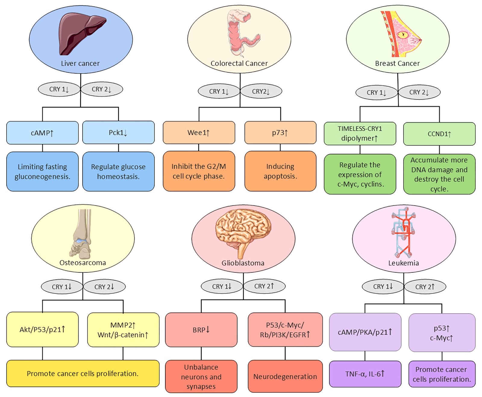
The circadian rhythm is a phenomenon in which physiological, behavioral, and biochemical processes within an organism naturally fluctuate over a period of approximately 24 hours. This phenomenon is ubiquitous in living organisms. Disruption of circadian rhythms in mammals leads to different diseases, such as cancer, and neurodegenerative and metabolic disorders. In specific tissues, numerous genes have been found to have circadian oscillations, suggesting a broad role for rhythm genes in the regulation of gene expression. This review systematically summarizes the role of cryptochromes (CRYs) in the initiation and progression of different types of cancer and discusses the relationships between clock genes and the tumor microenvironment (TME), as well as clock-based therapeutic strategies.

Read Full Abstract10.3724/abbs.2025025
Gastrodin inhibits reactive astrocyte-mediated inflammation in hypoxic-ischemic brain damage through S100B/RAGE-Smad3 signalingGraphical AbstractVerified
Acta Biochimica et Biophysica Sinica

Gastrodin inhibits reactive astrocyte-mediated inflammation in hypoxic-ischemic brain damage through S100B/RAGE-Smad3 signaling

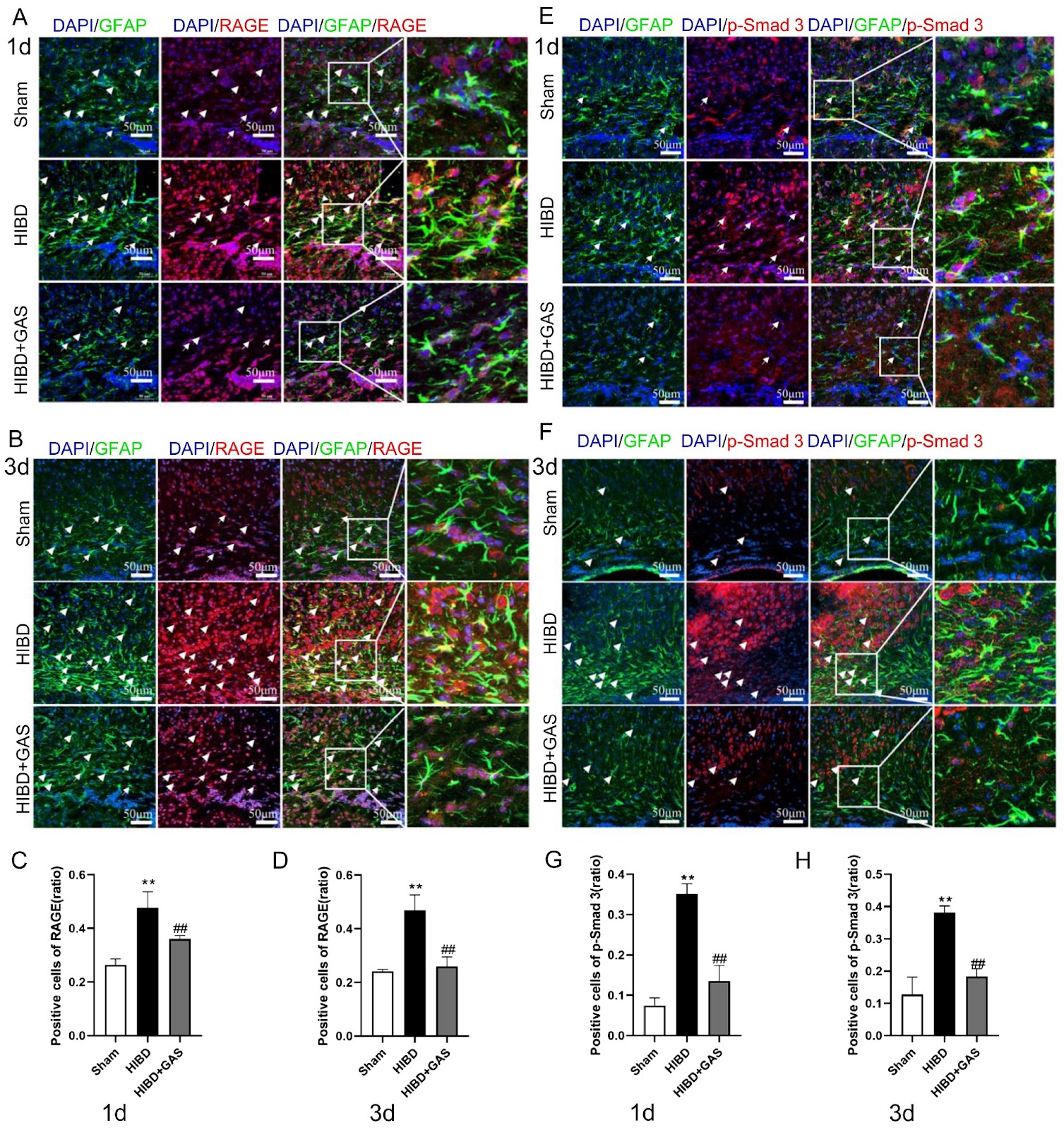
Activated astrocytes and their associated inflammatory responses play critical roles in the pathogenesis of hypoxic-ischemic brain damage (HIBD). Gastrodin (GAS), an anti-inflammatory herbal agent, is known to suppress microglial activation. Here, we investigate whether it exerts a similar effect on activated astrocytes and whether it acts through S100B/RAGE-Smad3 signaling. The expression changes of S100B/RAGE-Smad3 signaling pathway-related proteins, inflammatory factors and A1/A2 astrocyte markers were detected by ELISA, western blot analysis, immunofluorescence and immunohistochemistry. The results show that GAS decreases the expression of sRAGE in the brain tissue and S100B in the serum and brain tissue of HIBD mice. However, it promotes the expression of sRAGE in the serum of HIBD mice. Moreover, GAS inhibits the expressions of RAGE, p-Smad3, TNF-α, and C3 (A1 astrocyte marker), and promotes the expressions of S100A10 (A2 astrocyte marker) and BDNF in HIBD model mice, as well as in oxygen glucose deprivation (OGD)-treated TNC-1 astrocytes. The immunofluorescence and immunohistochemical results of RAGE and p-Smad3, as well as the immunofluorescence results of C3 and S100A10, reveal the same trend. Interestingly, FPS-ZM1 (a specific inhibitor of RAGE) inhibits the expressions of p-Smad3, TNF-α, C3, and S100A10, but promotes that of BDNF compared with those in the OGD group. The combination of GAS and FPS-ZM1 further decreases the expression of C3. These results indicate that GAS can inhibit the activation of Smad3 through S100B/RAGE signaling and regulate the expression of A1/A2-type astrocytes.

Read Full Abstract10.3724/abbs.2024235
D-CAPS: an efficient CRISPR-Cas9-based phage defense system for E. coliGraphical AbstractVerified
Acta Biochimica et Biophysica Sinica

D-CAPS: an efficient CRISPR-Cas9-based phage defense system for E. coli

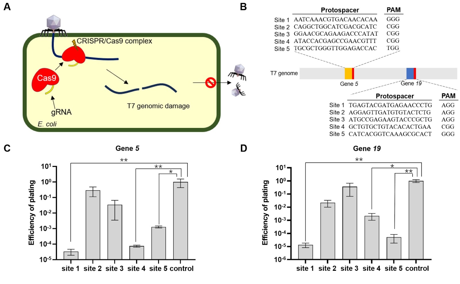
Escherichia coli is widely used in industrial chemical synthesis but faces significant challenges due to bacteriophage contamination, which reduces product quality and yield. Therefore, developing an efficient antiphage system is essential. In this study, we develop a CRISPR-Cas9-based antiphage system (CAPS) targeting essential genes of the T7 phage (gene 5 and gene 19) with single gRNAs transformed into MG1655 strains expressing Cas9. While CAPS provides limited resistance, with plating efficiencies ranging from 10–5 to 10–1, further optimization is needed. To enhance efficacy, we design a double-site-targeting CRISPR-Cas9-based antiphage system (D-CAPS). D-CAPS demonstrates complete resistance, with no plaques observed even at a high multiplicity of infection (MOI of 2), and growth curve analysis reveals that antiphage E. coli strains grow normally, similar to the wild-type strain, even at a high multiplicity of infection. Furthermore, D-CAPS is effective against BL21(DE3) strains, showing strong resistance and demonstrating its versatility across different E. coli strains. Protein expression analysis via green fluorescent protein confirms that E. coli carrying D-CAPS could maintain normal protein expression levels even in the presence of phages, comparable to wild-type strains. Overall, D-CAPS offers a robust and versatile approach to enhancing E. coli resistance to phages, providing a practical solution for protecting industrial E. coli strains and improving fermentation processes.

Read Full Abstract10.3724/abbs.2024208
Pregnancy-induced metabolic reprogramming in skeletal muscle: a multi-omics interrogation of transcriptional and metabolic adaptationsGraphical AbstractVerified
Acta Biochimica et Biophysica Sinica

Pregnancy-induced metabolic reprogramming in skeletal muscle: a multi-omics interrogation of transcriptional and metabolic adaptations

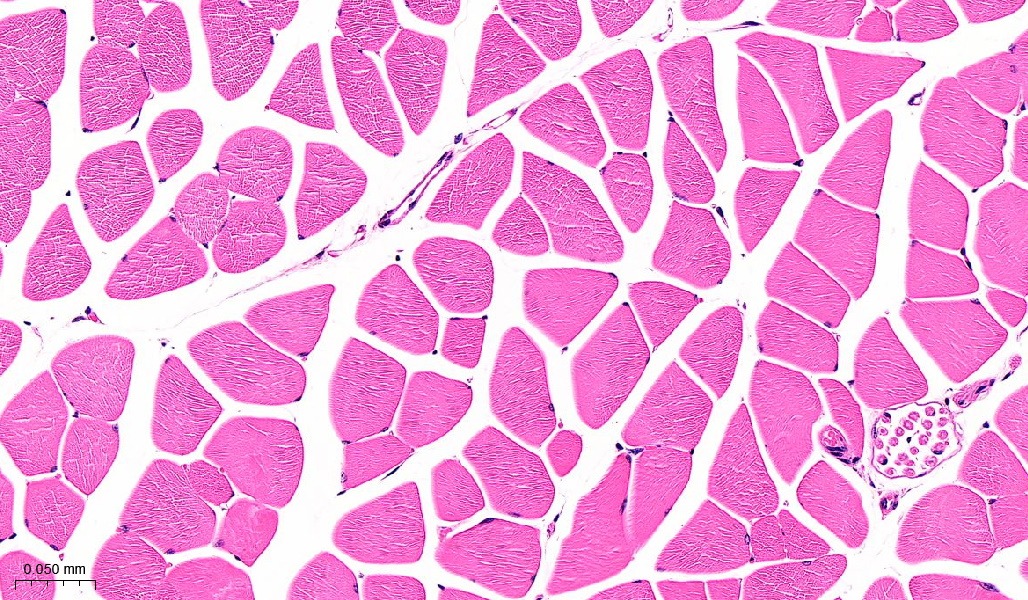
Pregnancy induces profound physiological adaptations to meet the dynamic nutritional demands of fetal development, including a deliberate reduction in maternal insulin sensitivity to ensure fetal glucose availability. However, excessive insulin resistance may precipitate gestational diabetes mellitus (GDM), increasing the risk of both obstetric complications and long-term metabolic disorders in mothers and offspring. Although the role of adipose tissue in pregnancy-associated metabolic adaptation has been extensively studied, the contribution of skeletal muscle remains poorly understood. Here, we systematically characterize pregnancy-induced molecular and metabolic changes in maternal skeletal muscle through multi-omics profiling. We use transcriptomic, metabolomic, computational single-cell deconvolution, and qPCR validation in an established C57BL/6J mouse pregnancy model (8-week-old females). Pregnancy triggers remarkable skeletal muscle remodelling, featuring histological reorganization with myofiber depletion and expanded endothelial compartments. Concurrent metabolic disturbances include insulin resistance, dysregulated TCA cycle activity, and impaired ubiquinone biosynthesis. This study represents a multi-omics-based systematic elucidation of pregnancy-induced maternal skeletal muscle adaptations. Our findings demonstrate that pregnancy induces profound structural reorganization and metabolic reprogramming in maternal skeletal muscle, characterized by prioritized fetal nutrient provision at the expense of maternal tissue utilization. These observations not only reveal previously unrecognized mechanisms of pregnancy-specific metabolic regulation but also, more importantly, establish a critical theoretical foundation for developing skeletal muscle-targeted intervention strategies to prevent gestational diabetes mellitus.

Read Full Abstract10.3724/abbs.2025199
The protective effect of naringenin on ulcerative colitis in mice through increasing Nrf2 pathway activityGraphical AbstractVerified
Acta Biochimica et Biophysica Sinica

The protective effect of naringenin on ulcerative colitis in mice through increasing Nrf2 pathway activity

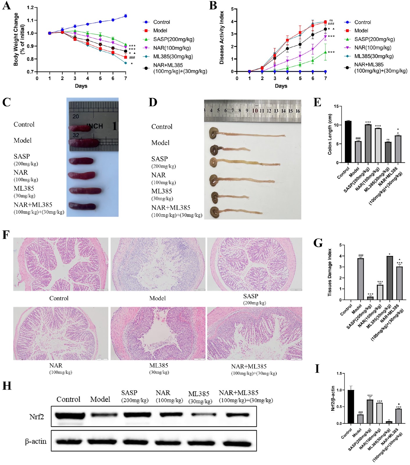
Ulcerative colitis (UC) is a chronic inflammatory disease with an increasing prevalence worldwide. Naringenin (NAR) has been proven effective in preventing UC, but its mechanism has not been fully elucidated. In this study, network pharmacology and bioinformatics methods are used to screen the genes associated with NAR and UC. A mouse model of dextran sulfate sodium (DSS)-induced UC is established. After treatment with NAR, the disease activity index (DAI) is scored, and colonic histopathology is observed via hematoxylin-eosin (HE) staining. The expressions of the nuclear factor erythroid 2-related factor 2 (Nrf2) signaling pathway and inflammation-related factors in the colons of UC mice are examined via western blot analysis and immunohistochemistry (IHC). The results of the animal experiments reveal that the model group of UC mice present the most severe weight loss and the highest DAI scores. After the administration of NAR, weight loss is alleviated, and DAI scores are reduced (P < 0.05). NAR improves pathological manifestations in the mouse colon, such as reducing inflammatory cell infiltration and restoring goblet cell loss (P < 0.05). NAR significantly increases the protein expression levels of Nrf2, heme oxygenase 1 (HO-1), and NAD(P)H dehydrogenase [quinone] 1 (NQO1) in the colon (P < 0.05) but decreases the protein expression levels of nuclear factor kappa-B (NF-κB), tumor necrosis factor-α (TNF-α), and interleukin-1β (IL-1β) (P < 0.05), thus alleviating the inflammatory response in UC model mice.

Read Full Abstract10.3724/abbs.2025026
Regulation of immune responses by a tumor necrosis factor in pearl oysters: insights from PmTNF gene expression and functionGraphical AbstractVerified
Acta Biochimica et Biophysica Sinica

Regulation of immune responses by a tumor necrosis factor in pearl oysters: insights from PmTNF gene expression and function

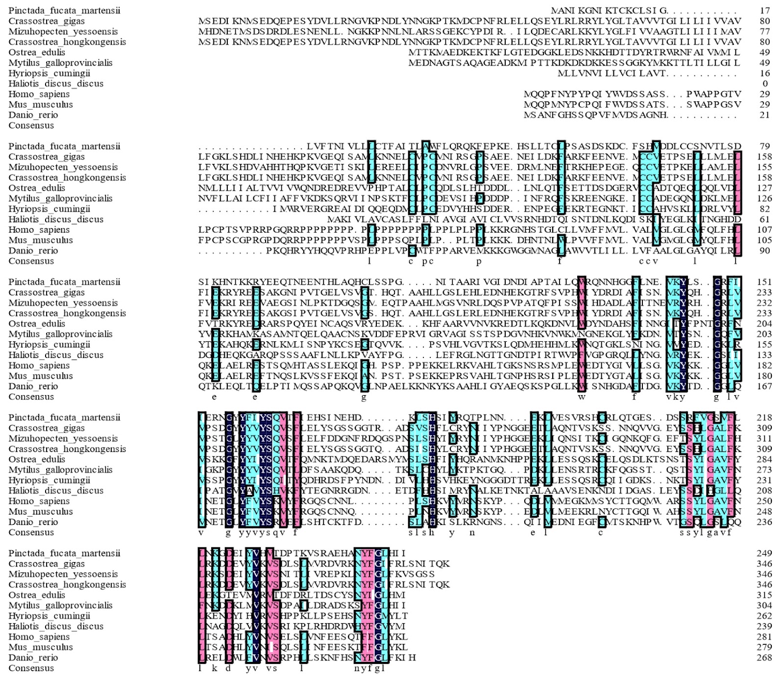
Tumor necrosis factor (TNF) is a multifunctional cytokine that regulates cellular processes such as inflammation, apoptosis, differentiation, and proliferation and activates various functions of the immune system. This article reports the discovery and characterization of a novel tumor necrosis factor gene in the pearl oyster Pinctada fucata martensii, which is named PmTNF. The deduced PmTNF protein sequence displays the typical structural characteristics of a TNF domain, and phylogenetic analysis of the sequences of PmTNF and its putative orthologs shows that they conform to the current taxonomy. Analysis of PmTNF mRNA expression via real-time PCR reveals its constitutive expression in all the examined tissues, with the highest expression in the gills. Furthermore, PmTNF expression in the gills varies upon exposure to pathogen-derived stimuli, with modest upregulation in response to lipopolysaccharides, but with significant downregulation in response to polyinosinic:polycytidylic acid. Nucleus insertion surgery induces an increase in PmTNF mRNA level in the gills at 12 h postoperation. Knocking down PmTNF through RNA interference significantly inhibits the expressions of immune-related genes in the NF-κB signaling pathway in the gills by 24 h (P < 0.05). The function of PmTNF is further characterized by studying the activity of an engineered recombinant PmTNF protein (rPmTNF) in vivo. Upon nuclear insertion, treatment with rPmTNF for 6 h upregulates several genes in the NF-κB pathway. Similarly, rPmTNF increases the activities of the antioxidant enzymes, including superoxide dismutase, glutathione and peroxidase, which reflect the total antioxidant capacity. Collectively, these results indicate that PmTNF participates in pearl oyster immunity by modulating the NF-κB pathway and activating the antioxidant defense system.

Read Full Abstract10.3724/abbs.2025003
Celastrol alleviates SGLT2 inhibitor-induced diabetic hyperketonemia by inhibiting hepatic ketogenesisGraphical AbstractVerified
Acta Biochimica et Biophysica Sinica2026

Celastrol alleviates SGLT2 inhibitor-induced diabetic hyperketonemia by inhibiting hepatic ketogenesis

SGLT2 inhibitor (SGLT2i)-induced diabetic hyperketonemia is a life-threatening acute complication of diabetes. While celastrol has been reported to have beneficial effects on obesity, its potential role in ketogenesis remains unclear. In this study, celastrol administration significantly attenuates the fasting-induced increase in blood β-hydroxybutyrate levels. Moreover, a 7-day course of celastrol (1 mg/kg/day) leads to reductions in body weight and fat mass. Mechanistically, celastrol specifically downregulates HMGCS2 expression and suppresses hepatic ketogenesis through the inhibition of PPARα expression in the short term (≤ 2 days). However, after prolonged treatment for 7 days, celastrol modulates both PPARα and serum free fatty acid (FFA) levels. Furthermore, the anti-ketogenic effect of celastrol is abolished in Pparα⁻/⁻ mice. Importantly, celastrol effectively ameliorates SGLT2i-induced hyperketonemia. In summary, celastrol curbs hepatic ketone overproduction in a PPARα-dependent manner, indicating its protective potential against SGLT2i-induced hyperketonemia.

Read Full Abstract10.3724/abbs.2026117
Atrial APD prolongation caused by the upregulation of RAGE and subsequent INaL increase in diabetic patientsGraphical AbstractVerified
Acta Biochimica et Biophysica Sinica

Atrial APD prolongation caused by the upregulation of RAGE and subsequent INaL increase in diabetic patients

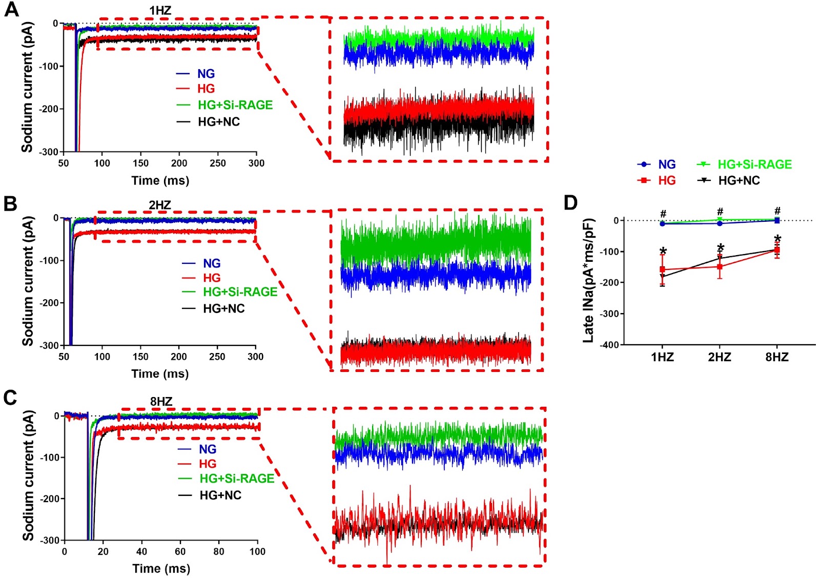
Diabetes mellitus (DM) is a risk factor for the development of atrial fibrillation (AF). The action potential duration (APD) has been demonstrated to be prolonged in the atrium of diabetic mice. In contrast, the APD is generally shortened in AF patients. It is unclear what change occurs in the atrial APD of diabetic patients. In this study, we explore the APD change of atrial myocytes from diabetic patients and the underlying molecular mechanisms. The whole-cell patch-clamp technique is used to detect single-cell electrical activity in diabetic and nondiabetic human samples. The results show that both APD50 and APD90, the APD at 50% and 90% repolarization, are increased in diabetic patients compared with those in nondiabetic controls. The density of late sodium current (INaL) in the atrial myocytes of diabetic patients is greater than that in the myocytes of nondiabetic patients. The expression of receptor for advanced glycation end products (RAGE) is increased in the atria of diabetic patients. In cultured HL-1 cells, high glucose (HG) treatment increases INaL, and the expression of RAGE prolongs APD. The siRNA-mediated knockdown of RAGE reduces the INaL and shortens the APD. The APD is prolonged in the atria of diabetic patients because of the upregulation of RAGE and the subsequent increase in INaL. Our findings provide novel insights into atrial electrical remodeling in diabetic patients.

Read Full Abstract10.3724/abbs.2025018
AKR1C3 protects cardiomyocytes against hypoxia-induced cell apoptosis through the Nrf-2/NF-κB pathwayGraphical AbstractVerified
Acta Biochimica et Biophysica Sinica

AKR1C3 protects cardiomyocytes against hypoxia-induced cell apoptosis through the Nrf-2/NF-κB pathway

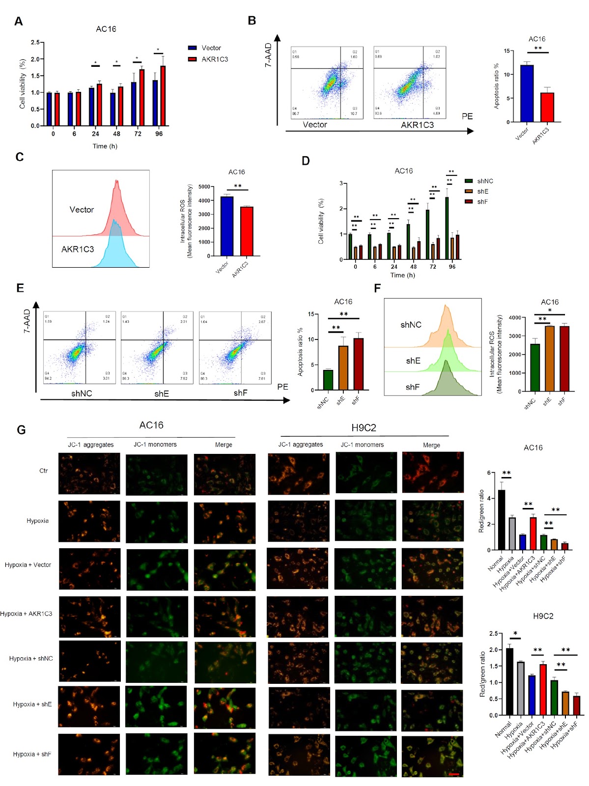
Hypoxia-induced apoptosis plays a critical role in the progression of various cardiac diseases, such as heart failure and acute myocardial infarction (AMI). Aldosterone reductase 1C3 (AKR1C3), a member of the aldo-keto reductase superfamily, participates in the metabolism of steroid hormones and redox reactions in vivo. Imbalances in prostaglandin levels have been linked to coronary events. However, the function and molecular mechanism by which AKR1C3 influences AMI are not yet fully understood. This study aims to investigate the role of AKR1C3 in hypoxia-induced myocardial cell damage and elucidate its mechanism. Our findings reveal that a hypoxic microenvironment triggers cardiomyocyte apoptosis and elevates AKR1C3 expression in H9C2 and AC16 cells, as well as in cardiac tissue from rats and mice with AMI. The overexpression of AKR1C3 promotes cardiomyocyte proliferation and cell vitality, whereas the silencing of AKR1C3 exerts the opposite effects in vitro. AKR1C3 protects cardiomyocytes against hypoxia-induced cell apoptosis by reducing ROS levels, preventing mitochondrial damage, and maintaining the oxygen consumption rate (OCR) and ATP production; conversely, AKR1C3 knockdown leads to adverse outcomes. Moreover, the application of a ROS inhibitor (MitoQ10) mitigates the increase in mitochondrial ROS in cardiomyocytes induced by AKR1C3 knockdown under hypoxic conditions. Mechanically, AKR1C3 increases Nrf-2 expression through the ubiquitin-proteasome pathway in cardiomyocytes and subsequently inhibits the NF-κB signaling pathway, thereby inhibiting Bax/caspase-3 signaling. Collectively, these results suggest that AKR1C3 prevents hypoxia-induced cardiomyocyte injury by modulating the Nrf-2/NF-κB axis, suggesting new insights into the mechanisms underlying myocardial protection.

Read Full Abstract10.3724/abbs.2024230
Corrigendum to: Vitamin B6 prevents Isocarbophos-induced posterior cerebral artery injury in offspring rats through up-regulating S1P receptor expressionGraphical AbstractVerified
Acta Biochimica et Biophysica Sinica

Corrigendum to: Vitamin B6 prevents Isocarbophos-induced posterior cerebral artery injury in offspring rats through up-regulating S1P receptor expression

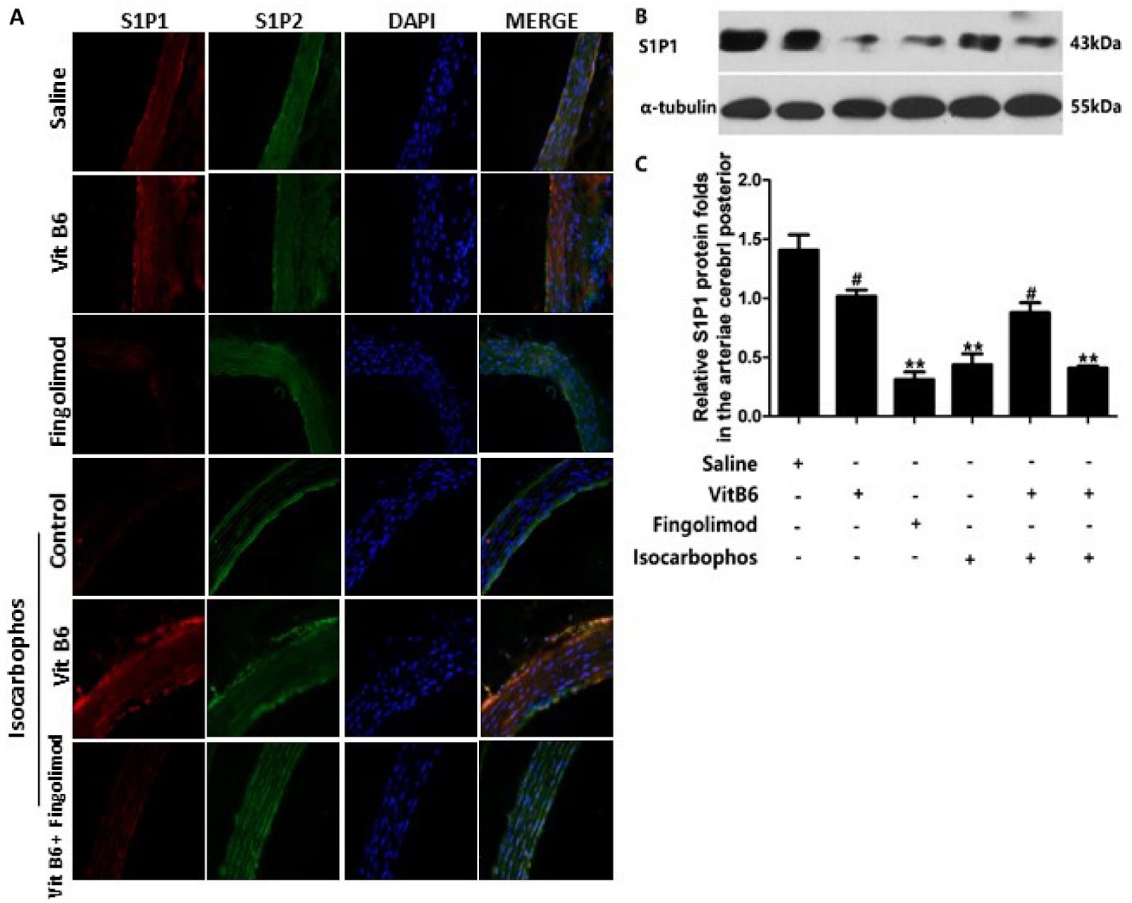
This corrigendum corrects errors in the original article 'Vitamin B6 prevents Isocarbophos-induced posterior cerebral artery injury in offspring rats through up-regulating S1P receptor expression' published in Acta Biochim Biophys Sin 2021, 53(12): 1691–1701. The errors were found in Figure 2B (Vit B6 + Fingolimod), Figure 5 (Saline), and Figure 7 (Isocarbophos/Control). The correct figures are shown. The authors apologize for the error. The corrigendum does not affect the interpretation of data and conclusions.

Read Full Abstract10.3724/abbs.2025088
circDCBLD2 regulates the Th1/Th2 immune balance via the miR-26a-5p/PTEN axisGraphical AbstractVerified
Acta Biochimica et Biophysica Sinica

circDCBLD2 regulates the Th1/Th2 immune balance via the miR-26a-5p/PTEN axis

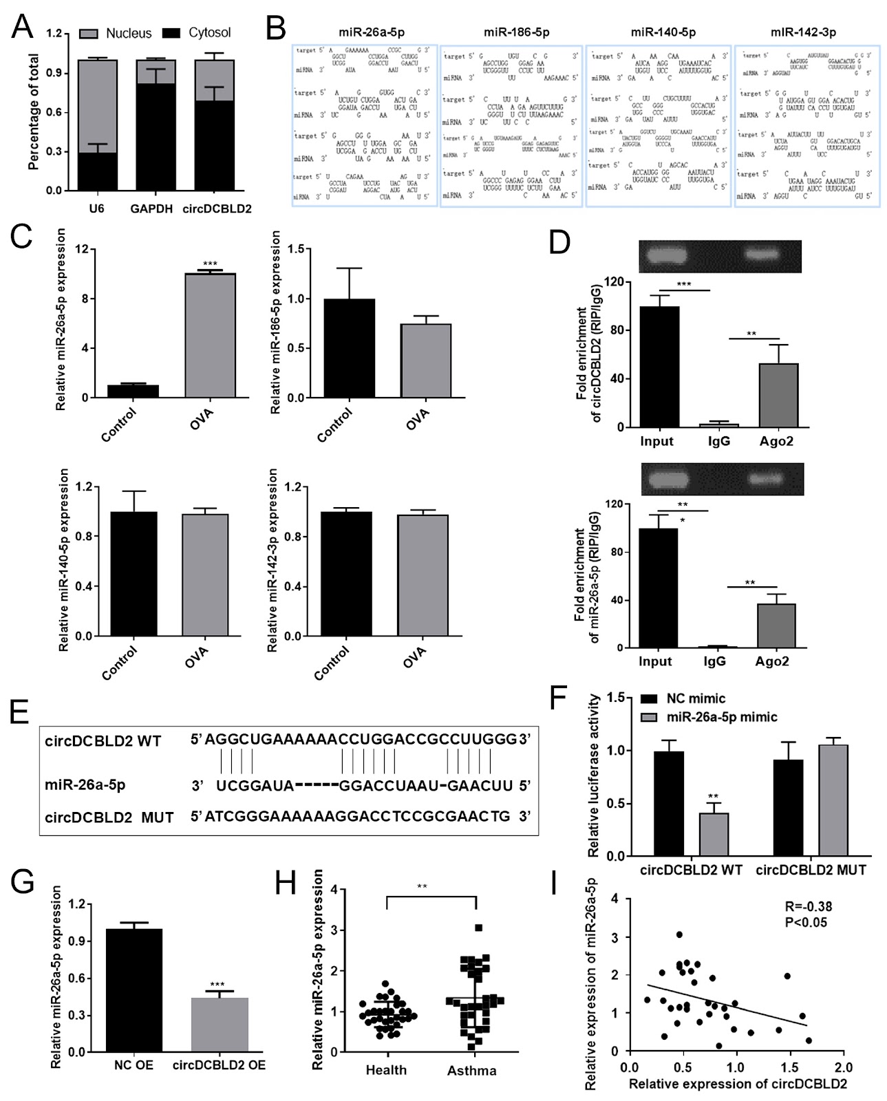
Asthma is a prevalent chronic respiratory disease in children. Recently, adjusting the Th1/Th2 imbalance has become a significant focus in asthma immunotherapy. The present study aims to investigate the roles and mechanisms of circDCBLD2 in maintaining the Th1/Th2 immune balance. CircDCBLD2 is downregulated in CD4+ T cells from asthmatic patients and in CD4+ T cells from an OVA-induced asthmatic mouse model. Additionally, circDCBLD2 levels are significantly decreased in the PBMCs of asthmatic mice. The expression of circDCBLD2 is positively correlated with the Th1 cytokines IFN-γ and IL-2 but negatively correlated with the Th2 cytokines IL-4 and IL-13. Flow cytometry and ELISA analyses demonstrate that circDCBLD2 overexpression increases the proportion of Th1 cells (CD4+IFN-γ+) and the levels of Th1 cytokines while decreasing the proportion of Th2 cells (CD4+IL-4+) and the levels of Th2 cytokines. Furthermore, circDCBLD2 overexpression alleviates the asthma phenotype in OVA-induced mice, reduces the infiltration of inflammatory cells in the lungs, and corrects the Th1/Th2 imbalance. Mechanistically, circDCBLD2 is found to target miR-26a-5p. Rescue experiments indicate that circDCBLD2 regulates the Th1/Th2 immune balance by targeting miR-26a-5p. Additionally, PTEN has been identified as a direct target of miR-26a-5p. The overexpression of PTEN partially reverses the effects of miR-26a-5p on the Th1/Th2 immune balance. These findings indicate that circDCBLD2 increases the proportion of Th1 cells and decreases the proportion of Th2 cells via the miR-26a-5p/PTEN axis, providing a promising target for asthma treatment.

Read Full Abstract10.3724/abbs.2025178
A Mycobacterium tuberculosis multi-epitope DNA vaccine encoding adaptive immune antigens provokes IFNγ/Th1 immunity and confers potential protectionGraphical AbstractVerified
Acta Biochimica et Biophysica Sinica

A Mycobacterium tuberculosis multi-epitope DNA vaccine encoding adaptive immune antigens provokes IFNγ/Th1 immunity and confers potential protection

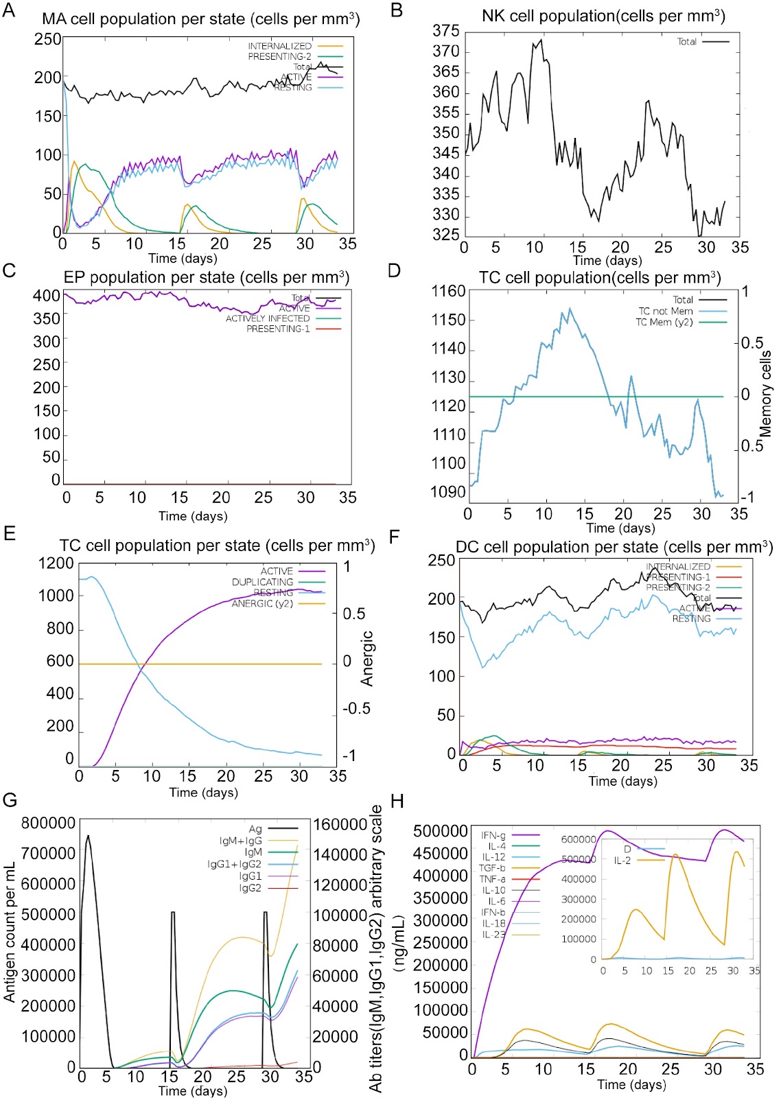
Tuberculosis (TB), caused by Mycobacterium tuberculosis (MTB), remains a significant global health threat. However, the licensed Bacille Calmette-Guérin (BCG) vaccine provides only limited protection in adults, underscoring the urgent need for more effective preventive strategies. Recent studies have shown that multi-epitope DNA vaccines are superior to traditional vaccines in terms of immunogenicity, safety and stability. In this study, we develop a multi-epitope DNA vaccine that contains CD8+ T-cell epitopes, CD4+ T-cell epitopes, and B-cell epitopes using bioinformatics tools. These epitopes are derived from three genome-encoded proteins, ESAT-6, Rv2660c, and RpfB, which exhibit stage-specific immunodominance in the early, resting, and convalescent stages of MTB infection. Using reverse vaccinology and computational immunomodulation, we demonstrate that the multiepitope vaccine increases antigen-specific antibody titres, activates CD8+ T and CD4+ T cells, and enhances IFN-γ secretion. In vitro validation studies in HEK293T cells confirm high-yield expression of multi-epitope-encoded antigens, whereas in vivo immunization experiments reveal significant expansion of NK cells and Th1-polarized lymphocytes, with concomitant upregulation of pro-inflammatory mediators. Collectively, these results highlight the potent activation of adaptive immunity through Th1-driven mechanisms and IFN-γ-mediated mycobacterial clearance, which are crucial for defending against MTB.

Read Full Abstract10.3724/abbs.2025152
Histone acetylases are required for iron homeostasis in yeastGraphical AbstractVerified
Acta Biochimica et Biophysica Sinica

Histone acetylases are required for iron homeostasis in yeast

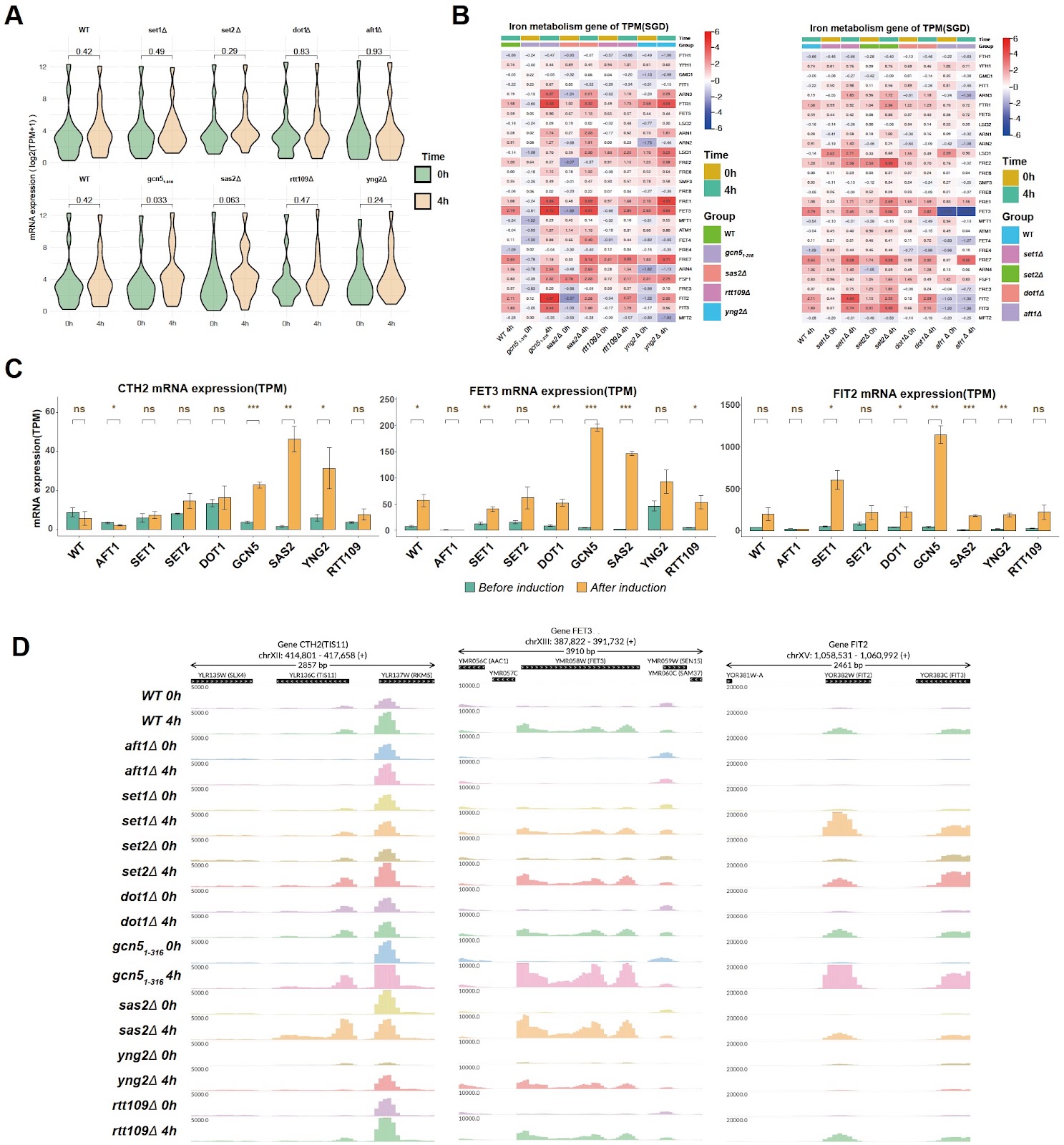
Iron, an ancient and essential transition metal, is involved in various biological functions, including oxygen transport, DNA synthesis, heme production, and iron-sulfur clusters, which participate in electron transport, DNA repair, and other cellular processes. However, excessive iron can lead to oxidative stress, lipid peroxidation, and cell damage. Thus, maintaining the iron content within an appropriate safe range and maintaining the balance of iron metabolism play crucial roles in both cellular function and human health [1]. An important aspect of maintaining the balance of iron homeostasis is the regulation of the iron uptake system. In Saccharomyces cerevisiae, cells can either obtain iron from the external environment via the non-reducing siderophore transport system or transport iron from the extracellular space to the intracellular space via the reducing iron transport system [2]. Iron uptake system-related genes are regulated mainly by the transcription factor Aft1p. During iron deficiency, Aft1p translocates into the nucleus, binds to genes involved in iron metabolism, and regulates the expressions of genes involved in iron uptake systems [3]. In addition, when there is a problem in the synthesis of iron-sulfur clusters in the mitochondria, such as the lack of the iron chaperone Yfh1p, which promotes the synthesis of iron-sulfur clusters, the transcription and nuclear entry of the transcription factor Aft1p are also activated, thereby regulating the expressions of iron metabolism-related genes [4]. In addition to transcription factors, gene expression is also regulated by histones and their modifications at the epigenetic level. For example, histone H3K4 methylation is related to gene activation, H3K36 methylation plays an important role in the elongation of transcription, and histone acetylation results in the loss of nucleosome structure and facilitates gene expression [5]. Therefore, histone modifications should also play important roles in the regulation of iron homeostasis. The relationship between histone modifications and iron homeostasis has been reported in the literature. For example, the DNA methylation-binding protein MBD5 can change histone acetylation in the promoter region of the ferritin gene by recruiting the histone acetylase KAT2A protein [6]. Histone acetylation has also been reported to be reduced in iron-deficient environments [7,8], and direct effects of histone acetylation on iron homeostasis gene loci have also been reported both in C. albicans [9] and mammals [10]. More recently, the histone H3-H4 tetramer was found to be a copper reductase enzyme, and H3-mediated Cu+ toxicity is a major determinant of the cellular functional pool of iron-sulfur (Fe-S) clusters [11,12]. However, information on the role of histone modifications in the regulation of iron homeostasis is limited. The mechanism by which and how histone modifications are involved in the transcriptional regulation of iron uptake-related genes or the iron deficiency response require further investigation. To determine whether histone acetylation and methylation are involved in the iron deficiency response, we first deleted the histone acetylase genes, including GCN5 (histones H2B and H3 N-terminal lysine acetylase, partial deletion of the ADA2 interaction sequence [13]), RTT109 (H3 lysine 9 and 56 acetylase), SAS2 (H4 lysine 16 acetylase), and YNG2 (subunit of the histone acetyltransferase complex NuA4 for acetylation of histone H4 or histone H2A) in the wild-type (WT) strain, as well as the histone methyltransferases SET1 (H3 lysine 4 methyltransferase), SET2 (H3 lysine 36 methyltransferase) and DOT1 (H3 lysine 79 methyltransferase). The genomic deletions were confirmed by colony PCR and genomic coverage analysis, as depicted in Supplementary Figures S1 and S2. The sensitivity of single histone modification enzyme mutants to iron deficiency induced by the iron chelator bathophenanthroline disulfonate (BPS) was tested. As shown in Supplementary Figure S3, the histone acetylation-related mutants gcn51–316, rtt109Δ, sas2Δ, and yng2Δ did not exhibit significant growth defects compared with the WT on the YPD + BPS plate. None of the histone methyltransferase knockout strains presented significant growth defects. As a positive control, the iron-responsive transcription factor gene AFT1 knockout strain grew slowly on YPD + BPS plates. It is possible that histone modifications do not have a strong effect on the equilibrium status of iron deficiency but still regulate transcription induction during the iron deficiency response. To investigate the role of histone modifications during the induction of the iron deficiency response, the expressions of iron response genes in the wild-type and mutant strains before and 4 h after BPS treatment were examined.
Read Full Abstract10.3724/abbs.2025040
The D826V point mutation in IREB2 causes early-onset neurodegeneration in miceGraphical AbstractVerified
Acta Biochimica et Biophysica Sinica

The D826V point mutation in IREB2 causes early-onset neurodegeneration in mice

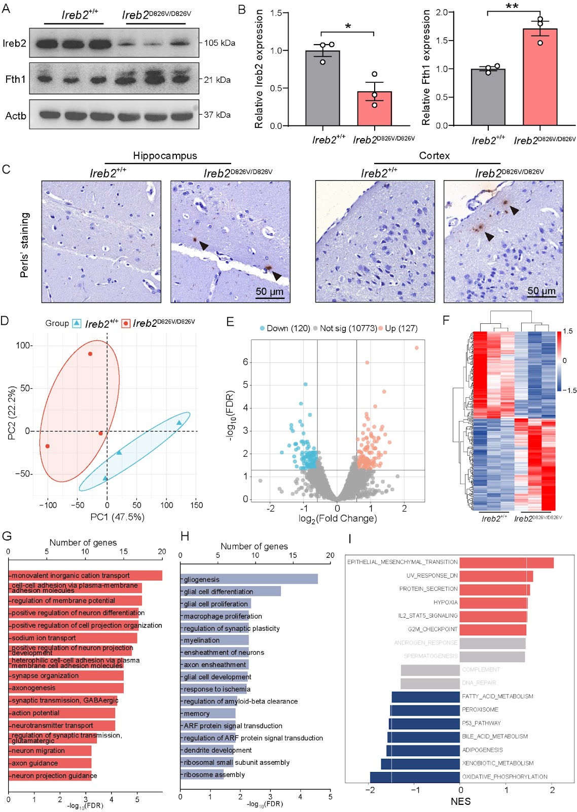
The iron regulatory protein IREB2 (Iron Responsive Element Binding Protein 2) plays a crucial role in maintaining cellular iron homeostasis through the posttranscriptional regulation of genes involved in iron metabolism. Mutations in the IREB2 gene have been linked to NDCAMA (OMIM#618451), a rare genetic neurological disorder characterized by early-onset neurodegeneration, choreoathetoid movements, and microcytic anemia. However, the absence of an IREB2-mutated animal model has left the underlying pathogenic mechanisms poorly understood. To investigate this, we establish a CRISPR-Cas9-mediated Ireb2D826V/D826V mouse model, which carries the c.2477A>T (p.D826V) pathogenic variant in IREB2 identified in a Chinese pedigree with NDCAMA. Behavioral studies, including the Morris water maze (MWM), open field test (OFT), and Y-maze, reveal significant neurobehavioral deficits, such as impaired spatial learning and memory and reduced motor activity, in Ireb2D826V/D826V mice. Furthermore, we observe increased microglial activation and decreased dendritic spine density in the hippocampus, along with impaired long-term potentiation (LTP) and elevated paired-pulse facilitation (PPF), indicating synaptic dysfunction. Mechanistically, Ireb2D826V/D826V mice present reduced Ireb2 protein levels, dysregulated iron metabolism, and an altered expression profile associated with neurological function. This study elucidates the molecular mechanisms underlying NDCAMA and establishes Ireb2D826V/D826V mice as a model for iron metabolism-driven neurodegeneration. This finding links the instability of IREB2 to synaptic failure and neuroinflammation, highlighting potential therapeutic implications for neurodegenerative diseases.

Read Full Abstract10.3724/abbs.2025176
Corrigendum to: Stattic sensitizes osteosarcoma cells to epidermal growth factor receptor inhibitors via blocking the interleukin 6-induced STAT3 pathwayGraphical AbstractVerified
Acta Biochimica et Biophysica Sinica

Corrigendum to: Stattic sensitizes osteosarcoma cells to epidermal growth factor receptor inhibitors via blocking the interleukin 6-induced STAT3 pathway

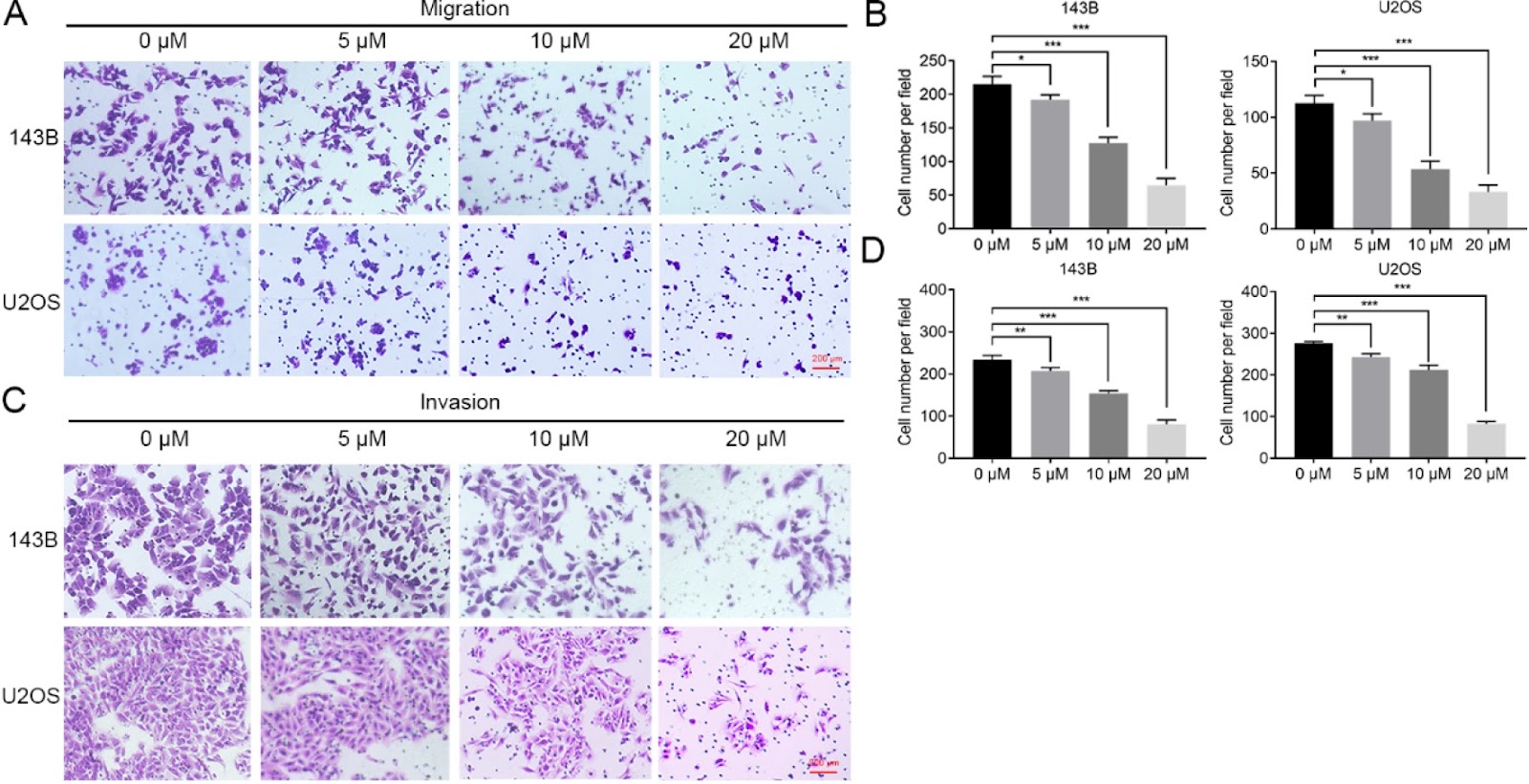
This is a corrigendum to the original article published in Acta Biochim Biophys Sin (Shanghai) 2021, 53(12): 1670–1680. In the original version, errors were found in Figure 2 and Figure 6. The correct figures are shown in this corrigendum. The authors apologize for the error.

Read Full Abstract10.3724/abbs.2025079
Biochemical and structural studies of NFIA and NFIC reveal a conserved mechanism for specific DNA recognition and provide insight into potential pathogenicity of disease-associated mutationsGraphical AbstractVerified
Acta Biochimica et Biophysica Sinica

Biochemical and structural studies of NFIA and NFIC reveal a conserved mechanism for specific DNA recognition and provide insight into potential pathogenicity of disease-associated mutations

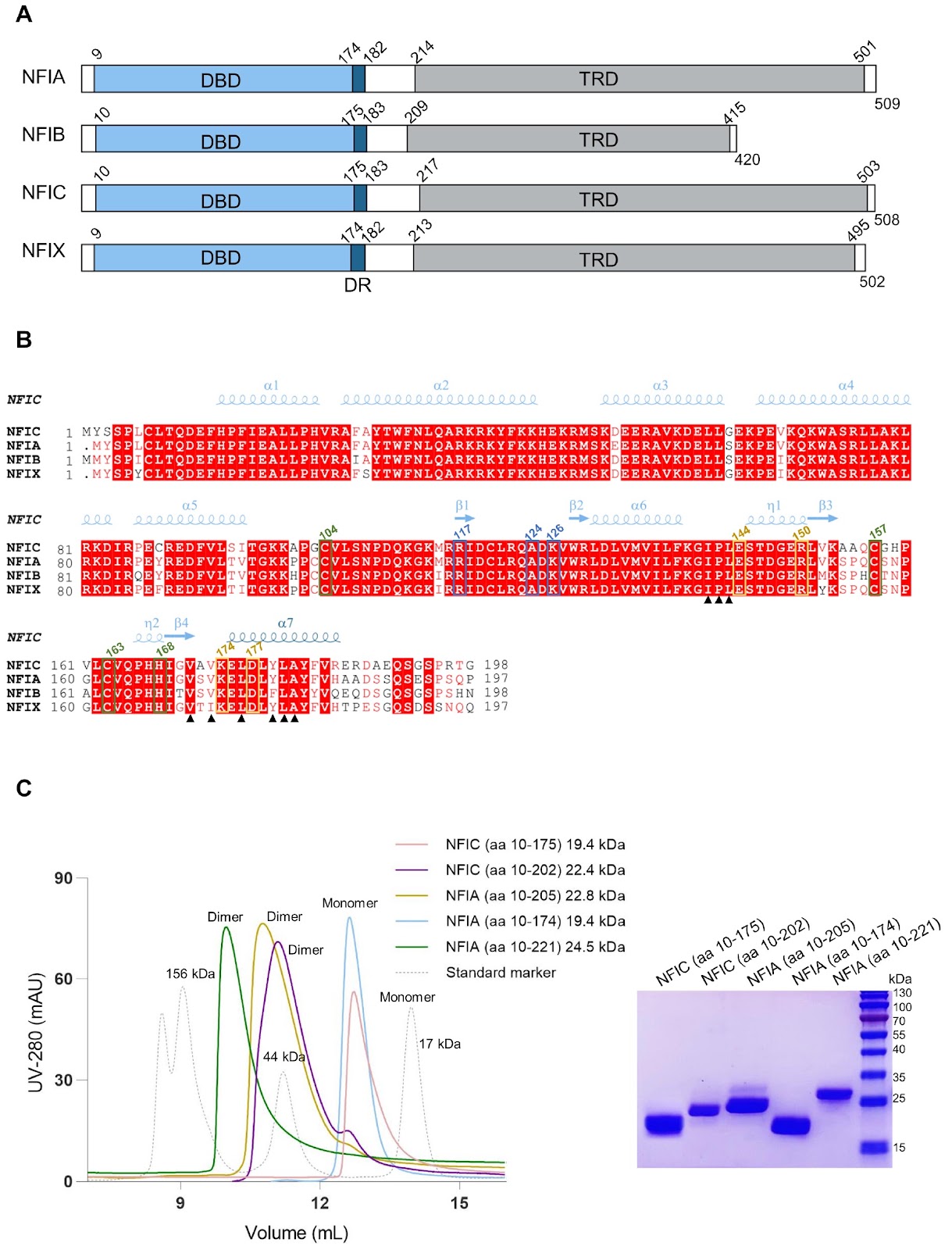
Nuclear factor I (NFI) transcription factors play essential roles in multiple aspects of nervous system development, including radial glia maturation, neurogenesis, gliogenesis, and brain morphogenesis. Numerous NFI variants have been identified in individuals with neurodevelopmental disorders, yet the molecular basis of their pathogenicity remains unclear. The absence of resolved NFI-DNA complex structures continues to impede mechanistic insights and therapeutic exploration. Here, we define the oligomeric states of NFIA and NFIC, and determine the crystal structures of the NFIC homodimer, as well as the NFIA and NFIC monomers lacking their dimerization region, in complexes with double-stranded DNAs. Structural analysis reveals the molecular mechanism underlying NFI dimerization and recognition of a dyad-symmetric TGGCA(N3)TGCCA sequence motif, and demonstrates that dimerization enhances both DNA-binding affinity and specificity of NFI proteins. The functional importance of key NFI residues and DNA bases involved in the protein-DNA interaction is further validated by mutagenesis and binding assays. Additionally, we systematically evaluate the effects of the neurodevelopmental disorders-associated NFI mutations on DNA binding of NFIA, providing insights into their potential pathogenic mechanisms. Together, our findings elucidate the structural basis of NFI dimerization and dyad-symmetric DNA recognition and highlight pathogenic variants for further mechanistic studies in neurodevelopmental disorders.

Read Full Abstract10.3724/abbs.2025236
Similarities and differences in the response and molecular characteristics of peripheral sensory neurons associated with pain and itchGraphical AbstractVerified
Acta Biochimica et Biophysica Sinica

Similarities and differences in the response and molecular characteristics of peripheral sensory neurons associated with pain and itch

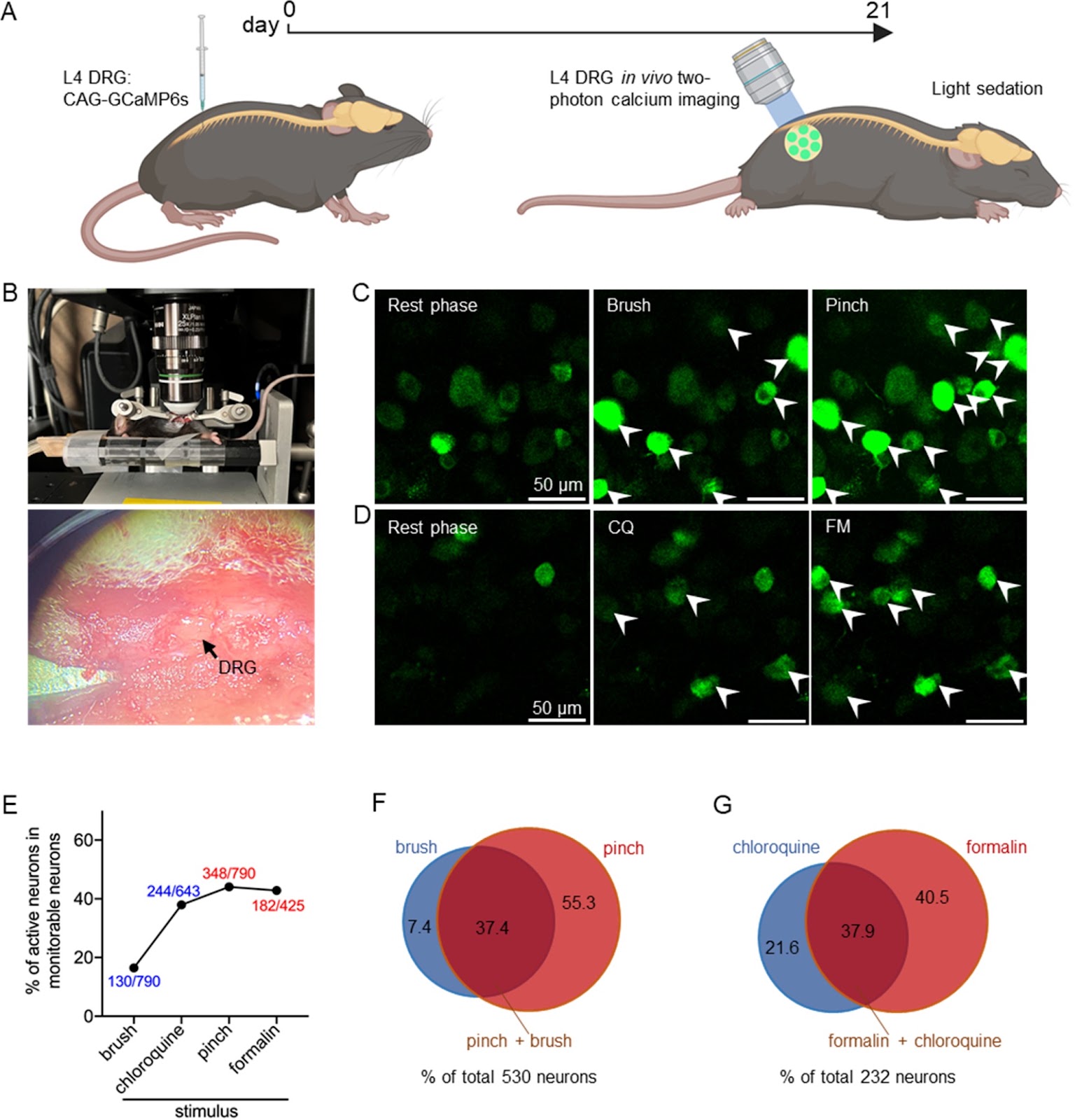
Dorsal root ganglion (DRG) neurons are responsible for the primary detection and transmission of peripheral noxious stimuli, mainly pain and itch. However, as two distinct noxious sensations, how DRG neurons respond differently to and code pain and itch is still an attractive topic. Here, we investigate the response and activation spectrum of DRG neurons under peripheral pain and itch stimuli using in vivo two-photon calcium imaging and find differences in the response intensity to pain and itch between multisensory neurons (both pain and itch) and single-sensory neurons (either pain or itch). In addition, single-cell RNA sequencing (scRNA-seq) is used to reveal the heterogeneity of distinct subpopulations on the basis of their expressions of pain- or itch-related marker genes and to determine the similarities and differences in their transcriptomic changes under chronic pain and itch. Our results show that primary sensory neurons with different sensory patterns respond differently to the same nociceptive stimuli. Additionally, distinct clusters of neurons exhibit unique transcriptomic changes in the development of chronic pain and itch, which may offer new insights for treating these conditions.

Read Full Abstract10.3724/abbs.2024202
Gut-brain axis and exosome-mediated communication in postoperative cognitive dysfunction associated with colorectal cancerGraphical AbstractVerified
Acta Biochimica et Biophysica Sinica

Gut-brain axis and exosome-mediated communication in postoperative cognitive dysfunction associated with colorectal cancer

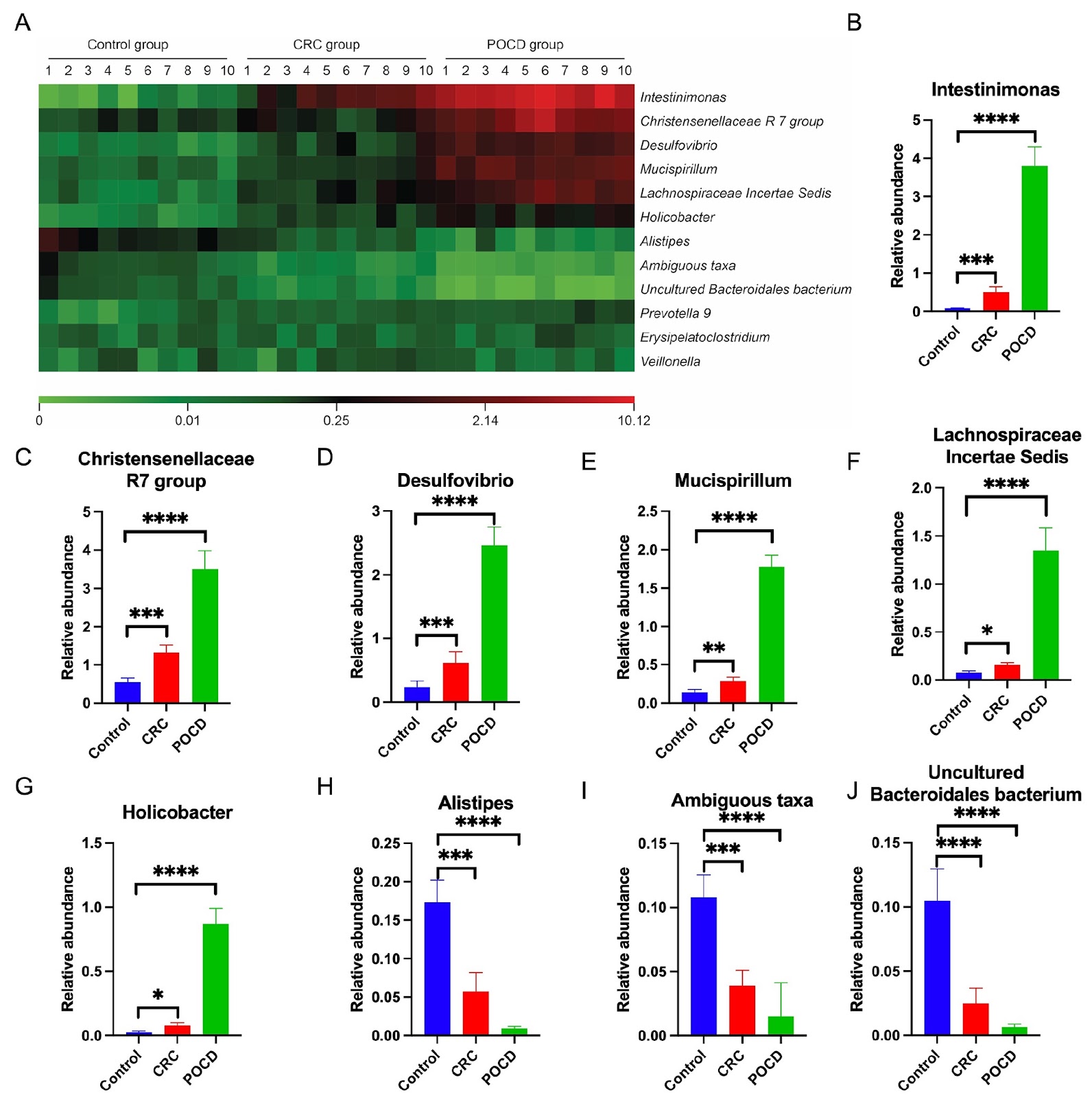
Postoperative cognitive dysfunction (POCD) is a serious complication in patients undergoing colorectal cancer (CRC) surgery. It is characterized by significant impairments in memory, information processing and attention, and may also result in mood and personality changes, thereby increasing the risk of postoperative mortality. Currently, there are no effective interventions available, highlighting the need for further investigation into its pathogenesis. While the current literature has identified an association between gut microbiota dysregulation and cognitive deficits, the precise mechanisms involved remain insufficiently understood. This study hypothesizes that exosome-like (Exos-like) nanoparticles derived from the gut microbiota contribute to POCD by modulating autophagy-dependent ferroptosis in hippocampal neurons. In a rat model of CRC, significant alterations in the gut microbiota composition, including reduced microbial diversity and changes in the abundance of key taxa, are observed. Exosomes derived from these microbiota enhance neuronal uptake and trigger markers of ferroptosis, as evidenced by increased expressions of ATG5 and COX2, along with decreased levels of GPX4 and FTH1. These findings establish a mechanistic link between microbial dysbiosis, ferroptosis, and cognitive decline in POCD, providing new insights into potential therapeutic targets for CRC-associated POCD.

Read Full Abstract10.3724/abbs.2025151
Long noncoding RNA LINC02432 inhibits papillary thyroid cancer via promoting ferroptosisGraphical AbstractVerified
Acta Biochimica et Biophysica Sinica

Long noncoding RNA LINC02432 inhibits papillary thyroid cancer via promoting ferroptosis

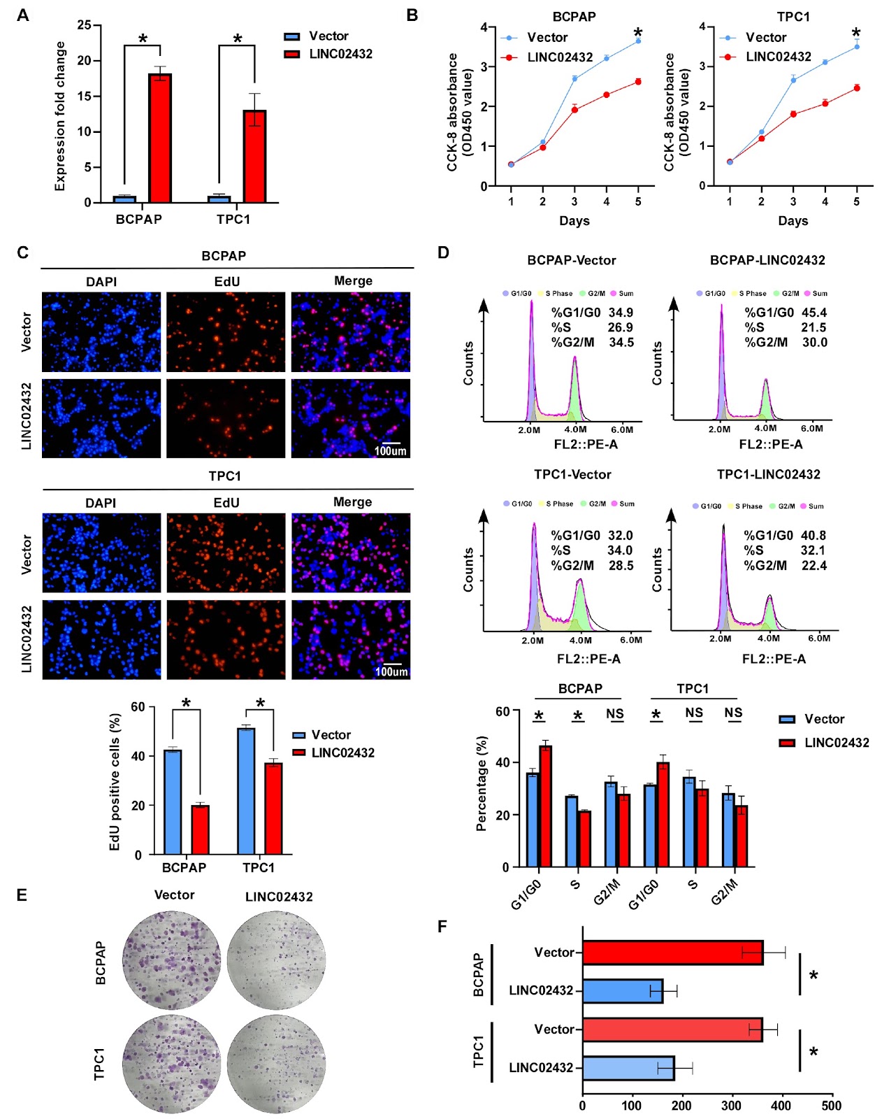
Long noncoding RNAs (lncRNAs) are essential in regulating the development and progression of different types of cancer. However, our insights into their impact and mechanisms in papillary thyroid carcinoma (PTC) are still insufficient. In this study, we investigate the effects of the ferroptosis-associated long noncoding RNA LINC02432 on PTC, which recognizes ferroptosis as a critical mechanism in cancer biology and lncRNAs as significant factors in various malignancies. To identify lncRNAs associated with ferroptosis in PTC, we conduct bioinformatics analyses and perform functional assays to evaluate the biological impact of LINC02432 on PTC cells, as well as its relationship with ferroptosis. Mechanistic studies employ methods such as western blot analysis, flow cytometry, and real-time PCR. Our analysis of transcriptome data from TCGA reveals that LINC02432 is the only lncRNA consistently identified by all 10 machine learning methods used, and its expression is significantly downregulated in PTC. Overexpression of LINC02432 in PTC cells inhibits cell proliferation and migration while promoting ferroptosis through inactivation of the NRF2 pathway. LINC02432 knockdown in PTC cells yields the opposite result. These findings highlight the potential of LINC02432 as a tumor suppressor in PTC progression, offering new insights into the mechanisms underlying the development and progression of this malignancy.

Read Full Abstract10.3724/abbs.2025172
Yaf9 conditionally contributes to cell size control in Candida albicansGraphical AbstractVerified
Acta Biochimica et Biophysica Sinica

Yaf9 conditionally contributes to cell size control in Candida albicans

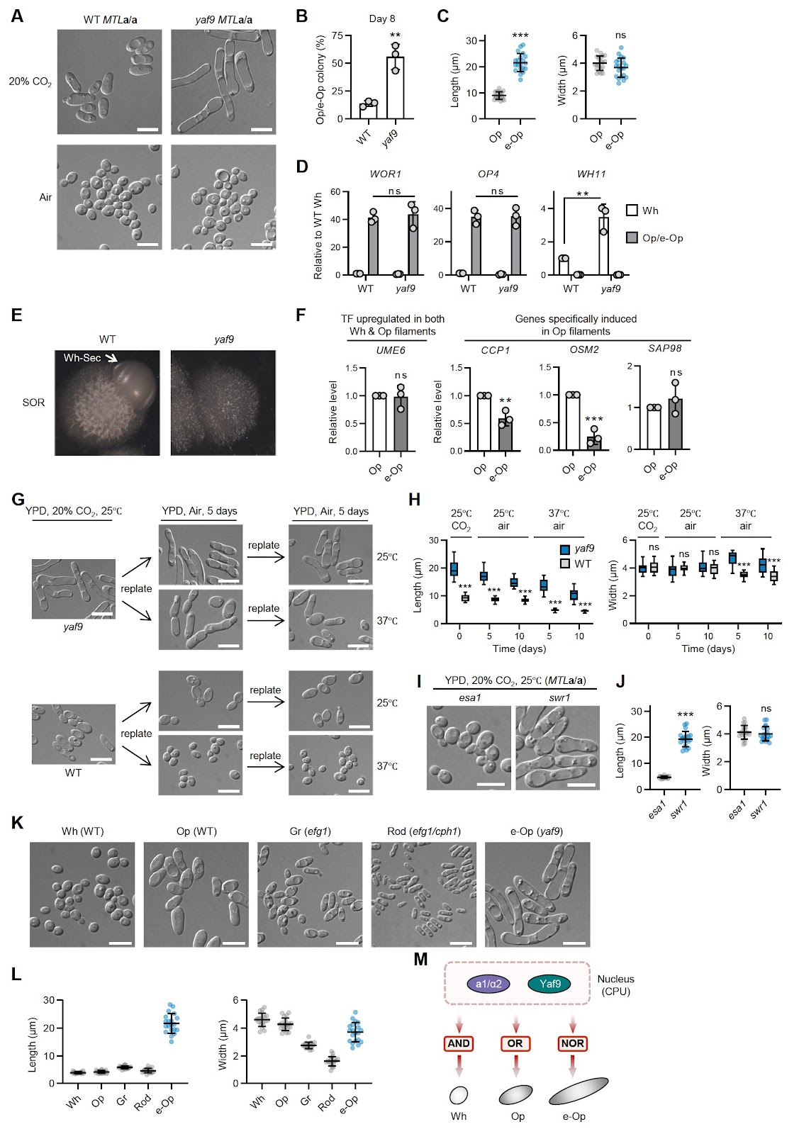
Candida albicans is an opportunistic fungal pathogen renowned for its ability to transition between distinct phenotypic states, such as the yeast-hyphae transition and the white-opaque switching. This morphological plasticity allows the organism to adapt to various host environments and evade immune responses. The white state is characterized by yeast-like cells with high proliferative capacity, whereas the opaque state features elongated cells with enhanced mating ability. The regulation of white-opaque switching is primarily controlled by a complex network of transcription factors. White-Opaque Regulator 1 (Wor1) serves as a master regulator crucial for the establishment and maintenance of the opaque state by activating the expression of genes required for opaque cell formation [1–3]. Conversely, the Mating-Type Like (MTL) locus in C. albicans acts as a critical barrier to white-opaque switching. The genes present at this locus strictly repress the white-to-opaque transition by the formation of a1/α2 complex; therefore, only MTLa/a or MTLα/α strains frequently switch to the opaque state [4,5]. Although the MTLa/α lab strain CAI4 is typically locked in the white state, some MTLa/α clinical isolates can switch to opaque [6]. Several genes were found to modulate this repression. For example, loss of HBR1, which is an activator of MTLALPHA1 and MTLALPHA2 gene expression, enables switching in MTLa/α cells [7]. Deletion of transcriptional repressors of the opaque state such as TUP1 also facilitates white-to-opaque switching [8]. The SWR1 complex incorporates H2A.Z into chromatin, and loss of Swr1 enhances switching and stabilizes the opaque state in MTL homozygous cells [9]. Our previous work revealed that the NuA4 histone acetyltransferase complex and the SWR1 complex merge into a supercomplex via Yaf9 in white-state yeast cells in C. albicans [10]. Here, we first tested whether Yaf9 is involved in white-to-opaque switching in MTLa/α heterozygous cells. The knockout of the YAF9 gene was validated by genotyping and qRT-PCR, confirming its loss at both the genomic and transcriptional levels (Supplementary Figure S1). The yaf9 null mutant cells were spread onto YPD plates and incubated in 20% CO2 at 25°C. After eight days of growth, sectors containing opaque cells were observed (Figure 1A). The frequency of opaque cell formation in the yaf9 mutant exceeded that in wild-type (WT) cells overexpressing WOR1 (Figure 1B). qRT-PCR analysis confirmed significant upregulation of opaque cell-specific markers, including WOR1 and OP4, in yaf9 mutant opaque cells, whereas the white cell marker WH11 was downregulated (Figure 1C). To examine whether YAF9 deletion affects the expression of MTL genes, we performed qRT-PCR for MTLA1 and MTLALPHA2 in white WT cells and in both white and opaque yaf9 mutant cells. The expression of both genes remained unchanged in yaf9 mutant cells (Figure 1D), suggesting that Yaf9-mediated repression of white-to-opaque switching occurs independently of MTL gene regulation. As Yaf9 is a component of the NuA4 and SWR1 complexes, we next investigated the roles of the NuA4 core enzyme Esa1 and the SWR1 core enzyme Swr1 in white-to-opaque switching in MTLa/α heterozygous cells. As shown in Figure 1E (upper panel), esa1 cells failed to switch to the opaque form under 20% CO2 stimulation, indicating that Esa1 activity is essential for opaque cell formation under the tested conditions. In contrast, swr1 cells readily underwent white-to-opaque switching (Figure 1E, lower panel), similar to the yaf9 mutant. These results indicate that Yaf9 functions as a repressor of white-to-opaque switching and that its deletion bypasses the repression imposed by the MTLa/α configuration. We then examined the role of YAF9 in white-to-opaque switching in MTLa/a cells, where MTL repression is removed. In air, yaf9 cells remained white; however, when exposed to 20% CO2, they frequently (> 50%) switched to the opaque form, which occurred at a significantly higher frequency than WT cells (Figure 2A,B). Notably, yaf9 cells exhibited a novel elongated opaque morphology, which we term e-Op cells. Quantification revealed that e-Op cells had similar width but were two to three times longer than WT opaque cells (Figure 2C). At the transcriptional level, e-Op cells displayed comparable upregulation of WOR1 and OP4 and downregulation of WH11 (Figure 2D). Notably, WH11 expression in yaf9 white cells was slightly higher than that in WT white cells. Like white cells, opaque cells are also capable of forming filaments under specific conditions [11]. To determine whether e-Op cells represent a filamentous form of opaque cells, we examined their gene expression and morphological stability. Multiple lines of evidence indicate that e-Op cells are distinct from these filamentous forms. First, when cultured on SOR medium, which promotes filamentous g

Read Full Abstract10.3724/abbs.2025180
COCA-seq: genome-wide mapping of O-GlcNAc-associated open chromatinGraphical AbstractVerified
Acta Biochimica et Biophysica Sinica

COCA-seq: genome-wide mapping of O-GlcNAc-associated open chromatin

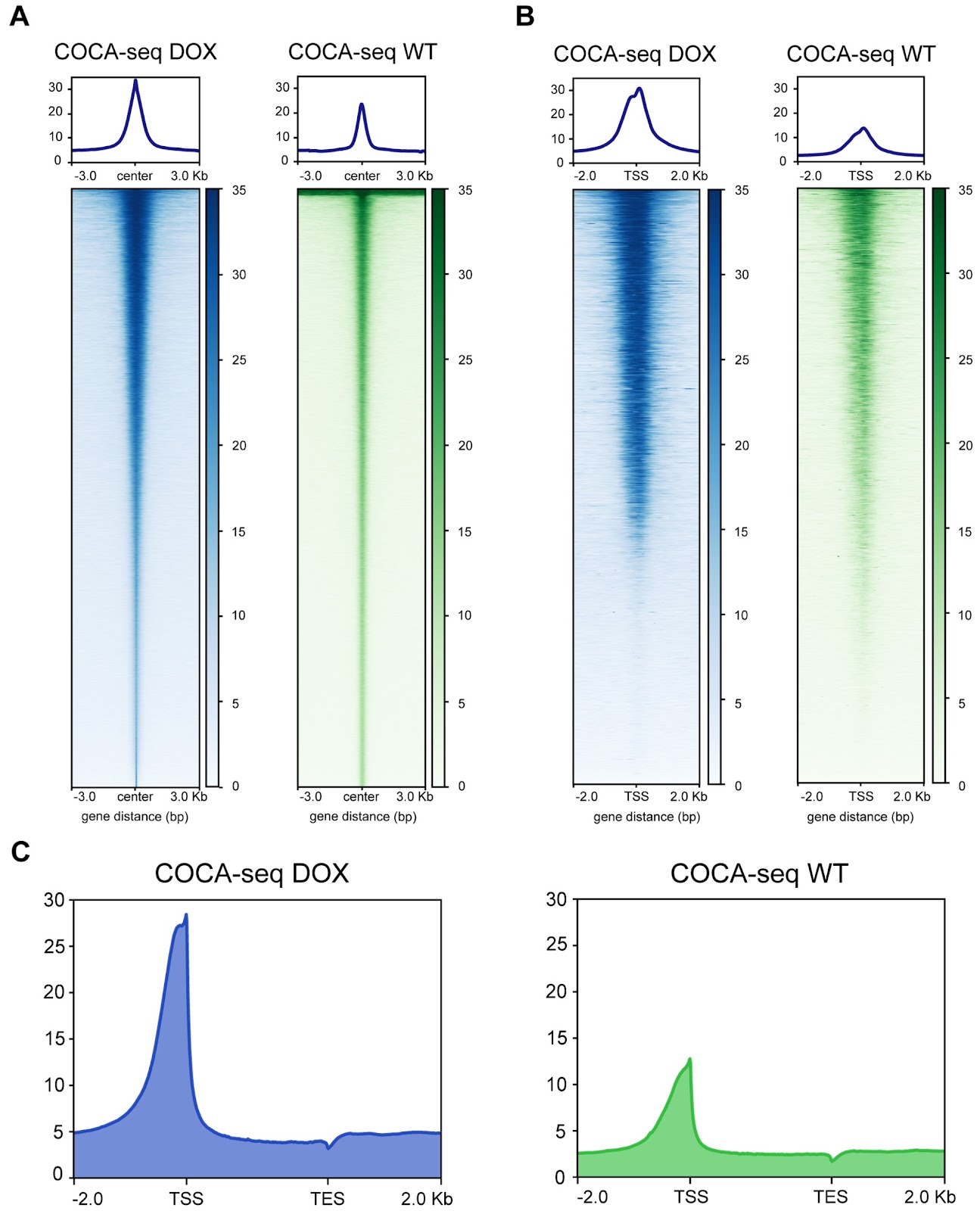
O-GlcNAcylation, a prevalent reversible post-translational modification, intricately alters non-histone proteins, influencing the organization of gene transcriptional regulation within the accessible chromatin environment. This nucleoplasmic landscape, characterized by histone-free regions, fundamentally enables O-GlcNAc-mediated modulation through dynamic accessibility. However, unraveling the O-GlcNAc-open chromatin interplay that governs sophisticated transcriptional regulatory networks remains constrained by current techniques, which lack the resolution to probe this spatiotemporal crosstalk. Here, we report a general strategy to systematically and chemoselectively profile O-GlcNAc-associated chromatin accessibility on a genome-wide scale (COCA-seq). Through comprehensive validation across low- and high-throughput levels, we demonstrate COCA-seq’s dual fidelity in both O-GlcNAc chemoselectivity and open chromatin specificity. We employ it to delve into doxorubicin resistance for breast cancer, scrutinizing pivotal regulatory genes and transcription factors implicated in this complex biological event. By integrating bulk RNA-seq with COCA-seq, we offer a multiomics perspective, shedding light on related biological processes and pathways like drug efflux and stress homeostasis, thereby uncovering potential mechanisms by which O-GlcNAc-associated open chromatin orchestrates tumor drug resistance. COCA-seq emerges as a general and versatile tool across various biological contexts, poised to reveal the landscape of O-GlcNAc-associated open chromatin regions across the genome and decipher the significance of glycosylation behind it.

Read Full Abstract10.3724/abbs.2025207