🧬 SinoBioData Academic Portal
📚 Peer-Reviewed Translated Literature

All Biomedical & Clinical Articles (Page 47)

Browse complete peer-reviewed translations from top Chinese biomedical, oncology, and genomics journals. Read verified previews and download full authentic clinical reports.

Published Research Papers

Showing 24 of 1542 peer-reviewed translated articles (Page 47 of 65)

CD47-mediated tumor microenvironment remodeling: a central mechanism in immune evasionGraphical AbstractVerified
Acta Biochimica et Biophysica Sinica

CD47-mediated tumor microenvironment remodeling: a central mechanism in immune evasion

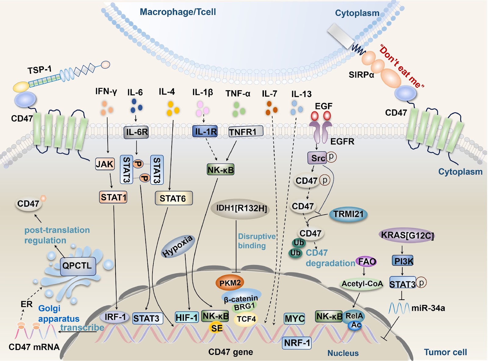
Immune evasion is a crucial strategy for tumor growth and survival, with the tumor microenvironment facilitating tumor immune evasion and cancer progression. CD47, a transmembrane protein highly expressed in various cancer cell types, interacts with its ligands SIRPα and TSP-1 to induce immune tolerance, enabling tumor cells to evade immune surveillance and phagocytosis by immune cells. Understanding the pathways driving CD47 signaling and related activation factors is essential. In this review, we discuss the interactions between CD47 and its ligands SIRPα and TSP-1; their roles in inhibiting the functions of immune cells (macrophages, dendritic cells (DCs), glial cells, T cells, NK cells, etc.); and the mechanisms involved. Furthermore, we also explore the influence of factors within the tumor microenvironment, including TNF-α, IFN-γ, ILs, HIF-1, oncogenes, isocitrate dehydrogenase 1, metabolic enzymes, and exosomes, on CD47-mediated immune evasion. Recent monoclonal antibody drugs targeting CD47 for cancer treatment have shown side effects and cause economic losses. Researchers can explore alternative approaches, such as designing targeted drugs with minimal side effects or investigating other related molecules or pathways. Combination therapy and further research into the molecular mechanisms of CD47 could offer new directions for antitumor drug development.

Read Full Abstract10.3724/abbs.2025071
FOXD3 promotes homologous recombination repair and genomic stability by facilitating MRE11-mediated DNA end resectionGraphical AbstractVerified
Acta Biochimica et Biophysica Sinica

FOXD3 promotes homologous recombination repair and genomic stability by facilitating MRE11-mediated DNA end resection

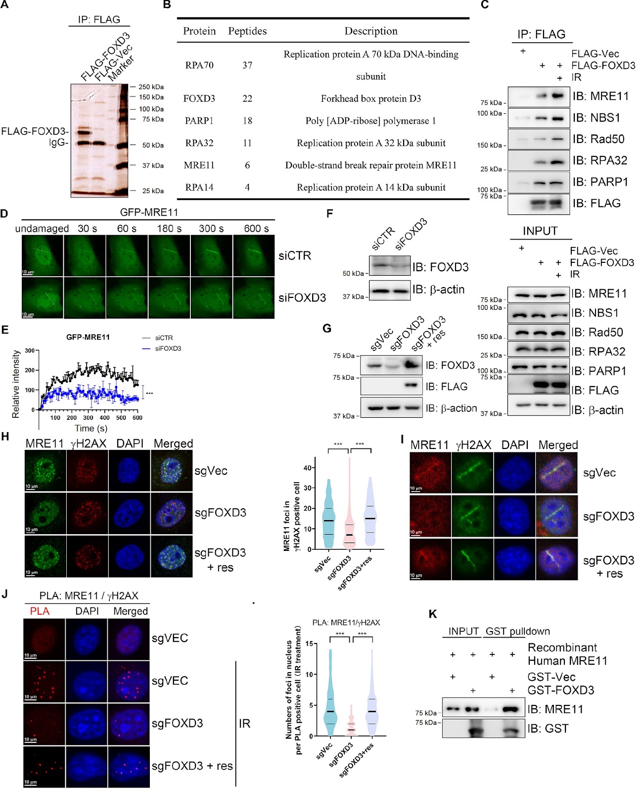
Homologous recombination (HR) is crucial for the high-fidelity repair of DNA double-strand breaks (DSBs), ensuring the maintenance of genome stability. In this study, we show that FOXD3 interacts with poly (ADP-ribose) polymerase 1 (PARP1) and is recruited to DSBs in a PARP1-dependent manner. FOXD3 directly binds to the DSB repair protein MRE11 and promotes its recruitment to DSB sites, ensuring proper end resection. Inhibition of FOXD3 expression compromises HR-mediated DSB repair and chromosome stability and sensitizes cancer cells to ionizing radiation. Collectively, our findings demonstrate that FOXD3 promotes HR-mediated DSB repair and genome stability.

Read Full Abstract10.3724/abbs.2025063
ATF3 triggers M2 macrophage polarization to protect against pulp inflammation through WNT4 regulationGraphical AbstractVerified
Acta Biochimica et Biophysica Sinica

ATF3 triggers M2 macrophage polarization to protect against pulp inflammation through WNT4 regulation

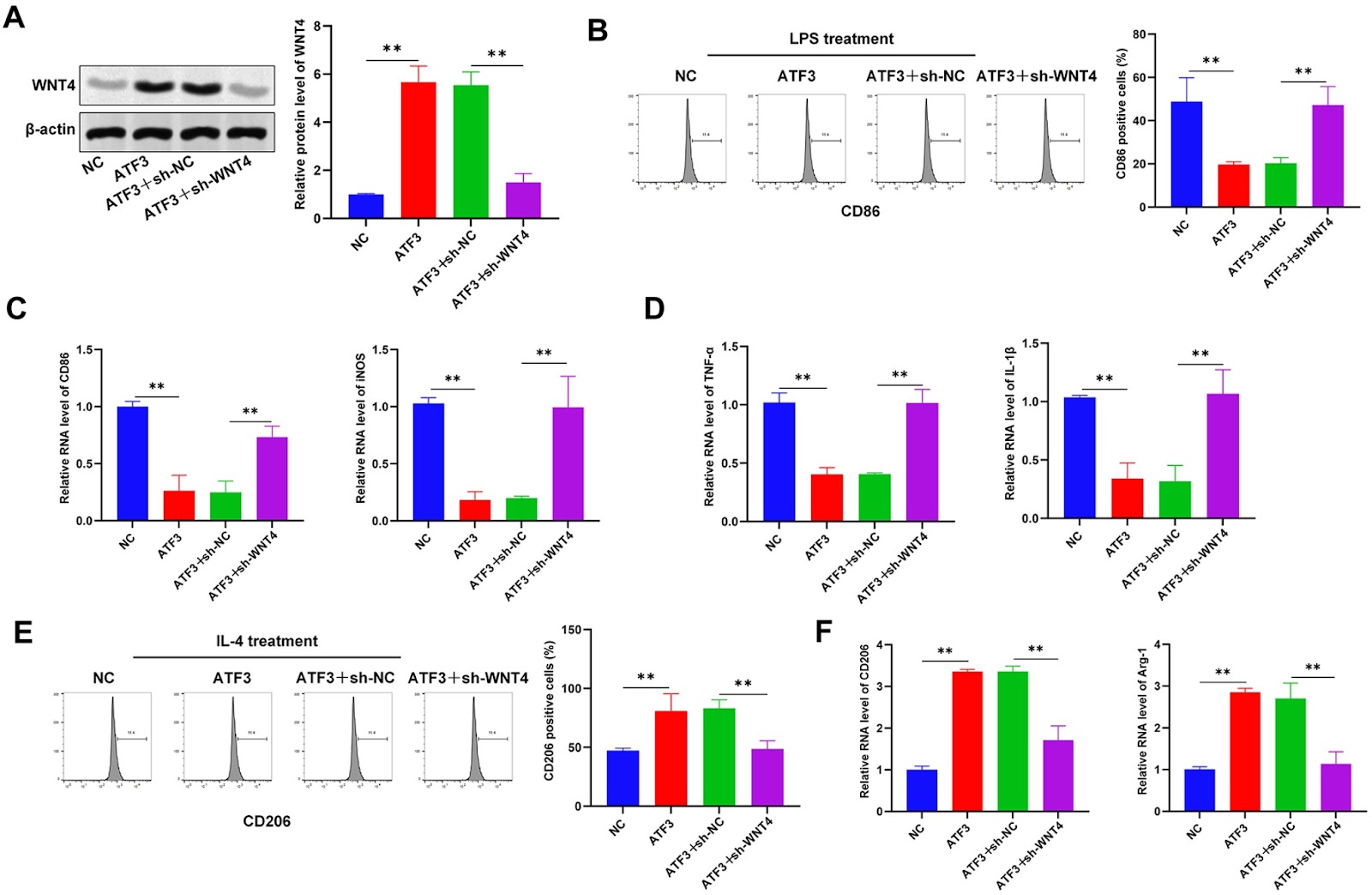
Pulpitis is a common inflammatory oral disease that can lead to pulp necrosis. The aim of this study is to investigate the expression and regulatory mechanisms of ATF3, a potential therapeutic marker, in pulpitis. A mouse pulpitis model with different degrees of inflammation is established, and the expression of ATF3 in pulpitis is explored. The histological features of healthy pulp and pulpitis are analyzed by HE staining, and classical inflammatory factors are detected by immunohistochemistry (IHC). In an in vitro study, we investigate the role of ATF3 in the regulation of WNT4 transcription and explore the effects of the ATF3/WNT4 axis on the polarization of RAW264.7 macrophages, the inflammatory response and the osteogenic differentiation of human dental pulp stem/stromal cells (hDPSCs). Our results show that ATF3 is expressed at low levels in inflamed pulp tissues; overexpression of ATF3 reduces the area of pulp necrosis, decreases the level of pro-inflammatory factors, and promotes macrophage polarization toward the M2 type. Furthermore, we reveal that ATF3 binds to the WNT4 promoter region and positively regulates the expression of WNT4 and that ATF3 downregulates M1 markers and increases the expression of M2 markers by regulating WNT4 expression. In addition, ATF3 promotes the osteogenic differentiation of dental pulp stem cells. In summary, this study reveals that ATF3 promotes M2 macrophage polarization by regulating WNT4, which in turn inhibits pulpal inflammatory responses and promotes the osteogenic differentiation of dental pulp stem cells. These findings suggest that ATF3 may be a potential target for pulpitis treatment.

Read Full Abstract10.3724/abbs.2025005
Schisandrin A ameliorates the diabetes-associated memory impairment by alleviating inflammation and ferroptosisGraphical AbstractVerified
Acta Biochimica et Biophysica Sinica

Schisandrin A ameliorates the diabetes-associated memory impairment by alleviating inflammation and ferroptosis

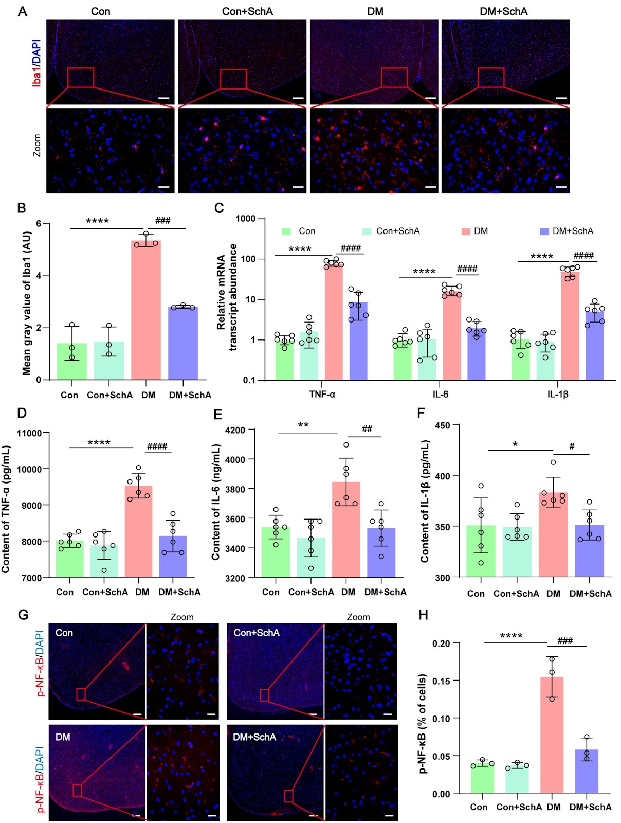
Schisandrin A (SchA), a bioactive lignan that was isolated from the dried fruit of Schisandra chinensis, has attracted much attention because of its diverse spectrum of pharmacological effects. The aim of this study is to clarify the function of SchA in diabetes-related fear memory impairment and its molecular mechanisms. Rats are randomly assigned to 4 groups: the control group (Con group), the DM group, the DM + SchA group, and the Con + SchA group. The results demonstrate that SchA treatment improves insulin sensitivity, reduces blood glucose, and significantly reduces memory impairment. SchA treatment also prevents histological damage, enhances synaptic protein production, and significantly decreases Aβ42 formation in the diabetic prefrontal cortex. Further research reveals that SchA therapy decreases microglial activation and the expression levels of variables linked to inflammation while increasing the phosphorylation of proteins implicated in the insulin resistance signaling pathway. Furthermore, in the prefrontal cortex of diabetic rats, SchA decreases ferroptosis by increasing the protein expressions of GPX4, SLC7A11, Nrf2, HO-1, and SIRT1. Overall, our findings suggest that SchA may lessen diabetes-associated fear memory impairment symptoms by, most likely, lowering ferroptosis and inflammatory responses in the prefrontal brain of diabetic rats. SchA may be a useful therapy for diabetes, including memory impairment.

Read Full Abstract10.3724/abbs.2025070
Long noncoding RNA HNF1A-AS1 promotes ovarian cancer growth and M2 macrophage polarization by counteracting miR-214-mediated suppression of semaphorin 4D signalingGraphical AbstractVerified
Acta Biochimica et Biophysica Sinica

Long noncoding RNA HNF1A-AS1 promotes ovarian cancer growth and M2 macrophage polarization by counteracting miR-214-mediated suppression of semaphorin 4D signaling

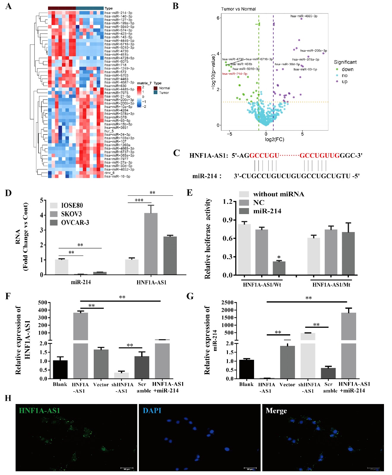
To determine whether lncRNA HNF1A-AS1 affects epithelial ovarian cancer (EOC) growth and macrophage polarization through miR-214/SEMA4D, the endogenous HNF1A-AS1 and miR-214 levels in human EOC cell lines are compared with those in normal ovarian epithelial IOSE80 cells. HNF1A-AS1 is overexpressed or silenced to investigate whether HNF1A-AS1 regulates miR-214/SEMA4D in SKOV3 cells and xenograft tumors, as well as the phenotypic switching of THP-1 cells. Compared with IOSE80 cells, EOC cells present significantly higher HNF1A-AS1 level and lower miR-214 level. Fluorescence in situ hybridization reveals predominant cytoplasmic localization of HNF1A-AS1, supporting its role as a competing endogenous RNA. HNF1A-AS1 and miR-214 antagonize each other in SKOV3 cells. In vitro, HNF1A-AS1 inhibits SKOV3 apoptosis and promotes migration and invasion. HNF1A-AS1 overexpression enhances miR-214 downstream of SEMA4D/PLEXIN-B1/TIAM1/RAC signaling, but miR-214 mimics significantly reverses this effect. Compared with control tumors, xenograft tumors derived from HNF1A-AS1-overexpressing SKOV3 cells present increased tumor growth, attenuated miR-214 expression, and activated SEMA4D/PLEXIN-B1/TIAM/RAC signaling. Knockdown of HNF1A-AS1 has the opposite effects. Additionally, HNF1A-AS1 promotes M2 phenotypic switching in THP-1 cells, which could be reversed by miR-214 overexpression or SEMA4D silencing. Our study suggests that by antagonizing miR-214, HNF1A-AS1 activates the SEMA4D/PLEXIN-B1/TIAM/RAC pathway, facilitating EOC growth and potentially promoting M2 macrophage polarization in the tumor microenvironment. HNF1A-AS1 represents a compelling therapeutic target for treating EOC.

Read Full Abstract10.3724/abbs.2025208
ArfGAP2 deficiency ameliorates autoinflammation by regulating STING signaling and proton channel activityGraphical AbstractVerified
Acta Biochimica et Biophysica Sinica

ArfGAP2 deficiency ameliorates autoinflammation by regulating STING signaling and proton channel activity

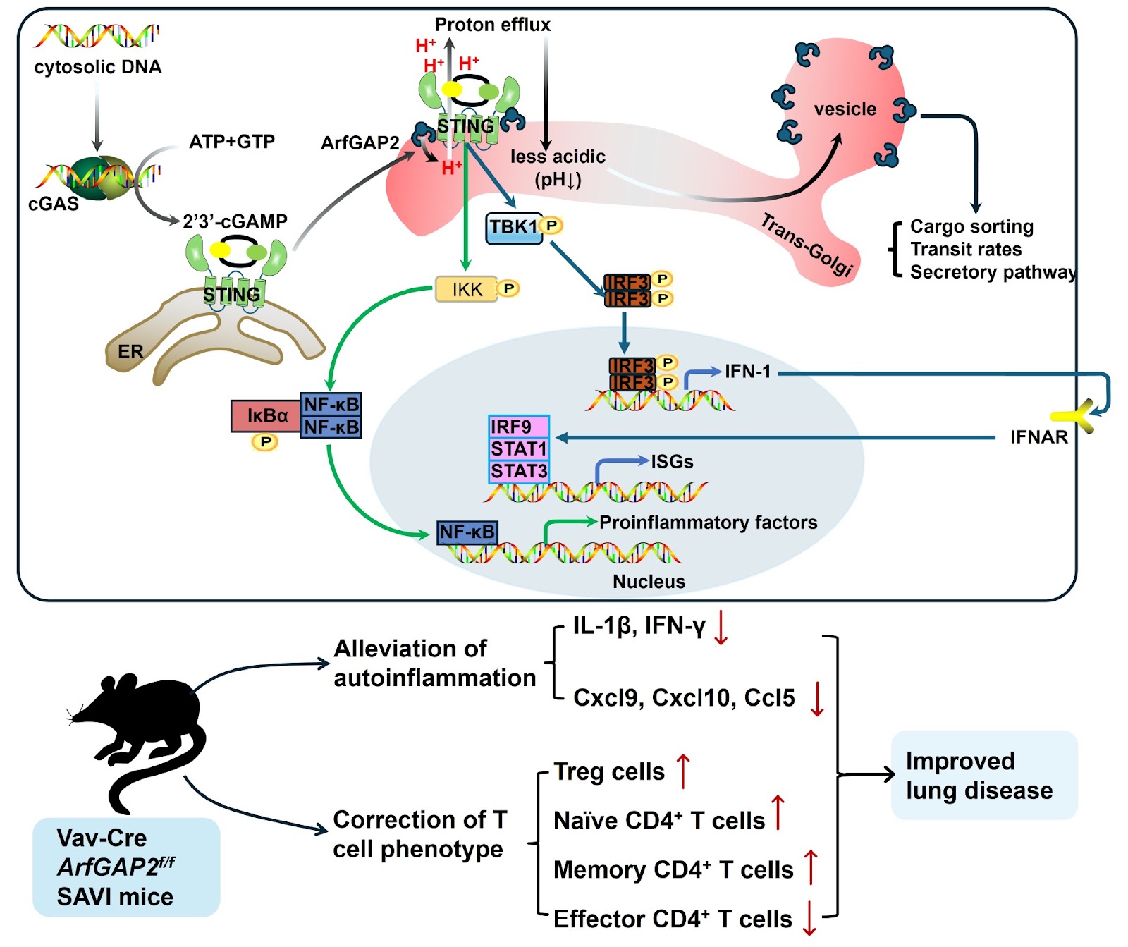
The cGAS-STING pathway is a critical regulator of innate immunity. When cyclic GMP-AMP synthase (cGAS) detects aberrant cytosolic DNA, it synthesizes the second messenger 2′3′-cGAMP, which binds and activates stimulator of interferon genes (STING) on the endoplasmic reticulum (ER). Activated STING then translocates to the Golgi apparatus, where it recruits and mutually phosphorylates TANK-binding kinase 1 (TBK1) and interferon regulatory factor 3 (IRF3). Phosphorylated IRF3 dimerizes and translocates to the nucleus, where it drives the production of type 1 interferons (IFN-1). In addition to being activated by IFN-1, STING also engages the nuclear factor kappa-B (NF-κB) pathway to induce the production of proinflammatory cytokines and chemokines. Moreover, IFN-1 signaling upregulates the expressions of interferon-stimulated genes (ISGs) through the IFN-α receptor (IFNAR). STING-associated vasculopathy with onset in infancy (SAVI) is a severe pediatric autoimmune disease caused by heterozygous gain-of-function mutations in STING, such as the N154S variant in humans and N153S in mice. Initially, classified as an interferonopathy due to constitutive activation of the STING pathway, SAVI is characterized by systemic inflammation, vasculopathy, interstitial lung disease, T-cell cytopenia, skin ulcerations, and premature death. However, recent studies challenge this paradigm, demonstrating that SAVI pathology develops independently of IFNAR-1 and IRFs (IRF3 and IRF7) [1,2], suggesting that alternative mechanisms drive disease progression. Emerging evidence indicates that STING restricts microbial infection through noncanonical autophagy and cell death pathways [3,4]. These functions may depend on its recently identified role as a proton channel in the Golgi apparatus [4,5]. Xun et al. [4] demonstrated that ligand-bound STING forms an ion channel in its transmembrane domain, facilitating proton efflux from post-Golgi vesicles and inducing Golgi deacidification. This raises a critical question: could STING-mediated Golgi deacidification be a potential mechanism underlying SAVI pathogenesis? A recent study by Poddar et al. [6] identified ADP ribosylation factor GTPase-activating protein 2 (ArfGAP2), which is involved in coatomer protein-1 (COP-1) coating in Golgi vesicles, as a key regulator of both STING signaling and proton channel activity, offering novel therapeutic insights for SAVI. First, to elucidate the role of STING in SAVI pathogenesis, they conducted a genome-wide CRISPR-Cas9 screen in T cells resistant to chronic STING activation and identified ArfGAP2 as a critical STING modulator among multiple Golgi-related proteins. Further investigation revealed that genetic ablation of ArfGAP2 in Jurkat T cells significantly attenuated STING-mediated ISG induction. Compelling evidence indicates that STING activation is associated with its subcellular location [7]. While ArfGAP family proteins typically regulate Golgi membrane trafficking, vesicle transport, and cargo sorting [8], Poddar et al. [6] surprisingly reported that ArfGAP2 enhances STING-mediated ISG induction and promotes LC3 lipidation without altering STING palmitoylation or its Golgi localization. Further experiments revealed that ArfGAP2 enhances STING signaling and IFN-β secretion in mouse bone marrow-derived macrophages (BMDMs). In addition to promoting IFN-1 induction, ArfGAP2 promotes the secretion of NF-κB-dependent proinflammatory cytokines activated by STING in THP-1 monocytes [6]. In addition to inducing ISGs and NF-κB signaling, activated STING acts as a proton channel triggering Golgi deacidification [4]. Given the well-established importance of the Golgi pH in regulating enzyme activity, protein modification, and membrane trafficking [9], researchers have further explored how STING and ArfGAP2 modulate cargo transport and secretion. They reported that the loss of ArfGAP2 impairs STING-mediated proton channel activity in the Golgi, leading to a lower luminal pH. Moreover, ArfGAP2-deficient cells presented significant alterations in the cell surface proteome upon STING activation, accompanied by altered sorting, secretion and trafficking rates of specific protein cargos in the Golgi [6].
Read Full Abstract10.3724/abbs.2025107
Intracellular acetyl phosphate modulates Escherichia coli pyruvate metabolismGraphical AbstractVerified
Acta Biochimica et Biophysica Sinica

Intracellular acetyl phosphate modulates Escherichia coli pyruvate metabolism

Lysine acetylation has been shown to be an abundant and vital post-translational modification (PTM) that utilizes acetyl phosphate (AcP) as one of the acetyl group donors in bacteria. The pyruvate dehydrogenase (PDH) complex catalyzes the conversion from pyruvate to acetyl coenzyme A (acetyl-CoA). Thus far, the connection between lysine acetylation and pyruvate metabolism has not been thoroughly investigated. In this study, we show that AcP could acetylate Escherichia coli pyruvate dehydrogenase (AceE) in vitro and in vivo, which could be reversed by protein lysine deacetylase (CobB). In vitro treatment of AceE with AcP also causes increased phosphorylation of the protein, whereas deleting ackA does not affect the phosphorylation of the protein. As a result, in vitro treatment of AceE by AcP leads to decreased enzymatic activity. In contrast, deleting ackA leads to increased acetylation and enzymatic activity of AceE, and deleting pta results in the decreased acetylation and enzymatic activity of AceE. As expected, deleting pta in E. coli causes pyruvate accumulation. Although deleting ackA also causes pyruvate accumulation, decreased expression of the two genes involved in pyruvate metabolism (ldhA and poxB) is observed in the mutant, indicating that AcP could affect pyruvate metabolism by other routes in addition to modulating the AceE activity. Thus, our results demonstrate that intracellular AcP could modulate pyruvate metabolism in E. coli. For the first time, a linkage between AcP-mediated protein lysine acetylation, pyruvate dehydrogenase activity, and pyruvate metabolism is established.

Read Full Abstract10.3724/abbs.2025068
Vesicle-mediated transport-related gene SEC23A promotes cell proliferation by regulating cell cycle leading to gastric cancer progressionGraphical AbstractVerified

Acta Biochimica et Biophysica Sinica

Vesicle-mediated transport-related gene SEC23A promotes cell proliferation by regulating cell cycle leading to gastric cancer progression

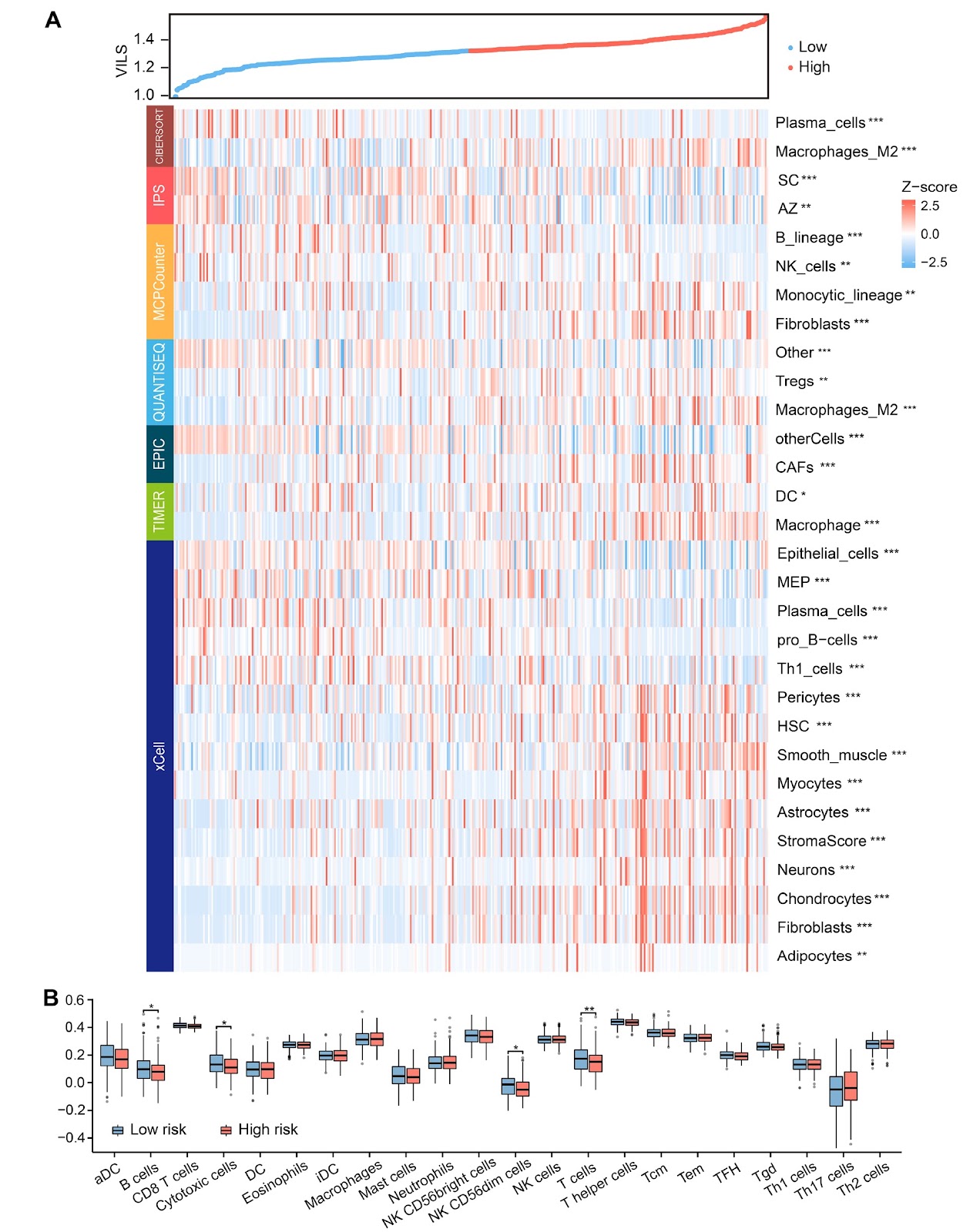
Gastric cancer (GC) is a highly prevalent and lethal gastrointestinal cancer. Dysregulation of vesicle-mediated transport-related genes (VMTRGs) is closely associated with tumorigenesis and disease progression. However, the prognostic value of VMTRGs in GC remains unclear. In this study, on the basis of our proteomics data and public databases, we identify differentially expressed VMTRGs in infiltrative-type GC with more metastases and recurrences identified by Ming’s classification. Least absolute shrinkage and selection operator (LASSO) regression identifies 3 VMTRGs (SEC23A, RAB31, and GABARAPL2) from 41 infiltrative-associated VMTRGs, based on which a risk model Vesicle-Infiltrative Lasso System (VILS) is constructed, and its effectiveness and potential importance are validated by immune microenvironment analysis and functional enrichment analysis. As an independent prognostic factor for GC, VILS, combined with other clinically independent prognostic factors to form a nomogram, is effective in predicting GC prognosis. The VILS high-risk group has higher M2 macrophage and cancer-associated fibroblast infiltration, and lower infiltration of Th1 cells and natural killer cells. SEC23A is highly expressed in GC tissues and cells. The importance of SEC23A in GC cells is evaluated by in vitro assays including colony formation assay and CCK-8 assay, and by in vivo assay using a subcutaneous xenograft mouse model. The results show that SEC23A promotes GC cell proliferation and tumor growth through regulation of the cell cycle in vitro and in vivo. VILS provides excellent prognostic prediction for GC patients and is correlated with antitumor immune cell infiltration. SEC23A, the dominant gene of VILS, is highly expressed in GC and promotes GC growth and malignant progression through various molecular mechanisms. Our study reveals the effect of SEC23A on the proliferation of gastric cancer cells for the first time. Therefore, SEC23A has the potential to be a new therapeutic target for the diagnosis and treatment of GC.

Read Full Abstract10.3724/abbs.2025051
Inhibition of HMOX1 alleviates diabetic cardiomyopathy by targeting ferroptosisGraphical AbstractVerified
Acta Biochimica et Biophysica Sinica

Inhibition of HMOX1 alleviates diabetic cardiomyopathy by targeting ferroptosis

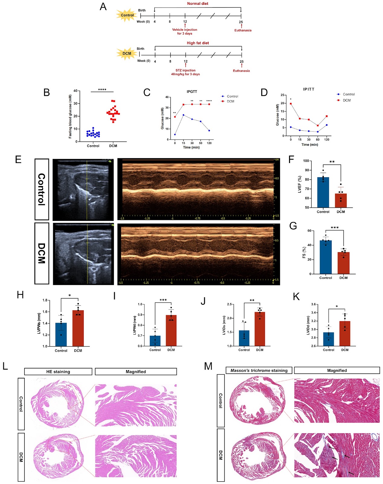
Diabetic cardiomyopathy (DCM) is an important complication of chronic diabetes mellitus. However, its pathologic process and pathogenesis have not been fully elucidated. This study aims to investigate the role of ferroptosis in DCM and clarify the effect of heme oxygenase-1 (HMOX1) on DCM by targeting ferroptosis. In vivo, an animal model of DCM is established by subjecting mice to a high-fat diet (HFD) combined with low-dose streptozotocin (STZ) injection. We induce an in vitro DCM model by exposing H9C2 cells to high glucose and palmitic acid. Transcriptome sequencing reveals that the differentially expressed genes (DEGs) are enriched primarily in fatty acid metabolism and mitochondrial fatty acid β-oxidation, which are closely related to ferroptosis. The experimental results show that the diabetic microenvironment induces ferroptosis both in vivo and in vitro. Western blot analysis reveals the decreased expressions of the antioxidant proteins GPX4, SLC7A11 and ferritin in the DCM group. However, qPCR demonstrates the elevated expressions of the ferroptosis markers PTGS2 and ACSL4. Biochemical indicators further support the occurrence of ferroptosis, with increased levels of malondialdehyde (MDA) and lactate dehydrogenase (LDH), along with decreased level of glutathione (GSH). In vitro, intervention with high glucose and palmitic acid in H9C2 cells results in ferroptosis, which is reversed by ferrostatin-1 (Fer-1). Results show the elevated expression of HMOX1 in DCM. Moreover, knockdown of HMOX1 ameliorates ferroptosis, thereby alleviating diabetic cardiomyopathy by reducing cardiac fibrosis and improving cardiac function. Our study elucidates the role of HMXO1 in DCM pathogenesis and provides a potential therapeutic strategy for clinical treatment.

Read Full Abstract10.3724/abbs.2024232
Effective resistance to UVB-induced skin damage through the encapsulation of chebulinic acid in mulberry-derived exosome-like nanoparticlesGraphical AbstractVerified
Acta Biochimica et Biophysica Sinica

Effective resistance to UVB-induced skin damage through the encapsulation of chebulinic acid in mulberry-derived exosome-like nanoparticles

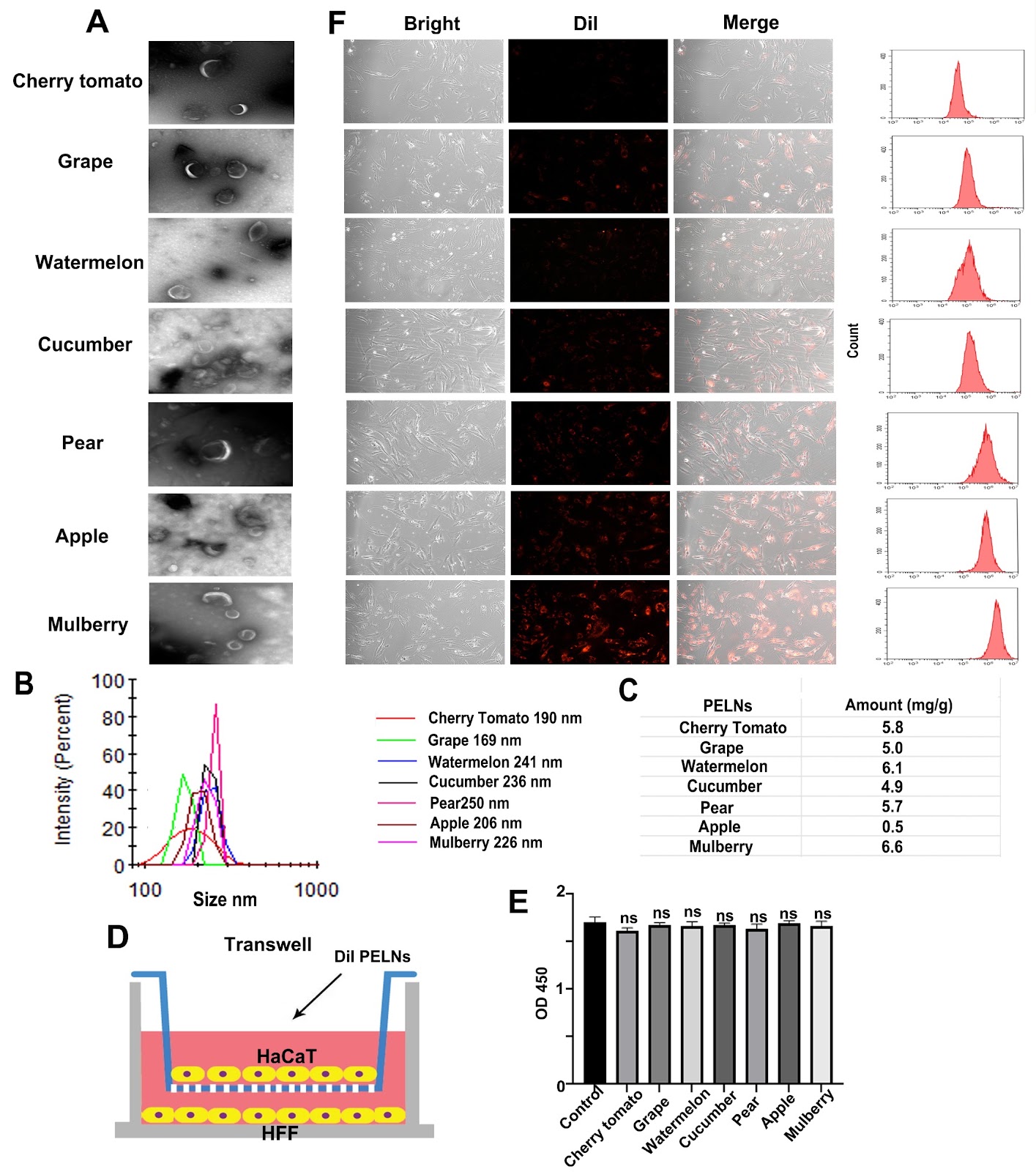
Ultraviolet-B (UVB) radiation induces significant skin damage by penetrating into the dermal layer, leading to reactive oxygen species (ROS) generation and triggering cellular necrosis and apoptosis. Conventional sunscreens focus primarily on UVB blocking but are limited in their ability to repair dermal damage due to insufficient permeability. In this study, we discover that chebulinic acid (CA), one of the principal monomers in Terminalia chebula Retz., has superior efficacy in promoting recovery from UVB-induced skin damage compared with other major monomers. Mechanistically, CA’s anti-UVB function involves regulating the expression of IL-6 and IFN-β through activation of the MAPK pathway. To overcome the formidable barrier posed by the skin, we identify mulberry exosome-like nanoparticles (MELNs) as an efficient transdermal delivery system and develop CA@MELNs loaded with CA. Furthermore, we demonstrate that the dissociative CA within the CA@MELNs delivery system significantly enhances both transdermal penetration and anti-UVB efficiency in vitro and in vivo. Our findings suggest the substantial potential of CA as an effective ingredient and CA@MELNs as a robust and accessible platform for mitigating UVB damage.

Read Full Abstract10.3724/abbs.2025205
MYB represses ζ-globin expression through upregulating ETO2Graphical AbstractVerified
Acta Biochimica et Biophysica Sinica

MYB represses ζ-globin expression through upregulating ETO2

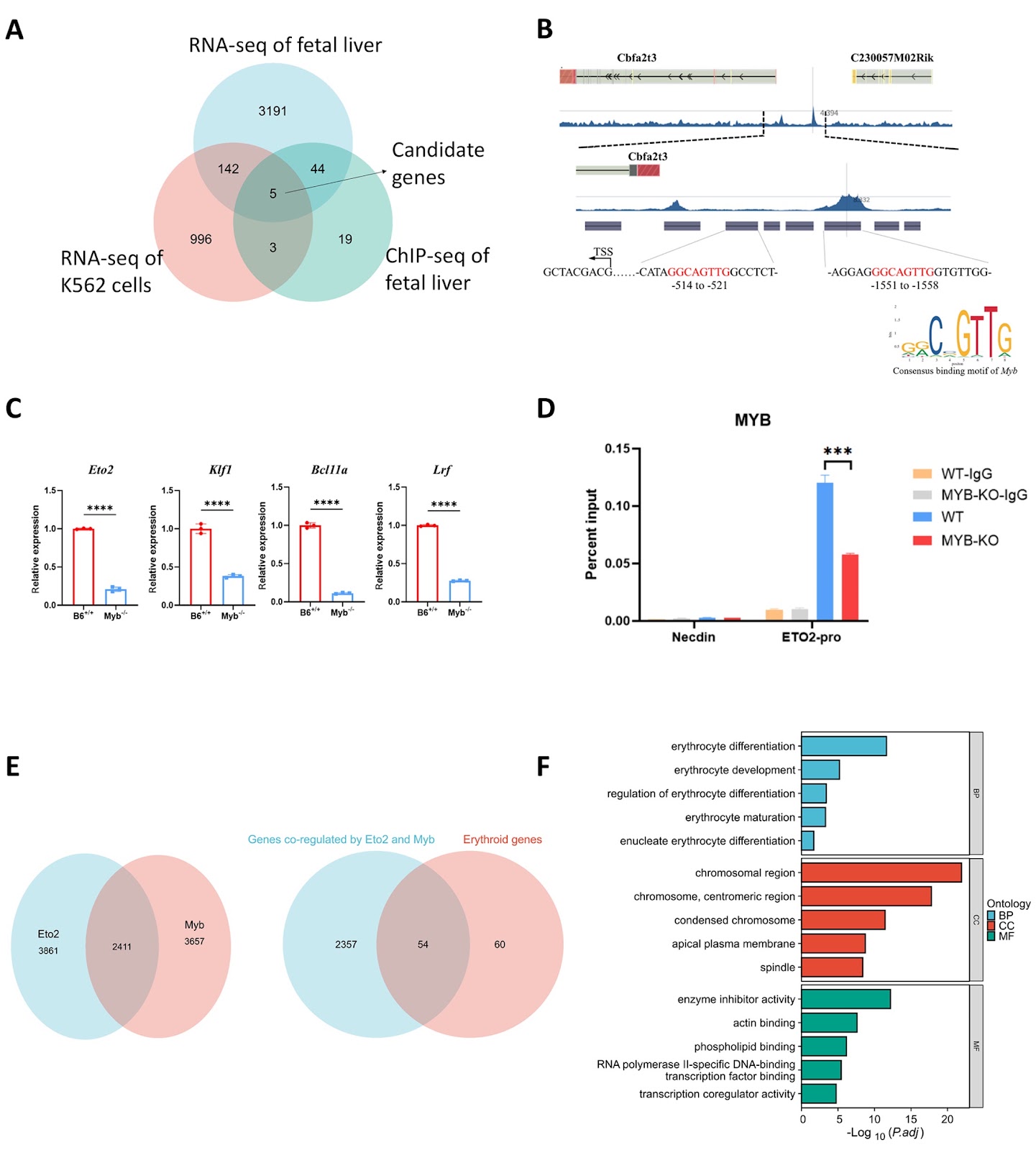
Reactivating the embryonic ζ-globin gene represents a potential therapeutic approach to ameliorate the severe clinical phenotype of α-thalassemia and sickle cell disease. The transcription factor MYB has been extensively proven to be a master regulator of the γ-globin gene, but its role in the regulation of ζ-globin remains incompletely understood. Here, we report a mechanistic study on the derepression of ζ-globin both in vivo and in vitro. We show that MYB depletion in mouse models and human hematopoietic stem cells leads to consistent and remarkable reactivation of ζ-globin. Furthermore, multiomics analysis and functional validation of MYB-knockout and wild-type cell lines reveal that ETO2 functions as a novel repressor of ζ-globin through coordination with NuRD nucleosome remodeling and the deacetylation complex to modulate histone deacetylation of ζ-globin. Additionally, we evaluate the clinical significance of these findings by knocking out ETO2 in primary CD34+ cells from nondeletional hemoglobin H patients, which results in a significant increase in ζ-globin expression. The RNA-seq data reveal that key erythroid genes are more co-regulated by Myb and Eto2 than by Myb and Klf1, highlighting a distinctly enhanced erythroid-specific transcriptional impact within the MYB-ETO2 regulatory axis. Compared with ETO2 knockout alone, codepletion of ETO2 and BCL11A did not significantly activate ζ-globin, suggesting that the MYB-ETO2 pathway primarily silences ζ-globin. Our study reveals a linear MYB-ETO2 signaling pathway crucial for ζ-globin repression and offers new targets for treating α-thalassemia and sickle cell disease.

Read Full Abstract10.3724/abbs.2024239
D-mannose suppresses the angiogenesis and progression of colorectal cancerGraphical AbstractVerified
Acta Biochimica et Biophysica Sinica

D-mannose suppresses the angiogenesis and progression of colorectal cancer

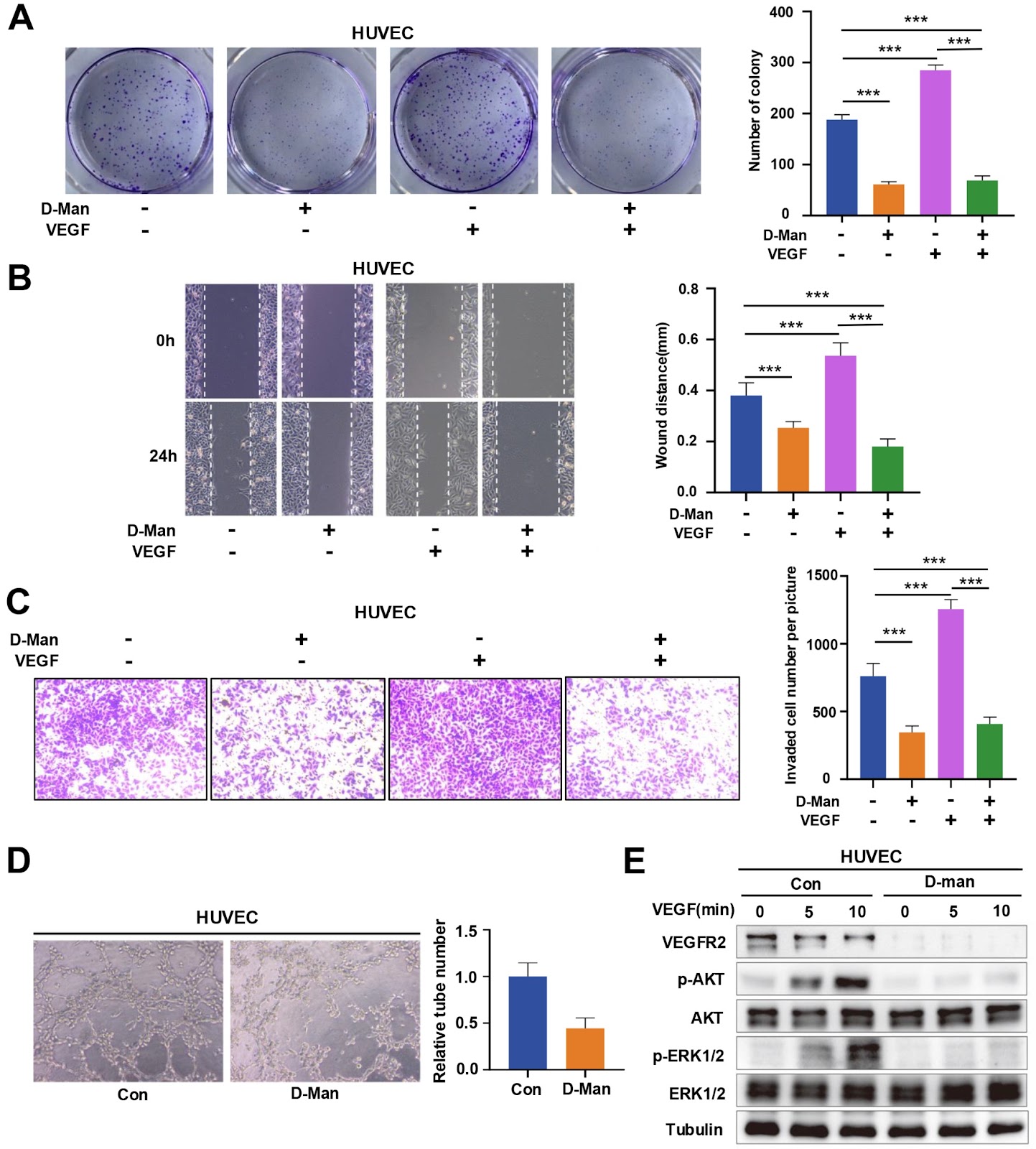
Angiogenesis is an important factor influencing the development of solid tumors, and vascular endothelial growth factor receptor-2 (VEGFR2) is a central regulator of angiogenesis. Antibodies and inhibitors against VEGFR2 have been widely used in various malignancies. However, the regulatory mechanism of VEGFR2 has not been fully clarified. Here, we show that D-mannose can significantly inhibit angiogenesis and tumor growth by degrading VEGFR2. Specifically, D-mannose inactivates GSK3β by promoting the phosphorylation of GSK3β at Ser9, enhances the nuclear translocation of TFE3, and promotes lysosomal biogenesis, thereby increasing the lysosome-mediated degradation of VEGFR2. Thus, D-mannose significantly inhibits the proliferation, migration, and capillary formation of human umbilical vein endothelial cells (HUVECs) in vitro. Oral administration of D-mannose dramatically inhibits angiogenesis and tumor growth in mice. Our findings reveal a previously unrecognized anti-tumor mechanism of D-mannose by destabilizing VEGFR2 and provide a new strategy for the clinical treatment of colorectal cancer (CRC).

Read Full Abstract10.3724/abbs.2025043
cGAS-STING pathway reprograms macrophage polarization and is highly expressed in responding tumors after neoadjuvant immunotherapy in head and neck carcinomaGraphical AbstractVerified
Acta Biochimica et Biophysica Sinica

cGAS-STING pathway reprograms macrophage polarization and is highly expressed in responding tumors after neoadjuvant immunotherapy in head and neck carcinoma

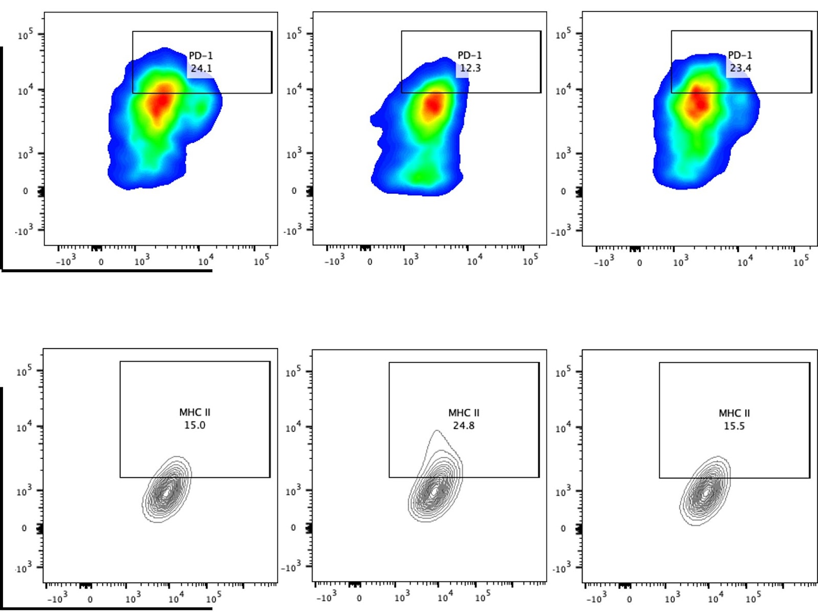
Given the critical role of the cGAS-STING pathway in antitumor immunity, this study investigates the functional role of STING in head and neck squamous cell carcinoma (HNSCC) to evaluate the therapeutic potential of STING agonists. Analysis of the TCGA-HNSC dataset reveals that elevated expression of the STING-encoding gene TMEM173 is significantly correlated with increased M1 macrophage infiltration and enrichment of macrophage polarization-related signaling pathways. In vitro experiments in which RAW 264.7 cells are co-cultured with tumor cell-conditioned medium demonstrate that the STING agonist MSA-2 effectively reprograms tumor-induced M2-polarized macrophages toward the M1 phenotype. This MSA-2-induced M1 polarization is accompanied by increased expressions of IFN-α, IFN-β, IFN-γ, TNF-α, and IL-6, while the STING inhibitor H-151 reverses these effects. Flow cytometry further reveals that MSA-2 treatment reduces PD-1 and increases MHC II expression on macrophages. Immunohistochemical analysis of clinical samples confirms that high STING expression is correlated with increased numbers of CD68⁺ and CD80⁺ (M1-like) macrophages. In support of translational relevance, analysis of single-cell RNA-seq data from HNSCC patients receiving neoadjuvant immunotherapy indicates that TMEM173 is expressed primarily in T cells and macrophages and that the cGAS-STING pathway score is significantly higher in patients who respond to treatment. Collectively, these findings provide systematic clinical and experimental evidence supporting the potential of STING agonists, such as MSA-2, to enhance antitumor immunity in HNSCC, particularly when combined with immunotherapy.

Read Full Abstract10.3724/abbs.2025209
Serum starvation induces density-dependent apoptosis via HIF-1 activation and JNK suppressionGraphical AbstractVerified
Acta Biochimica et Biophysica Sinica

Serum starvation induces density-dependent apoptosis via HIF-1 activation and JNK suppression

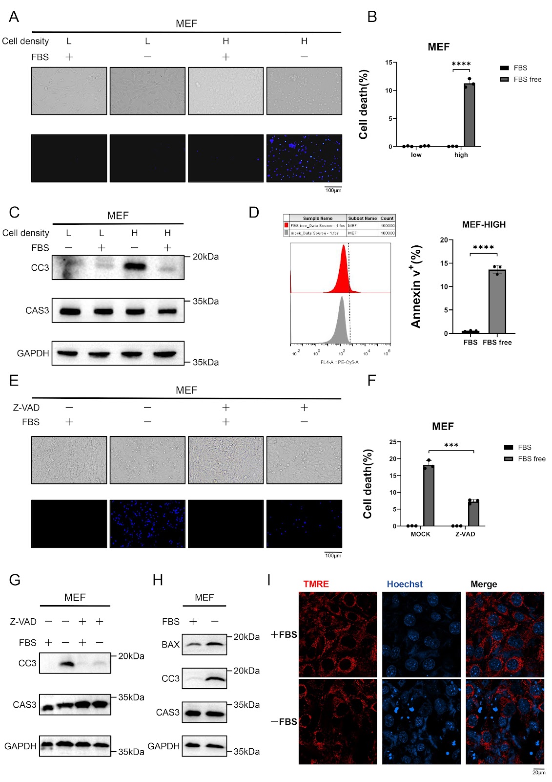
Serum deprivation is a well-established inducer of apoptosis, yet the molecular mechanisms governing this process remain incompletely understood. Here, we show that serum starvation selectively triggers intrinsic apoptosis in high-density murine embryonic fibroblasts (MEFs) through coordinated HIF-1α activation and JNK signaling suppression. Knockdown of HIF-1α abolishes caspase-3 activation and apoptosis induced by serum deprivation, whereas upregulation of HIF-1α in low-density cells recapitulates the apoptotic response observed in high-density cultures. Simultaneously, serum deprivation leads to the suppression of the JNK pathway, which contributes to apoptosis. Notably, combined HIF-1α activation and JNK inhibition in low-density cells fully mimics the apoptotic phenotype of high-density conditions, underscoring the interplay between these pathways. Together, these findings define a density-dependent apoptotic switch in which HIF-1α drives metabolic stress adaptation, whereas JNK suppression removes a critical survival signal, converging to promote mitochondrial-mediated cell death. This work provides a mechanistic framework for understanding nutrient stress-induced apoptosis and suggests potential therapeutic targets for diseases characterized by aberrant cell survival.

Read Full Abstract10.3724/abbs.2025161
The catalase gene CAT2 and its role in the virulence of one sub-cluster of Cryptococcus gattii VGI clinical isolatesGraphical AbstractVerified
Acta Biochimica et Biophysica Sinica

The catalase gene CAT2 and its role in the virulence of one sub-cluster of Cryptococcus gattii VGI clinical isolates

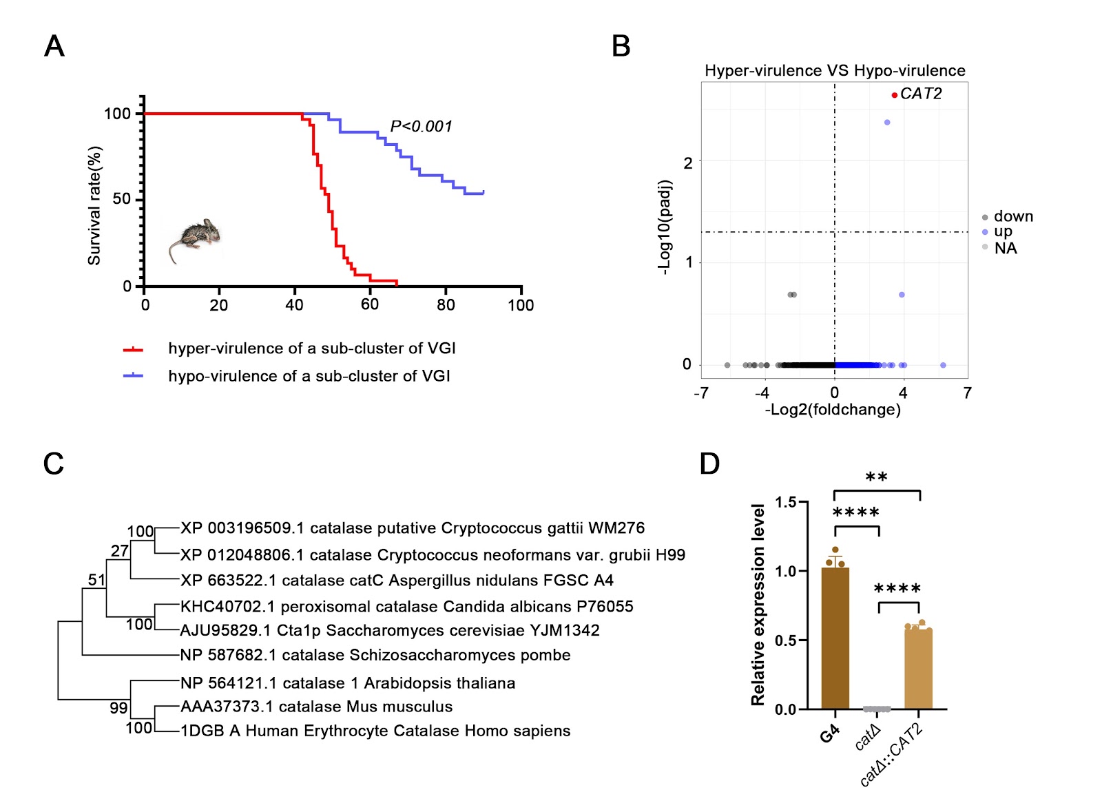
Cryptococcus gattii causes cryptococcosis and life-threatening cryptococcal meningitis. Currently, the pathogenic virulence mechanisms of C. gattii remain a significant area of ongoing research with considerable unexplored aspects. On the basis of our established research, a sub-cluster of strains with independent evolutionary relationships from WM276 in the phylogenetic analysis of VGI-type strains is identified. In vivo infection experiments on this sub-branch of strains reveal that there are hypervirulent strains and hypovirulent strains among these strains, and the virulence differences are significant (P < 0.001). Bioinformatic interrogation of differentially expressed genes reveals that the catalase-encoding gene CGB_J0620W, CAT2, is a pivotal virulence-associated gene. The hypervirulent clinical isolate G4 (G4-WT) is selected as the parental strain, from which an isogenic CAT2-knockout mutant (cat2Δ) is constructed via homologous recombination, which shows increased sensitivity to oxidative stress, as well as growth defects in response to hyperosmosis, 5-fluorocytosine, fluconazole and amphotericin B. The cat2Δ::CAT2 strain exhibits phenotypic restoration to wild type (WT). In the mouse experiments, significant differences in survival (P < 0.001), pulmonary fungal burden (P < 0.01), and alveolar structural damage are observed between the WT and cat2Δ strains, which are completely different from C. neoformans. Moreover, comparative transcriptome analysis is performed on the WT and cat2Δ strains, which reveals that enzymes encoded by CAT2 may be involved in oxidative stress, metabolism and sugar transport. In conclusion, this study may explain the differences in virulence among different genetic evolutionary processes of a sub-cluster of the VGI geneotype of C. gattii and provide a theoretical basis for targeted therapy in a specific genotype population in the future.

Read Full Abstract10.3724/abbs.2025170
NCOA6 knockdown enhances RSL3-induced ferroptosis in pancreatic cancer cells and increases the sensitivity to gemcitabineGraphical AbstractVerified
Acta Biochimica et Biophysica Sinica

NCOA6 knockdown enhances RSL3-induced ferroptosis in pancreatic cancer cells and increases the sensitivity to gemcitabine

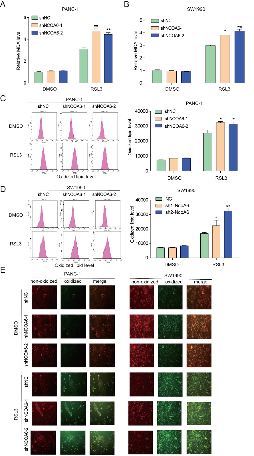
Ferroptosis is a type of programmed death characterized by iron-dependent lipid peroxidation, and targeting ferroptosis has been shown to efficiently kill highly aggressive cancer cells. Previously, we confirmed that nuclear receptors regulate ferroptosis in pancreatic cancer. However, whether nuclear receptor co-activators regulate ferroptosis is unclear. Here, we show that knocking down the nuclear receptor co-activator, NCOA6, enhances the sensitivity of pancreatic cancer cells to ferroptosis. Mechanistically, NCOA6 knockdown promotes the expression of ACSL4 while inhibiting the expression of SCD1, resulting in changes in lipid metabolism, sensitivity to RSL3-induced ferroptosis, and sensitivity to gemcitabine in pancreatic cancer. The relationships between NCOA6 and ACSL4 or SCD1 are further explored in clinical specimens. This study reveals that targeting NCOA6 might alleviate gemcitabine resistance in pancreatic cancer.

Read Full Abstract10.3724/abbs.2024221
Corrigendum to: Magnolol promotes the autophagy of esophageal carcinoma cells by upregulating HACE1 gene expressionGraphical AbstractVerified
Acta Biochimica et Biophysica Sinica

Corrigendum to: Magnolol promotes the autophagy of esophageal carcinoma cells by upregulating HACE1 gene expression

This is a corrigendum to the article 'Magnolol promotes the autophagy of esophageal carcinoma cells by upregulating HACE1 gene expression' published in Acta Biochimica et Biophysica Sinica 2024, 56(7): 1044–1054. In the original publication, the corresponding author's email address was personal. To comply with the institution's publishing policy, it has been changed from '[email protected]' to the institutional address '[email protected]'. The authors apologize for any confusion it may have caused.

Read Full Abstract10.3724/abbs.2025129
MAN1A1 promotes colorectal cancer liver metastasis by maintaining TGFBR2 protein stabilityGraphical AbstractVerified
Acta Biochimica et Biophysica Sinica

MAN1A1 promotes colorectal cancer liver metastasis by maintaining TGFBR2 protein stability

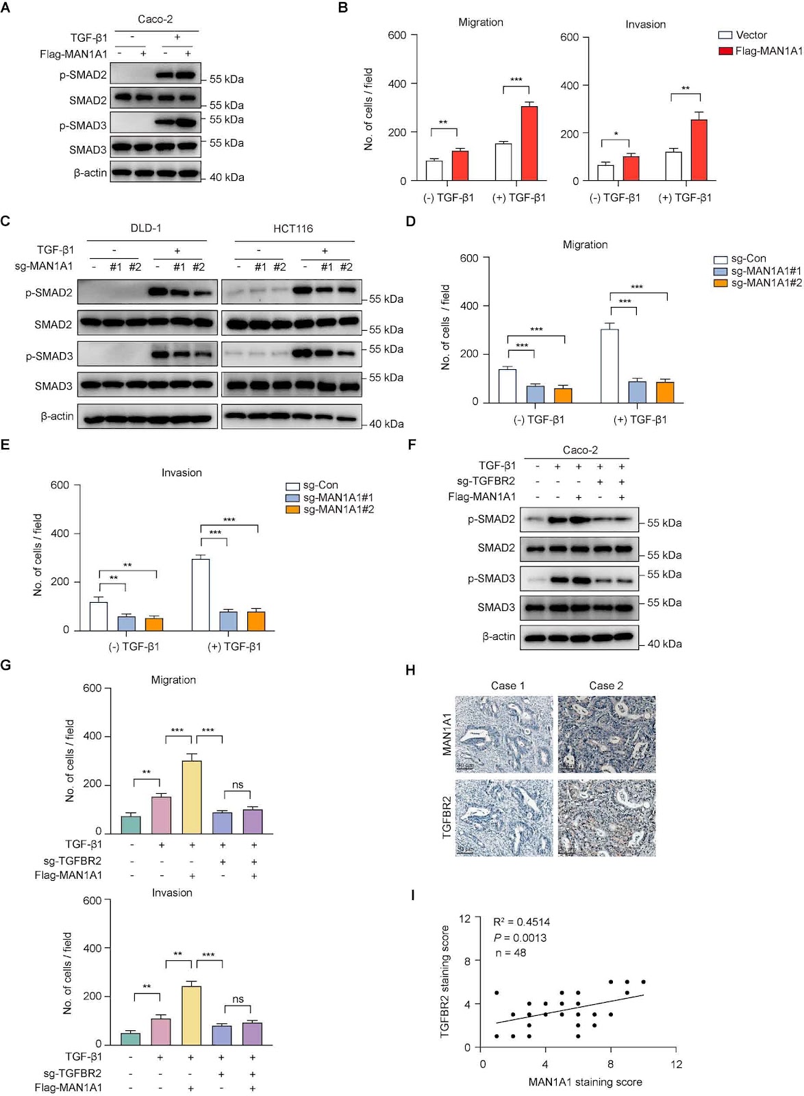
Emerging biochemical and genetic evidence has firmly established aberrant protein glycosylation as a critical regulator of oncogenic transformation, with glycocalyx remodeling profoundly influencing tumor microenvironment dynamics and metastatic progression. Despite the well-documented association between metastatic dissemination and poor clinical outcomes in patients with colorectal cancer, the underlying molecular mechanisms remain incompletely characterized. Through integrative analysis of single-cell RNA sequencing data from a public database, we identify the Golgi-resident α-1,2-mannosidase MAN1A1 as a consistently upregulated enzyme in malignant epithelial cells derived from colorectal cancer liver metastases. Clinically, elevated MAN1A1 expression is correlated with reduced overall survival, suggesting that MAN1A1 is both a prognostic biomarker and therapeutic target for colorectal cancer liver metastases. Genetic manipulation of MAN1A1 in colorectal cancer cells demonstrates that although the proliferation capacity of colorectal cancer cells remains unchanged, MAN1A1 overexpression significantly enhances migratory and invasive capacities in transwell assays, suggesting its specific involvement in metastatic progression. Mechanistic investigations reveal that MAN1A1 exerts its pro-metastatic effects by significantly prolonging the TGFBR2 protein half-life. Together, our work identifies MAN1A1 as both a prognostic biomarker and a promising therapeutic target, highlighting the critical role of glycan remodeling in the metastatic progression of colorectal cancer.

Read Full Abstract10.3724/abbs.2025164
GWAS study of myelosuppression among NSCLC patients receiving platinum-based combination chemotherapyGraphical AbstractVerified
Acta Biochimica et Biophysica Sinica

GWAS study of myelosuppression among NSCLC patients receiving platinum-based combination chemotherapy

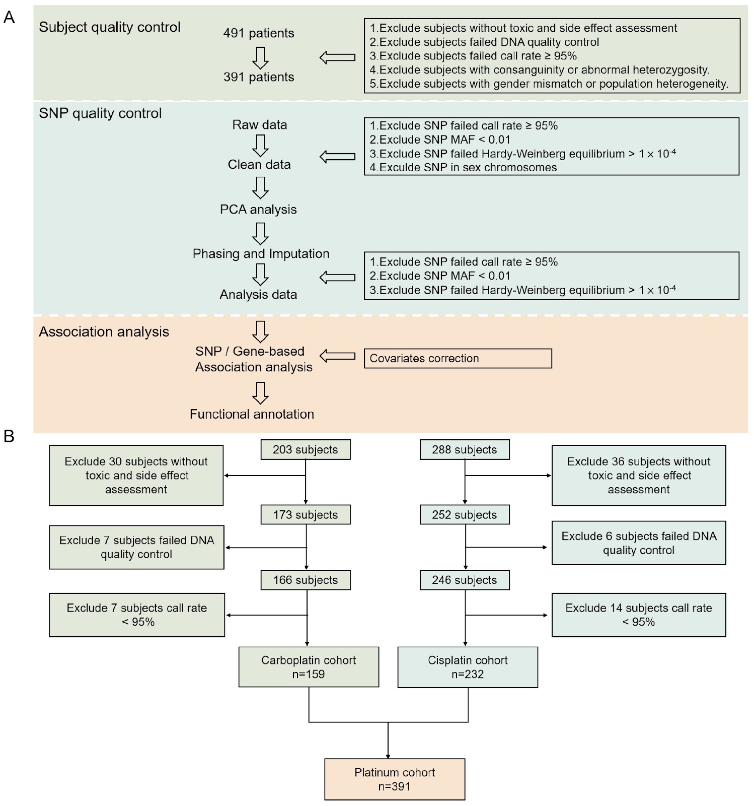
Platinum-based chemotherapy remains the mainstay for non-small cell lung cancer (NSCLC), but it frequently causes dose-limiting myelosuppression, with significant individual variability in susceptibility. However, the genetic basis of myelosuppression side effects remains elusive, greatly hindering personalized therapeutic approaches. In this study, we perform a comprehensive genome-wide association analysis on 491 NSCLC patients receiving platinum-based chemotherapy, examining 4,690,998 single-nucleotide polymorphisms (SNPs) to identify relevant genetic variants. LDBlockShow, FUMA, and MAGMA are utilized to explore linkage disequilibrium, expression quantitative trait loci (eQTLs), chromatin interaction, and conduct gene-based and gene set-based analysis of candidate SNPs. The GWAS results reveal that rs6856089 and its linked SNPs are significantly associated with platinum-based chemotherapy-induced myelosuppression. Specifically, patients with the A allele of rs6856089 have a significantly lower risk of myelosuppression [odds ratio (OR) = 0.1300, P = 7.59 × 10⁻⁸]. Furthermore, gene-based analysis reveals that EMCN (P = 2.47 × 10⁻⁵), which encodes endomucin, a marker for hematopoietic stem cells, might mediate myelosuppression. This study provides a scientific basis for the individual differences in platinum-based chemotherapy-induced myelosuppression.

Read Full Abstract10.3724/abbs.2025013
Knockdown of lncRNA XR_877193.1 suppresses ferroptosis and promotes osteogenic differentiation via the PI3K/AKT signaling pathway in SONFHGraphical AbstractVerified
Acta Biochimica et Biophysica Sinica

Knockdown of lncRNA XR_877193.1 suppresses ferroptosis and promotes osteogenic differentiation via the PI3K/AKT signaling pathway in SONFH

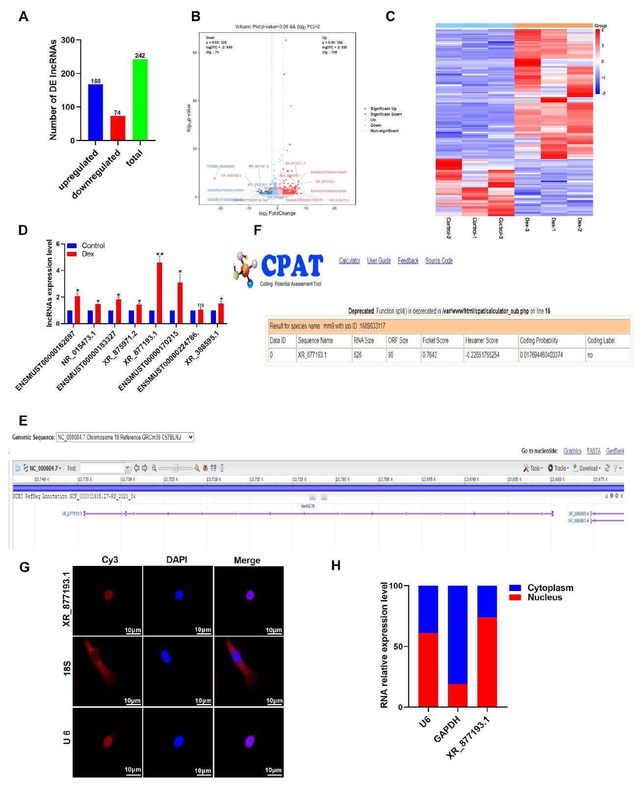
Ferroptosis is a novel form of regulated cell death characterized by the iron-dependent accumulation of lipid peroxides. Recent research has suggested that ferroptosis in osteoblasts contributes to steroid-induced osteonecrosis of the femoral head (SONFH). However, the relationship between ferroptosis and SONFH remains unclear. In this study, in vitro experiments show that dexamethasone (Dex) treatment reduces the expressions of key ferroptosis regulators, SLC7A11 and GPX4, in MC3T3-E1 cells. This reduction leads to a decrease in intracellular glutathione (GSH) levels, accompanied by elevated levels of total iron, malondialdehyde (MDA), and reactive oxygen species (ROS). Importantly, the ferroptosis inhibitor ferrostatin-1 (Fer-1) effectively reverses Dex-induced ferroptosis in MC3T3-E1 cells. Furthermore, RNA-seq analysis reveals that the long noncoding RNA (lncRNA) XR_877193.1 is significantly upregulated in Dex-treated MC3T3-E1 cells. Functional studies demonstrate that the knockdown of lncRNA XR_877193.1 promotes osteogenic differentiation by inhibiting Dex-induced ferroptosis in MC3T3-E1 cells, whereas its overexpression exacerbates cell death via ferroptosis. Kyoto Encyclopedia of Genes and Genomes (KEGG) enrichment analysis reveals that the differentially expressed lncRNA XR_877193.1 is enriched in ferroptosis-related pathways, including the PI3K/AKT signaling pathway. Moreover, PI3K/AKT inhibitors reverse ferroptosis in MC3T3-E1 cells inhibited by lncRNA XR_877193.1 knockdown. Collectively, our findings indicate that lncRNA XR_877193.1 knockdown exerts anti-ferroptosis effects by stimulating the PI3K/AKT signaling pathway, suggesting a promising therapeutic strategy for attenuating SONFH.

Read Full Abstract10.3724/abbs.2025014
CRATS: a one-pot CRISPR-Cas12b and RPA combined assay with a temperature switch for highly sensitive detection of monkeypox virusGraphical AbstractVerified
Acta Biochimica et Biophysica Sinica

CRATS: a one-pot CRISPR-Cas12b and RPA combined assay with a temperature switch for highly sensitive detection of monkeypox virus

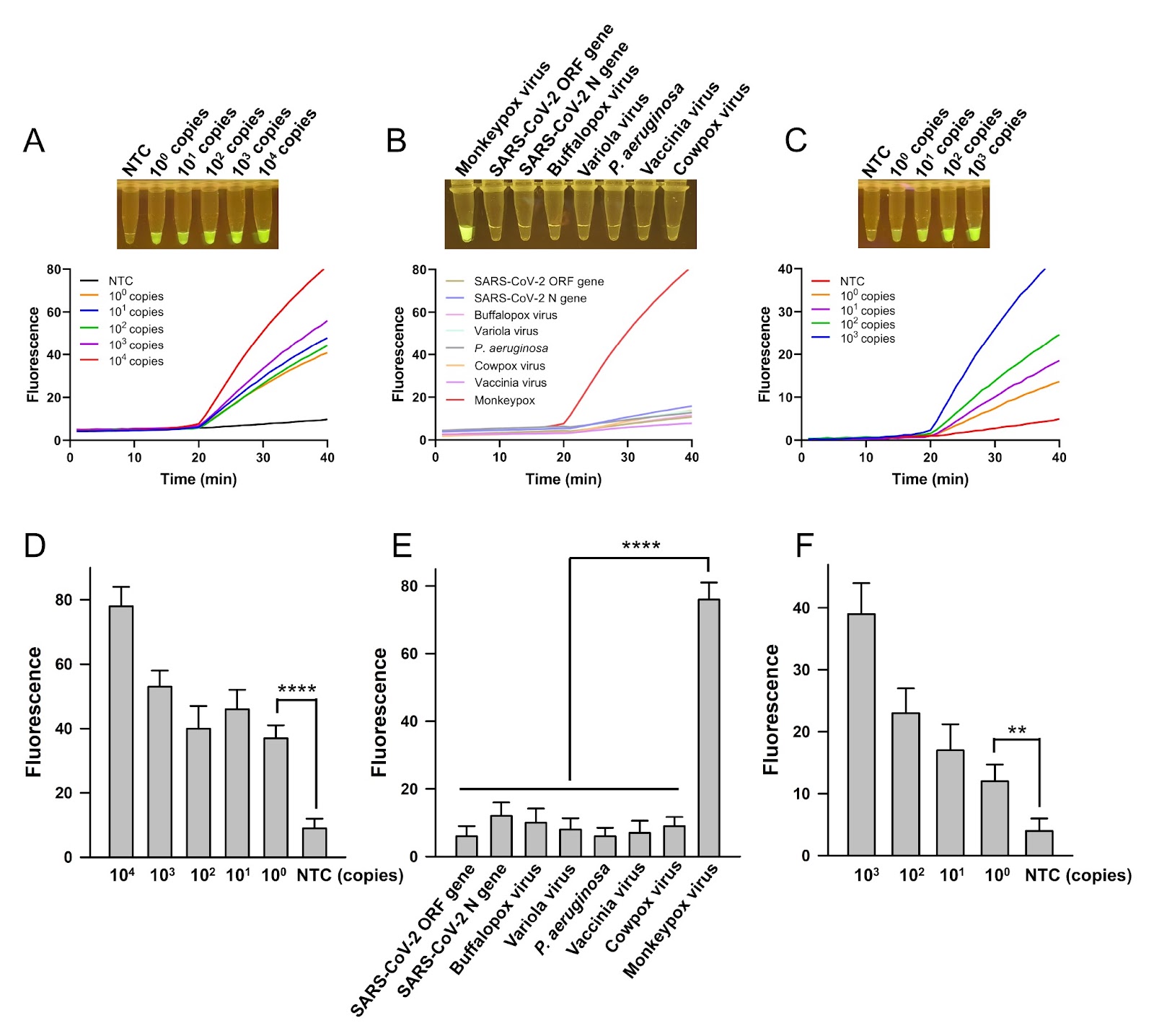
CRISPR-Cas nucleases have been extensively used in molecular detections, especially highly sensitive nucleic acid detections. In these detections, Cas nucleases are programmed by the guide RNA to respond to the detection targets and cleave the chemically labelled molecular beacons by the trans-cleavage activity to produce the detection signal. To improve sensitivity, nucleic acid amplification technologies are usually introduced to give a pre-amplification of the nucleic acid targets, increasing the detection sensitivity extraordinarily. Polymerase chain reaction (PCR) technology has been used for pre-amplification in laboratories, and isothermal amplification technologies are applied to meet point-of-care testing (POCT) needs because they avoid the use of sophisticated thermal cycling devices. The recombinase polymerase amplification (RPA) technology that amplifies nucleic acid targets isothermally at 37–42°C has been combined with CRISPR-Cas nucleases to establish advantageous nucleic acid detection assays, e.g., the SHERLOCK, which combines with Cas13a, and the DETECTR, which combines with Cas12a. It has been challenging to integrate Cas nucleases and RPA in a one-pot reaction system because the cleavage activity of Cas stimulated by even small amounts of the targets can interfere with amplification by digesting the primers or the newly amplified fragments. Thus, many assays based on Cas nucleases and RPA are in a two-step setting, with pre-amplification and Cas cleavage being isolated as two independent procedures. The two-step setting ensures that amplification and cleavage occur under favorable conditions but sacrifices operational convenience and introduces the risk of cross-contamination. In efforts to establish one-pot RPA-Cas assays, many strategies have been applied, including the use of photocontrolled guide RNA to activate the Cas nuclease at a preferred timepoint, the use of a suboptimal protospacer adjacent motif (PAM) to suppress Cas activity, the generation of dynamic aqueous multiphase with sucrose or glycerol to partially separate the two reactions, and extensive optimization of the RPA-CRISPR reaction system to achieve a subtle balance between the two reactions. In this study, a one-pot CRISPR-Cas12b and RPA combined assay with a temperature switch (CRATS) was established utilizing the reaction temperature difference between RPA and CRISPR-Cas12b cleavage. The Cas12b used in this study, AaCas12b, is a type V-B CRISPR‒Cas nuclease from Alicyclobacillus acidiphilus. It has a bi-lobed architecture consisting of an α-helical recognition lobe containing the REC domains and a nuclease lobe containing the WED, RuvC and Nuc domains. As a dual-RNA-guided DNA endonuclease, Cas12b can be guided by a chimeric single-guide (sg) RNA, and its trans-cleavage activity is specifically activated by the DNA target and results in nonspecific cleavage of single-stranded (ss) DNA molecules, which can be used to produce detection signals if the ssDNA is appropriately labelled as the molecular beacon. As the temperature for RPA is 37°C and the trans-cleavage of Cas12b is active at 60°C, CRATS uses temperature switching to adjust the major on-going reaction in the one-pot system and realizes sequential amplification of the target and cleavage reactions for signal detection. The detection target of this study, monkeypox virus, is an infectious pathogen that has caused the announcement of the Public Health Emergency of International Concern (PHEIC) by the World Health Organization (WHO) twice in recent years. In this one-pot CRATS assay, the reaction reagents of CRISPR-Cas12b and RPA are mixed in a single tube. After the addition of the sample containing the detection target, the reaction was carried out at 37°C for 20 min for amplification, followed by 60°C for 20 min for Cas12b cleavage. The fluorescently labelled molecular beacon is cleaved by Cas12b to release the FAM fluorophore from quenching, producing a fluorescence signal that is visualized under blue light. CRATS shows a high sensitivity of 100 copies of the target DNA per reaction and good specificity, providing a novel strategy of temperature switching to integrate CRISPR-Cas and RPA in a one-pot reaction system. Moreover, it provides a POCT-friendly tool for the detection of the important infectious pathogen monkeypox virus.

Read Full Abstract10.3724/abbs.2025016
Carfilzomib triggers cardiotoxicity by suppressing SENP1-mediated deSUMOylation of DDX17Graphical AbstractVerified
Acta Biochimica et Biophysica Sinica

Carfilzomib triggers cardiotoxicity by suppressing SENP1-mediated deSUMOylation of DDX17

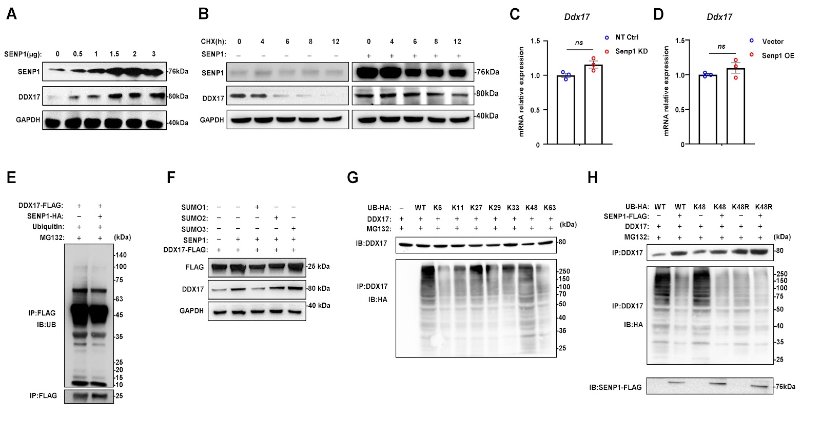
Carfilzomib (Cfz) is a second-generation proteasome inhibitor approved for the treatment of relapsed/refractory multiple myeloma (RRMM). Previous studies have shown that Cfz is associated with a higher incidence of severe adverse cardiac effects than bortezomib (Btz); however, the underlying mechanisms remain to be elucidated. The aim of this study is to identify key regulators of cardiotoxicity induced by Cfz and to investigate the mechanisms by which these factors exert their effects. We establish a mouse model of cardiac toxicity induced by Cfz and confirm the phenotype through cardiac functional analysis, morphology assessment, myocardial fibrosis, and apoptosis analysis. We subsequently perform RNA sequencing to identify differentially expressed genes (DEGs) and further validate their functions and mechanisms. We find that Cfz induces myocardial hypertrophy and myocardial injury, along with the suppression of SENP1 expression in mouse heart tissues and in vitro cultured neonatal rat cardiomyocytes. Suppression of SENP1 exacerbates Cfz-induced injury and remodeling in cardiomyocytes by directly binding to and deconjugating the SUMO1-mediated SUMOylation of the RNA helicase DDX17. This process leads to a reduction in K-48 ubiquitin-linked polyubiquitination and degradation of DDX17, resulting in increased expressions of anti-apoptotic genes and maintenance of mitochondrial homeostasis. Therefore, the overexpression of SENP1 using AAV vectors alleviates Cfz-induced cardiotoxicity in mice. In summary, our findings reveal a previously unknown role of the SENP1-DDX17 axis in protecting against cardiotoxicity induced by Cfz, providing a potential foundation for developing therapeutic strategies to mitigate cardiac side effects in the clinical management of MM patients.

Read Full Abstract10.3724/abbs.2025121
Essential role of the metabolite α-ketoglutarate in bone tissue and bone-related diseasesGraphical AbstractVerified
Acta Biochimica et Biophysica Sinica

Essential role of the metabolite α-ketoglutarate in bone tissue and bone-related diseases

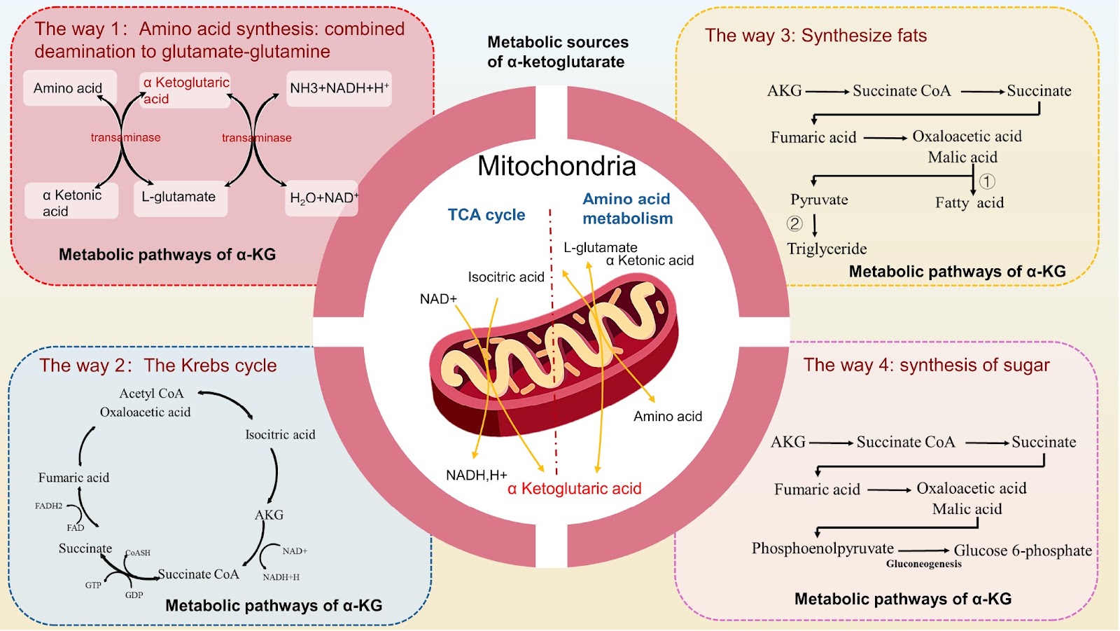
Bone metabolism in bone tissue is constantly maintained in a state of dynamic equilibrium. The mass of bone and joint tissues is determined by both bone formation and bone resorption. It is hypothesized that disrupted metabolic balance leads to osteoporosis, osteoarthritis, rheumatoid arthritis, and bone tumors. Such disruptions often manifest as either a reduction or abnormality in bone mass and are frequently accompanied by pathological changes such as inflammation, fractures, and pain. α-Ketoglutarate (α-KG) serves as a pivotal intermediate in various metabolic pathways in mammals, significantly contributing to cellular energy metabolism, amino acid metabolism, and other physiological processes. α-KG may be a therapeutic target for a variety of bone-related diseases, such as osteoporosis, osteoarthritis, and rheumatoid arthritis, because of its role in maintaining the metabolic balance of bone. After the application of α-KG, bone loss and inflammation in bone tissue are alleviated. This review focuses on the regulatory effects of α-KG on various cells in bone and joint tissues. Owing to the regulatory effect of α-KG on the balance of bone metabolism, the application of α-KG in the treatment of osteoporosis, osteoarthritis, rheumatoid arthritis, bone tumors, and other bone tissue diseases has been clarified.

Read Full Abstract10.3724/abbs.2025020
Investigation of the cardioprotective potential of dantrolene in mitigating arsenic-induced cardiac dysfunction in ratsGraphical AbstractVerified
Acta Biochimica et Biophysica Sinica

Investigation of the cardioprotective potential of dantrolene in mitigating arsenic-induced cardiac dysfunction in rats

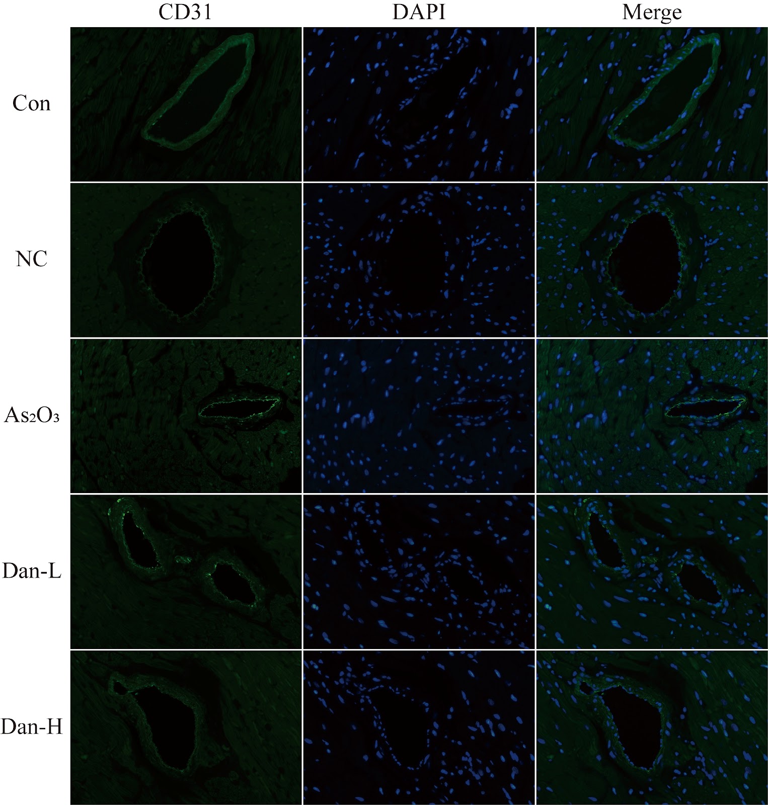
Arsenic, a toxic metalloid, exists in organic or inorganic states within the Earth’s seawater, river water, soil, atmosphere, food sources, and diverse biological tissues [1]. It poses a threat to the health of hundreds of millions of people globally [2]. Arsenic exposure has toxic effects on the cardiovascular system of organisms, thus endangering human health [3]. Research has indicated that the harmful effect of arsenic exposure on the heart is associated with abnormal calcium handling in myocardial cells [4]. The cardiac ryanodine receptor type 2 (RyR2) is a primary channel involved in the surface of the endoplasmic reticulum in cardiac myocytes that regulates the release of Ca2+ during the systolic phase [5]. The integrity of its function is crucial for maintaining calcium homeostasis in cardiac myocytes. However, when myocardial tissue is damaged and undergoes pathological changes, the spatial structure of the RyR2 protein becomes unstable and becomes excessively activated, thereby triggering Ca2+ leakage [6]. Dantrolene (Dan), which serves as a stabilizer of RyR1, is frequently employed in clinical settings for the treatment of malignant hyperpyrexia and relieves spastic muscle tension [7]. Previous studies have demonstrated that dantrolene also has a stabilizing effect on RyR2 [8]. Research has shown that dantrolene can prevent calcium leakage in myocardial cells by stabilizing the tertiary structure of the RyR2 protein and thereby inhibiting the pathological hyperactivity of RyR2 [9]. Therefore, this study hypothesizes that dantrolene, by virtue of this stabilizing effect, can alleviate myocardial injury caused by arsenic exposure to some extent and plays a role in protecting cardiac function. For this purpose, we established an arsenic exposure model and a Dan intervention arsenic exposure model to verify the protective effect of Dan on the myocardial tissue and cardiac function of arsenic-exposed rats.

Read Full Abstract10.3724/abbs.2025193