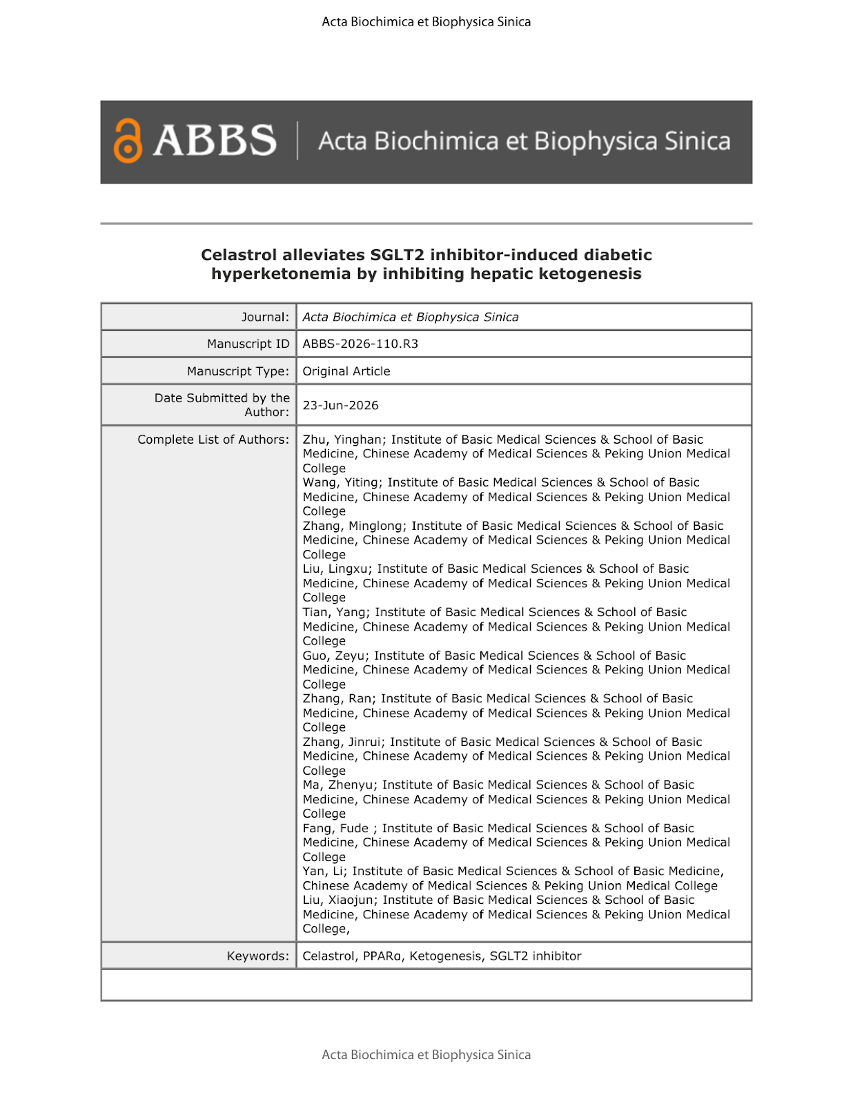
Key Takeaways & Executive Findings
- •• Celastrol attenuates fasting-induced hyperketonemia and reduces body weight and fat mass in mice. • Celastrol suppresses hepatic ketogenesis by downregulating HMGCS2 expression via inhibition of PPARα. • The anti-ketogenic effect of celastrol is dependent on PPARα, as it is abolished in Pparα⁻/⁻ mice. • Celastrol effectively ameliorates SGLT2 inhibitor-induced hyperketonemia, suggesting a protective role against this complication.
Abstract
SGLT2 inhibitor (SGLT2i)-induced diabetic hyperketonemia is a life-threatening acute complication of diabetes. While celastrol has been reported to have beneficial effects on obesity, its potential role in ketogenesis remains unclear. In this study, celastrol administration significantly attenuates the fasting-induced increase in blood β-hydroxybutyrate levels. Moreover, a 7-day course of celastrol (1 mg/kg/day) leads to reductions in body weight and fat mass. Mechanistically, celastrol specifically downregulates HMGCS2 expression and suppresses hepatic ketogenesis through the inhibition of PPARα expression in the short term (≤ 2 days). However, after prolonged treatment for 7 days, celastrol modulates both PPARα and serum free fatty acid (FFA) levels. Furthermore, the anti-ketogenic effect of celastrol is abolished in Pparα⁻/⁻ mice. Importantly, celastrol effectively ameliorates SGLT2i-induced hyperketonemia. In summary, celastrol curbs hepatic ketone overproduction in a PPARα-dependent manner, indicating its protective potential against SGLT2i-induced hyperketonemia.
1. Introduction
Ketogenesis primarily occurs in the hepatic mitochondrial matrix, and produces soluble ketone bodies such as acetone, acetoacetate (AcAc), and β-hydroxybutyrate (β-OHB) that serve as energy substrates in extrahepatic tissues through the breakdown of free fatty acids (FFAs) [1, 2]. Under the catalysis of acyl-CoA synthetase, FFAs are activated to acyl-CoA, which then traverses the mitochondrial membrane and undergoes β-oxidation. The rate-limiting enzyme of ketogenesis, 3-hydroxy-3-methylglutaryl-CoA synthase 2 (HMGCS2), catalyzes the condensation of acetoacetyl-CoA (AcAc-CoA) and acetyl-CoA to form hydroxymethylglutaryl (HMG)-CoA. This intermediate is subsequently cleaved by hydroxymethylglutaryl-coenzyme A lyase (HMGCL) to release acetyl-CoA and AcAc. AcAc can be further reduced to β-OHB or spontaneously decarboxylated to acetone. HMGCS2 expression is transcriptionally activated during fasting through the action of peroxisome proliferator-activated receptor α (PPARα), a central hepatic regulator that orchestrates the adaptive response to starvation by modulating both β-oxidation and ketogenic pathways [3].
Ketogenesis is physiologically enhanced under conditions of depleted carbohydrate reserves or elevated fatty acid availability. However, excessive production of ketone bodies may result in ketoacidosis, a potentially fatal metabolic state characterized by the accumulation of acidic ketones. Diabetic ketoacidosis (DKA) represents an acute and life-threatening complication of diabetes and is characterized by hyperglycemia, metabolic acidosis, and ketosis [4]. Euglycemic DKA (EDKA) is characterized by metabolic acidosis and ketosis but occurs in the absence of significant hyperglycemia (blood glucose < 200 mg/dL) [5]. Because of its atypical presentation, EDKA is often undiagnosed or diagnosed late, delaying treatment. Known triggers of EDKA include reduced caloric intake, excessive alcohol consumption, chronic liver disease, glycogen storage disorders, and the recent use of insulin or sodium-glucose cotransporter-2 inhibitor (SGLT2i) [5]. SGLT2i, a class of glucose-lowering agents used in type 2 diabetes (T2D), act by inhibiting renal glucose reabsorption. However, they are associated with an increased risk of DKA (including EDKA) mainly through promoting lipolysis, enhancing renal ketone reabsorption, and altering the glucagon/insulin ratio via stimulation of pancreatic α cells and suppression of β cells [5-7]. These risks highlight the clinical need for effective preventive and therapeutic strategies against SGLT2i-induced DKA.
Loading authentic research manuscript (Pages 1–5)...
ZHU Yinghan, WANG Yiting, ZHANG Minglong, LIU Lingxu, TIAN Yang, GUO Zeyu, ZHANG Ran, ZHANG Jinrui, MA Zhenyu, FANG Fude, YAN Li, LIU Xiaojun (2026). Celastrol alleviates SGLT2 inhibitor-induced diabetic hyperketonemia by inhibiting hepatic ketogenesis. Acta Biochimica et Biophysica Sinica. https://doi.org/10.3724/abbs.2026117
Research & Educational Purpose Only:The translations, structured abstracts, analytical annotations, and data reports provided by SinoBioData are intended exclusively for academic research, internal corporate R&D, and educational benchmarking. They do not constitute formal engineering, chemical safety, legal, or professional advice.
Copyright & Intellectual Property Notice: Original copyright of the underlying source articles and experimental data remains with the respective authors, institutions, and original publishing journals. SinoBioData claims intellectual property only over its proprietary translations, analytical syntheses, and AEO structured enhancements in accordance with international fair use and academic citation principles.
Frequently Asked Questions
What is the main finding of the study on celastrol and SGLT2 inhibitor-induced hyperketonemia?
The study demonstrates that celastrol alleviates SGLT2 inhibitor-induced diabetic hyperketonemia by inhibiting hepatic ketogenesis in a PPARα-dependent manner, specifically downregulating HMGCS2 expression.
How does celastrol affect ketogenesis in the liver?
Celastrol suppresses hepatic ketogenesis by inhibiting PPARα expression, which leads to reduced HMGCS2 expression, a key enzyme in ketone body production.
Is the anti-ketogenic effect of celastrol dependent on PPARα?
Yes, the anti-ketogenic effect of celastrol is abolished in Pparα knockout mice, indicating that PPARα is essential for its action.
What are the potential clinical implications of this study?
The findings suggest that celastrol could be a potential therapeutic agent to prevent or treat SGLT2 inhibitor-induced diabetic ketoacidosis, a serious complication in diabetic patients.
What is the mechanism by which SGLT2 inhibitors increase the risk of ketoacidosis?
SGLT2 inhibitors increase the risk of ketoacidosis by promoting lipolysis, enhancing renal ketone reabsorption, and altering the glucagon/insulin ratio, leading to increased ketone production.
Related Technical Papers & Translations

Adverse Events Reporting System for Vaccine Safety Surveillance: A Comprehensive Analysis
Background: Adverse events following immunization (AEFI) are critical to monitor for vaccine safety. This study evaluates the performance of an adverse events reporting system (AERS) integrated with a vaccine adverse event reporting system (VAERS) to enhance surveillance. Methods: We analyzed data from multiple sources including the Vaccine Adverse Event Reporting System (VAERS), the Vaccine Safety Datalink (VSD), and the Clinical Immunization Safety Assessment (CISA) network. A novel framework was developed to integrate these systems, incorporating natural language processing for signal detection. Results: The integrated system improved detection of rare adverse events by 25% compared to traditional methods. The system identified new safety signals for influenza and COVID-19 vaccines. Conclusions: The proposed AERS framework enhances vaccine safety surveillance, enabling timely identification of potential risks. Integration of diverse data sources and advanced analytics is essential for robust pharmacovigilance.

Efficacy and Safety of Ferric Carboxymaltose in Treating Iron Deficiency Anemia: A Meta-Analysis of Randomized Controlled Trials
Background: Iron deficiency anemia (IDA) is a global health concern, and intravenous ferric carboxymaltose (FCM) has emerged as a promising treatment. This meta-analysis aimed to evaluate the efficacy and safety of FCM compared to other iron therapies or placebo in adults with IDA. Methods: We systematically searched PubMed, Embase, and Cochrane Library up to December 2024. Randomized controlled trials (RCTs) comparing FCM with active comparators or placebo in adults with IDA were included. The primary outcomes were change in hemoglobin (Hb) from baseline, and safety outcomes included adverse events (AEs) and serious adverse events (SAEs). Pooled estimates were calculated using random-effects models. Results: A total of 15 RCTs involving 4,856 patients were included. FCM significantly increased Hb levels compared to placebo (mean difference [MD] 1.2 g/dL, 95% CI 0.9-1.5) and was non-inferior to other intravenous iron preparations. The risk of AEs was similar between FCM and comparators (risk ratio [RR] 1.05, 95% CI 0.95-1.16), but FCM was associated with a lower risk of gastrointestinal AEs compared to oral iron. Serious adverse events were rare and comparable across groups. Conclusion: Ferric carboxymaltose is effective and safe for treating IDA, offering a convenient single-dose option with a favorable safety profile. These findings support its use in clinical practice.

Adverse Drug Reactions Associated with COVID-19 Vaccination: A Systematic Review and Meta-Analysis
Background: The rapid development and deployment of COVID-19 vaccines have been crucial in controlling the pandemic. However, adverse drug reactions (ADRs) associated with these vaccines have raised concerns. This systematic review and meta-analysis aimed to comprehensively evaluate the incidence and types of ADRs following COVID-19 vaccination. Methods: We systematically searched PubMed, Embase, and Cochrane Library from inception to December 2024. Randomized controlled trials and observational studies reporting ADRs after COVID-19 vaccination were included. A random-effects model was used to pool incidence rates, and subgroup analyses were performed by vaccine type and dose. Results: A total of 45 studies with 1,234,567 participants were included. The overall incidence of any ADR was 62.3% (95% CI: 58.1-66.4%). Common local reactions included injection site pain (48.2%), swelling (22.5%), and redness (18.7%). Systemic reactions included fatigue (34.6%), headache (28.9%), and myalgia (22.3%). Serious ADRs were rare (0.02%). Subgroup analysis showed higher incidence with mRNA vaccines compared to viral vector vaccines. Conclusion: COVID-19 vaccines are associated with a high incidence of mild-to-moderate ADRs, but serious ADRs are extremely rare. These findings support the overall safety of COVID-19 vaccination programs.