🧬 SinoBioData Academic Portal
📚 Peer-Reviewed Translated Literature

All Biomedical & Clinical Articles (Page 46)

Browse complete peer-reviewed translations from top Chinese biomedical, oncology, and genomics journals. Read verified previews and download full authentic clinical reports.

Published Research Papers

Showing 24 of 1542 peer-reviewed translated articles (Page 46 of 65)

LPS mediates cuproptosis and inflammation in THP-1 macrophages through HKDC1Graphical AbstractVerified
Acta Biochimica et Biophysica Sinica

LPS mediates cuproptosis and inflammation in THP-1 macrophages through HKDC1

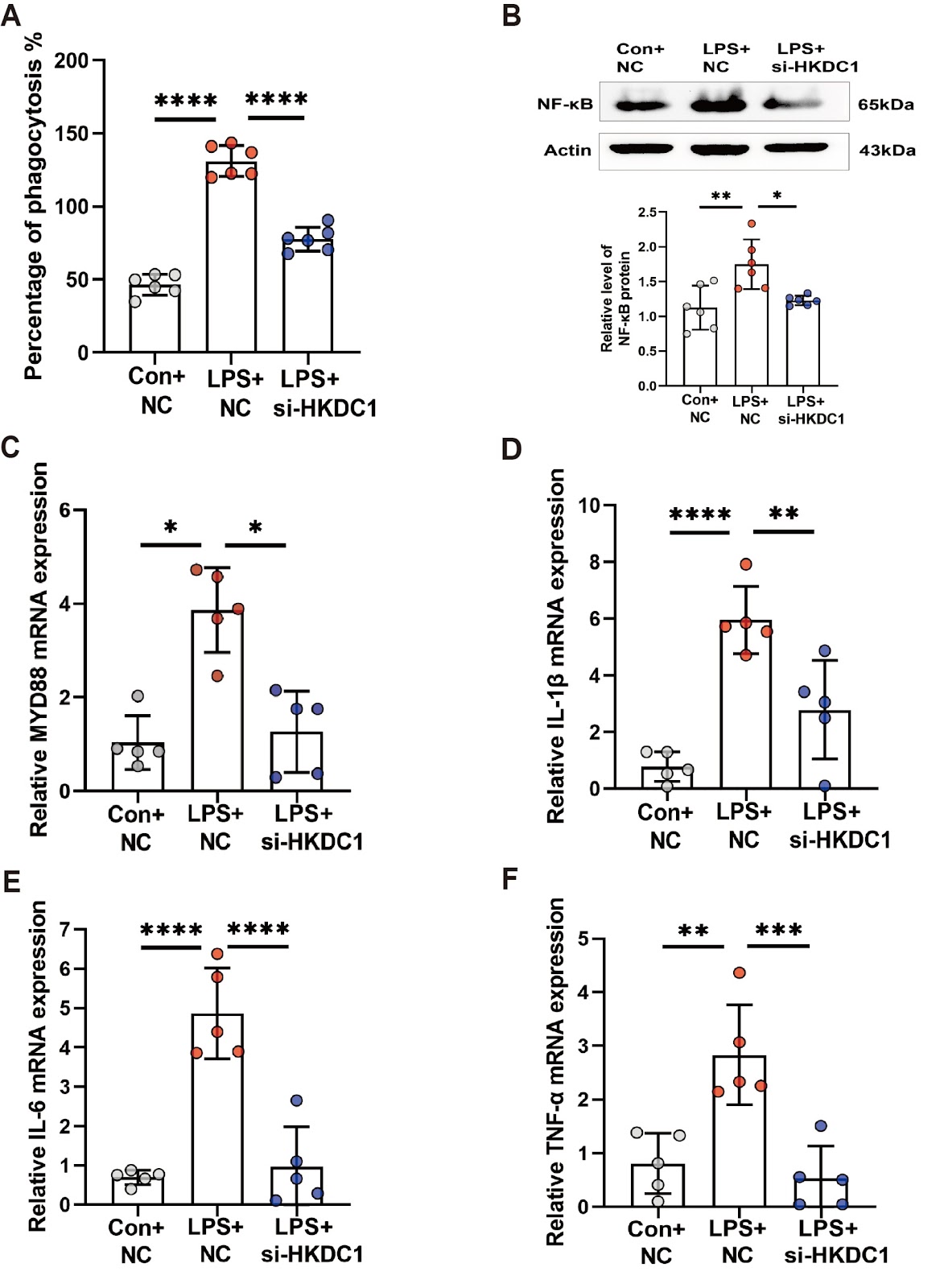
Cuproptosis is a recently identified form of copper-driven cell death characterized by the aggregation of acylated proteins and proteotoxic stress in the mitochondrial tricarboxylic acid cycle, which plays a role in inflammation. Recent studies suggest that hexokinase structural domain protein 1 (HKDC1), a fifth hexokinase, is involved in regulating mitochondrial function. However, the role of HKDC1 in cuproptosis and LPS-induced macrophage inflammation remains unclear. Here, we assess macrophage plasticity using CCK8 viability assays and phagocytosis activity experiments in an in vitro inflammatory model of THP-1 cells. We measure the levels of inflammatory factors and cuproptosis-related proteins using western blot analysis and RT-qPCR. Additionally, we examine the expression and localization of the HKDC1 protein using ChIP-qPCR and immunofluorescence staining. We find that LPS promotes the expressions of inflammatory factors and decreases cuproptosis levels in THP-1-derived macrophages while also activating glycolysis and inducing the expression of HKDC1 via the Toll-like receptor 4 (TLR4) receptor. We further demonstrate that HKDC1 knockdown inhibits glycolysis and induces cuproptosis. Mechanistically, we provide the first evidence that LPS promotes the binding of Yin Yang 1 (YY1) to the HKDC1 promoter, thereby regulating HKDC1 transcription. HKDC1 interacts with heat shock cognate B (HSCB) and ferredoxin 1 (FDX1), leading to increased intracellular copper levels and subsequent cuproptosis. HKDC1 knockdown in vivo alleviates acute sepsis by activating copper-dependent cell death pathways. Collectively, our findings suggest that LPS mitigates cuproptosis and promotes inflammation via HKDC1, suggesting a new cuproptosis-dependent anti-inflammatory strategy.
Read Full Abstract10.3724/abbs.2025089
Expression characteristics of serum exosomal microRNAs in patients with liver injury induced by anti-tuberculosis drugsGraphical AbstractVerified
Acta Biochimica et Biophysica Sinica

Expression characteristics of serum exosomal microRNAs in patients with liver injury induced by anti-tuberculosis drugs

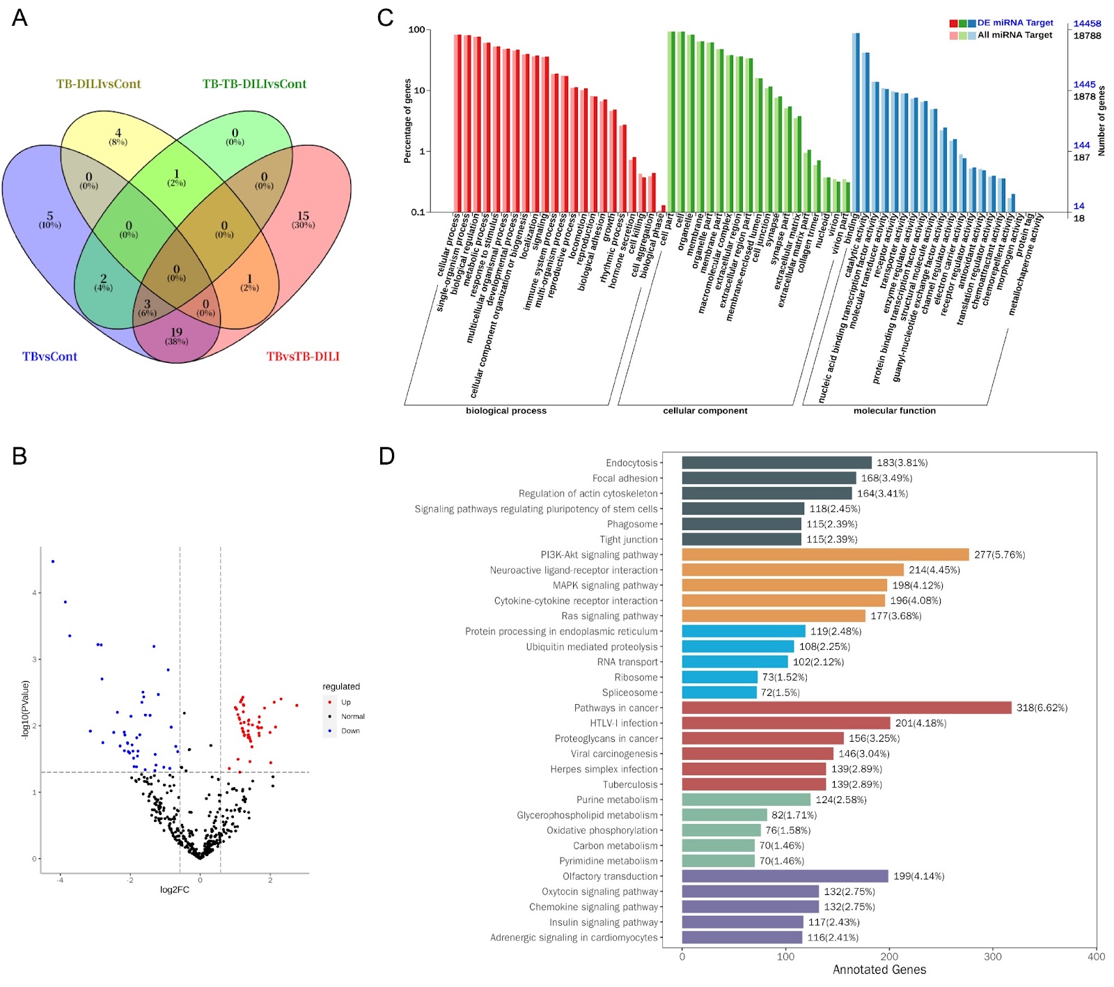
Drug-induced liver injury (DILI) caused by anti-tuberculosis drugs is a serious clinical problem that can lead to acute liver failure and even death. Current diagnosis relies on lagged indicators such as serum transaminase levels, which rise only 48–72 hours after liver injury. This study is the first to systematically analyze the microRNA expression profile of serum exosomes in patients with anti-tuberculosis drug-induced liver injury (TB-DILI) to discover early diagnostic markers. A total of 12 tuberculosis patients and 6 normal controls were included. Serum exosomes were isolated and characterized, and small RNA sequencing identified 701 miRNAs, with 128 differentially expressed between TB-DILI and TB groups. Notably, miR-122-5p was upregulated and has shown early warning value. Target gene prediction and enrichment analysis revealed involvement in GTPase activity regulation, cell migration, and BMP signaling. These findings suggest that exosomal miRNAs, particularly miR-122-5p, may serve as early diagnostic biomarkers for TB-DILI.

Read Full Abstract10.3724/abbs.2025242
High-resolution imaging atlas reveals the context-dependent role of pancreatic sympathetic innervation in diabetic miceGraphical AbstractVerified
Acta Biochimica et Biophysica Sinica

High-resolution imaging atlas reveals the context-dependent role of pancreatic sympathetic innervation in diabetic mice

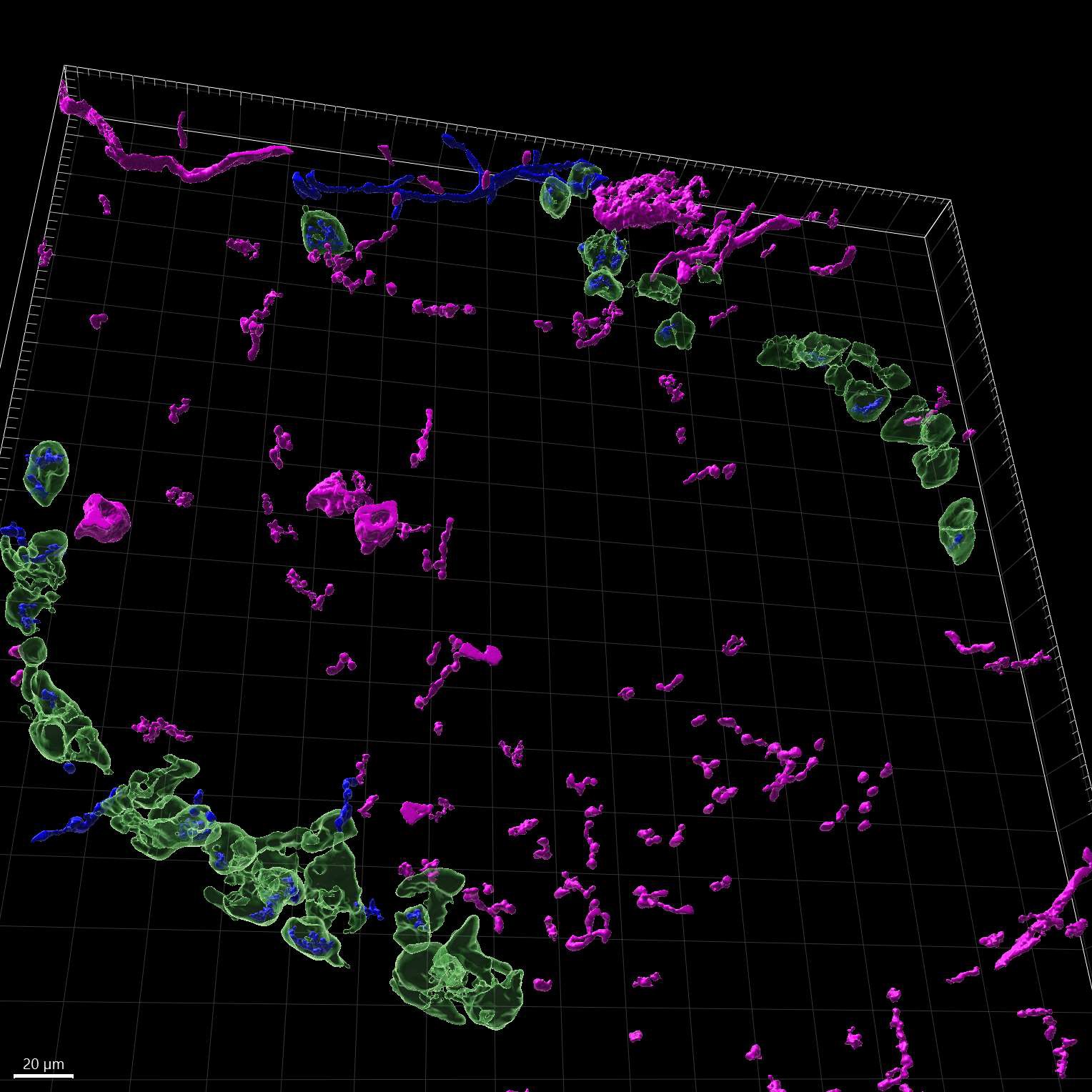
A better understanding of how sympathetic nerves impact pancreatic function is helpful for understanding diabetes. However, there is still uncertainty and controversy surrounding the roles of sympathetic nerves within the pancreas. To address this, we utilize high-resolution imaging and advanced three-dimensional (3D) reconstruction techniques to study the patterns of sympathetic innervation and morphology in the islets of adult wild-type (WT) and diabetic mice. Our data show that more than ~30% of α/β-cells are innervated by sympathetic nerves in both WT and diabetic mice. Additionally, sympathetic innervated α/β-cells are reduced in diet-induced obese (DIO) mice, whereas sympathetic innervated β-cells are increased in db/db mice. In addition, in situ chemical pancreatic sympathetic denervation (cPSD) improves glucose tolerance in WT and db/db mice but decreases glucose tolerance in DIO mice. In situ cPSD also enhances insulin sensitivity in diabetic mice without affecting WT mice. Overall, our findings advance our understanding of diabetes by highlighting the distinctive impact of pancreatic sympathetic innervation on glucose regulation.

Read Full Abstract10.3724/abbs.2024215
Single-cell transcriptomics reveals apolipoprotein A4-mediated metabolic-immune reprogramming in lymphocytes during early obesity-related chronic kidney diseaseGraphical AbstractVerified
Acta Biochimica et Biophysica Sinica

Single-cell transcriptomics reveals apolipoprotein A4-mediated metabolic-immune reprogramming in lymphocytes during early obesity-related chronic kidney disease

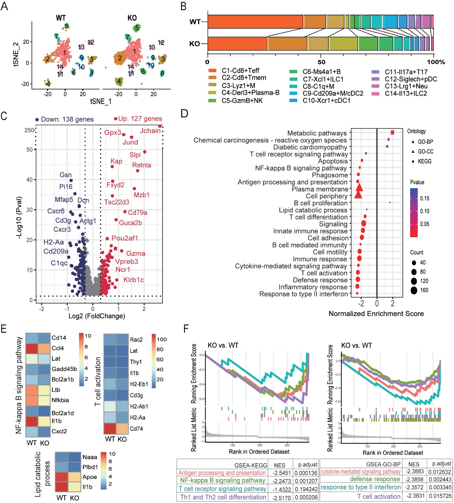
Obesity-induced metabolic inflammation is a key driver of chronic kidney disease (CKD), with immune dysregulation, particularly among lymphocytes, contributing to early disease pathology. To explore the role of apolipoprotein A4 (Apoa4) in regulating immune cell metabolism and function, we establish high-fat diet-induced obese (DIO) models using wild-type and Apoa4-knockout (KO) mice. KO mice exhibit exacerbated insulin resistance and renal lipid accumulation. Single-cell RNA sequencing reveals that Apoa4 deletion remodeled the renal immune-metabolic landscape. This remodeling broadly compromises the immune functions of T, NK, and B cells, even as it expands the proportions of cytotoxic Gzma+ NK cells and Derl3+ plasma cells. Mechanistically, Apoa4 deletion aggravates metabolic dysregulation and oxidative stress and downregulates the expression levels of key effector genes, including Ifng and Il1b. Furthermore, the regulatory network activities of key transcription factors, such as Lef1 and Runx3 in Cd8+ T cells; Irf8, T-bet, and Eomes in NK cells; and Tcf4, Lmo2, and Xbp1 in B cells, are perturbed. CellChat analysis predicts disruptions in pro-inflammatory (IFN-II and IL-1), immunoregulatory (FASLG), and metabolic regulatory (ENHO and ANGPTL) signaling, alongside enhanced IL-2-mediated suppression. These findings are corroborated by flow cytometry, immunofluorescence staining, and qPCR. Our results establish Apoa4 as a crucial regulator of lymphocyte metabolic and immune homeostasis in the early stages of obesity-associated CKD.

Read Full Abstract10.3724/abbs.2025171
Quantitative liquid chromatography-tandem mass spectrometric analysis of 11dH-TXB2 and creatinine in urineGraphical AbstractVerified
Acta Biochimica et Biophysica Sinica

Quantitative liquid chromatography-tandem mass spectrometric analysis of 11dH-TXB2 and creatinine in urine

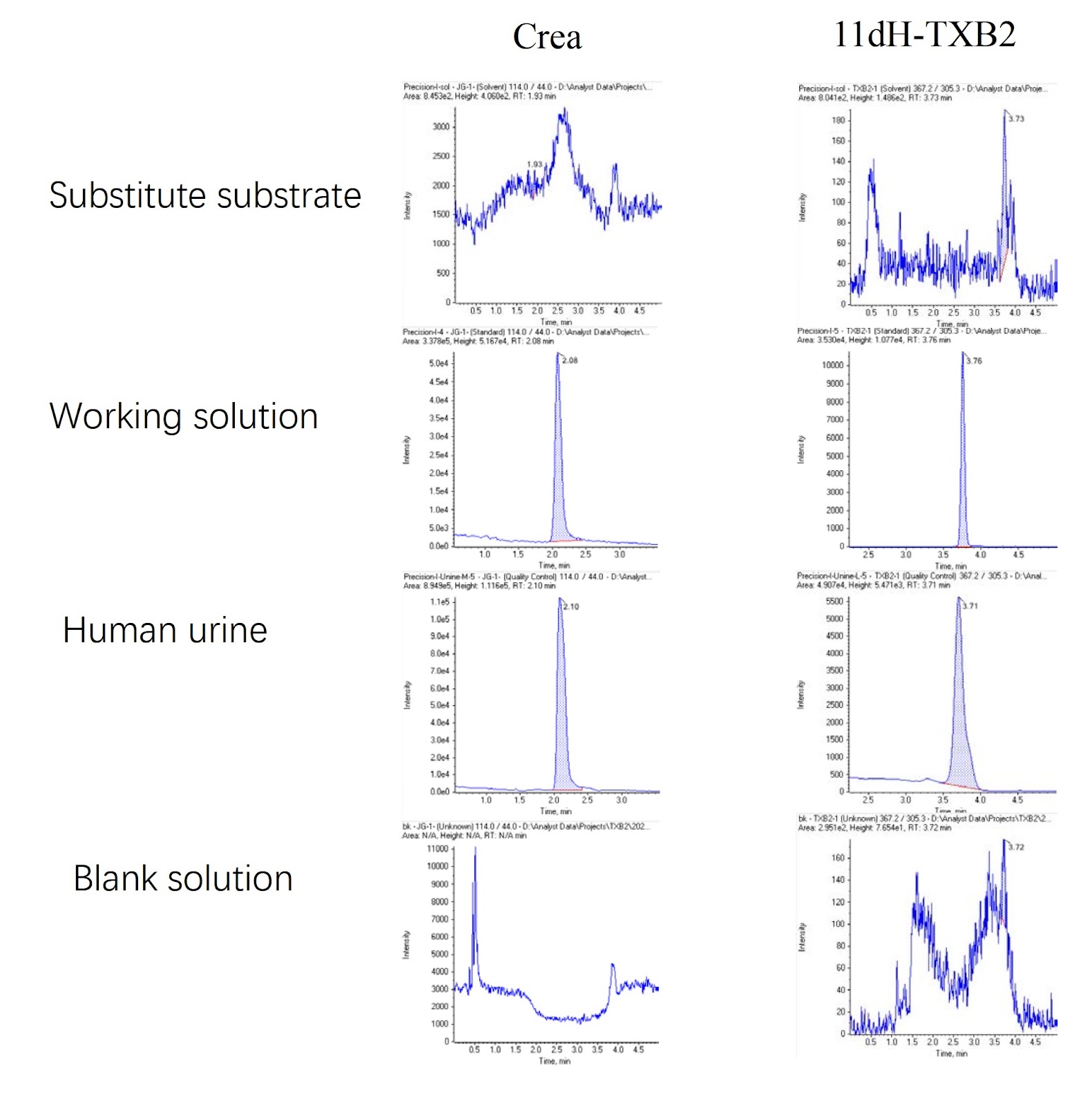
Platelets circulate in an inactive form in the body until they contact with defective areas of endothelial cells or encounter a clotting cascade [1]. Activated platelets release and express bioactive substances and acquire the ability to bind plasma fibrinogen. Abnormal activation of platelets is involved in atherosclerosis and thrombosis [2,3]. When platelets are stimulated and activated, phospholipase A2 is activated at the same time, which then cleaves membrane phospholipids and frees arachidonic acid (AA) [4]. The latter catalyzes thromboxane A2 (TXA2) via thromboxane synthetase [5], which is induced by the cyclooxygenase COX-1 to produce prostaglandins G2 and H2 [6]. TXA2 is highly unstable, with a half-life of only 30 s, and it is rapidly hydrolyzed to relatively stable thromboxane B2 (TXB2), which is then converted in the liver to 11-dehydrothromboxane B2 (11dH-TXB2), which has a longer half-life and is excreted in the urine [7]. Dehydrothromboxane B2 is the final stable metabolite of thromboxane A2, which is derived only from arachidonic acid metabolism and can represent thromboxane A2 level in the body [8]. Specifically, by inhibiting the action of COX-1, the most important enzyme in the process of arachidonic acid metabolism, aspirin inhibits the production of thromboxane A2, that is, the concentration of TXA2 affects the effect of aspirin on platelet aggregation. However, the half-life of TXA2 (including the metabolic intermediate TXB2) is too short to be accurately measured, so the detection of its metabolic end product 11dH-TXB2 can very accurately reflect the sensitivity of the body to aspirin [7]. The concentration of 11dH-TXB2 in the serum correlates well with the concentration of 11dH-TXB2 in the urine, so the determination of 11dH-TXB2 in the urine can more effectively reflect the production of TXA2 in vivo [8]. The 11dH-TXB2 concentration needs to be corrected with the urinary creatinine concentration to rule out the effects of the urine concentration and renal function, so random urine samples can be used for testing [6]. Aspirin can acetylate serine at the key site of cyclooxygenase and thus irreversibly inhibits the activity of COX-1, reduces the synthesis of TXA2, and blocks the production of TXA2 and its induced platelet aggregation. Low-dose aspirin (30–75 mg/day) can effectively inhibit 95% of COX-1 activity [7]. Since the production of TXA2 in serum is largely dependent on platelet COX-1 (a therapeutic target of aspirin), 11dH-TXB2 can be used as a monitor for aspirin-induced platelet inhibition [3]. Creatinine is a metabolic byproduct of muscle metabolism that is primarily excreted via glomerular filtration, and its level is indicative of renal function [9]. The 24-h creatinine clearance can also be used to determine the integrity of the sample or to correct the urine sample concentration with the creatinine ratio [10]. Currently, creatinine detection methods include the Jaffe method, enzymolysis spectrophotometry, HPLC, capillary electrophoresis, capillary zone electrophoresis, gas chromatography tandem mass spectrometry (GC-MS) and liquid chromatography tandem mass spectrometry (LC-MS/MS) [11]. At present, there is no method for the simultaneous detection of 11dH-TXB2 and creatinine. When both analytes are needed, separate tests must be performed, increasing the workload and sample volume requirements. Thus, developing a method that enables the concurrent quantification of 11dH-TXB2 and creatinine in a single assay remains a critical challenge. The aim of this study was to provide a method for the simultaneous detection of 11dH-TXB2 and creatinine and to alleviate the problem that 11dH-TXB2 and creatinine cannot be simultaneously detected. By developing a standardized quantitative approach for measuring 11dHTXB2 and creatinine in human urine, this study aims to provide reliable concentration data, thereby facilitating further clinical research and methodology optimization.

Read Full Abstract10.3724/abbs.2025055
Unique gene patterns lead to distinct functional phenotypes and chemosensitivity profiles among subclones obtained from a single glioblastoma cell lineGraphical AbstractVerified
Acta Biochimica et Biophysica Sinica

Unique gene patterns lead to distinct functional phenotypes and chemosensitivity profiles among subclones obtained from a single glioblastoma cell line

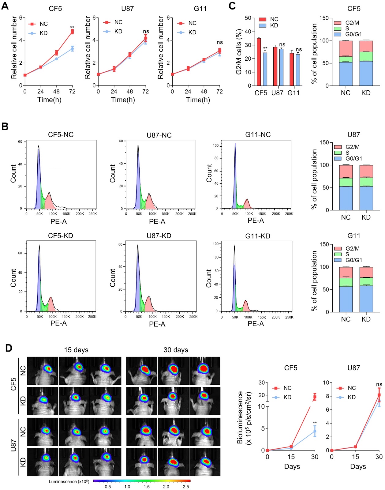
One of the characteristics of malignant tumors is heterogeneity, which refers to the molecular or genetic differences among progeny cells during tumor growth. This heterogeneity contributes to variations in the tumor growth rate, invasive ability, drug sensitivity, and prognosis. To gain a deeper understanding of the molecular background underlying tumor heterogeneity, we construct monoclonal cell lines derived from the glioblastoma (GBM) cell line U87-MG by limiting dilution assays. The selected CF5 and G11 subclones exhibit completely different cell morphologies and, more importantly, distinct functional phenotypes. CF5 exhibits stronger proliferative properties and chemoresistance, whereas G11 shows greater motility and invasion. Transcriptomic sequencing reveals great differences in gene expression among the CF5, G11, and U87 cell lines, and downregulated genes in individual clones are significantly enriched in gene sets related to extracellular matrix function. ITGA11 and ITGA6, as research subjects, are demonstrated to exclusively regulate functional phenotypes and chemotherapy sensitivity in CF5 or G11 cells. In U87 cells, combined knockdown of these two genes significantly inhibits tumor growth and increases chemotherapy sensitivity, but knockdown of either gene alone does not. In summary, these data reveal that even under uniform growth conditions, the heterogeneity of tumor cells and their diverse genetic backgrounds remain significant and persistent. This finding is crucial for accurately identifying tumor-related genes and their functional phenotypes, and a thorough understanding of the genetic and molecular background underlying tumor heterogeneity is essential for comprehensive cancer treatment.

Read Full Abstract10.3724/abbs.2025091
ISGylation: is our genome yearning for such a modification?Graphical AbstractVerified
Acta Biochimica et Biophysica Sinica

ISGylation: is our genome yearning for such a modification?

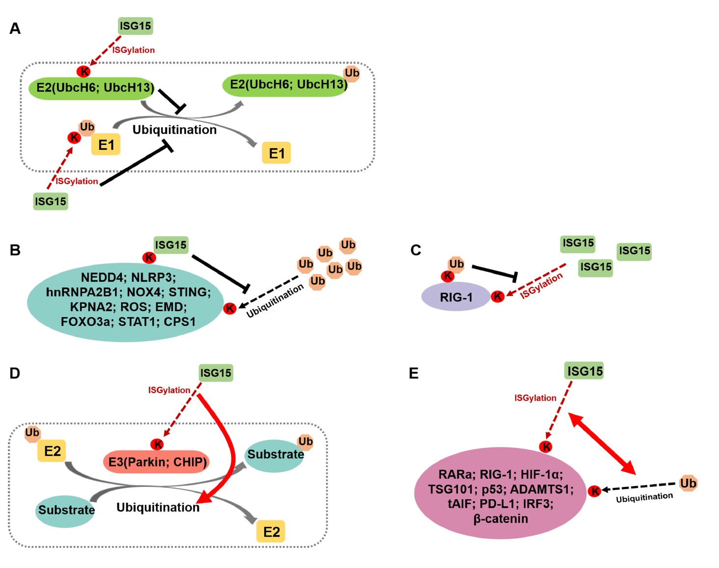
ISGylation is the post-translational modification of protein substrates covalently conjugated with the ubiquitin-like protein, interferon-stimulated gene 15 (ISG15). Although initially linked to antiviral immunity, recent evidence highlights important roles for ISGylation in various biological processes, such as maintaining genomic stability, promoting tumourigenesis, and being involved in other pathological conditions. In this review, we examine the molecular mechanisms underlying ISGylation, its interplay with other post-translational modifications, and its involvement in diverse biological and pathological processes. We propose future research directions to advance the field and discuss how ISGylation might be harnessed to ensure human health, particularly genome instability-associated diseases.

Read Full Abstract10.3724/abbs.2025028
Withaferin A combined with ricolinostat: a potent synergistic therapy for cervical cancer through regulating p53 ubiquitination and acetylationGraphical AbstractVerified
Acta Biochimica et Biophysica Sinica

Withaferin A combined with ricolinostat: a potent synergistic therapy for cervical cancer through regulating p53 ubiquitination and acetylation

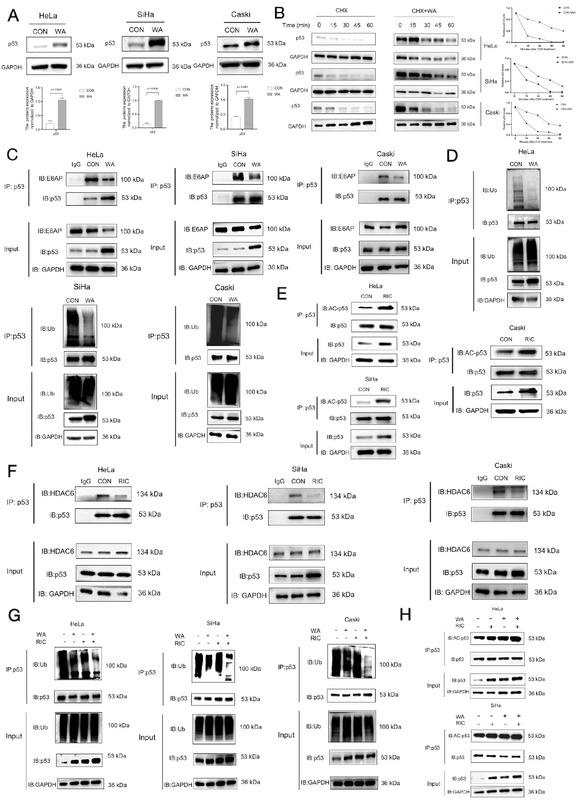
As a classic tumor suppressor gene, p53 has been extensively studied since its discovery in the mid-1980s. Research findings have revealed that p53 protein expression is suppressed in various cancers [1]. For example, in cervical cancer, p53 predominantly exists in a wild-type form to maintain its biological function [2]. Nevertheless, its tumor-suppressive activity is significantly impaired because of rapid protein degradation, short half-life, and low levels. Post-translational modifications (PTMs) of p53, such as ubiquitination, acetylation, phosphorylation and methylation, are critical regulators of its stability, activity, conformation, localization, and interactions with cofactors [3]. Among these, ubiquitination and acetylation play central roles in controlling p53 protein stability and activity [4]. Therefore, targeting p53 PTMs to modulate its ubiquitination and acetylation levels represents an effective strategy to increase its stability and tumor-suppressive function, offering a promising avenue for cervical cancer drug development. In 99% of cervical cancers (high-risk human papillomavirus-positive), E3 ubiquitin ligase E6-associated protein (E6AP) mediates the ubiquitination degradation of p53 [5], whereas histone deacetylase 6 (HDAC6) deacetylates p53. In this study, we explored the possibility of combining the natural product withferin A (WA) with the HDAC6 inhibitor ricolinostat (RIC) to treat cervical cancer cells, with a focus on the ubiquitination and acetylation of p53 and the consequences for its stability. These results suggested that the combination of WA and RIC is more effective than either treatment alone in inhibiting the degradation and increasing the stability of p53, thereby synergistically slowing the onset and progression of cervical cancer.

Read Full Abstract10.3724/abbs.2025048
VSIG2 hinders gastric cancer progression by suppressing ANXA2-mediated NF-κB pathway activationGraphical AbstractVerified
Acta Biochimica et Biophysica Sinica

VSIG2 hinders gastric cancer progression by suppressing ANXA2-mediated NF-κB pathway activation

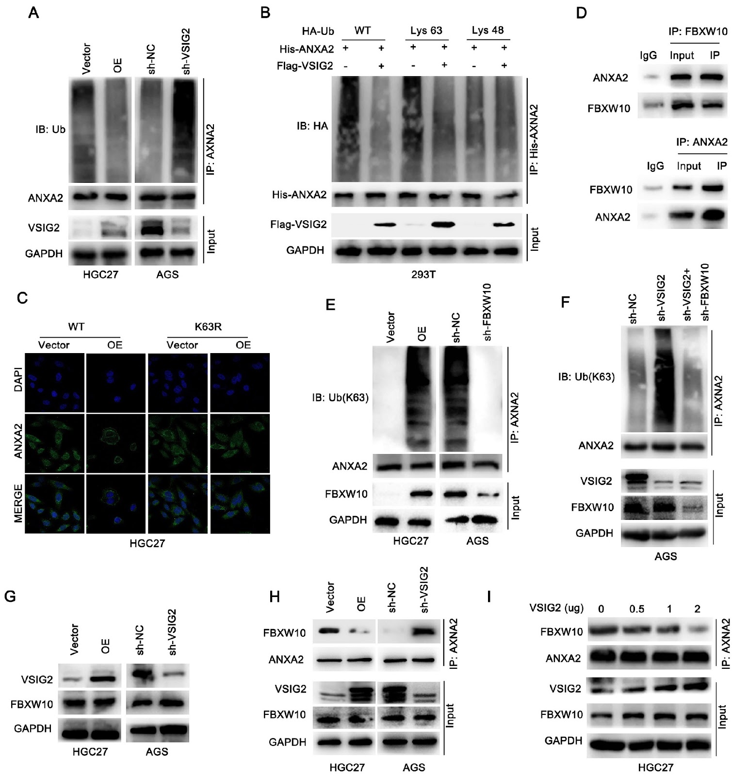
As the fifth most common cancer and the third leading cause of cancer death worldwide, gastric cancer (GC) has long been a serious global health challenge. The purpose of this study was to explore the expression of V-set and immunoglobulin domain containing 2 (VSIG2) in GC and to elucidate its role in GC progression and related mechanisms. Western blot analysis, qRT-PCR and immunohistochemical (IHC) staining are used to detect the expression of VSIG2 in GC cells and tissues. Kaplan-Meier survival curve analysis is performed. The effects of VSIG2 on biological effects related to GC progression in vitro are detected by CCK-8, EdU, Transwell and wound healing assays and in vivo by a nude mouse subcutaneous tumor model and a liver metastasis model. Mechanistically, co-immunoprecipitation, immunofluorescence and ubiquitination experiments are used to explore the regulatory effect of VSIG2 on ANXA2 and the regulatory effect between FBXW10 and ANXA2. VSIG2 is abnormally expressed at low levels in patients with GC and is associated with patient prognosis. Low VSIG2 expression is closely related to tumor size, lymph node metastasis, TNM stage and vascular invasion in GC patients. Functionally, in vitro and in vivo experiments reveal that VSIG2 could inhibit the growth, proliferation and metastasis of GC. Mechanistically, VSIG2 and ANXA2 interact directly in GCs and co-localize at the cell membrane. Further exploration reveals that highly expressed VSIG2 competes with FBXW10 for binding to ANXA2 and relies on FBXW10-mediated K63 polyubiquitination of ANXA2 to induce membrane localization of ANXA2 and further inactivate NF-κB, thereby suppressing GC progression. In summary, VSIG2 is expressed at abnormally low levels in patients with GC, and its low expression is associated with poor patient prognosis. VSIG2 can inhibit the proliferation and migration of GC via the ANXA2/NF-κB pathway. This study elucidates a new mechanism by which VSIG2 inhibits GC progression, which may provide a new perspective for the diagnosis and treatment of GC patients.

Read Full Abstract10.3724/abbs.2025202
Caveolin-1-deficient fibroblasts promote migration, invasion, and stemness by activating the TGF-β/Smad signaling pathway in breast cancer cellsGraphical AbstractVerified
Acta Biochimica et Biophysica Sinica

Caveolin-1-deficient fibroblasts promote migration, invasion, and stemness by activating the TGF-β/Smad signaling pathway in breast cancer cells

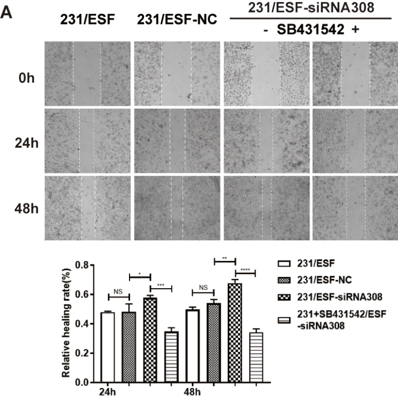
This is a corrigendum to the article 'Caveolin-1-deficient fibroblasts promote migration, invasion, and stemness by activating the TGF-β/Smad signaling pathway in breast cancer cells' published in Acta Biochim Biophys Sin 54: 1587–1598. The authors identified inaccuracies in the preparation of several figures (Figure 2D, 4A, and 5A) and have replaced them with corrected versions. The errors are strictly confined to figure presentation and do not impact the underlying data, statistical analysis, or main conclusions. The authors apologize for the oversight.

Read Full Abstract10.3724/abbs.2025233
New feature of hMEIOB and hSPATA22 binding to ssDNA from a single-molecule perspectiveGraphical AbstractVerified
Acta Biochimica et Biophysica Sinica

New feature of hMEIOB and hSPATA22 binding to ssDNA from a single-molecule perspective

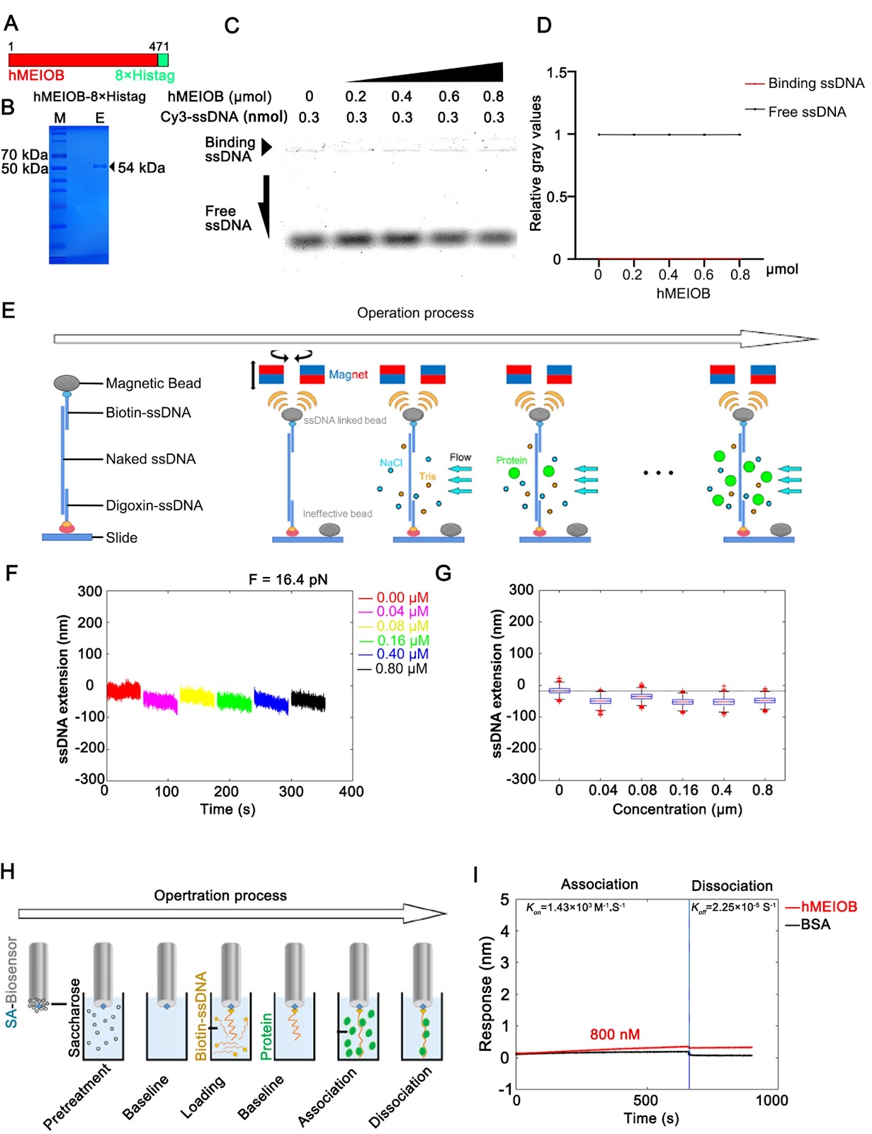
MEIOB and SPATA22 are gonad-specific proteins that function in meiosis recombination. Mutations in these two proteins cause oligospermia or azoospermia in human males. It has been reported that the heterodimer composed of MEIOB and SPATA22 recognizes and binds to the single-strand DNA (ssDNA) protected by the replication protein A (RPA) complex to promote DNA damage repair during homologous recombination. However, the amino acid sequences of the two proteins are inconsistent in humans and rodents, which leads to functional differences in meiosis. In this study, human-derived MEIOB (hMEIOB) and SPATA22 (hSPATA22) are expressed and purified for electrophoretic mobility shift assay (EMSA), magnetic tweezer (MT) assay and bio-layer interferometry (BLI) assay to analyze the ssDNA binding patterns. The results show that hMEIOB has low ssDNA-binding affinity and stability alone, but hSPATA22 binds to ssDNA faster and more stably and promotes ssDNA condensation. Strong binding affinity and stability to ssDNA are present when the hMEIOB-hSPATA22 heterodimer is formed. Moreover, we find that multiple hMEIOB-hSPATA22 heterodimers spontaneously aggregate in vitro. hRPA complex weakens the binding affinity of hMEIOB, hSPATA22 and hMEIOB-hSPATA22 heterodimer to ssDNA, and it can also bind to hSPATA22 and hMEIOB-hSPATA22 heterodimer in vitro, which might be related to the proven function of RPA complex to protect ssDNA and recruit proteins related to DNA damage repair during meiosis. Overall, this study is the first time to elucidate the binding patterns of the hMEIOB and hSPATA22 to ssDNA in vitro, and to verify the relationship between the RPA complex and meiosis-related proteins, MEIOB and SPATA22, from single-molecule perspective.

Read Full Abstract10.3724/abbs.2025057
The dual role of RNA-binding proteins: promotion of tumorigenesis, drug resistance, and emerging therapeutic targetsGraphical AbstractVerified
Acta Biochimica et Biophysica Sinica

The dual role of RNA-binding proteins: promotion of tumorigenesis, drug resistance, and emerging therapeutic targets

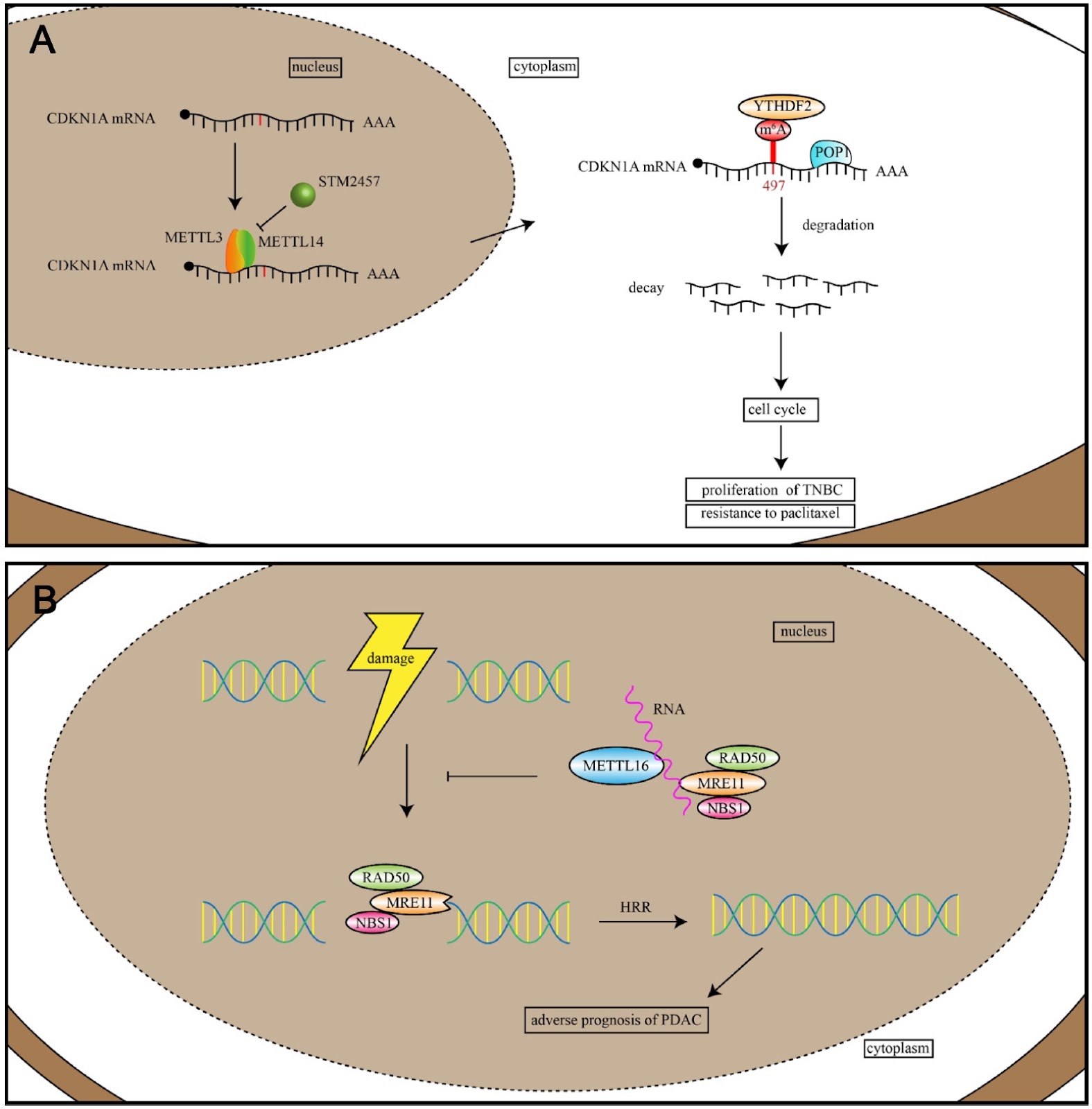
Cancer is a complex and multifaceted disease characterized by a multitude of molecular factors. RNA-binding proteins (RBPs) have emerged as pivotal regulators of tumor development, progression, and chemoresistance through their interactions with target transcripts. These interactions regulate a multitude of processes, including alternative splicing, cleavage and polyadenylation, RNA localization, translation, N6-methyladenosine (m6A) RNA modification, and DNA double-strand break repair. The RBP family comprises over 2000 proteins and plays a critical role in oncogene expression, invasion, metastasis, and inhibition of apoptosis. However, the mechanisms by which RBPs selectively recognize RNAs remain an active area of research. In this review, we examine recent advancements in understanding RNA-binding domains and the RNA processes regulated by RBPs in tumorigenesis, summarize and highlight the roles of RNA-binding domains in cancers and the molecular mechanisms of RBPs in chemotherapy resistance, discuss the potential of targeting RBPs for cancer therapy and review RBPs that are dysregulated in cancers. Additionally, we highlight recently developed tools for predicting RBP-RNA binding activities to provide valuable support for ongoing research efforts.

Read Full Abstract10.3724/abbs.2025099
Construction of an ASFV proteome library via multiple optimization strategies for high-throughput analysisGraphical AbstractVerified
Acta Biochimica et Biophysica Sinica

Construction of an ASFV proteome library via multiple optimization strategies for high-throughput analysis

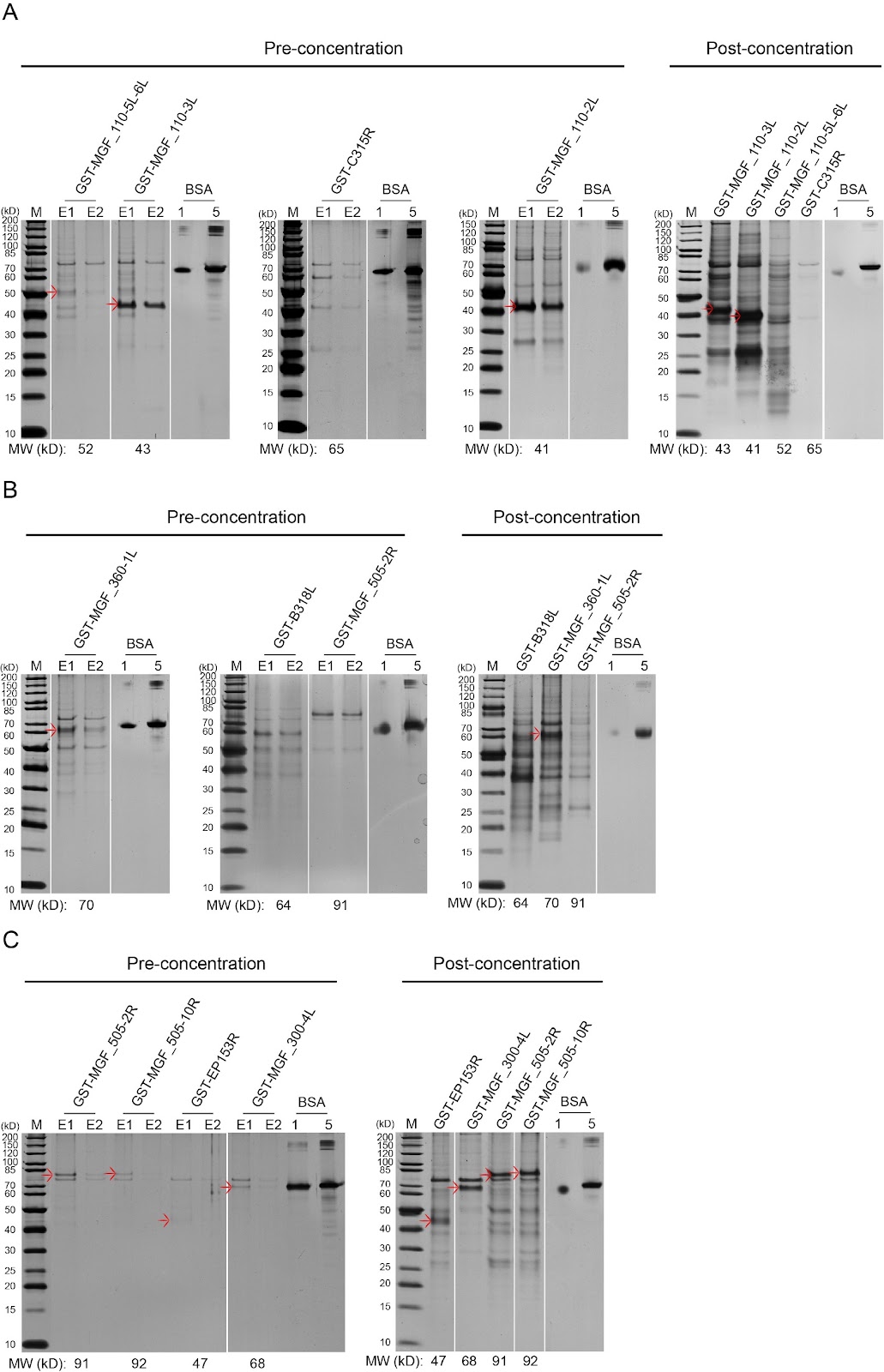
African swine fever virus (ASFV) is a large and structurally complex DNA virus encoding more than 160 proteins, including more than 68 structural proteins. A protein library covering recombinant ASFV proteins is fundamentally important for studies on protein function, antigenicity, vaccine development, and virus-host interactions. Here, to construct an ASFV protein library, we add a glutathione S-transferase (GST) tag at the N-terminus of each ASFV protein to facilitate solubilization and purification and express the recombinant proteins in the yeast host. By optimizing codons, expression vectors and strains and conditions of expression and purification, we achieve satisfactory protein yields for analytical applications and maximized access to the whole proteome of ASFV, with coverage of ca. 95%. Using the library, a protein chip is constructed and used to screen for interactions between ASFV and swine proteins (e.g., IRF3, p65, and IκBα). The ASFV protein library lays the groundwork for understanding and combatting ASFV. The methods for constructing the library are instructive for generating other protein libraries for high-throughput applications.

Read Full Abstract10.3724/abbs.2025125
Osteocalcin carboxylation/undercarboxylation levels and gene variants associated with type 2 diabetes mellitus in the Chinese Han populationGraphical AbstractVerified
Acta Biochimica et Biophysica Sinica

Osteocalcin carboxylation/undercarboxylation levels and gene variants associated with type 2 diabetes mellitus in the Chinese Han population

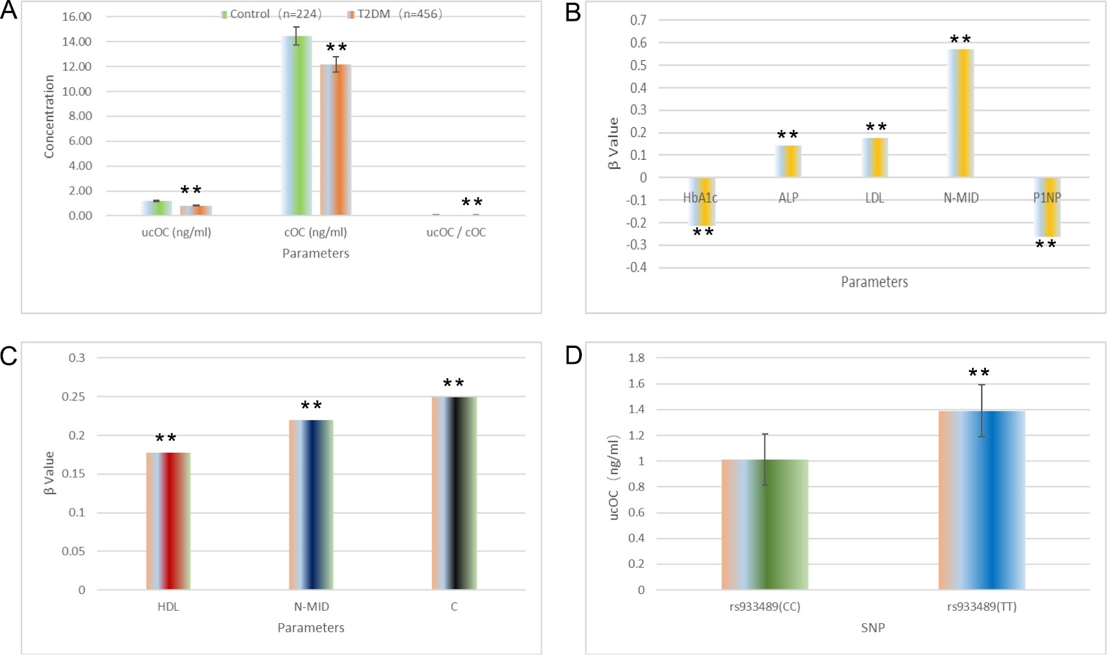
Type 2 diabetes mellitus (T2DM) is an endocrine metabolic disorder characterized by insulin secretion dysfunction and/or insulin resistance. Osteocalcin (OC), or bone γ-carboxyglutamic acid protein (BGP), is a bone matrix protein predominantly produced by osteoblasts. Vitamin K-dependent carboxylation converts OC into gamma-carboxyglutamic acid (Gla)-rich carboxylated osteocalcin (cOC), which binds to hydroxyapatite and can be decarboxylated to undercarboxylated osteocalcin (ucOC) under acidic conditions. While cOC influences bone formation and mineralization, ucOC regulates energy metabolism. This study aimed to explore the associations between T2DM, serum OC levels (including cOC and ucOC), and OC gene polymorphisms in the Chinese Han population. T2DM patients and a healthy cohort, all of Han ethnicity, were categorized into a T2DM group (n = 456) and a control group (n = 224). Serum levels of cOC and ucOC were determined via ELISA. Insulin sensitivity was assessed via HOMA-IR, and pancreatic β-cell function via HOMA-β. Nine SNPs in the OC gene were genotyped via SNaPshot. Compared with controls, T2DM patients presented significantly lower levels of ucOC, cOC, and ucOC/cOC ratio. Additionally, T2DM subjects had elevated BMI, HbA1c, HOMA-IR, ALP, TG, HDL, and LDL levels, with decreased HOMA-β, ALT, AST, hsCRP, and FFA levels. In terms of bone metabolism, T2DM patients presented increased blood phosphorus, ICTP, P1NP, and 25(OH)D levels and decreased blood calcium, N-MID, PTH, and β-CTX levels. Associations between serum cOC and ucOC and various factors were analyzed. In the T2DM group, cOC was inversely correlated with HbA1c and P1NP, and positively correlated with ALP, LDL, and N-MID. ucOC was positively associated with N-MID. In controls, cOC was positively correlated with HDL, N-MID, and PINP, while ucOC correlated with PINP. The study also examined SNPs in the OC gene and their relationships with serum cOC and ucOC.

Read Full Abstract10.3724/abbs.2025060
HDAC11 in ovarian granulosa cells coordinates LH in the maturation of oocytes in Tan sheepGraphical AbstractVerified
Acta Biochimica et Biophysica Sinica

HDAC11 in ovarian granulosa cells coordinates LH in the maturation of oocytes in Tan sheep

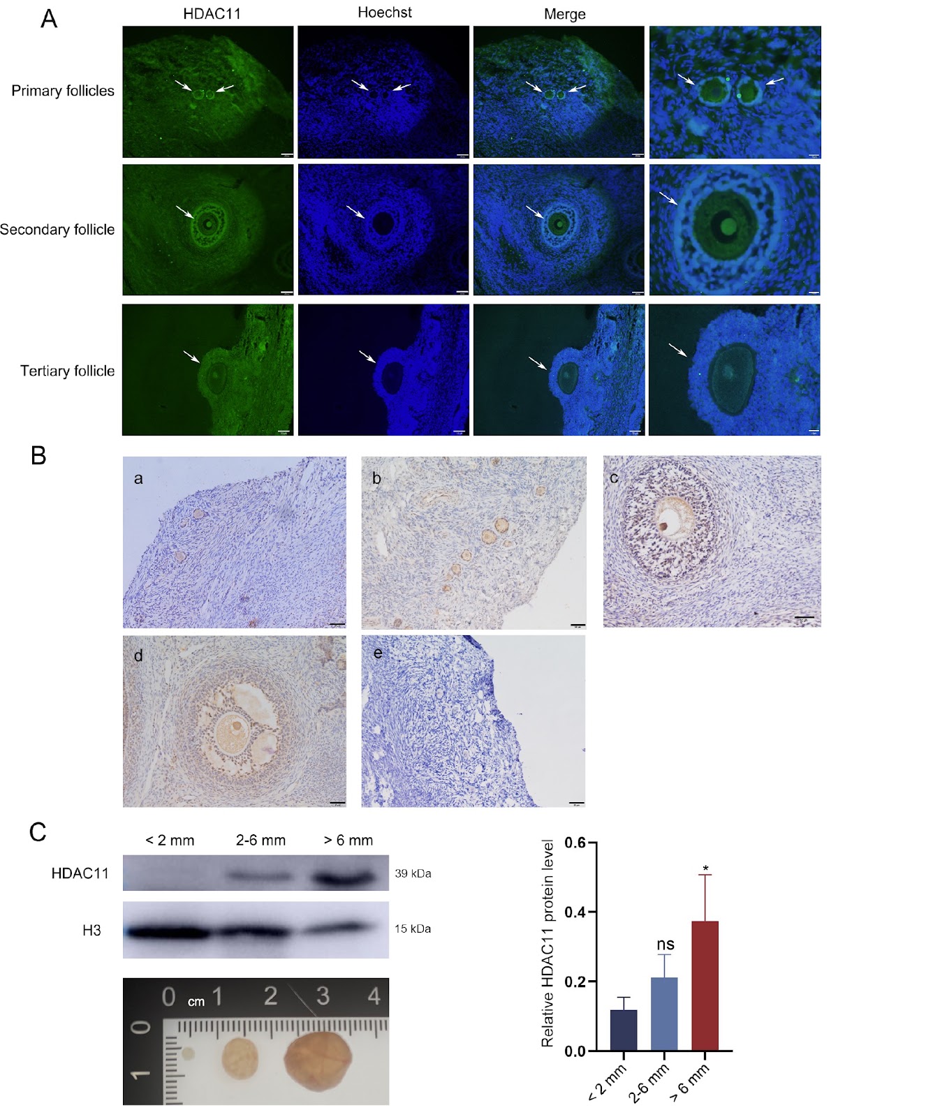
Oocyte maturation plays an important role in supporting mammalian reproduction. Histone deacetylase 11 (HDAC11), the only member of the class IV histone deacetylase family and the smallest histone deacetylases (HDACs), has been shown to regulate oocyte maturation in mice and pigs. However, the epigenetic effects of HDACs in follicular granulosa cells in response to LH induction remain elusive in sheep. In this study, the effects of follicular somatic cell-derived HDAC11 on oocyte maturation in Tan sheep are evaluated. The expression changes of HDAC11 and related proteins are detected by means of immunofluorescence, immunohistochemistry, western blot analysis and enzyme-linked immunosorbent assay. Our results indicate that the level of HDAC11 in follicular granulosa cells as well as oocytes in Tan sheep increases with the growth and maturation of the follicles. Specific inhibition of HDAC11 by SIS17 remarkably reduces the oocyte maturation rate under LH supplementation in vitro. Accordingly, the acetylation level of H3K9 in granulosa cells is increased, while the EGF-like growth factor AREG is remarkably decreased. Furthermore, inhibition of HDAC11 markedly decreases the level of YAP1, which is a negative regulator of AREG in granulosa cells. Conclusively, HDAC11 in the granulosa cells of Tan sheep contributes to the LH induced production of AREG during oocyte in vitro maturation by decreasing the level of H3K9 acetylation and increasing the level of YAP1.

Read Full Abstract10.3724/abbs.2025036
Increased neutrophil senescence is associated with impaired immunosuppressive activity in systemic lupus erythematosusGraphical AbstractVerified
Acta Biochimica et Biophysica Sinica

Increased neutrophil senescence is associated with impaired immunosuppressive activity in systemic lupus erythematosus

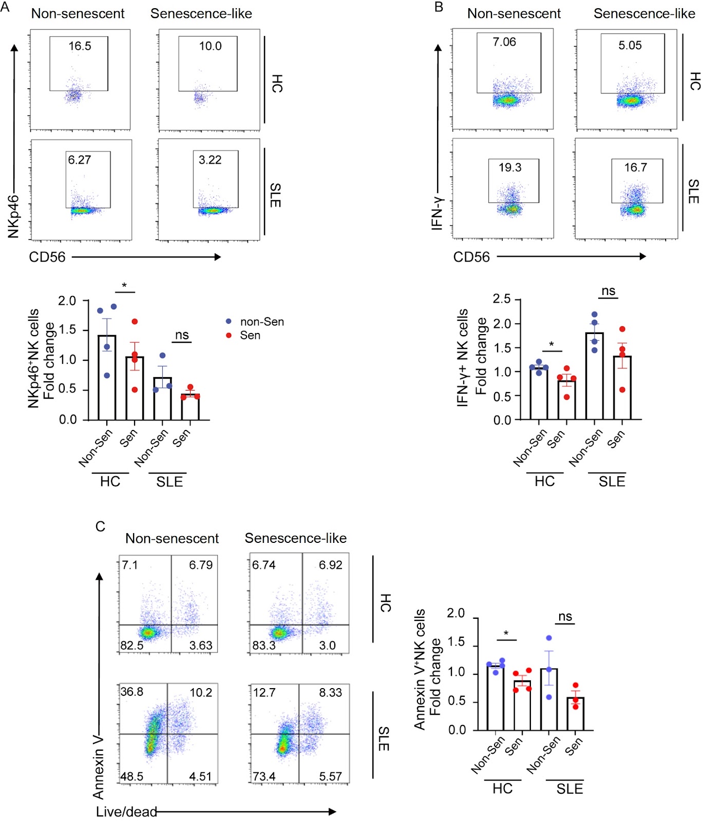
Systemic lupus erythematosus (SLE) is an autoimmune disease characterized by a complex pathogenesis that was previously thought to involve primarily adaptive immunity. Emerging evidence underscores the role of neutrophils in shaping immune dysregulation and inducing organ damage in lupus. This study aims to investigate the dynamics of neutrophil senescence and its relationship with lupus, an area that remains poorly understood. Here, we identify a significantly elevated proportion of CXCR4hiCD62Llo senescence-like neutrophils in the peripheral blood of SLE patients compare to that in the healthy donors. Increased numbers of senescence-like neutrophils are positively correlated with SLE disease activity and autoantibody production in SLE patients. In addition, senescence-like neutrophils derived from SLE patients exhibit an impaired ability to suppress the proinflammatory activity of natural killer (NK) cells and CD4+ T cells. Further mechanistic exploration suggests that these senescence-like neutrophils might exert their immunosuppressive effects via reactive oxygen species (ROS) production under physiological conditions. Our results demonstrate that senescence-like neutrophils could serve as biomarkers for assessing the disease activity of SLE. The compromised immunosuppressive function of senescence-like neutrophils provides a new perspective on SLE pathophysiology and may pave the way for the development of novel therapies.

Read Full Abstract10.3724/abbs.2025047
KARs negatively regulate the immune response in lampreyGraphical AbstractVerified
Acta Biochimica et Biophysica Sinica

KARs negatively regulate the immune response in lamprey

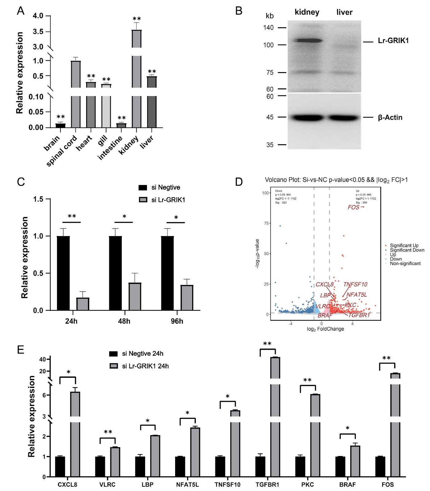
Kainate receptors (KARs) are one of the ionotropic glutamate receptor (iGluR) families, and their antagonists are being investigated for the treatment of several neurological disorders, including Alzheimer’s disease, a neurodegenerative condition, etc. As early as 1990, Bettler et al. [1] first cloned the GRIK1 subunit of KARs, marking a pivotal advancement in understanding these receptors. Members of the iGluR family have been identified in other jawed vertebrates and exhibit conserved structural features. However, research into iGluRs in jawless vertebrates has been limited. Owing to the unique evolutionary position of lampreys, their iGluRs might also present functions distinct from those of jawed vertebrates; therefore, it is particularly important to study iGluRs in lampreys. In this study, we identified four homologous subunits of iGluRs in lampreys, including Lr-GRIA2, Lr-GRIA4, Lr-GRIK1 and Lr-GRIN2B. Lampreys occupy a unique evolutionary position, making phylogenetic analysis of iGluR subunits between lampreys and other species essential for understanding iGluR evolution. Given the distinctive functional characteristics of iGluR family members, particularly KAR subtypes, we focused on the functional validation of Lr-GRIK1. First, we confirmed the expression of Lr-GRIK1 in lampreys and examined its expression profiles across various tissues via qPCR and western blotting. To elucidate the functional role of Lr-GRIK1 in lampreys, we used an siRNA to silence Lr-GRIK1. We subsequently conducted transcriptome sequencing of both the silenced and control groups to construct and analyze their expression profiles. Our analysis revealed differential expression of genes enriched in pathways related to signal transduction and the immune system, highlighting potential roles of Lr-GRIK1 beyond traditional neurotransmission functions. Unlike in jawed vertebrates, transcriptome enrichment provides a new direction for understanding the function of Lr-GRIK1. Therefore, we monitored the changes in Lr-GRIK1 expression in the kidney tissue of lampreys after stimulation. In addition, we confirmed that Lr-GRIK1 affects the expression levels of immune-related molecules during the immune response process. These findings provide insights into the broader functional significance of Lr-GRIK1 in the biology of lampreys.

Read Full Abstract10.3724/abbs.2025094
Nobiletin suppresses nasopharyngeal carcinoma by regulating the KEAP1/NRF2/ARE pathwayGraphical AbstractVerified
Acta Biochimica et Biophysica Sinica

Nobiletin suppresses nasopharyngeal carcinoma by regulating the KEAP1/NRF2/ARE pathway

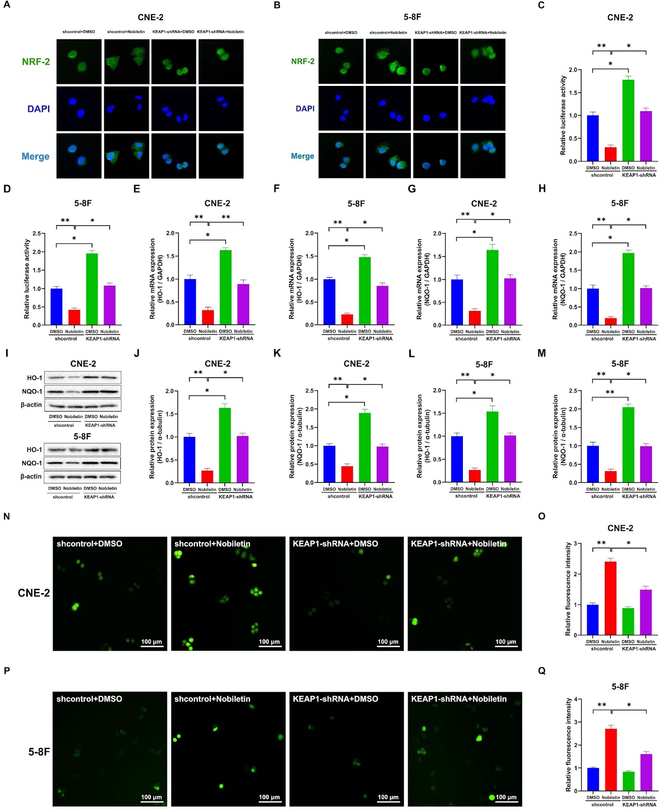
Nasopharyngeal carcinoma (NPC) ranks among the most prevalent malignancies, particularly in East Asia and Southeast Asia. Nobiletin (NOB), an exclusive polymethoxyflavonoid derived from citrus peel, exhibits diverse physiological properties, notably its potent anticancer activity. Kelch-like ECH-associated protein 1 (KEAP1), the repressor protein regulating the nuclear factor erythroid 2-related factor 2 (NRF2) transcription factor, has emerged as a promising strategy for addressing oxidative stress in various diseases. The KEAP1/NRF2/ARE signal is a fundamental pathway within the cellular homeostatic defense system. This study robustly demonstrates the chemopreventive potential of NOB through comprehensive in vitro and in vivo assessments using subcutaneous tumor mouse models. Furthermore, our groundbreaking findings reveal that NOB effectively hinders the migration and invasion capacities of CNE-2 and 5-8F (NPC) cells in a dose- and time-dependent manner. Mechanistically, NOB, a potent KEAP1 activator, significantly disrupts the NRF2/ARE signaling pathway by accelerating the proteasomal degradation of NRF2 and suppressing its nuclear translocation. Consequently, this cascade reduces the expressions of ARE-driven genes and antioxidant enzymes, thereby increasing intracellular reactive oxygen species (ROS) levels and increasing antitumor immunity. Moreover, the sensitivity induced by NOB is markedly diminished in CNE-2 cells following the gene silencing of KEAP1. These findings underscore the pivotal role of NOB in activating KEAP1. Overall, KEAP1 has emerged as a compelling target for potential malignancy treatment in nasopharyngeal carcinoma cell lines. Our results suggest the promising application of NOB as a natural sensitizer in chemotherapy, opening avenues for promising therapeutic interventions.

Read Full Abstract10.3724/abbs.2025113
Triptonide facilitates autophagy-mediated apoptosis in esophageal squamous cell carcinoma by targeting the AMPK-mTOR-ULK1 axisGraphical AbstractVerified
Acta Biochimica et Biophysica Sinica

Triptonide facilitates autophagy-mediated apoptosis in esophageal squamous cell carcinoma by targeting the AMPK-mTOR-ULK1 axis

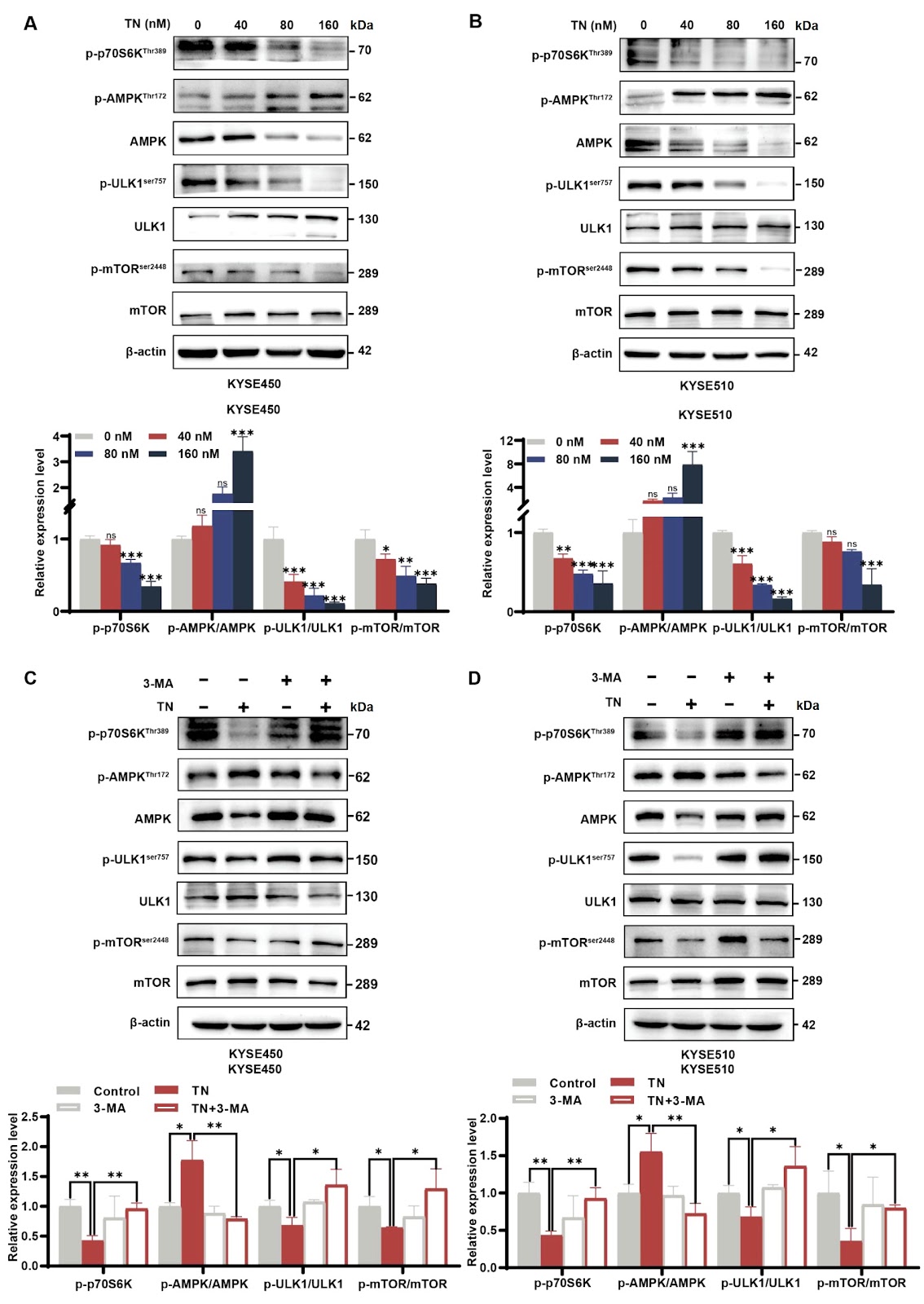
Triptonide (TN) is a small-molecule compound initially derived from Tripterygium wilfordii Hook. f used in traditional Chinese medicine. However, its potential antitumor mechanisms are still far from adequately understood. The purpose of this research is to elucidate the antitumor and pharmacological effects of TN on esophageal squamous cell carcinoma (ESCC). Functional assays, such as CCK-8 and colony formation assays, are used to evaluate the effects of TN on KYSE450 and KYSE510 cells. Subsequently, western blot analysis, Hoechst 33258 staining, flow cytometric analysis, autophagic flux detection, and transmission electron microscopy (TEM) are used to determine the effects of TN on apoptosis and autophagy in ESCC cells. Additionally, the autophagy inhibitor 3-methyladenine (3-MA) and the AMPK inhibitor dorsomorphin (Compound C, CC) are administered to explore the molecular mechanisms and crucial pathways in ESCC cells. Our findings provide strong evidence that TN induces autophagy-dependent apoptosis by targeting the AMPK-mTOR-ULK1 axis in ESCC cells. Collectively, this study sheds light on the anticancer mechanisms of TN in esophageal squamous cell carcinoma and suggests that TN is a promising candidate for the antitumor phytomedicine.

Read Full Abstract10.3724/abbs.2025056
Melatonin attenuates kidney injury by alleviating lysosomal damage in diabetic kidney diseaseGraphical AbstractVerified
Acta Biochimica et Biophysica Sinica

Melatonin attenuates kidney injury by alleviating lysosomal damage in diabetic kidney disease

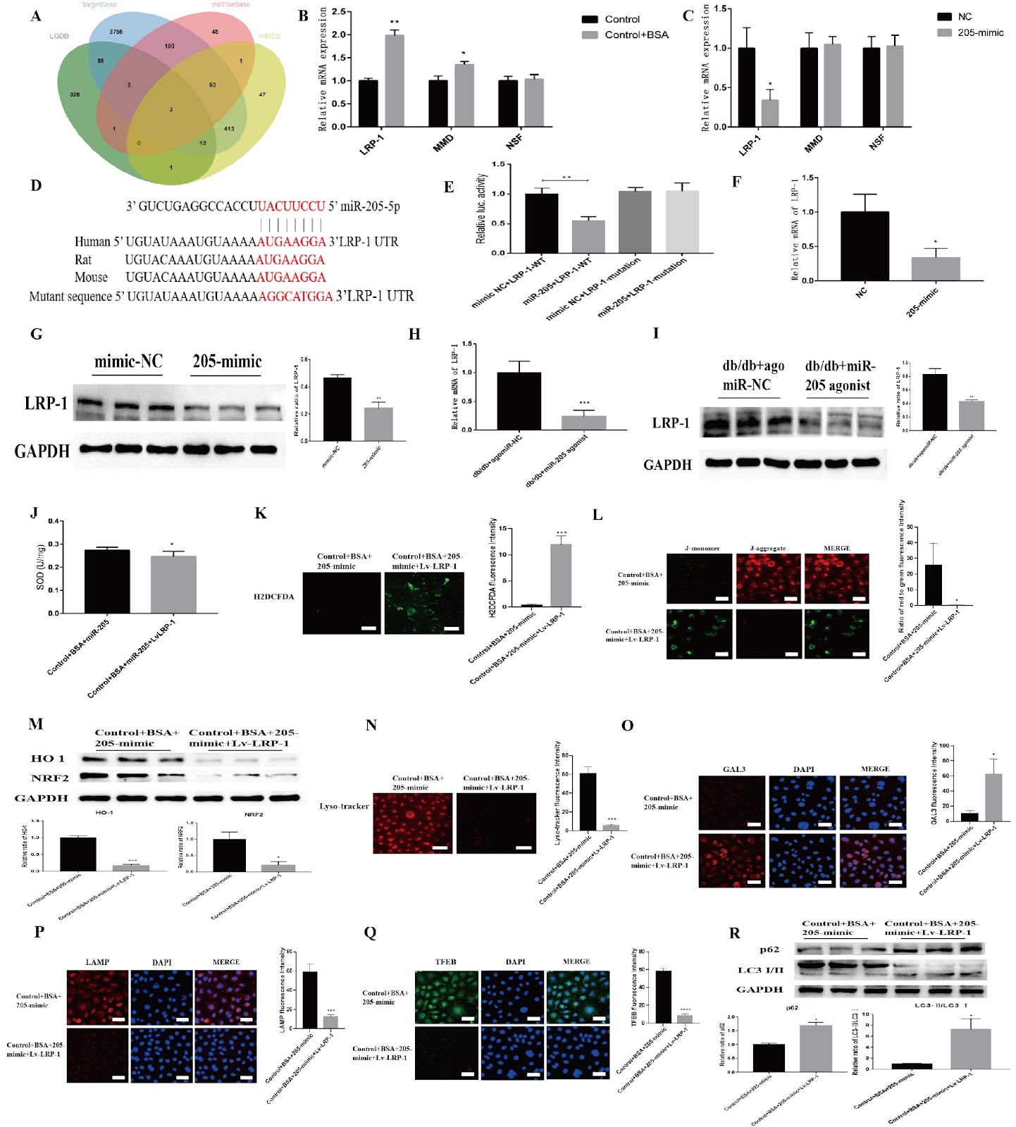
Proteinuria-induced damage to renal tubular epithelial cells is one of the main causes of diabetic kidney disease (DKD), and the clearance of overloaded albumin by lysosomes is crucial for maintaining the homeostasis of renal tubular epithelial cells. Therefore, lysosomal damage is closely related to the pathogenesis of DKD, but effective prevention and treatment measures are still lacking. Melatonin (MLT) is secreted by the pineal gland and can not only regulate circadian rhythms but also maintain lysosomal homeostasis. In this study, we demonstrate the presence of significant lysosomal damage in the renal tubules of DKD patients, which causes autophagy impairment and a concomitant oxidative stress imbalance; however, MLT can upregulate transcription factor EB (TFEB) to improve lysosomal damage and restore the biosynthesis of this organelle. Mechanistically, MLT may protect lysosomes via the upregulation of TFEB and the miR-205-5p-LRP-1 pathway in renal tubules, thus improving autophagy dysfunction and oxidative imbalance in DKD.

Read Full Abstract10.3724/abbs.2025034
Nanchangmycin suppresses influenza A virus infection by blocking endosomal acidificationGraphical AbstractVerified
Acta Biochimica et Biophysica Sinica

Nanchangmycin suppresses influenza A virus infection by blocking endosomal acidification

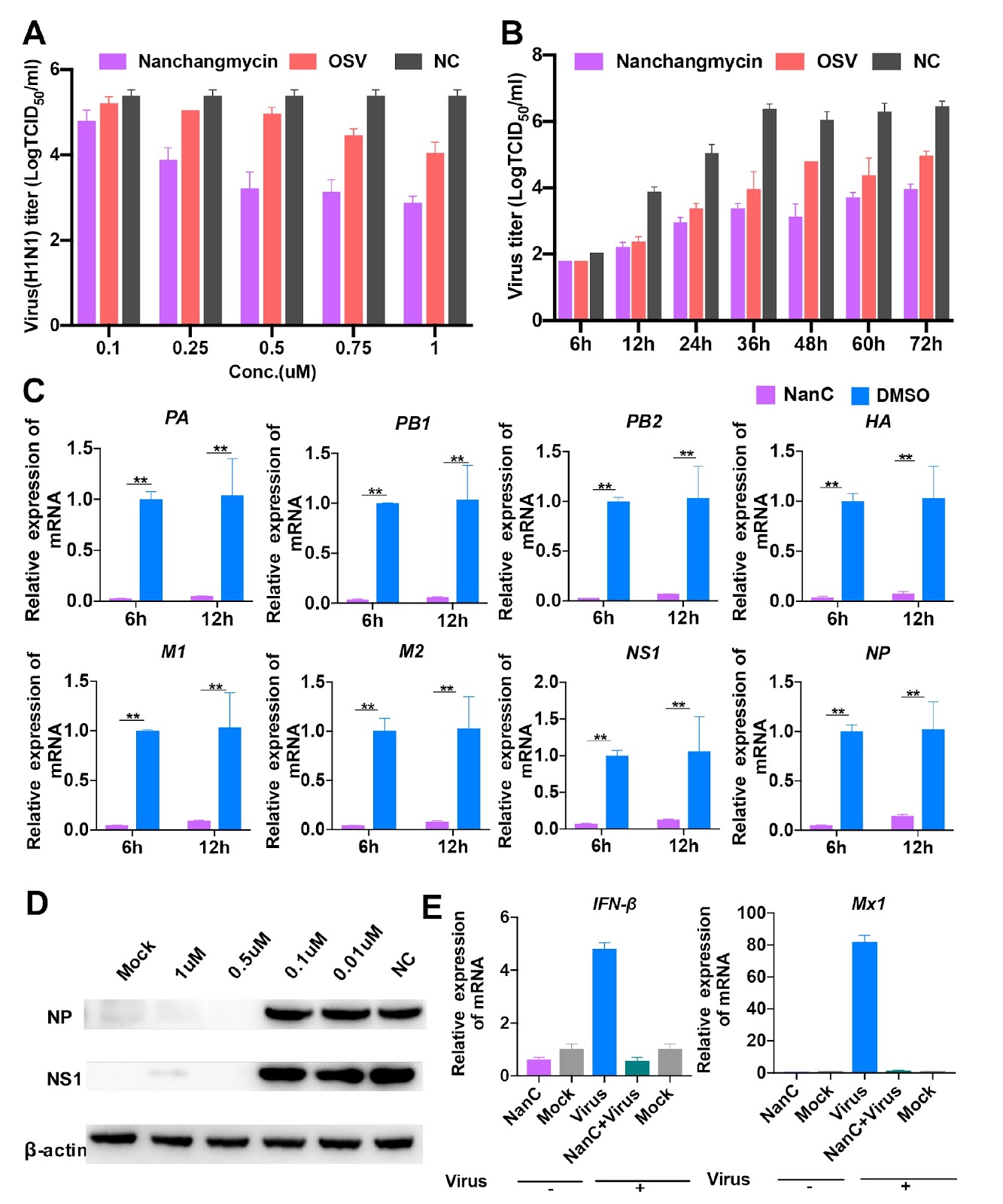
Influenza A viruses (IAVs) constitute a major threat to human and animal health. Currently, M2 ion-channel inhibitors, neuraminidase (NA) inhibitors, RNA polymerase inhibitors, and cap-dependent endonuclease inhibitors have been applied clinically as therapeutics against IAVs. However, IAVs possess adaptive mutations to these inhibitors, especially M2 ion channel and NA inhibitors. Thus, novel antiviral agents should be developed. In the present study, we screen approximately 5500 compounds and identify an IAV inhibitor, nanchangmycin, which possesses a robust antiviral activity both in vitro and in vivo. In addition, it exhibits broad-spectrum antiviral activity for additional virus infections, including pseudorabies virus, herpes virus, porcine epidemic diarrhea virus, porcine reproductive and respiratory syndrome virus. Most importantly, it has antiviral activity against oseltamivir-resistant strains in sub-μM ranges and promotes the survival of MDCK cells infected with the oseltamivir-resistant influenza A virus strain. Further studies reveal that it blocks the nuclei migration of viral nuclear proteins (NPs), resulting in NP accumulation in the cytoplasm, particularly within perinuclear endosomes. Also, it inhibits IAVs by blocking endosomal acidification. Overall, nanchangmycin has the potential to be developed as an anti-influenza agent.

Read Full Abstract10.3724/abbs.2025102
PDGFC secreted by cancer-associated fibroblasts promotes epithelial-mesenchymal transition and immunosuppression in lung adenocarcinomaGraphical AbstractVerified
Acta Biochimica et Biophysica Sinica

PDGFC secreted by cancer-associated fibroblasts promotes epithelial-mesenchymal transition and immunosuppression in lung adenocarcinoma

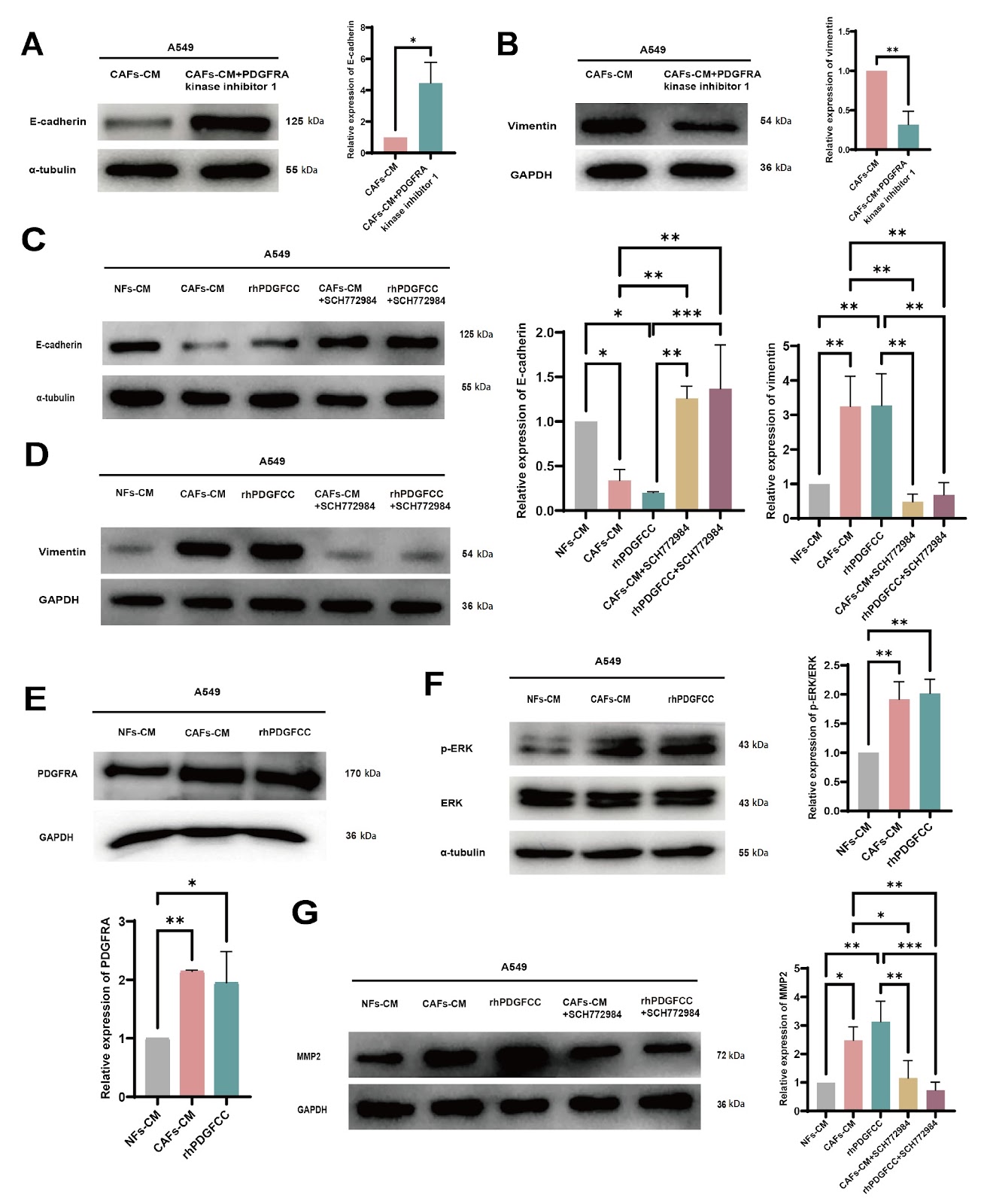
This study elucidates the mechanisms by which cancer-associated fibroblast (CAF)-derived platelet-derived growth factor C (PDGFC) promotes the progression of lung adenocarcinoma (LUAD) and explores the impact of PDGFC on immune regulation within the tumor microenvironment (TME). Our results show that there is higher expression of PDGFC in CAFs than in nontumor tissue fibroblasts (NFs) and that higher expression of PDGFC is correlated with poor prognosis in LUAD patients. Furthermore, CAF-derived PDGFC promotes epithelial-mesenchymal transition (EMT) in cancer cells as well as matrix metalloproteinase 2 (MMP2) expression through the PDGF receptor A (PDGFRA)-mitogen-activated protein kinase/extracellular signal-regulated kinase (MAPK/ERK) pathway. Moreover, our study demonstrates that CAF-derived PDGFC is essential for the activation and infiltration of fibroblasts in the TME, as well as the inflammatory infiltration of different immune cell types and the immunosuppressive conditions within the TME. In particular, PDGFC induces increased PDGFRA expression in both tumor cells and fibroblasts, which can lead to reciprocally positive feedback to accelerate malignant tumor progression. This discovery provides a novel TME-targeted strategy for LUAD treatment.

Read Full Abstract10.3724/abbs.2025042
CAR-macrophages: a new chapter in cancer immunotherapyGraphical AbstractVerified
Acta Biochimica et Biophysica Sinica2026

CAR-macrophages: a new chapter in cancer immunotherapy

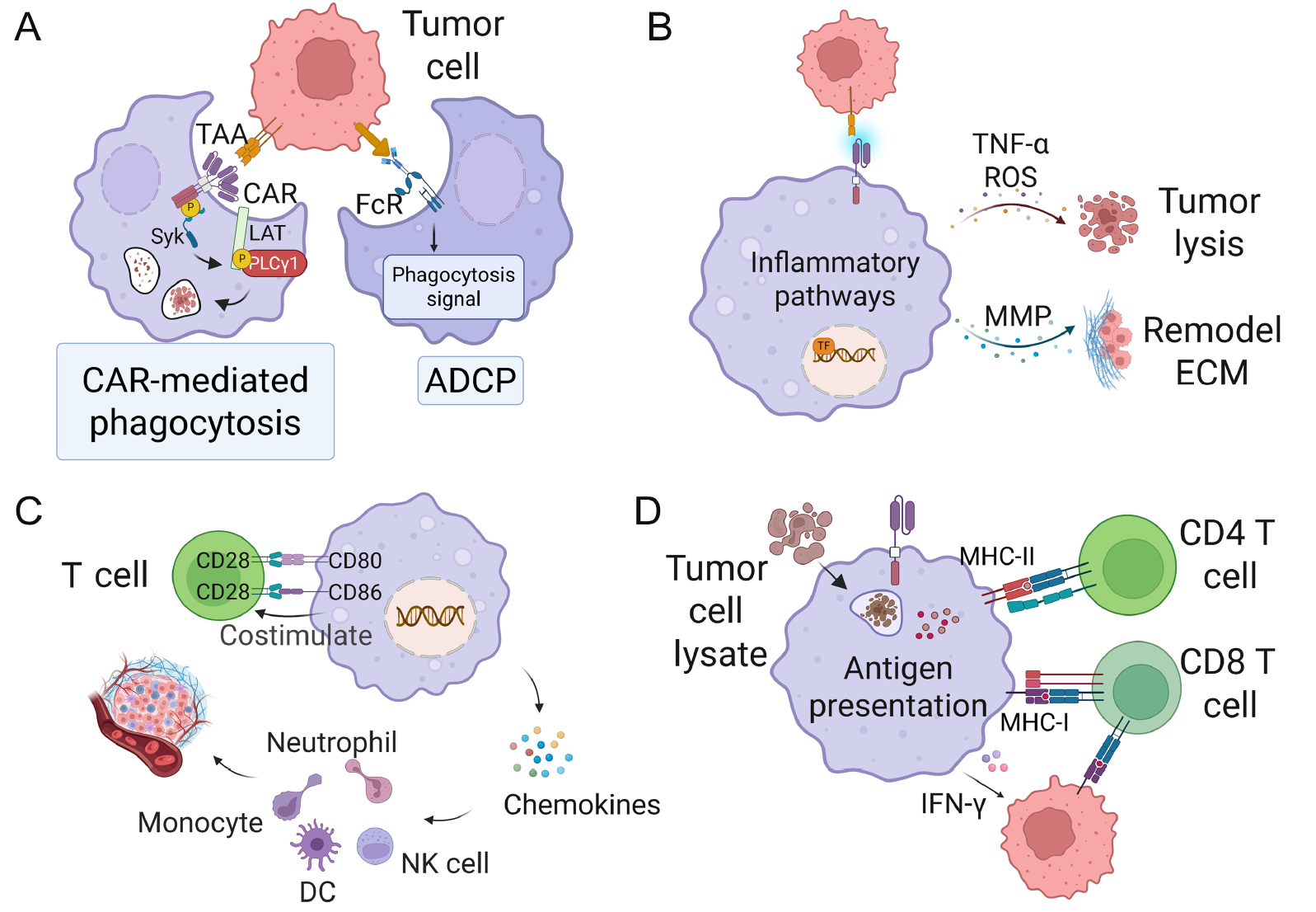
Chimeric antigen receptor T (CAR-T) cell therapy achieves remarkable success in hematological cancers, but its efficacy is severely limited in solid tumors by formidable obstacles including physical barriers, the highly immunosuppressive tumor microenvironment (TME), and antigen escape. To address these persistent challenges, chimeric antigen receptor-macrophage (CAR-M) therapy emerges as a promising alternative, leveraging intrinsic advantages of macrophages like unparalleled tumor infiltration, powerful phagocytosis, and high plasticity. The evolution of CAR-M is primarily defined by the intracellular signaling domain. CAR-M exerts its anti-tumor effects through multifaceted mechanisms, including direct enhanced phagocytosis and tumor cell killing, TME remodeling by repolarizing to a pro-inflammatory M1-like phenotype, releasing anti-tumor effectors, and degrading the extracellular matrix (ECM), and the activation of adaptive immunity via efficient antigen presentation. Despite its promise, CAR-M faces hurdles such as TME physical barriers and the potential for M2-like re-education. Current optimization strategies focus on enhancing tumor infiltration, overcoming immunosuppression with “armored” CAR-Ms, and improving safety with suicide switches. Encouraging pre-clinical data accelerates CAR-M into early-phase clinical trials for solid tumors, and the platform’s utility is also being explored beyond oncology in infectious, autoimmune, and neurodegenerative diseases.

Read Full Abstract10.3724/abbs.2026017
TLR4 mediates lipotoxic β-cell dysfunction by inhibiting the TMEM24/PI3K/AKT pathwayGraphical AbstractVerified
Acta Biochimica et Biophysica Sinica

TLR4 mediates lipotoxic β-cell dysfunction by inhibiting the TMEM24/PI3K/AKT pathway

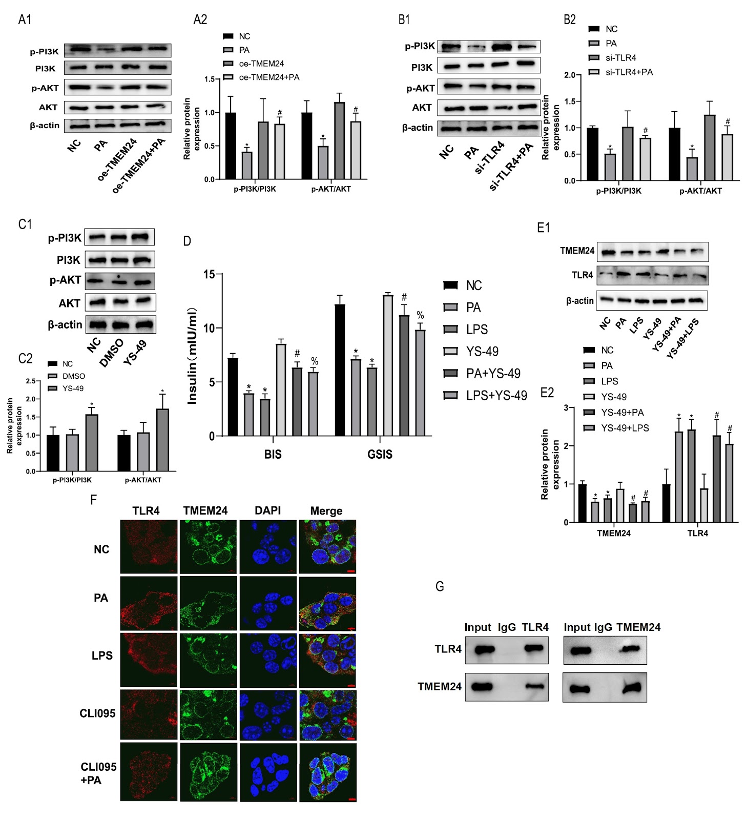
Immune imbalance is the core pathophysiological mechanism of the deterioration of β-cell function driven by lipid metabolism disorders. Toll-like receptor 4 (TLR4) inflammatory signaling is a key pathway that mediates lipotoxic injury in β-cells, but the underlying mechanism needs to be further elucidated. Transmembrane protein 24 (TMEM24) is a key transporter that regulates pulsatile insulin secretion, but its pathophysiology in lipotoxicity remains unclear. In this study, we investigate whether TLR4-mediated lipotoxicity is affected by the inhibition of TMEM24 expression. The PPI network shows that TLR4 is associated with both insulin secretion and ER stress proteins in islets from obese rats. Using in vitro lipotoxic β-cell models, we found that TMEM24 is the target signal of palmitic acid (PA)-induced insulin secretion impairment in islet β-cells, and TLR4 plays a mediating role in this process. Mechanistically, TLR4 mediates lipotoxicity by binding to TMEM24 and downregulating its protein expression to suppress PI3K/AKT signaling, leading to β-cell dysfunction. TLR4 knockout ameliorates islet function impairment through TMEM24/PI3K/AKT signaling in HFD-induced obese rats. Taken together, our results show that TLR4 mediates lipotoxicity in islet β-cells by inhibiting the TMEM24/PI3K/AKT pathway, and the mechanism of TLR4-mediated lipotoxicity is elucidated from the perspective of insulin vesicular secretion.

Read Full Abstract10.3724/abbs.2025045