🧬 SinoBioData Academic Portal
šŸ“š Peer-Reviewed Translated Literature

All Biomedical & Clinical Articles (Page 49)

Browse complete peer-reviewed translations from top Chinese biomedical, oncology, and genomics journals. Read verified previews and download full authentic clinical reports.

Published Research Papers

Showing 24 of 1542 peer-reviewed translated articles (Page 49 of 65)

Cannabidiol alleviates the inflammatory response in rats with traumatic brain injury through the PGE2-EP2-cAMP-PKA signaling pathwayGraphical AbstractVerified
Acta Biochimica et Biophysica Sinica

Cannabidiol alleviates the inflammatory response in rats with traumatic brain injury through the PGE2-EP2-cAMP-PKA signaling pathway

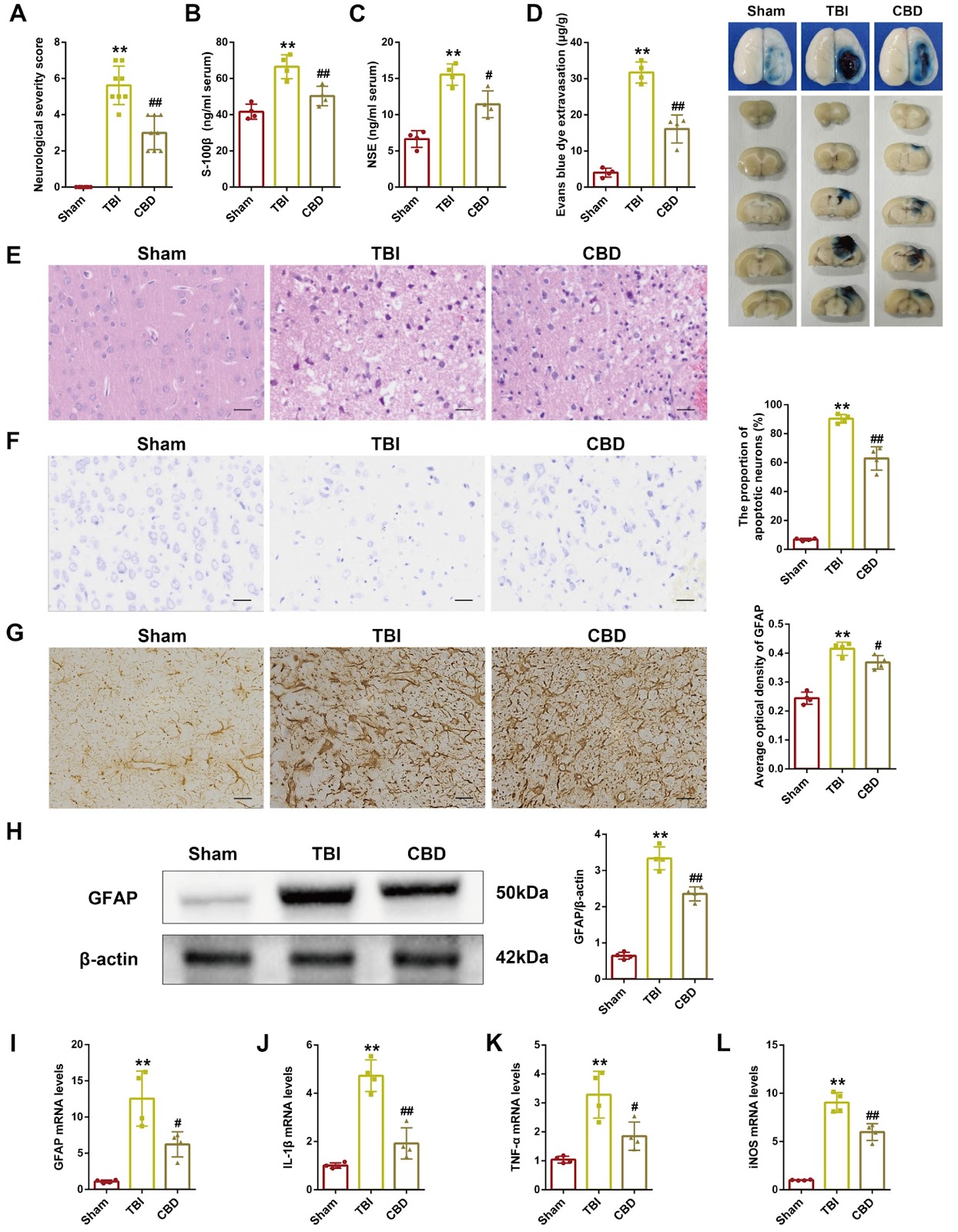
Traumatic brain injury (TBI) is a recognized global public health problem. However, there are still limitations in the available therapeutic approaches and a lack of clinically effective drugs. Therefore, an in-depth exploration of the secondary pathological mechanism of TBI and the identification of new effective drugs are urgently needed. Cannabidiol (CBD), a component derived from the cannabis plant, has potential therapeutic effects on neurological diseases and has received increasing attention. However, few reports on CBD intervention in TBI patients exist. Here, we use the Feeney free-fall method to establish a rat TBI model. CBD significantly improves neurological deficit scores, neuronal damage and blood-brain barrier permeability in rats and significantly inhibits the expressions of the brain injury markers S-100β and NSE. Mechanistically, CBD attenuates TBI-induced astrocyte activation, reduces inflammation, and attenuates the expressions of inflammatory prostaglandin system indicators. The use of TG6-10-1 (EP2 inhibitor) and H-89 (PKA inhibitor) indicates that CBD attenuates TBI-induced neurological damage via the PGE2-EP2-cAMP-PKA signaling pathway. Overall, this research provides a novel drug candidate for the treatment of clinical brain trauma.

Read Full Abstract10.3724/abbs.2024183
Cancer-specific bivalent promoters featuring low-level H3K27me3 signals favor active transcription and govern the cancer cell state transitionGraphical AbstractVerified
Acta Biochimica et Biophysica Sinica

Cancer-specific bivalent promoters featuring low-level H3K27me3 signals favor active transcription and govern the cancer cell state transition

Bivalent chromatin maintains genes in low-expression, poised states in embryonic stem cells (ESCs). However, bivalent promoters correlate with the transcriptional activation of oncogenic programs in malignancies, a seemingly contradiction that remains to be resolved. Here, we identify a class of cancer-specific bivalent promoters (CSBPs) through the integration of a system-level longitudinal framework. Compared with ESCs, CSBPs are characterized by lower and narrower H3K27me3 deposition alongside abundant H3K4me3, thus permitting the persistent expression of genes critical for cancer stem cell (CSC) formation and maintenance, as exemplified by SOX9. The generation of CSBPs is essentially induced by the acquisition of H3K27me3 during cell state transition, which is mediated by specific binding of PRC2.1 and the de novo recruitment of PRC2.2. Notably, disrupting the bivalency of CSBPs significantly increases H3K4me3 levels, leading to hyperactivation of CSBPs and eventually inhibiting clonal expansion of CSCs and impairing tumorigenesis. Our study not only helps explain the puzzle of transcriptionally active bivalent genes in cancer but also provides insights into the development of therapies targeting phenotypic plasticity.

Read Full Abstract10.3724/abbs.2025234
Metabolic analysis and identification of potential biomarkers of early-stage melanoma lung metastasisGraphical AbstractVerified
Acta Biochimica et Biophysica Sinica

Metabolic analysis and identification of potential biomarkers of early-stage melanoma lung metastasis

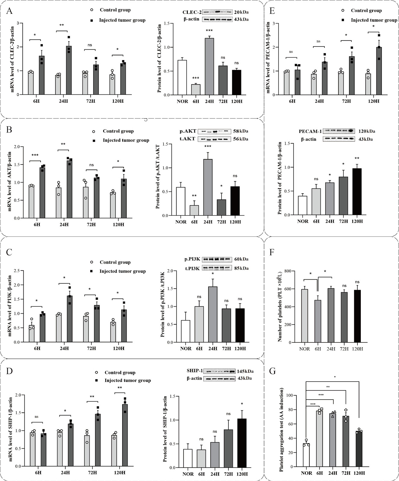
Tumor cells exhibit a notable ability to adapt to constantly changing microenvironments and possess distinct metabolic traits during metastasis. This study aims to establish a melanoma lung metastasis model in mice to elucidate the metabolic mechanisms involved in early-stage metastasis prior to treatment. The male C57BL/6 mice are divided into five groups based on time intervals of 6, 24, 72, and 120 h post-injection (SKCM-M groups) of melanoma cells, as well as a normal control group (NOR group). Our results demonstrate that platelet activation mainly occurs in the initial phases of metastasis to help tumor cells survive. NMR-based metabolomics analysis of mouse lung tissues identifies distinct metabolites and pathways associated with early-stage metastasis, revealing significant alterations in energy and amino acid metabolism during tumor progression. Further analysis indicates that methylxanthine and allantoin could serve as potential biomarkers for monitoring the early progression of tumor metastasis in cancer patients, providing novel insights into early diagnostic strategies for lung metastasis.

Read Full Abstract10.3724/abbs.2025136
Melanoma-derived versican reactivates tumor-associated macrophages by upregulating pyruvate carboxylase through TLR2-MyD88-RelB axis under normoxiaGraphical AbstractVerified
Acta Biochimica et Biophysica Sinica

Melanoma-derived versican reactivates tumor-associated macrophages by upregulating pyruvate carboxylase through TLR2-MyD88-RelB axis under normoxia

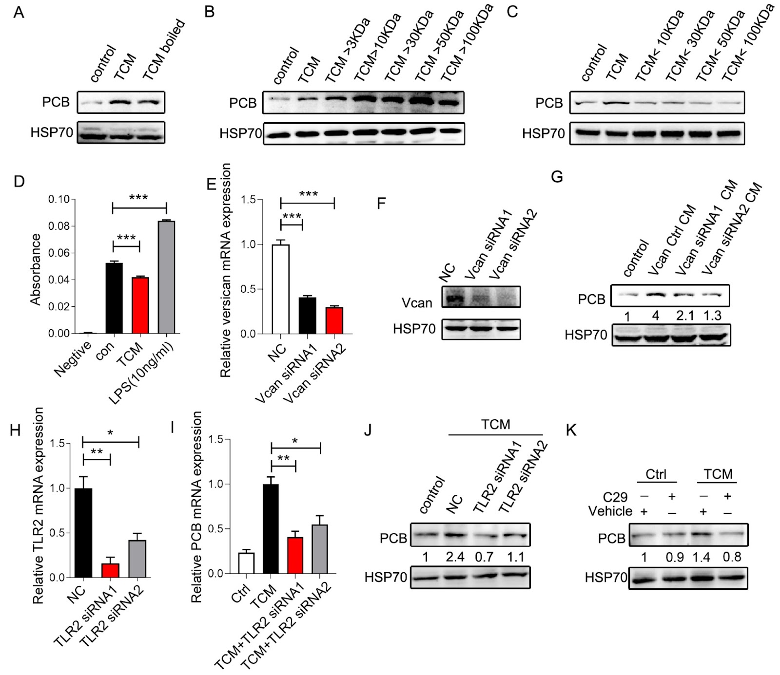
Relieving hypoxia in the tumor microenvironment (TME) promotes innate and adaptive immunity. Our previous research demonstrated that reoxygenation of the TME promotes the phagocytosis and tumor-killing functions of tumor-associated macrophages (TAMs) by upregulating pyruvate carboxylase (PCB). However, the mechanism remains obscure. In the present study, we find that versican derived from melanoma cells binds to TLR2 and activates the downstream transcription factor RelB, which transcribes PCB under normoxia. Blocking the versican-TLR2-MyD88-RelB axis not only reverses the upregulation of PCB in TAMs but also hinders the clearance of tumor cells by TAMs. Our work suggests a pathway that modulates the functions of TAMs under normoxia, which could be harnessed for strengthening anti-tumor immunity.

Read Full Abstract10.3724/abbs.2025011
TRIM21 promotes type I interferon by inhibiting the autophagic degradation of STING via p62/SQSTM1 ubiquitination in systemic lupus erythematosusGraphical AbstractVerified
Acta Biochimica et Biophysica Sinica

TRIM21 promotes type I interferon by inhibiting the autophagic degradation of STING via p62/SQSTM1 ubiquitination in systemic lupus erythematosus

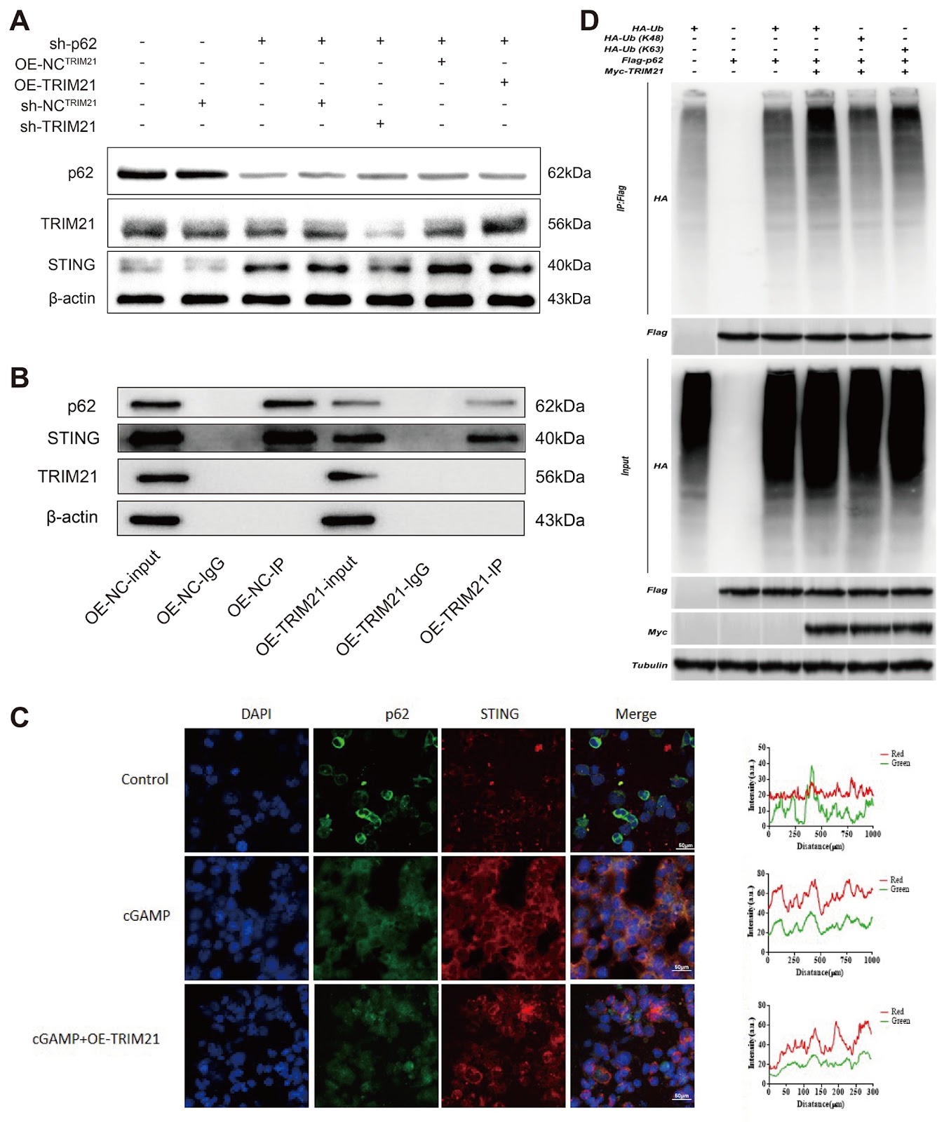
The cGAS-STING signaling pathway serves as a pivotal surveillance mechanism for cytosolic double-stranded DNA (dsDNA) detection in mammalian systems. While STING-mediated type I interferon production is crucial for host defense, sustained activation of this pathway contributes to autoimmune pathologies, including systemic lupus erythematosus (SLE). Maintaining immune homeostasis requires precise regulation of STING activity to prevent hyperactivation. Our study identifies TRIM21 as a novel positive regulator of cGAS-STING signaling in SLE pathogenesis. Our results demonstrate that TRIM21 overexpression stabilizes STING by suppressing autophagic degradation, whereas TRIM21 depletion accelerates this clearance process. Mechanistically, TRIM21 catalyzes the K63-linked polyubiquitylation of the selective autophagy receptor p62/SQSTM1, disrupting its interaction with STING. This post-translational modification prevents the sequestration of STING into autophagosomes, thereby stabilizing the adaptor protein and amplifying downstream type I interferon responses. Our findings reveal a previously unrecognized regulatory circuit in which TRIM21 orchestrates cross-talk between ubiquitin signaling and autophagy to control STING turnover. The TRIM21-p62 axis represents a potential therapeutic target for attenuating pathological interferon production in STING-dependent autoimmune disorders. This work advances our understanding of immune regulation by demonstrating how E3 ligase-mediated ubiquitin modifications modulate cargo recognition in selective autophagy pathways. The identified mechanism provides new insights into the molecular interplay between protein ubiquitylation and autophagic degradation in maintaining the innate immune balance, offering novel perspectives for developing targeted therapies against interferonopathies associated with cGAS-STING hyperactivity.

Read Full Abstract10.3724/abbs.2025046
p53-dependent chromatin relaxation is required for DNA double-strand break repairGraphical AbstractVerified
Acta Biochimica et Biophysica Sinica

p53-dependent chromatin relaxation is required for DNA double-strand break repair

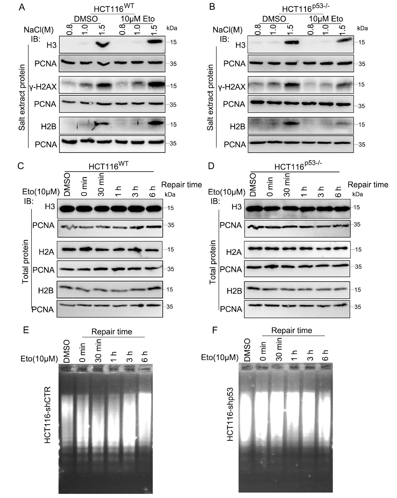
The tumor suppressor p53, an indispensable nuclear transcription factor, plays a central role in orchestrating cellular responses when DNA damage occurs. In this study, we demonstrate that in the initial phases of DNA double-strand break (DSB) repair, p53 is rapidly recruited to sites of damage and the surrounding chromatin, where it enhances DSB repair efficiency. This enhancement occurs through the modulation of chromatin dynamics and the promotion of a more relaxed chromatin configuration, a process influenced by p53 in response to DSB-inducing factors such as etoposide, ultraviolet radiation, and nucleases. These results underscore the pivotal function of p53 as a rapid responder to DSBs, delineating a significant departure from its traditionally recognized role as a downstream transcriptional regulator in DNA damage repair processes. This study emphasizes that the direct engagement of p53 in DNA repair through chromatin structure regulation extends beyond its established involvement in UV irradiation-induced nucleotide excision repair (NER), demonstrating analogous mechanistic attributes in the context of DSB repair. This newly illuminated perspective enhances our understanding of the multifaceted roles of p53 in genome stability and integrity.

Read Full Abstract10.3724/abbs.2025008
The DNA-PKcs-primary cilia axis maintains ionizing radiation-induced senescence in tumor cellsGraphical AbstractVerified
Acta Biochimica et Biophysica Sinica

The DNA-PKcs-primary cilia axis maintains ionizing radiation-induced senescence in tumor cells

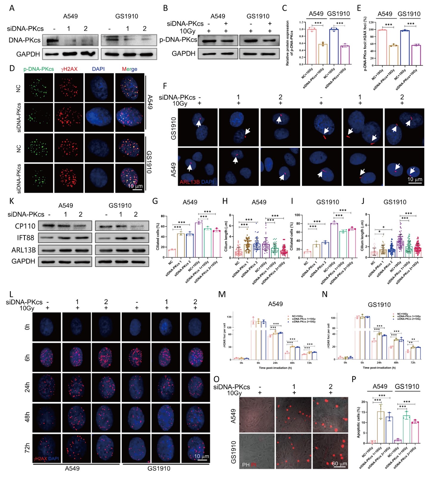
Senescence is a cellular response closely associated with genotoxic stress and plays a critical role in determining cell fate following irradiation exposure. Primary cilia, which are sensory organelles on the cell surface, detect and transmit diverse signaling cues. However, the relationship between primary cilia and senescence in long-term cell fate decisions after ionizing radiation remains poorly understood. Here, we show that the DNA-dependent protein kinase catalytic subunit (DNA-PKcs) colocalizes with centromeres during various stages of mitosis, whereas during interphase, phosphorylated DNA-PKcs (p-DNA-PKcs) is confined to the nucleus in tumor cells. Following irradiation exposure, primary cilia are formed and persistently maintained at high levels in senescent tumor cells. Inhibition of DNA-PKcs enhances primary cilia formation, whereas combined inhibition with siDNA-PKcs and irradiation reduces cilia generation. Moreover, chloral hydrate-induced primary cilia removal results in senescent cell death and decreases p-DNA-PKcs protein expression. Notably, treatment with the apoptosis inducer ABT263 also leads to increased cell death and decreased incidence of primary cilia. Inhibition of either primary cilia or DNA-PKcs further enhances the radiosensitivity of tumor cells. These findings suggest that DNA-PKcs contributes to primary cilia formation after irradiation and plays a critical role in both the induction and maintenance of cellular senescence.

Read Full Abstract10.3724/abbs.2025168
CD98hc, a novel of galectin-8 receptor, binds to galectin-8 in an N-glycosylation-dependent mannerGraphical AbstractVerified
Acta Biochimica et Biophysica Sinica

CD98hc, a novel of galectin-8 receptor, binds to galectin-8 in an N-glycosylation-dependent manner

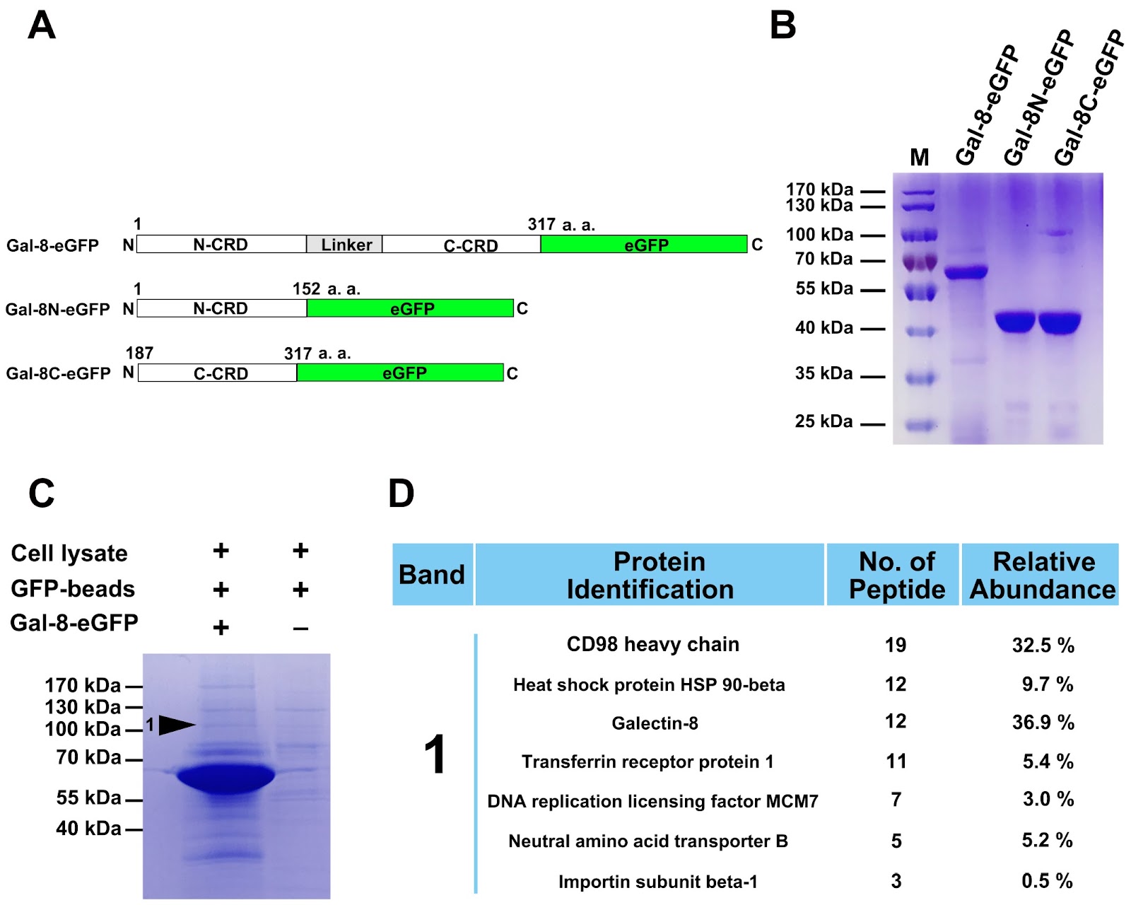
Glycan-mediated recognition plays a critical role in facilitating cell-cell and cell-matrix interactions. Galectin-8 (Gal-8), classified as a ā€˜tandem-repeat’ type of galectin, binds to cell surface glycans to modulate various cellular functions, including cell adhesion, migration, apoptosis, pathogen recognition, autophagy, and immunomodulation. Despite the known function of Gal-8 in binding to various glycosylated proteins, only a few interactions have been reported to date. In this study, mass spectrometry is used to identify CD98hc as a novel binding partner for Gal-8. Both the N-terminal and C-terminal carbohydrate recognition domains (CRDs) of Gal-8 (Gal-8N and Gal-8C) bind to CD98hc, an interaction that is specifically inhibited by lactose but not sucrose, as confirmed by pull-down assays. The binding affinity between CD98hc and Gal-8 measured by microscale thermophoresis (MST) is 1.51 ± 0.17 μM. In addition, Gal-8N and Gal-8C have the binding affinities of 0.22 ± 0.03 μM and 10.68 ± 1.69 μM, respectively. Gal-8N and Gal-8C are both involved in the recognition and binding process of CD98hc. Furthermore, both full-length Gal-8 and its individual CRDs bind specifically to N-glycosylated glycans on CD98hc, as demonstrated by the use of tunicamycin to inhibit N-glycosylation in cells. In addition, Gal-8 and its individual CRDs can pull down glycosylated CD98hc-ED but not free CD98hc-ED in vitro, indicating that the binding of Gal-8 to glycosylated CD98hc-ED is N-glycosylation-dependent. Overall, our findings establish CD98hc as a novel binding partner for Gal-8 and provide insights for further exploration of the diverse biological functions of Gal-8.

Read Full Abstract10.3724/abbs.2024182
Myocyte enhancer factor 2A orchestrates vascular redox homeostasis via direct transcriptional activation of SIRT1Graphical AbstractVerified
Acta Biochimica et Biophysica Sinica

Myocyte enhancer factor 2A orchestrates vascular redox homeostasis via direct transcriptional activation of SIRT1

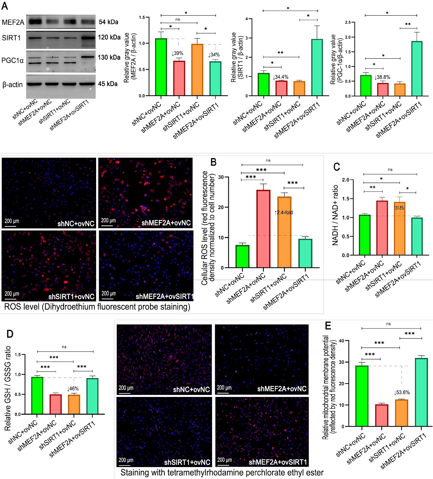
Myocyte enhancer factor 2A (MEF2A), a transcription factor implicated in coronary artery disease, remains unexplored in vascular redox regulation. To address this gap and overcome the limitations of current antioxidant therapies, we investigate the role of MEF2A in oxidative defense via human umbilical vein endothelial cells (HUVECs) and murine models. Adenoviral vectors encoding MEF2A-specific shRNAs or mRNAs are used to silence or overexpress MEF2A in HUVECs. For in vivo validation, endothelial-targeted MEF2A knockdown is achieved via AAV1-shRNA delivery in mice fed with a high-fat diet. Systemic redox status is assessed by measuring reactive oxygen species (ROS), glutathione homeostasis (GSH/GSSG ratio), the NADH/NAD+ balance, the mitochondrial membrane potential (ΔΨm), and 8-hydroxy-2′-deoxyguanosine (8-OHdG). Mechanistic insights are derived from immunofluorescence, qPCR, western blotting, and dual-luciferase reporter assays. MEF2A silencing induces redox imbalance, characterized by elevated ROS, a reduced GSH/GSSG ratio, and ΔΨm collapse. Conversely, MEF2A overexpression synergizes with SIRT1 to restore the glutathione pool, maintain NAD+ homeostasis, and suppress ROS under oxidative stress. Chromatin immunoprecipitation confirms that MEF2A directly binds to two cis-elements in the SIRT1 promoter, driving transcriptional activation. In vivo, MEF2A-deficient mice present increased vascular oxidative damage, as indicated by elevated DNA damage marker (8-OHdG) and ROS levels. The downregulation of SIRT1/PGC-1α in MEF2A-silenced cells is verified in vivo. Our findings establish MEF2A as a master regulator of endothelial redox defense via the SIRT1-PGC-1α axis, providing a mechanistic foundation for the treatment of oxidative cardiovascular disorders. This work suggests that pharmacological MEF2A activation is a novel strategy for precision antioxidant therapy in vascular medicine.

Read Full Abstract10.3724/abbs.2025163
Antibody-oligonucleotide conjugates for spatial proteomics: principles, applications, and challengesGraphical AbstractVerified
Acta Biochimica et Biophysica Sinica

Antibody-oligonucleotide conjugates for spatial proteomics: principles, applications, and challenges

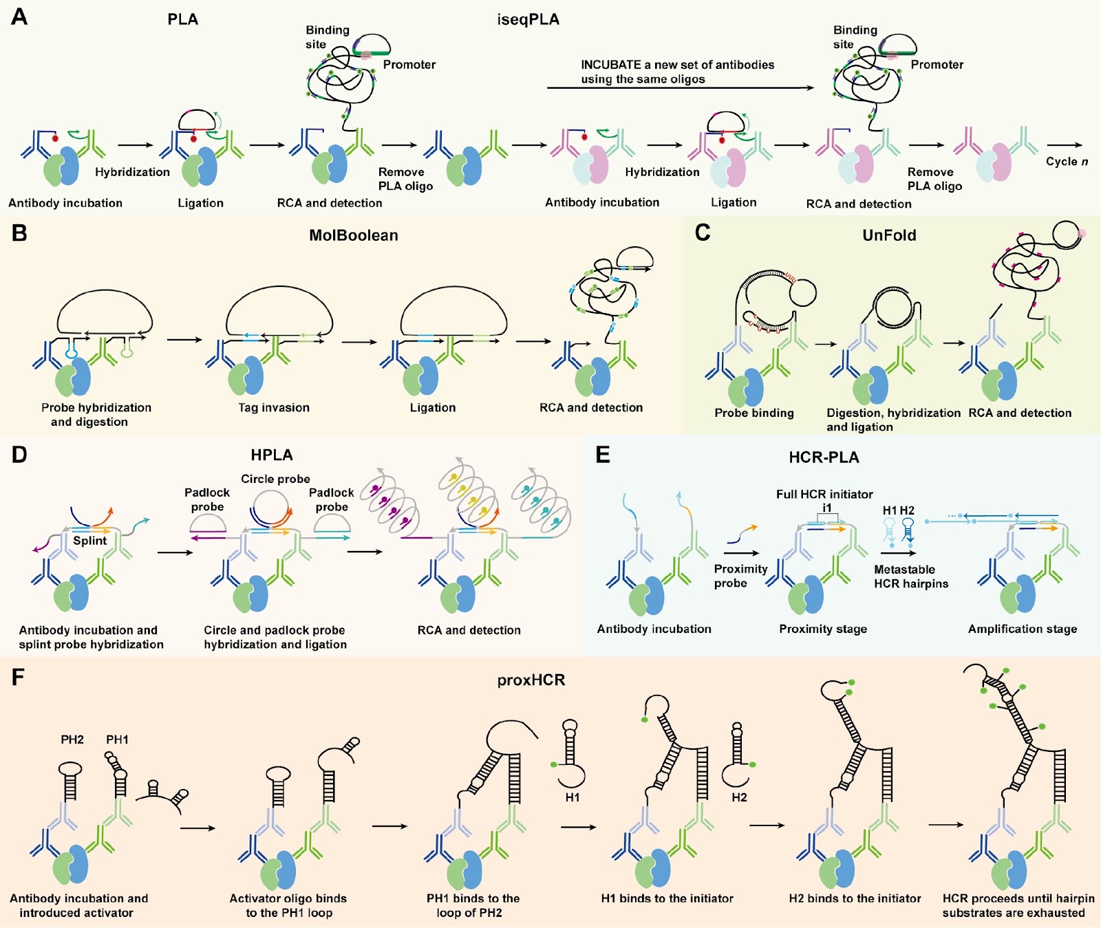
Spatial biology aims to elucidate cellular organization, function, and interactions within native tissue contexts, offering key insights into both normal physiology and disease. Spatial proteomics complements this by enabling high-resolution mapping of protein localization and abundance, directly reflecting functional cellular states. Unlike transcriptomics, which infers potential activity, proteomics captures actual molecular functions, including post-translational modifications and dynamic interactions. However, in situ protein profiling poses significant challenges, as proteins cannot be directly sequenced or easily targeted via nucleic acid hybridization. Antibody-oligonucleotide conjugates (AOCs) address this limitation by converting protein recognition into a DNA-based readout, thereby enabling sensitive and scalable detection. In this review, we outline the core principles of AOC-based spatial proteomic technologies, including multiplexed protein analysis, in situ protein-protein interactions, and integration with other biomolecular data. We highlight their applications in decoding tissue complexity and disease pathology and examine key technical challenges that remain. Overall, AOCs offer distinct advantages, including DNA-mediated signal amplification, spatially resolved proteomic profiling, and compatibility with multi-omics approaches, positioning them as powerful platforms in the advancement of spatial biology.

Read Full Abstract10.3724/abbs.2025212
Directly targeting G-quadruplexes contributes to the anti-multiple myeloma efficacy of Epimedokoreanin BGraphical AbstractVerified
Acta Biochimica et Biophysica Sinica

Directly targeting G-quadruplexes contributes to the anti-multiple myeloma efficacy of Epimedokoreanin B

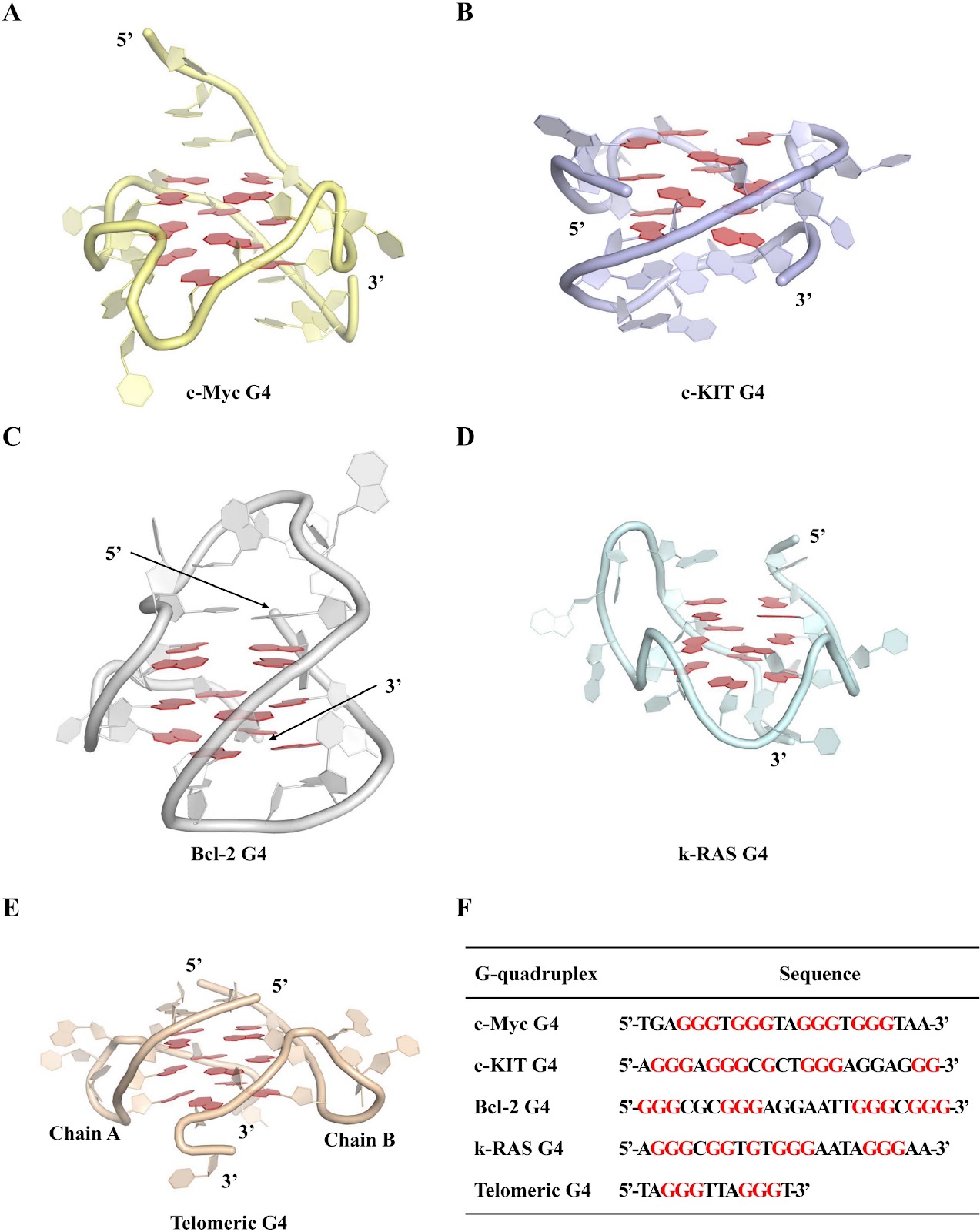
Multiple myeloma (MM) is a hematological malignancy for which novel therapeutic strategies are urgently needed. Epimedokoreanin B (EKB), an isoprenylated flavonoid compound derived from the medicinal plant Epimedium koreanum, has demonstrated promising antitumor activity. However, its effects on MM have not been previously investigated. This study explores the anti-MM activity and the molecular interaction mechanisms between EKB and G-quadruplexes (G4) through a combination of biological activity assessments and computer-aided methodologies. EKB exhibits potent cytotoxicity against the MM cell lines U266 and RPMI-8226, with IC50 values of 5.28 μM and 6.81 μM, respectively. It induces apoptosis in a concentration-dependent manner and specifically stabilizes the G4 structures of oncogenes such as c-Myc, c-KIT, Bcl-2, and k-RAS, as confirmed by BG4 immunofluorescence staining and fluorescence resonance energy transfer (FRET) assays. Additionally, EKB significantly suppresses the mRNA and protein expression levels of these genes in myeloma cells. Computational studies, including molecular docking, molecular dynamics (MD) simulations, and MM/GBSA calculations, confirm the strong binding affinity and stabilizing effects of EKB on G4s, revealing a mechanism involving Ļ€-Ļ€ stacking and hydrogen bonding. This discovery underscores the unique ability of EKB to increase the stability of G4 structures, which are critical for regulating gene expression and inhibiting cancer cell proliferation. This research highlights the therapeutic potential of EKB in targeting these specific molecular structures, thereby offering a more effective approach to managing MM.

Read Full Abstract10.3724/abbs.2025110
Gut microbiota and bile acids changes in MASLD mice model with hepatic PLD1 knockoutGraphical AbstractVerified
Acta Biochimica et Biophysica Sinica

Gut microbiota and bile acids changes in MASLD mice model with hepatic PLD1 knockout

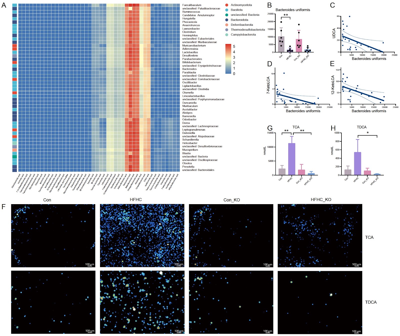
Hepatocyte phospholipase D1 (PLD1) knockout alleviates metabolic dysfunction-associated steatotic liver disease (MASLD) in mice, but the underlying mechanism is largely unknown. In this study, the mice are divided into four groups: Con (wild-type mice with normal control diet), HFHC (wild-type mice with high-fat diet), Con_KO (hepatocyte PLD1-knockout mice with normal control diet), and HFHC_KO (hepatocyte PLD1-knockout mice with high-fat diet). Intestinal contents of mice are analyzed via metagenomics and metabolomics, and the liver bile acids are assessed by mass spectrometry imaging. The results show that at the phylum level the abundance of Bacillota in the intestines of MASLD model mice is significantly increased, whereas that of Bacteroidota significantly is decreased. However, after the deletion of hepatocyte PLD1, Pseudomonadota and Candidatus Bathyarchaeota are significantly decreased in the MASLD model mice. At the species level, compared with that in the Con group, the abundance of Faecalibaculum rodentium is significantly increased in the HFHC group, whereas hepatocyte PLD1 knockout causes the abundances of Desulfovibrionaceae bacterium LT0009 and Lachnospiraceae bacterium 10-1 to be significantly decreased. In terms of intestinal bile acids, the levels of two bile acids (hyodeoxycholic acid and glycolithocholic acid) differ between the HFHC_KO group and the HFHC group. Association analysis shows that Faecalibaculum co-occurs with DCA, βMCA, ΩMCA and αMCA, while probiotic Bacteroides uniformis is significantly correlated with UDCA, 12-KetoLCA, and 7-KetoLCA. Finally, mass spectrometry imaging reveals that the TCA and TDCA contents in the liver are significantly decreased after PLD1 knockout in hepatocytes. These findings demonstrate that hepatocyte PLD1 knockout alters the gut microbiota and bile acids profiles, suggesting that PLD1 deficiency may modulate MASLD progression by changing intestinal microbiota-bile acid homeostasis.

Read Full Abstract10.3724/abbs.2025183
Explore antibody repertoire in the era of AIGraphical AbstractVerified
Acta Biochimica et Biophysica Sinica

Explore antibody repertoire in the era of AI

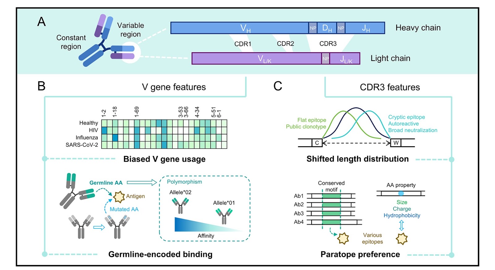
The diverse antibodies of adaptive immunity comprise an antibody repertoire that combats various pathogens. This repertoire is shaped by both intrinsic antibody gene diversification and extrinsic cellular selection. Conversely, an antibody repertoire contains multiple layers of immunological information, including the history of pathogen exposure. High-throughput sequencing-based antibody repertoire cloning approaches have revealed unexpected features of adaptive immunity. However, our understanding of antibody repertoire data is still in its infancy. In this review, we introduce the emerging concepts and discuss the application of deep learning approaches to understanding antibody repertoires. First, we introduce the definition and functional features of antibody clonotype. Next, we review the evolution of antibody clonotypes and discuss potential antibody repertoire-directed vaccination approaches. Lastly, we summarize the application of deep learning in predicting antibody binding, generating specific antibodies, and making immunologic diagnoses. Recently, artificial intelligence (AI) has made revolutionary progress in biology. Leveraging high-dimensional antibody repertoire information, deep learning models have the potential to transform our understanding of antibody repertoire.

Read Full Abstract10.3724/abbs.2025230
The TCF7L2/miR-206/Cofilin1 axis promotes the metastasis of bladder cancer cells by regulating the formation of invadopodiaGraphical AbstractVerified
Acta Biochimica et Biophysica Sinica

The TCF7L2/miR-206/Cofilin1 axis promotes the metastasis of bladder cancer cells by regulating the formation of invadopodia

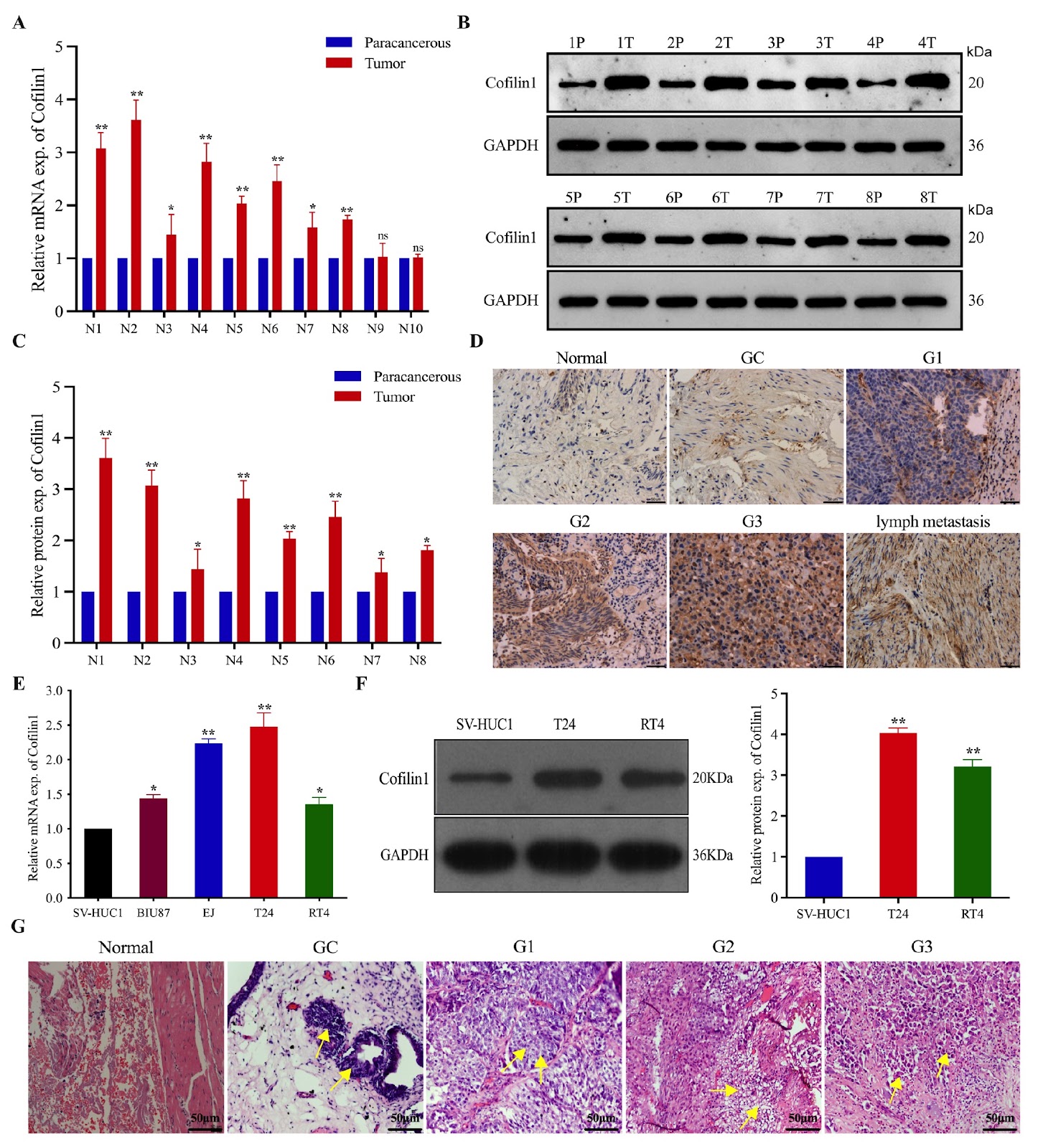
Bladder cancer (BCa) is one of the most common malignant tumors of the urinary system, but its pathogenesis is still unclear. T1G3 BCa is particularly invasive and relapses readily after treatment, with progression to invasive cancer or distant metastasis. Therefore, identification of the molecular mechanism by which it invades and metastasizes to guide treatment and predict patient prognosis is needed. Cofilin1 plays an important role in regulating gene expression and the invasiveness of tumors. In this study, we show that Cofilin1 is highly expressed in BCa and lymph nodes with metastasis, which is positively related to the grade of BCa, and is significantly related to clinicopathological parameters and cancer-specific survival. Phenotypic analysis reveals that Cofilin1 knockout inhibits the proliferation and migration of BCa cells, whereas Cofilin1 overexpression promotes the opposite phenotype. Cofilin1 binds to cortactin, thereby reducing the expression of F-actin and promoting the formation of invadopodia in BCa cells. Further experiments reveal that TCF7L2 can bind to the promoter of Cofilin1 and transactivate it, promoting a malignant phenotype. TCF7L2 may also reverse the inhibitory effect of miR-206 on the binding of Cofilin1 and cortactin and promote the metastasis of BCa by inhibiting the transcription maturation of miR-206. This study confirms that Cofilin1 is an oncogene in T1G3 BCa, and the TCF7L2/miR-206/Cofilin1 signaling pathway plays an important role in the formation of invadopodia in BCa.

Read Full Abstract10.3724/abbs.2025114
Annexins: central regulators of plant growth and stress signalingGraphical AbstractVerified
Acta Biochimica et Biophysica Sinica

Annexins: central regulators of plant growth and stress signaling

Annexins are a family of multifunctional calcium-dependent and phospholipid-binding proteins that are widely distributed in the plant kingdom. They have a highly conserved evolutionary history that dates back to single-celled protists. Plant annexins, as soluble proteins, can flexibly bind to endomembranes and plasma membranes, exhibiting unique calcium-dependent and calcium-independent characteristics. Members of the annexin family have diverse functions, including binding to F-actin, participating in ATP and GTP hydrolysis, and even serving as peroxidases or cation channels. Annexins play pivotal roles in plant growth and stress signaling. They can respond sensitively to environmental, metabolic, and developmental signals, thereby affecting cytoskeleton remodeling and exocytosis mechanisms. Plant annexin gene families have been successfully identified in multiple species, and their expression and intracellular localization are precisely regulated by developmental processes and environmental factors. Although research on plant annexins has aroused great interest, their depth and breadth still need further expansion compared with those of animal annexins. This article provides a comprehensive and in-depth review of the characteristics and functions of plant annexin families, revealing their core roles in plant growth and adaptation, and yielding valuable references and insights for future research.

Read Full Abstract10.3724/abbs.2024228
Scutellarin inhibits ferroptosis by promoting cellular antioxidant capacity through regulating Nrf2 signalingGraphical AbstractVerified
Acta Biochimica et Biophysica Sinica

Scutellarin inhibits ferroptosis by promoting cellular antioxidant capacity through regulating Nrf2 signaling

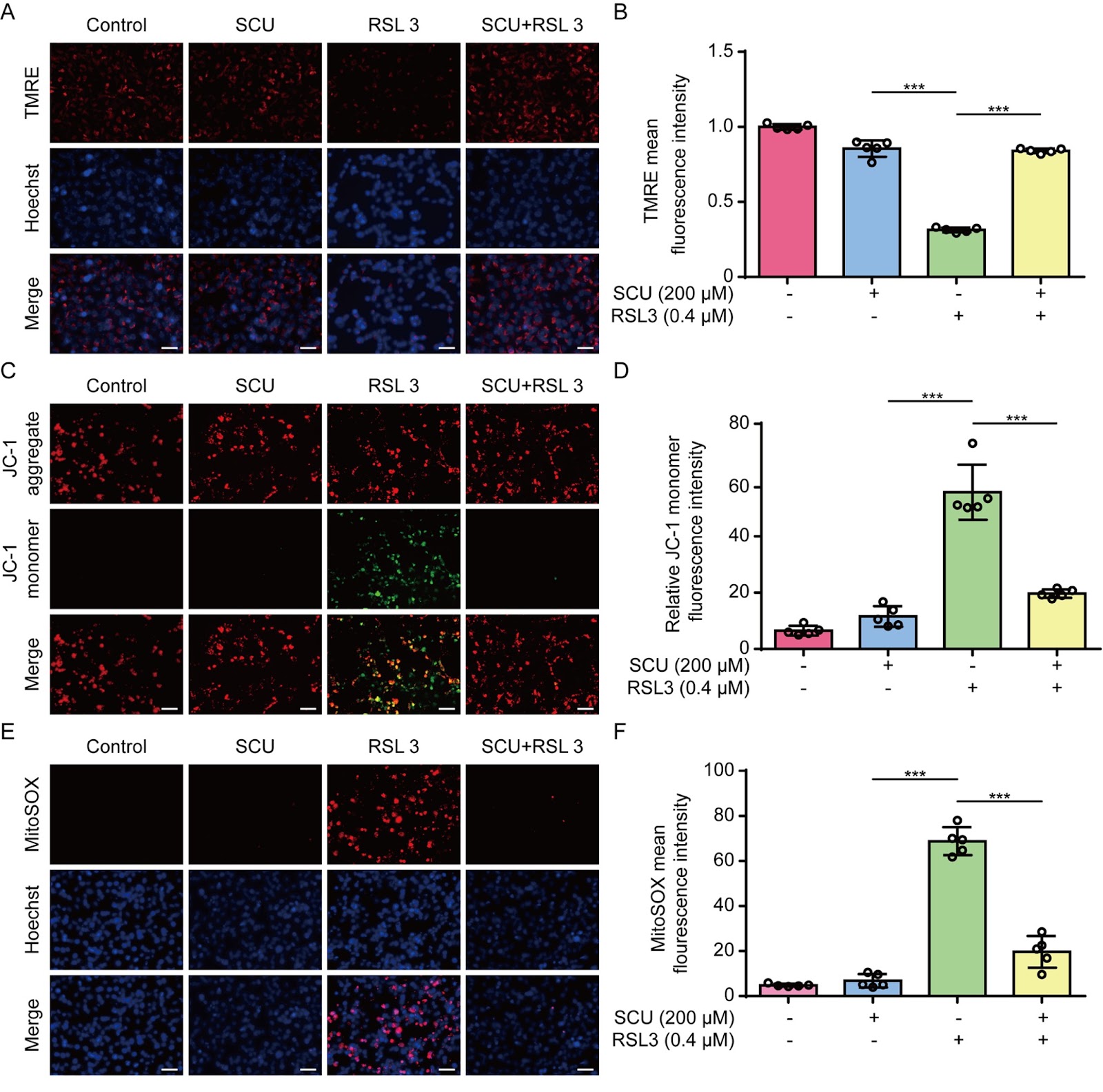
Ferroptosis is a lytic form of regulated cell death that is driven by iron-dependent lipid peroxidation and has been implicated in various diseases, including acute kidney injury (AKI). Scutellarin is a flavonoid isolated from Erigeron breviscapus (Vant.) Hand.-Mazz. and possesses various pharmacological activities, including anti-inflammatory and antioxidative properties. However, it is unclear whether scutellarin can inhibit ferroptosis and mitigate related diseases. In this study, we show that scutellarin can inhibit ferroptosis in both human HK-2 cells and mouse bone marrow-derived macrophages stimulated with RSL3 or erastin. Mitochondrial dysfunction and reactive oxygen species generation are counteracted by scutellarin treatment, suggesting the involvement of its antioxidative activity. Furthermore, scutellarin increases the nuclear levels of Nrf2 and the expressions of its target genes, including HO-1 and GPX4. Scutellarin-mediated inhibition of ferroptosis and increases in these proteins are abrogated by co-treatment with brusatol, an Nrf2 inhibitor, indicating an essential role for Nrf2 in this process. In a mouse model of folic acid-induced AKI, scutellarin mitigates acute renal damage, as revealed by histopathological analysis and serum blood urea nitrogen and creatinine assays. Folic acid-induced acute renal injury is associated with increased ferroptosis, as revealed by elevated level of 4-hydroxynonenal (4-HNE), a surrogate marker of ferroptosis, which is diminished by scutellarin co-treatment. Specifically, the elevated 4-HNE levels in macrophages (MAC-2 positive) and other renal cells are suppressed by scutellarin. Overall, scutellarin can inhibit ferroptosis both in cultured cells and in a mouse model of AKI by regulating Nrf2 signaling.

Read Full Abstract10.3724/abbs.2025112
Structural basis for suramin binding to the C-terminal domain of the SARS-CoV-2 nucleocapsid proteinGraphical AbstractVerified
Acta Biochimica et Biophysica Sinica

Structural basis for suramin binding to the C-terminal domain of the SARS-CoV-2 nucleocapsid protein

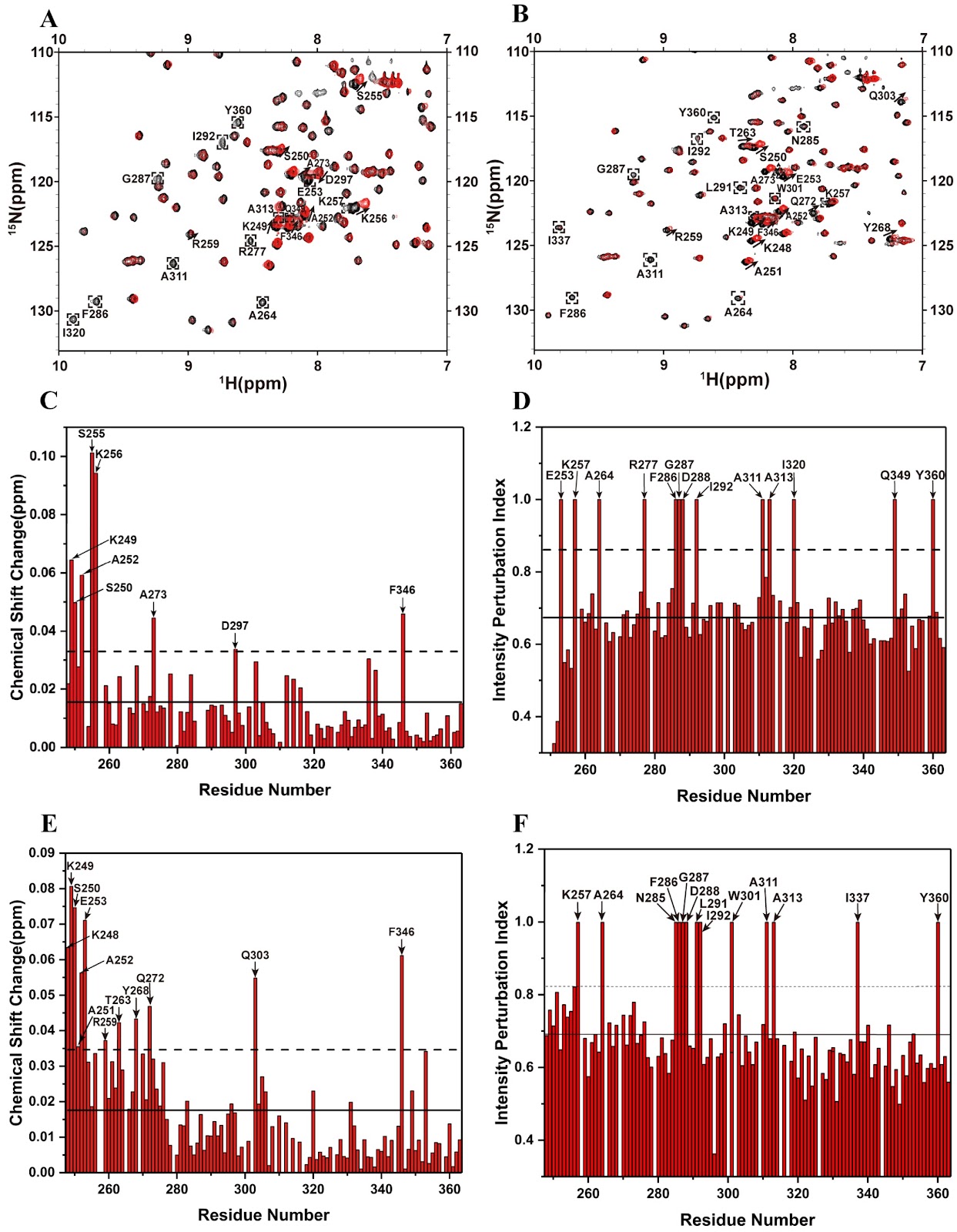
The global threat posed by COVID-19 persists, largely due to the high mutability of SARS-CoV-2 and the limited availability of effective antiviral therapeutics. The nucleocapsid (N) protein of SARS-CoV-2 is an attractive drug target because of its high degree of sequence conservation and essential role in viral replication. In this study, we show that suramin, a polysulfonated antiviral compound, binds to the C-terminal domain (N-CTD) of the N protein and interferes with its interaction with RNA. Biolayer interferometry (BLI) shows that suramin has a higher binding affinity for N-CTD (Kd, 3.30 μM) than for RNA (Kd, 10.12 μM). Electrophoretic mobility shift assays (EMSAs) further confirms that suramin effectively displaces RNA from N-CTD. NMR titration experiments and site-directed mutagenesis identify the α1-Ī·1 helix (residues 248–262) as the primary suramin binding region, with residues K256, R259 and R262 playing critical roles in ligand recognition. In addition, NMR relaxation and model-free analyses reveal that the α1-Ī·1 helix is highly flexible on the picosecond to nanosecond timescale, a dynamic feature that likely facilitates ligand binding. Furthermore, ITC and EMSA experiments demonstrate that suramin can bind to the full-length N protein at multiple sites and dissociate RNA from the N protein. Taken together, these findings provide structural and biophysical insights into the mechanism of action of suramin and establish a rational basis for the development of targeted antiviral therapies against SARS-CoV-2.

Read Full Abstract10.3724/abbs.2025182
Combined ex vivo and in vivo evaluation of dolutegravir embryotoxicity: NTDs and yolk sac vascular abnormalitiesGraphical AbstractVerified
Acta Biochimica et Biophysica Sinica

Combined ex vivo and in vivo evaluation of dolutegravir embryotoxicity: NTDs and yolk sac vascular abnormalities

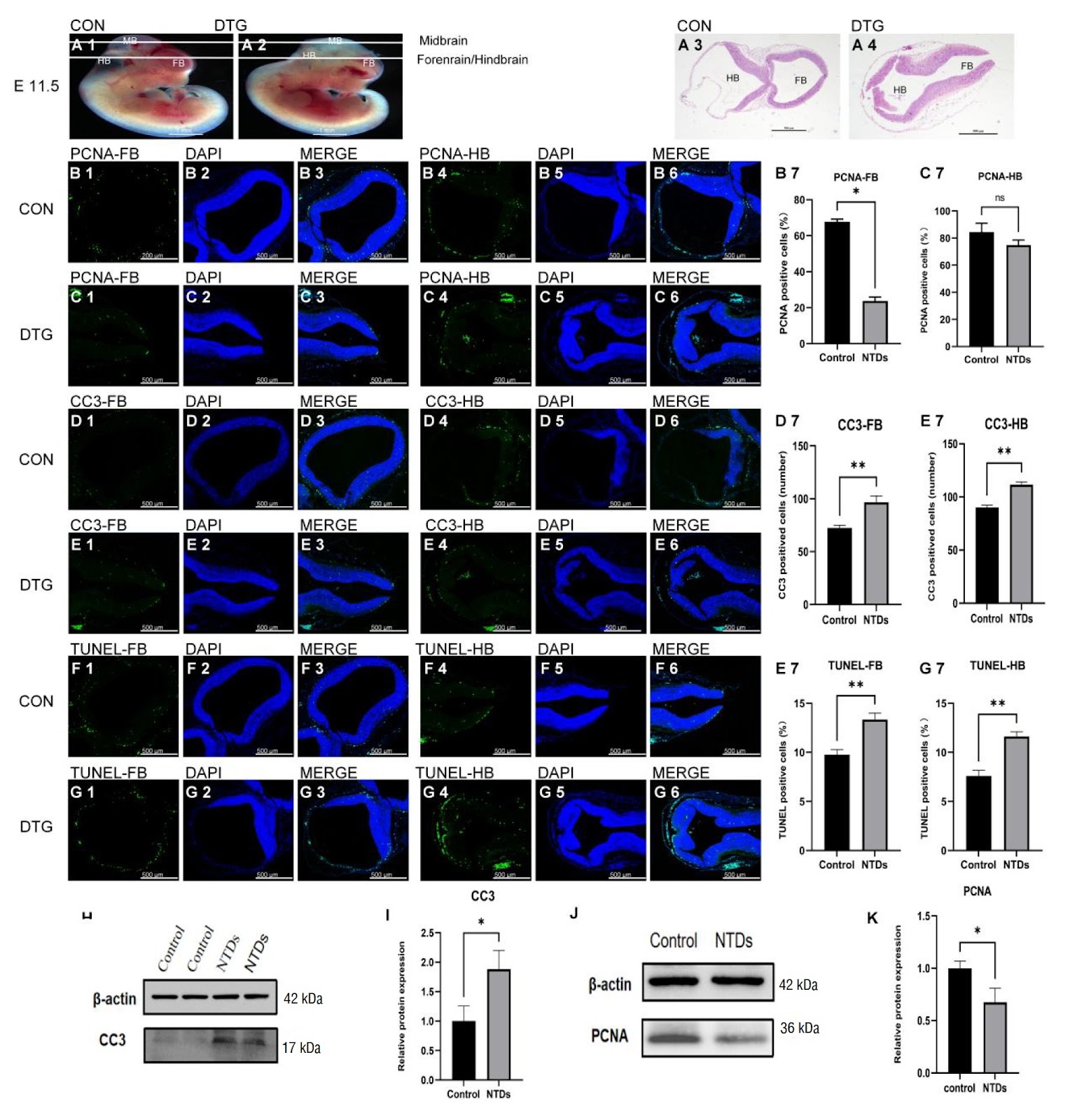
Dolutegravir (DTG) disrupts mouse embryonic development in a dose-dependent manner, culminating in neural-tube defects (NTDs). Using whole embryo culture (WEC), mouse embryos at embryonic day 8.5 (E8.5) are cultured for 24–48 h with 8, 10, or 12 μM DTG. The results reveal that higher DTG concentrations dose-dependently disrupt yolk sac development and markedly increase the frequency of NTDs. In vivo NTD models are generated by intraperitoneally injecting DTG at a dose of 7.5 mg/kg, and the resulting embryos exhibit disrupted yolk sac blood circulation, embryonic growth restriction, and malformations. Mechanistic studies suggest that DTG contributes to NTDs by inducing apoptosis: DTG exposure activates the Nrf2-SOD1/CAT antioxidant axis, yet it culminates in increased apoptosis and suppressed proliferation, ultimately impairing yolksac vasculogenesis and neuralepithelial closure, thereby producing NTDs. This study provides new evidence for assessing the potential risk of DTG in embryonic development and highlights the need to re-evaluate its clinical safety in future applications.

Read Full Abstract10.3724/abbs.2025142
Exploring DNA topoisomerase II alpha in adrenocortical carcinoma through multi-omics analysis: a potential biomarker and therapeutic targetGraphical AbstractVerified
Acta Biochimica et Biophysica Sinica

Exploring DNA topoisomerase II alpha in adrenocortical carcinoma through multi-omics analysis: a potential biomarker and therapeutic target

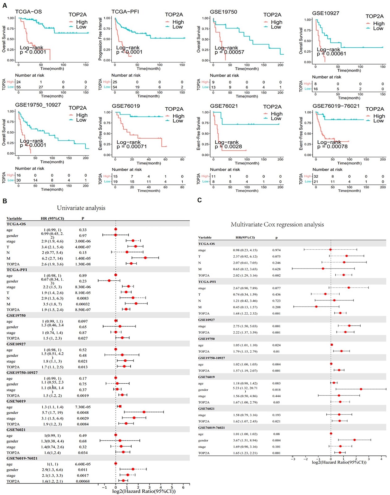
Adrenocortical carcinoma (ACC) is a rare but aggressive cancer. Recent studies identified DNA Topoisomerase II Alpha (TOP2A) as a potential biomarker for ACC, which can provide new avenues for targeted therapy and improve clinical outcomes. This study aims to elucidate the role of TOP2A in ACC by exploring its prognostic value and identifying inhibitors for ACC therapy. Utilizing RNA sequencing data, mutation data, and clinical information from The Cancer Genome Atlas (TCGA-ACC) and additional datasets from the Gene Expression Omnibus (GEO), differential expression and prognostic analyses are conducted to assess the significance of TOP2A in ACC. Immunohistochemistry and cell assays, including cell viability, colony formation, and transwell assays, are conducted to validate the oncogenic effects of TOP2A. The ā€œIOBRā€ R package is used to examine the relationship between TOP2A expression and CD8+ T-cell infiltration. The CMap platform is used to identify potential TOP2A inhibitors. In vivo assays verify the therapeutic effect of TOP2A inhibitors on ACC. Our findings indicate that TOP2A is significantly overexpressed in ACC and is associated with poor prognosis. Immunohistochemistry and cell assays confirm the oncogenic role of TOP2A. Furthermore, distinct gene expression patterns related to different TOP2A expression levels are identified, influencing the response to immunotherapy. Potential inhibitors targeting TOP2A are discovered, and the therapeutic effects of resminostat and etoposide are confirmed via in vivo assays, suggesting new therapeutic strategies for ACC treatment. In conclusion, TOP2A serves as a crucial biomarker in ACC and is associated with adverse clinical outcomes and a diminished immune response. The identification of potential inhibitors against TOP2A opens new avenues for the development of targeted therapies for ACC patients.

Read Full Abstract10.3724/abbs.2025144
Immunopathological characteristics and therapeutic effects of UC-MSCs in a pigeon breeder’s lung mouse modelGraphical AbstractVerified
Acta Biochimica et Biophysica Sinica

Immunopathological characteristics and therapeutic effects of UC-MSCs in a pigeon breeder’s lung mouse model

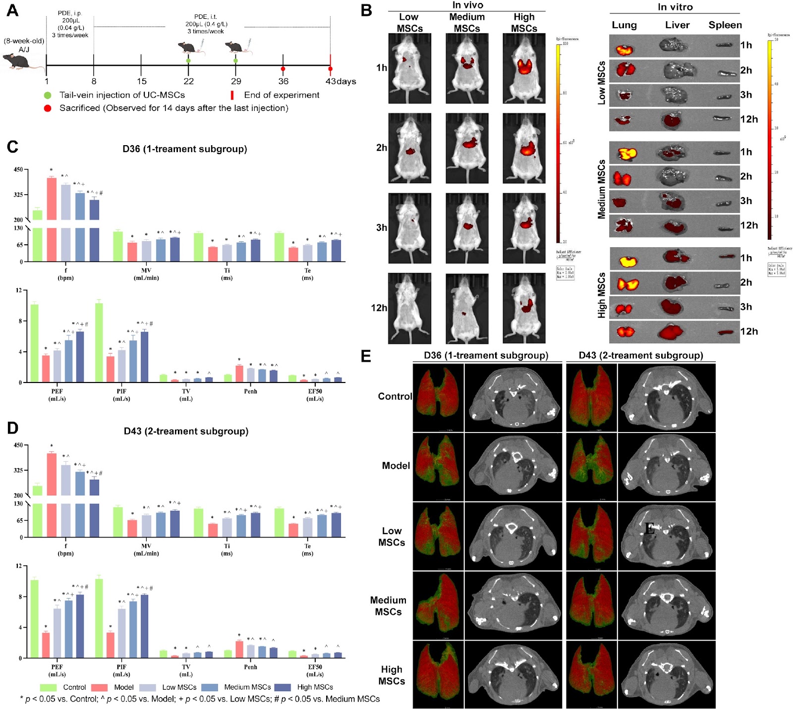
Hypersensitivity pneumonitis (HP), including pigeon breeder’s lung (PBL), often progresses from acute inflammation to fibrosis, impairing lung function and limiting targeted therapeutic strategies. Mechanistic studies on PBL progression are limited by the lack of preclinical animal models and a predominant focus on patient data. This study explores the immunopathological characteristics of all stages of PBL in mice and evaluates the therapeutic potential of human umbilical cord-derived mesenchymal stem cells (UC-MSCs) during the non-fibrotic stage. PBL models are created in A/J mice through tracheal instillation of pigeon dropping extract (PDE) protein powder. Different doses (0.4 Ɨ 106, 0.8 Ɨ 106, and 1.6 Ɨ 106 cells per animal) and frequencies (1–2 times) are administered to the model. The immunopathological characteristics of PBL and the therapeutic effects of UC-MSCs are assessed using micro-CT, pulmonary function, histopathology, cell counts in BALF, HYP levels, inflammatory factor levels, immunohistochemistry, and fibrosis marker expression in lung tissues. The results show that PDE exposure consistently impairs pulmonary function and increases the levels of inflammation and fibrosis markers as the disease progresses. Model mice experience non-fibrotic stages (acute inflammation) from days 0–36, mild fibrosis from days 37–77, and severe fibrosis from day 78 onwards. UC-MSCs, particularly at the highest dose (1.6 Ɨ 106 cells), effectively treat non-fibrotic PBL by improving pulmonary function (lung ventilation area recovers) and reducing inflammation and fibrosis. This study successfully establishes PBL mouse models reflecting both the acute (inflammatory) and chronic (fibrotic) stages, and UC-MSCs have the potential to delay fibrosis, providing new therapeutic options for PBL and other inflammation-induced lung fibrotic diseases.

Read Full Abstract10.3724/abbs.2025010
Rapid detection of Escherichia coli in bloodstream infection via CRISPR-Cas9 engineered reporter phage T7::Nluc and microfluidic chip platformGraphical AbstractVerified
Acta Biochimica et Biophysica Sinica

Rapid detection of Escherichia coli in bloodstream infection via CRISPR-Cas9 engineered reporter phage T7::Nluc and microfluidic chip platform

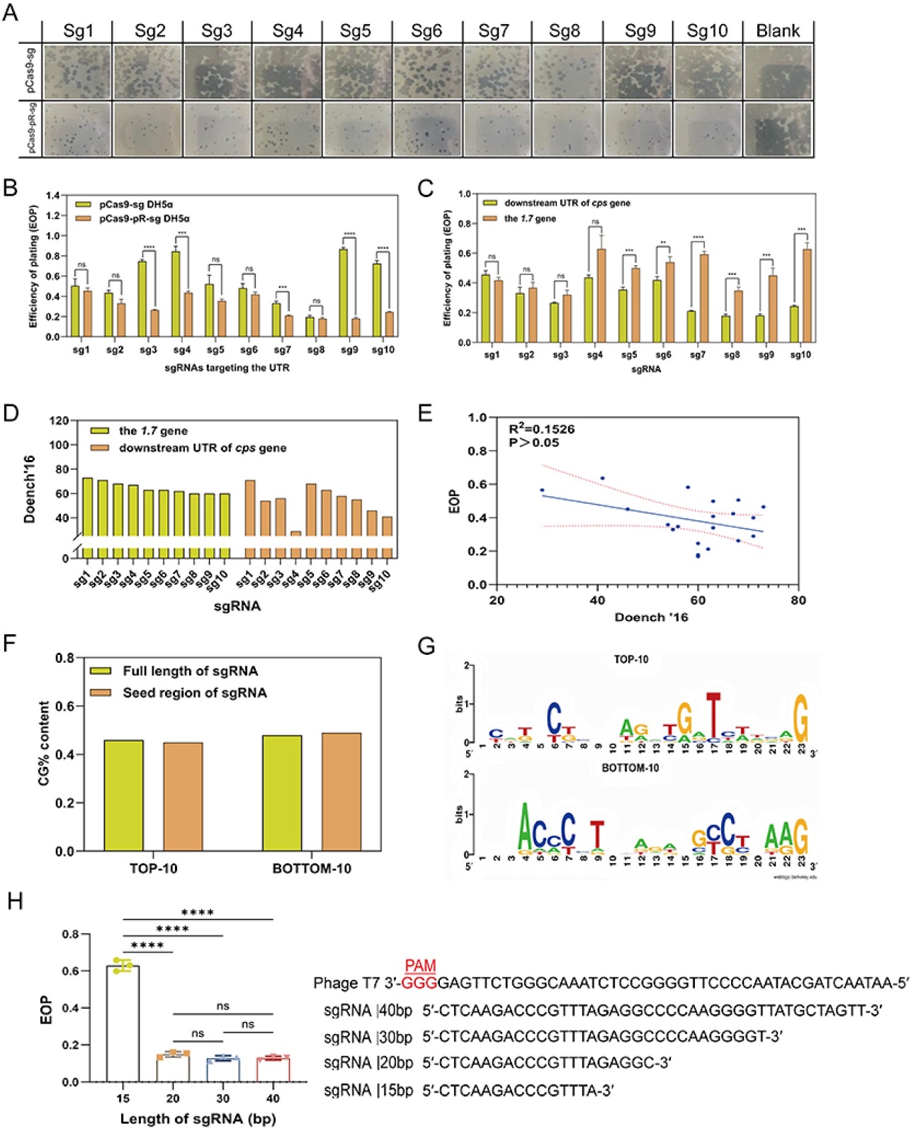
Rapid identification of pathogens responsible for bloodstream infection is critical for early intervention and effective treatment. Reporter phages, which are known for their exceptional sensitivity and specificity in pathogen detection, have garnered significant interest. In this study, we systematically evaluate phage genome editing strategies that combine homologous recombination with the CRISPR-Cas9 system. We investigate the impacts of homologous arm length, sgRNA activity, target site, and plasmid interactions on editing efficiency. Our results demonstrate that successful genome editing depends on both sufficient cleavage pressure and optimal homologous arm length, particularly when using low-activity sgRNAs. On the basis of these findings, we develop a highly efficient gene editing strategy TPMSR (triple-plasmid-mediated synchronous recombination) that overcomes the limitations of conventional methods that rely on high-activity sgRNA and restricted editing sites. Using the TPMSR strategy, we integrate the Nluc gene into phage T7, generating the reporter phage T7::Nluc, which is then incorporated into a microfluidic chip. Validation with 51 clinical isolates demonstrates outstanding sensitivity, specificity, and accuracy in detecting Escherichia coli in blood within 1.5 h at concentrations less than 30 CFU/mL. This study presents a robust strategy for phage genome engineering and develops a promising method for the rapid diagnosis of bloodstream infections caused by E. coli.

Read Full Abstract10.3724/abbs.2025150
Targeting USP2 induces degradation of PML-RARα with or without drug-resistant mutations in acute promyelocytic leukemiaGraphical AbstractVerified
Acta Biochimica et Biophysica Sinica

Targeting USP2 induces degradation of PML-RARα with or without drug-resistant mutations in acute promyelocytic leukemia

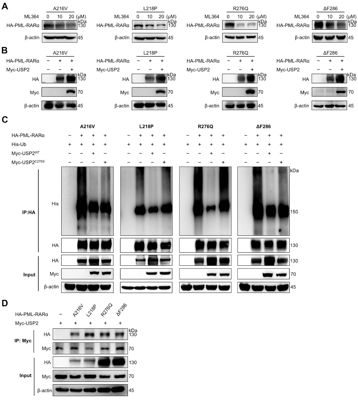
Despite the high efficacy of all-trans retinoic acid (ATRA) and arsenic trioxide (ATO) in treating acute promyelocytic leukemia (APL), approximately 10%–20% of patients develop drug resistance due to mutations in PML-RARα and other factors. Here, we find that inhibition of USP2 with ML364 or USP2 silencing reduces PML-RARα protein levels in both ATRA-sensitive and ATRA-resistant APL cells, and this effect is reversed by proteasome inhibition. Conversely, USP2 overexpression enhances PML-RARα stability. Mechanistically, USP2 interacts with and deubiquitinates PML-RARα, including its drug-resistant mutants. Consistent with PML-RARα degradation, ML364 treatment significantly induces apoptosis in APL cell lines and primary leukemia cells. In conclusion, this study identifies USP2 as a novel deubiquitinating enzyme for PML-RARα and highlights USP2 inhibition as a potential therapeutic strategy for APL with PML-RARα mutations.

Read Full Abstract10.3724/abbs.2025135
Iron overload mediates cytarabine resistance in AML by inhibiting the TP53 signaling pathwayGraphical AbstractVerified
Acta Biochimica et Biophysica Sinica

Iron overload mediates cytarabine resistance in AML by inhibiting the TP53 signaling pathway

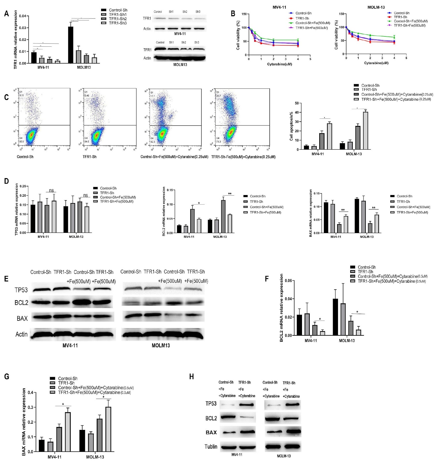
Currently, chemotherapy remains the primary treatment for acute myeloid leukemia (AML). Drug resistance in AML cells is a critical factor contributing to the failure of chemotherapy remission and subsequent relapse. Iron overload frequently occurs in AML patients because of hematopoietic suppression or supportive blood transfusion therapy. Previous studies have indicated that iron overload may promote the progression of AML; however, the underlying mechanisms remain unclear. Our results demonstrate that, compared with TP53-wild-type AML cells, TP53-mutant AML cells exhibit increased resistance to cytarabine-induced cytotoxicity. Moreover, reducing TP53 expression in wild-type AML cells diminishes their sensitivity to cytarabine. The TP53 signaling pathway is essential for mediating cytarabine-induced apoptosis in AML cells. In this study, an iron overload model in AML cells via the use of ferric citrate is constructed. Our data indicate that iron overload can suppress the TP53/BCL2/BAX signaling pathway, counteracting cytarabine-induced apoptosis. In TP53 wild-type AML cells, TFR1 participates in iron-mediated resistance to cytarabine by regulating the entry of iron into the cells. These findings provide a foundation for further exploration of the molecular mechanisms involved in AML resistance to cytarabine.

Read Full Abstract10.3724/abbs.2025027
A novel method to increase transgene expression and the stability of gene therapy-associated episomal vectorsGraphical AbstractVerified
Acta Biochimica et Biophysica Sinica

A novel method to increase transgene expression and the stability of gene therapy-associated episomal vectors

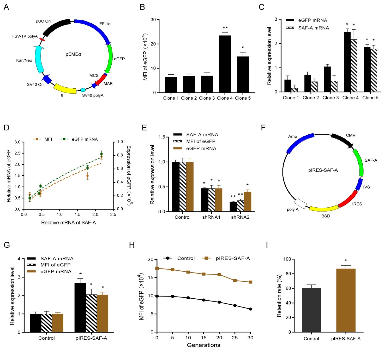
Non-viral episomal vectors offer a safe and attractive alternative to viral and integrated vectors by avoiding insertional mutagenesis and position effects, making them ideal expression vectors for gene therapy. The first non-viral episomal vector, pEPI-1, which is based on the full-length scaffold/matrix attachment region (S/MAR), was established by Piechaczek et al. The full-length S/MAR element interacts with the nuclear matrix via the matrix protein, e.g. SAF-A, thereby maintaining mitotic stability and transgene expression. Several strategies, including optimization of the vector backbone and promoter and incorporation of chromatin-modifying elements, have been used to increase expression levels and stability. In our previous work, we constructed the novel vector pEGFP-C1-M on the basis of S/MAR characteristic motifs (only 375 bp). This vector, which is shorter than the prototype episomal vector pEPI-1, resulted in relatively higher transgene expression. Building on the pEGFP-C1-M vector, we further constructed the episomal vector pEMEα with the EF-1α promoter and demonstrated that pEMEα maintained higher transgene expression, stability and copy number. The transgene expression levels of episomal vectors are correlated with gene copy number, that is, the number of plasmid episomes on the host cell chromosome. Previous studies have shown that the episomal maintenance of pEPI-1 vectors is mediated primarily by SAF-A. While the role of SAF-A in maintaining mammalian pEPI-1 episomal vectors has been well established, it remains unknown whether the overexpression of SAF-A promotes transgene expression and stability and whether the 375 bp MAR characteristic sequence retains its interaction with SAF-A. In the present study, we first evaluated whether transgene expression is positively correlated with the expression level of SAF-A. The non-viral episomal vector pEMEα was used as the gene of interest (GOI) vector and was subsequently transfected into CHO-K1 cells using the Lipofectamine 2000 reagent. The cells were cultured in medium containing 800 μg/mL geneticin (G418) 48 h post-transfection, and the G418 concentration was then reduced to 400 μg/mL to obtain monoclonal cell lines using the limiting dilution method. Five monoclonal cell clones were selected, and the eGFP expression levels, measured as the mean fluorescence intensity (MFI), were (6.5 ± 1.0) Ɨ 104, (6.8 ± 0.9) Ɨ 104, (7.0 ± 1.4) Ɨ 104, (23.5 ± 1.2) Ɨ 104 and (14.9 ± 0.17) Ɨ 104 for Clones 1–5, respectively. qPCR analysis of Clones 1–5 revealed that the relative mRNA levels of SAF-A and eGFP were 0.16 ± 0.13, 0.43 ± 0.11, 0.46 ± 0.23, 2.17 ± 0.41, 1.78 ± 0.15 and 0.51 ± 0.16, 0.71 ± 0.12, 1.05 ± 0.09, 2.47 ± 0.14, and 1.86 ± 0.10, respectively. Our results indicated that eGFP mRNA and protein expression levels are positively correlated with SAF-A mRNA level. To further verify the relationship between SAF-A expression and transgene expression, two shRNA plasmids targeting SAF-A (shRNA1: 5′-GCCACCTGTTGAAGAAGAAGA-3′, and shRNA2: 5′-GCTGGAGGAAGAGCTTCTTAT-3′) which were obtained from Shanghai GenePharma Co., Ltd. were designed and transfected into stable cell pools with the pEMEα vector. qPCR analysis revealed that the relative SAF-A mRNA levels in the shRNA1 and shRNA2 vectors were 0.47 ± 0.01 and 0.19 ± 0.02, respectively, indicating successful downregulation of SAF-A expression. Moreover, flow cytometry and qPCR revealed that, compared with those in the control group, the relative protein and mRNA levels of eGFP were reduced by 0.47- and 0.23-fold, and 0.47- and 0.40-fold in the pools of cells transfected with the shRNA1 and shRNA2 vectors, respectively. On the basis of the above results, the SAF-A overexpression vector pIRES-SAF-A was constructed and transfected into CHO-K1 cells, and the cells were cultured in blasticidin-containing medium 48 h after transfection to obtain stable cell pools. The stable cell pools overexpressing SAF-A were subsequently transfected with the pEMEα vector. Stable cell pools coexpressing SAF-A and GOI were selected, and the relative mRNA levels of SAF-A and eGFP were analyzed. qPCR analysis revealed that the relative mRNA levels of SAF-A and eGFP in the pools of cells overexpressing SAF-A were 2.69-fold and 2.05-fold higher than those in the control group, respectively. Flow cytometry also revealed a 2.07-fold increase in MFI in stable cell pools overexpressing SAF-A compared with the control group. To assess the long-term stability of transgene expression, we measured the MFI in st
Read Full Abstract10.3724/abbs.2025104