🧬 SinoBioData Academic Portal
📚 Peer-Reviewed Translated Literature

All Biomedical & Clinical Articles (Page 45)

Browse complete peer-reviewed translations from top Chinese biomedical, oncology, and genomics journals. Read verified previews and download full authentic clinical reports.

Published Research Papers

Showing 24 of 1542 peer-reviewed translated articles (Page 45 of 65)

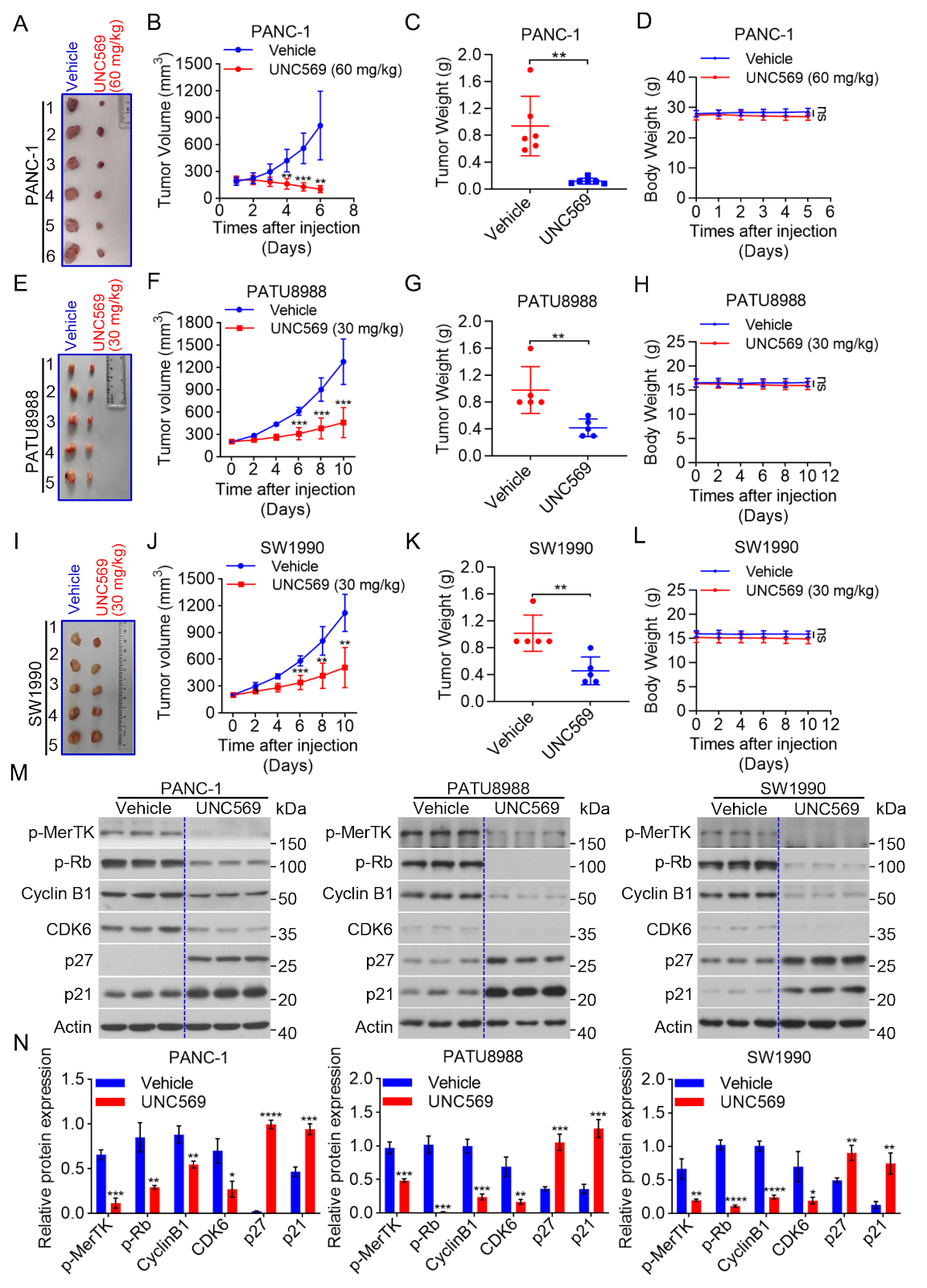
MerTK inhibition by UNC569 triggers DNA damage and JNK/p38 MAPK cascade-driven apoptosis in pancreatic cancerGraphical AbstractVerified
Acta Biochimica et Biophysica Sinica2026

MerTK inhibition by UNC569 triggers DNA damage and JNK/p38 MAPK cascade-driven apoptosis in pancreatic cancer

Pancreatic ductal adenocarcinoma (PDAC) is a malignancy with increasing mortality rates and remains a major clinical challenge due to its aggressive progression and limited therapeutic options. Therefore, the identification of early biomarkers and the development of effective targeted therapies are critically needed. MerTK, a receptor tyrosine kinase aberrantly expressed in various cancers, can be selectively inhibited by UNC569, a small-molecule antagonist with demonstrated efficacy in hematologic malignancies. This study shows that UNC569 potently suppresses PDAC cell proliferation and clonogenic growth, inhibits migration and invasion by attenuating epithelial-mesenchymal transition, and enhances the sensitivity of PDAC cells to Gemcitabine while promoting apoptosis. Mechanistically, UNC569 induces DNA damage-mediated G2/M phase arrest and activates JNK/p38 mitogen-activated protein kinase-dependent apoptotic signaling. Collectively, these results establish MerTK as a promising therapeutic target in PDAC and highlight the translational potential of UNC569 as a dual-pathway inhibitor for PDAC treatment.

Read Full Abstract10.3724/abbs.2026048
Metabolic Reprogramming in Cardiometabolic Syndrome: Mechanisms, Biomarkers, and Therapeutic ApproachesGraphical AbstractVerified
Acta Biochimica et Biophysica Sinica2026

Metabolic Reprogramming in Cardiometabolic Syndrome: Mechanisms, Biomarkers, and Therapeutic Approaches

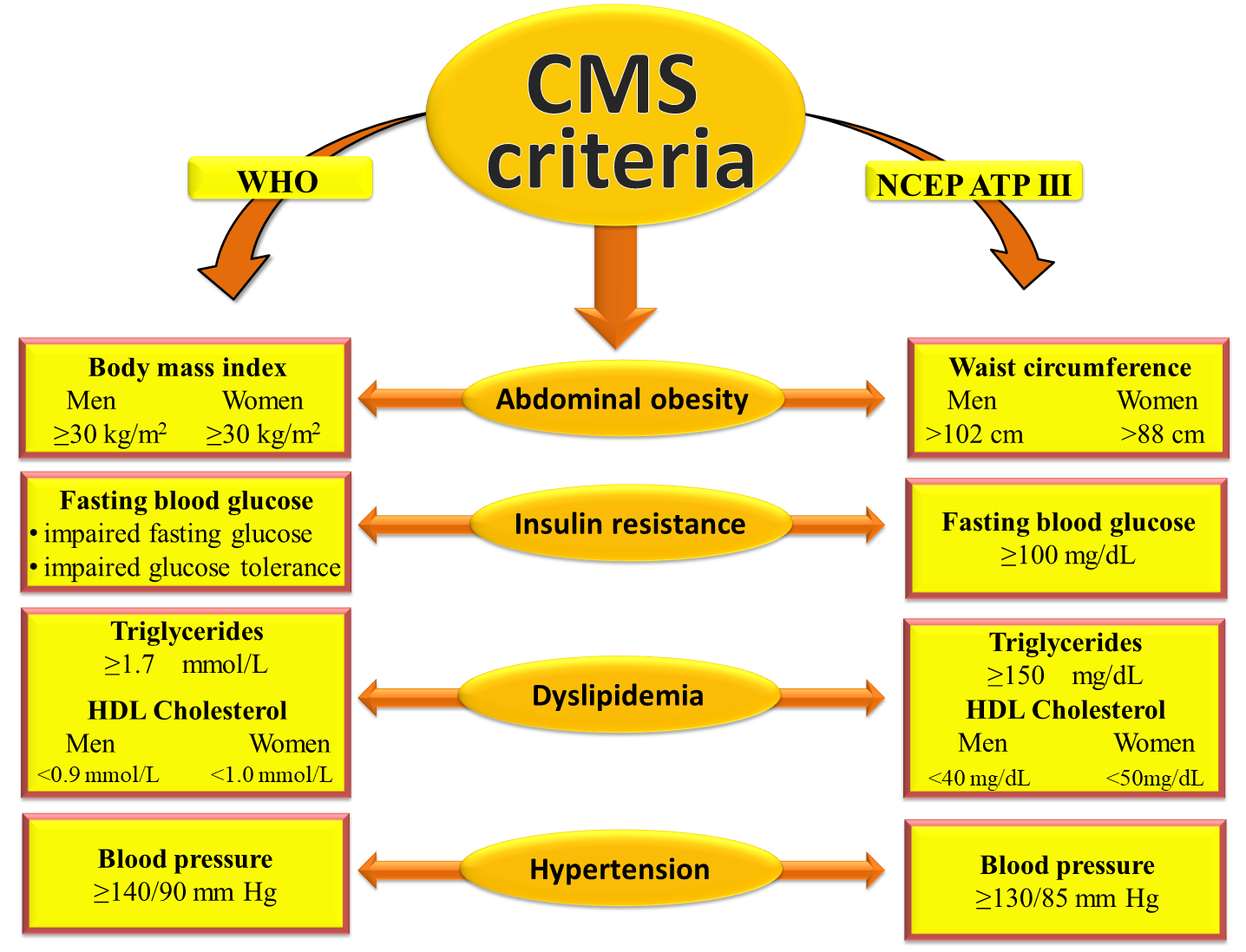
Cardiometabolic syndrome (CMS), a combination of central obesity, insulin resistance, dyslipidemia, and hypertension, accounts for a significant portion of the global incidence of type 2 diabetes and cardiovascular disease. Traditionally, hormonal and hemodynamic dysregulation have been considered the primary causes of CMS. However, increasing evidence shows that metabolic reprogramming, which involves long-lasting, tissue-specific changes in cellular metabolism, is a common cause of the initiation and progression of CMS. This review examines the systemic metabolic alterations, the molecular pathways facilitating these modifications, and the transformative impact of multiomics platforms on the discovery of novel biomarkers and therapeutic targets. We also discuss drugs that can help restore metabolic flexibility and stop disease progression.

Read Full Abstract10.3724/abbs.2025255
Bioinformatics classification of the MgtE Mg2+ channel and de novo protein design for the stabilization of its novel subclassGraphical AbstractVerified
Acta Biochimica et Biophysica Sinica

Bioinformatics classification of the MgtE Mg2+ channel and de novo protein design for the stabilization of its novel subclass

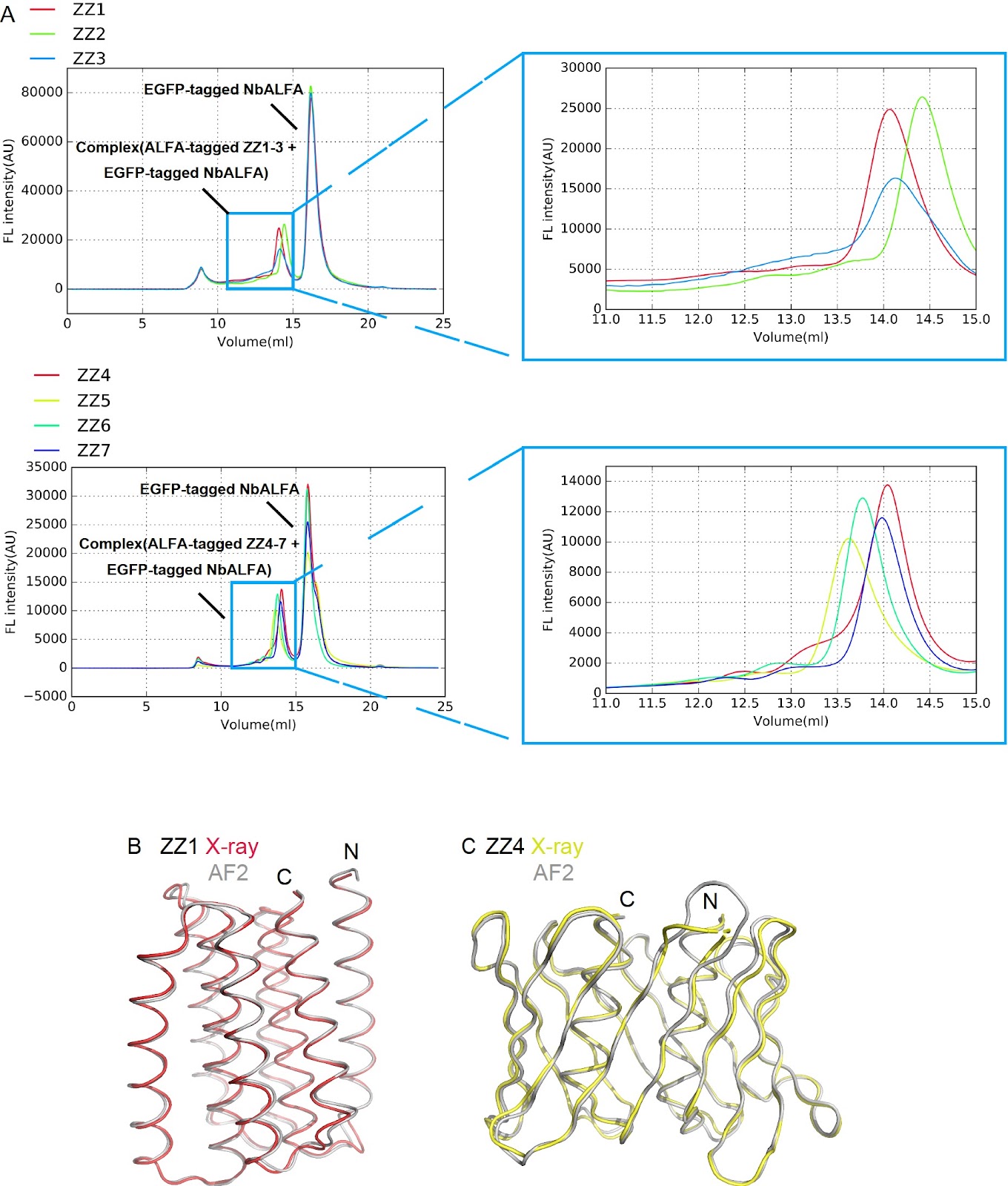
MgtE channels play crucial roles in Mg2+ homeostasis and are implicated in bacterial survival under antibiotic exposure. Previous structural and biophysical studies have focused predominantly on Thermus thermophilus MgtE, leaving the structural and mechanistic diversity of MgtE family proteins largely unexplored. In this study, via a genome mining approach, we identify diverse MgtE homologs, including a novel subclass termed the “mini-N type”, which lacks the canonical cytoplasmic N and CBS domains but possesses a unique small N-like domain. Despite extensive expression screening, mini-N-type homologs cannot be stably purified. To address this issue, we design a series of de novo proteins and determine their crystal structures. A selected de novo protein is fused to a mini-N-type MgtE, enabling successful purification and preliminary cryo-EM imaging. Our findings demonstrate that de novo-designed protein fusions serve as powerful tools for stabilizing and purifying otherwise unstable membrane proteins, opening new avenues for structural and functional studies of otherwise inaccessible membrane proteins.

Read Full Abstract10.3724/abbs.2025224
ATP-citrate lyase: carcinogenesis and therapeutic advancesGraphical AbstractVerified
Acta Biochimica et Biophysica Sinica2026

ATP-citrate lyase: carcinogenesis and therapeutic advances

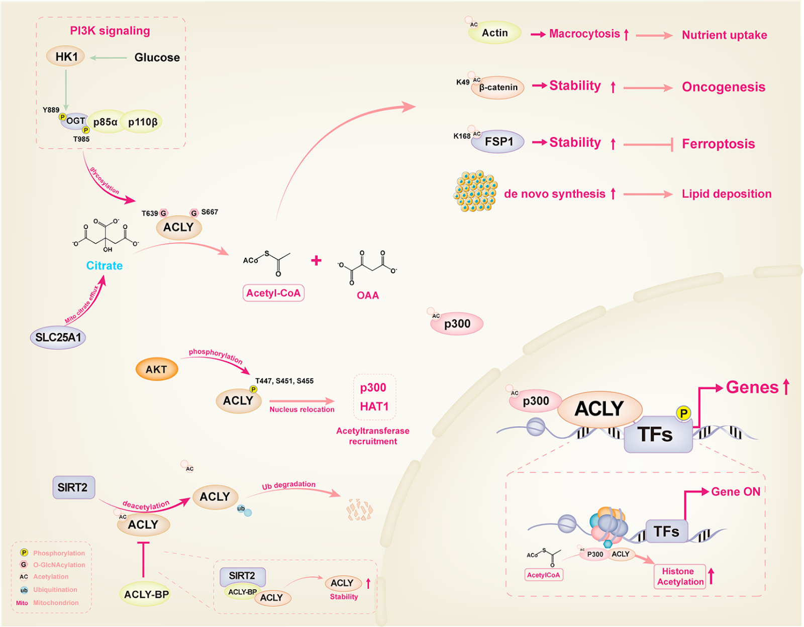
ATP citrate lyase (ACLY) is involved in acetyl-coenzyme A synthesis and protein acetylation, thereby increasing lipid metabolism and altering protein metabolism to affect cellular metabolism. Additionally, ACLY is associated with various biological and pathological functions, especially regarding tumorigenesis. It facilitates the progression of various cancer types, including liver, lung, breast, prostate, and colorectal cancers. Mechanisms underlying ACLY-mediated carcinogenesis are under investigation and may not be limited to energy metabolism and biosynthesis. Acetylation modification of specific signaling molecules and transcription factors is considered a potential mechanism of ACLY-mediated tumorigenesis and offers novel insights and potential targets for the clinical treatment of tumors. Furthermore, the antitumor effect of pharmacological ACLY-inhibiting agents, including various small molecules or naturally active compounds, has been reported, albeit their practical application in clinical settings remains limited. This study aims to comprehensively review the oncogenic role of ACLY, with a focus on major collaborators and regulatory genes.

Read Full Abstract10.3724/abbs.2026028
Atractylenolide I mitigates Alzheimer’s disease pathology in ApoE–/– mice via ARG1/nNOS axis and lipid homeostasis regulationGraphical AbstractVerified
Acta Biochimica et Biophysica Sinica

Atractylenolide I mitigates Alzheimer’s disease pathology in ApoE–/– mice via ARG1/nNOS axis and lipid homeostasis regulation

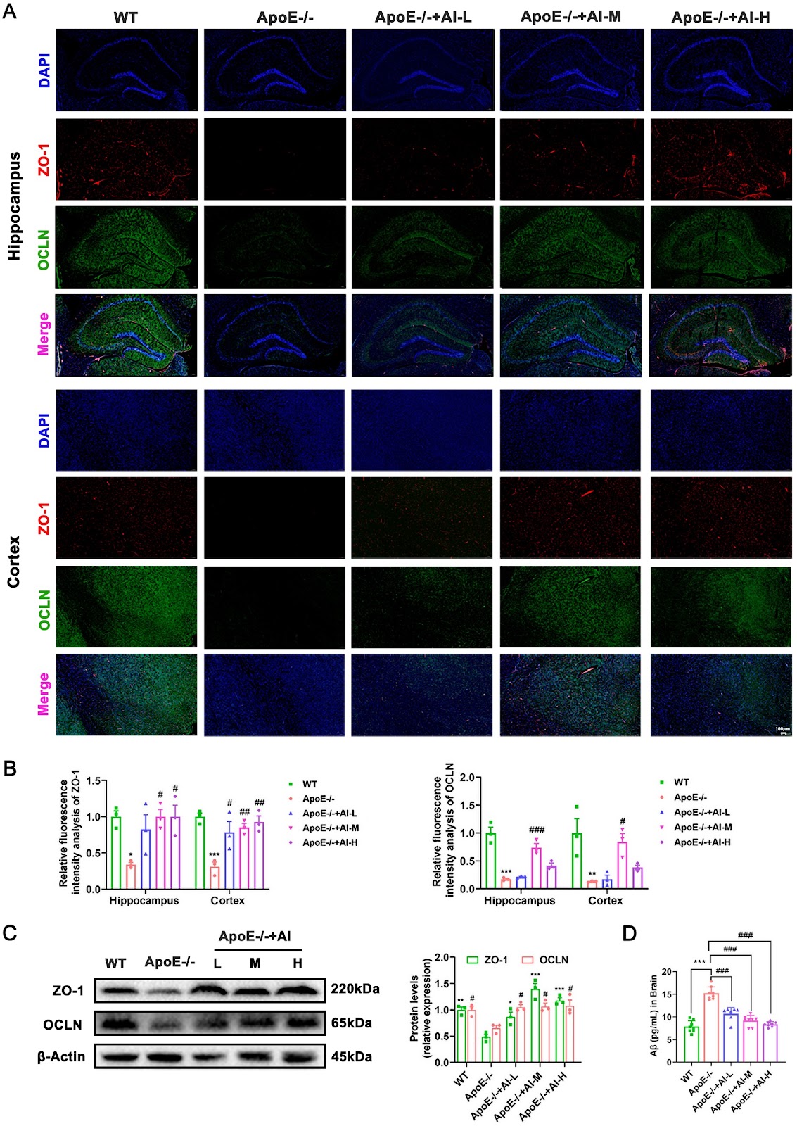
Apolipoprotein E (ApoE) serves as a critical molecular nexus between Alzheimer’s disease (AD) and atherosclerosis, two age-associated inflammatory disorders that share vascular pathology, amyloid-beta (Aβ) deposition, and lipid dysregulation. Atractylenolide I (AI), a promising therapeutic candidate derived from Atractylodes macrocephala Koidz., exhibits multimodal bioactivities with demonstrated anti-inflammatory and neuroprotective properties. To explore its therapeutic potential against AD pathology, we use high-fat diet (HFD)-fed ApoE knockout (ApoE–/–) mice treated with or without AI for 12 weeks. Integrated bioinformatics analyses and experimental validation reveal that AI treatment markedly attenuates systemic lipid dyshomeostasis, particularly cerebral lipid deposition, suppresses neuroinflammation via downregulation of M1 macrophage polarization markers, and restores cognitive function through neuronal preservation in hippocampal regions. Mechanistically, AI orchestrates cholesterol efflux by up-regulating ATP-binding cassette transporter A1 (ABCA1) and liver X receptor (LXR) expression, while concurrently modulating the abundance of arginine biosynthesis metabolites (urea, malic acid, and creatinine) to rebalance neurovascular homeostasis. Notably, western blot and RT-qPCR analyses reveal that AI differentially regulates key enzymes including arginase 1 (ARG1) and simultaneously upregulates the expression of neuronal nitric oxide synthase (nNOS). Further molecular docking and surface plasmon resonance (SPR) analyses confirm the direct binding of AI to ARG1, indicating a novel neuroprotective mechanism involving the modulation of arginine metabolism. These findings delineate the pleiotropic effects of AI against AD pathology and establish a preclinical foundation for the development of AI-based therapeutics targeting neurodegenerative-cardiovascular comorbidities.

Read Full Abstract10.3724/abbs.2026055
Structural and functional insights into the distinct DNA recognition mechanisms of the terminase small subunit TerS from cyanophagesGraphical AbstractVerified
Acta Biochimica et Biophysica Sinica

Structural and functional insights into the distinct DNA recognition mechanisms of the terminase small subunit TerS from cyanophages

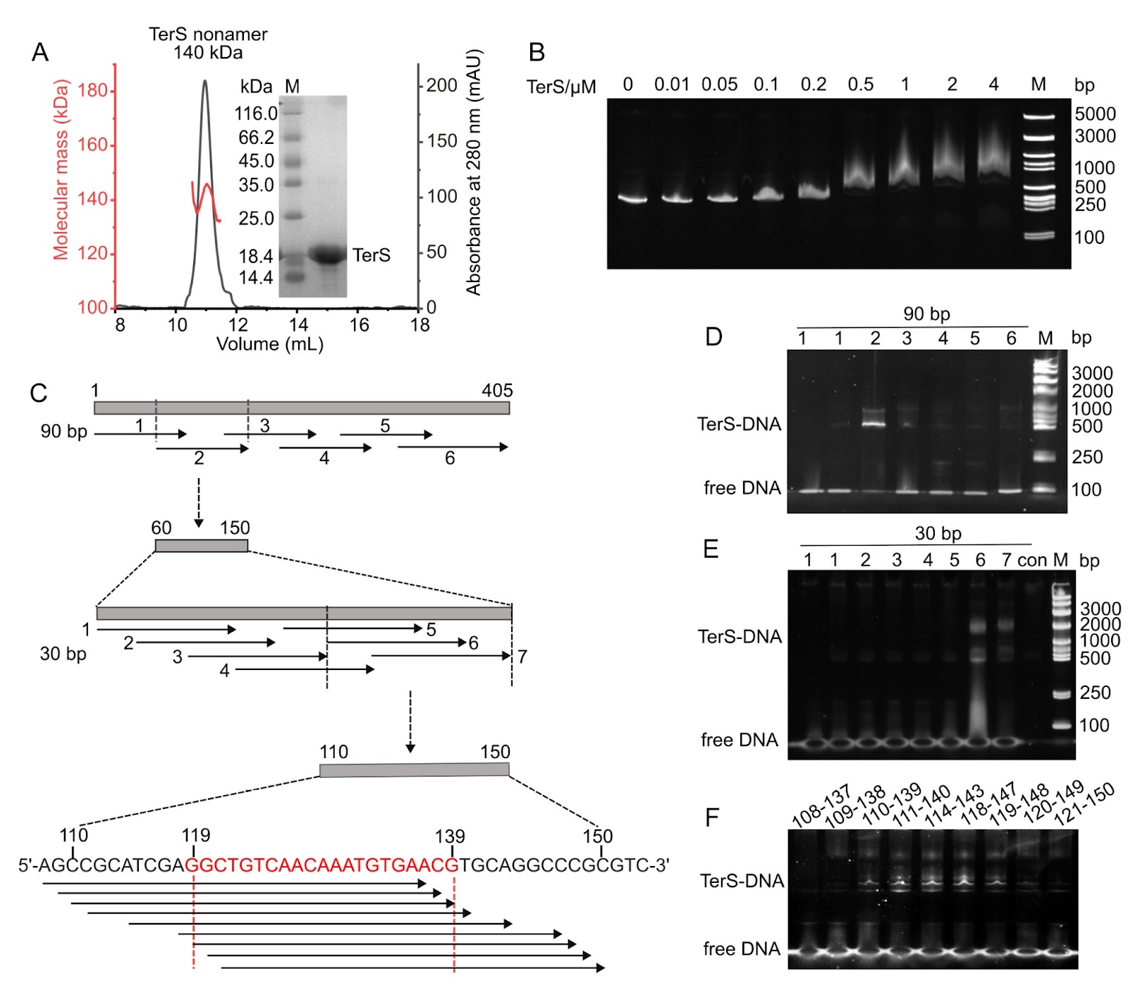
Efficient genome packaging is a critical step in the phage life cycle, directly influencing the viral maturation and infectivity. In tailed phages, this process is driven by a packaging motor composed of a portal protein and a terminase complex. The terminase complex usually consists of a large subunit (TerL) and a small subunit (TerS), which cooperate to recognize, cleave, and translocate genomic DNA into the capsid. However, due to the remarkable diversity and complexity of phage packaging systems, the molecular mechanisms governing TerS-mediated DNA recognition remain poorly understood. Here, we report the 3.51 Å cryo-electron microscopy structure of the TerS from the short-tailed cyanophage Pam5, which infects the host Pseudanabaena mucicola Chao 1806. Pam5 TerS assembles into a nonameric ring with a radially symmetric spiral architecture. Biochemical assays show that Pam5 TerS recognizes the genomic DNA via a specific interaction between the N-terminal helix-turn-helix (HTH) domain of TerS and a 21-bp DNA sequence within the terS gene. In contrast, the TerS from another short-tailed cyanophage, Pam1, which infects the same host, binds to DNA in a sequence-independent manner. These findings reveal that cyanophages, even infecting the same host, could adopt two distinct DNA recognition strategies: HTH-mediated sequence-dependent or sequence-independent modes. This work provides structural and mechanistic insights into the diverse DNA-recognition strategies of TerS and advances our understanding of the evolutionary plasticity of viral genome packaging mechanisms.

Read Full Abstract10.3724/abbs.2026042
Luteolin reprograms macrophage polarization via the STING-TBK1 pathway to accelerate bone repairGraphical AbstractVerified
Acta Biochimica et Biophysica Sinica2026

Luteolin reprograms macrophage polarization via the STING-TBK1 pathway to accelerate bone repair

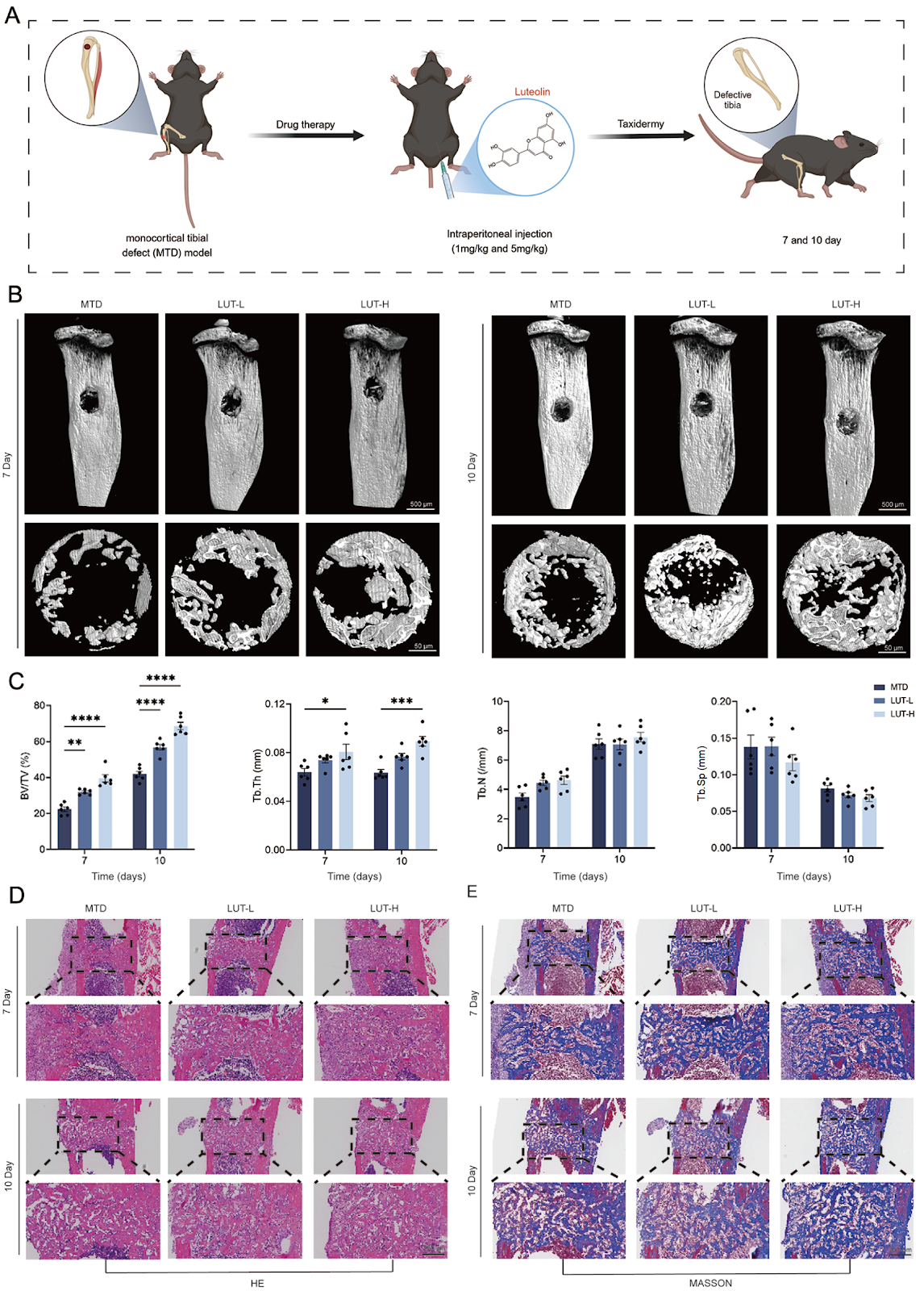
Macrophages play a pivotal role in bone regeneration, making their polarization a key target for immune regulation and therapeutic intervention. Modulating macrophage polarization represents a promising strategy for enhancing bone repair. Luteolin, a plant-derived flavonoid with well-documented anti-inflammatory properties, has been explored for its role in bone repair. However, its specific effects on macrophage polarization in bone repair remain unclear. This study investigates the role of luteolin in macrophage polarization and its underlying mechanisms. Our findings demonstrate that luteolin promotes M2 polarization while suppressing M1 polarization, as indicated by a reduction in the expression of pro-inflammatory markers, including IL-6 and iNOS, and an increase in the expression of anti-inflammatory factors, such as CD206, IL-10, and TGF-β. Mechanistically, luteolin inhibits STING oligomerization, thereby suppressing the STING-TBK1 pathway and mitigating downstream inflammatory responses. In vivo, in a mouse tibial bone defect model, luteolin effectively alleviates inflammation, facilitates angiogenesis, enhances collagen deposition, and improves bone density. Collectively, these findings highlight the potential of luteolin as a therapeutic agent for bone repair by modulating macrophage polarization and inhibiting STING-TBK1 signaling.

Read Full Abstract10.3724/abbs.2026021
Structural basis for the FOXM1 DNA binding domain to specific dsDNA substrateGraphical AbstractVerified
Acta Biochimica et Biophysica Sinica2026

Structural basis for the FOXM1 DNA binding domain to specific dsDNA substrate

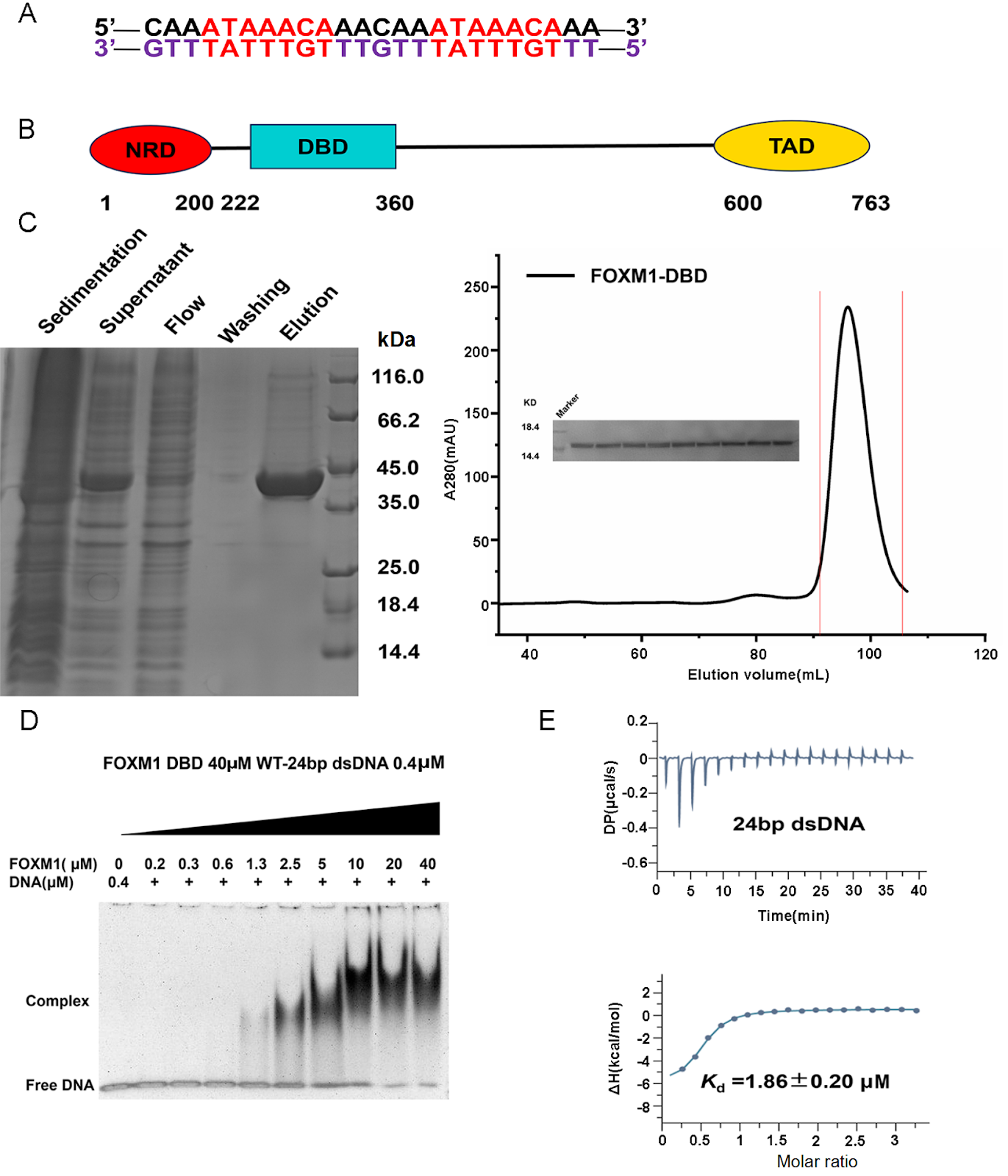
Forkhead box protein M1 (FOXM1) is a key transcription factor that regulates cell cycle progression and is frequently overexpressed in human cancers, driving tumor proliferation and therapy resistance. FOXM1 recognizes the canonical forkhead response element (FKH motif, RYAAAYA) through its conserved DNA-binding domain (DBD). Here, we report the high-resolution crystal structure of the FOXM1-DBD in complex with a double-stranded DNA substrate containing two FKH motifs. The structure reveals that FOXM1-DBD adopts the canonical winged-helix fold, with the third α-helix (α3) inserted into the DNA major groove to mediate sequence-specific recognition. Within this helix, Asn283, Arg286, and His287 form an essential triad that engages DNA bases through specific hydrogen bonds and hydrophobic interactions. Using structure-guided mutagenesis of key DNA-interacting residues combined with biophysical validation by isothermal titration calorimetry (ITC) and DNA binding assessment via electrophoretic mobility shift assay (EMSA), we confirm the functional importance of these residues and uncover position-dependent tolerance to base substitutions within the FKH motif. Furthermore, we demonstrate that FOXM1 overexpression promotes cell proliferation and upregulates the transcription of target genes in a DBD-dependent manner. Our findings provide a structural basis for understanding the DNA recognition mechanism of FOXM1 and offer mechanistic insights into how FOXM1 selectively binds to its genomic targets to regulate transcription.

Read Full Abstract10.3724/abbs.2026036
Caught the ‘Catch’ of midnolin: structural basis for broad substrate specificity in ubiquitin-independent proteasomal degradationGraphical AbstractVerified
Acta Biochimica et Biophysica Sinica

Caught the ‘Catch’ of midnolin: structural basis for broad substrate specificity in ubiquitin-independent proteasomal degradation

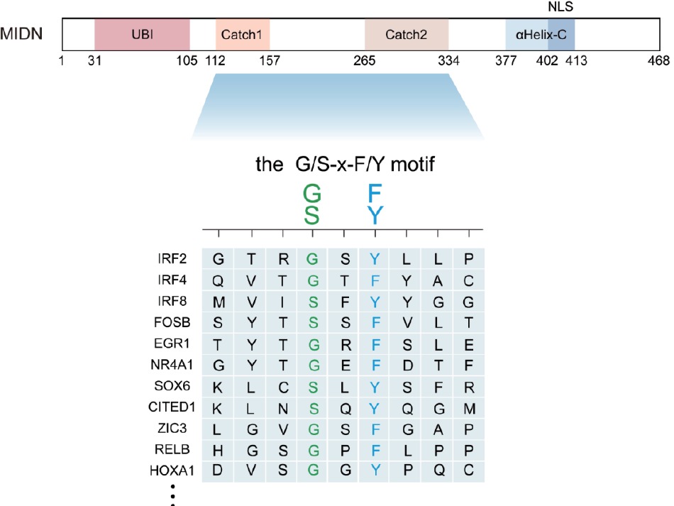
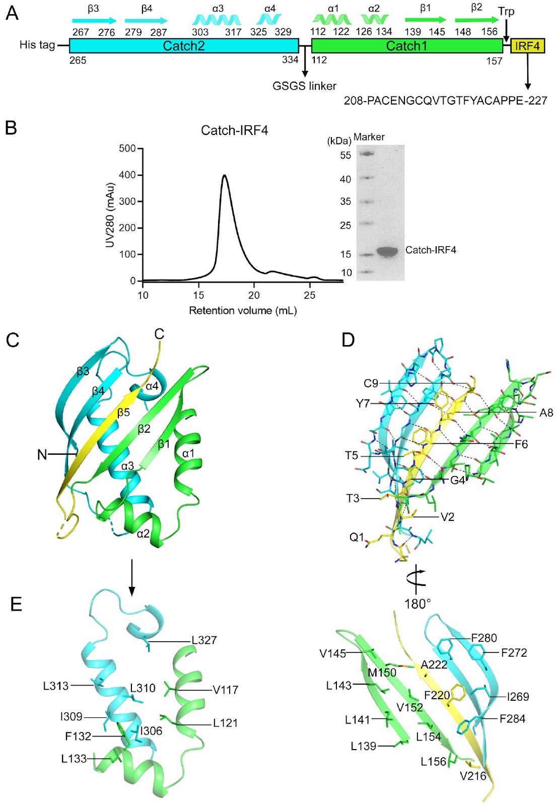
Protein homeostasis serves as the foundation for every cellular decision—division, differentiation, stress adaptation, or death—by precisely balancing the proteome across abundance, quality, spatial distribution, and temporal dynamics; its dysregulation drives numerous human pathologies, including cancers and neurological disorders. In the traditional ubiquitin-dependent degradation cascade, target proteins are marked by covalent attachment of polyubiquitin chains, a process requiring E1 activating enzymes, E2 conjugating enzymes, and E3 ligases that confer substrate specificity. This ubiquitin signal is then recognized by the 19S regulatory particle of the proteasome, which unfolds and translocates the tagged protein into the 20S core for proteolytic destruction. The ubiquitin-independent proteasomal degradation pathway mediated by midnolin (MIDN) has recently emerged as a distinct and biologically important mechanism for regulating nuclear protein turnover. While earlier genetic, biochemical, and cryo-electron microscopy studies established MIDN as a proteasome-associated adaptor for immediate-early gene (IEG) products, the molecular logic underlying its broad yet selective substrate recognition remains unresolved. In the current study, Zhong et al. provide a comprehensive structural and biochemical analysis of the MIDN Catch domain bound to IRF4-derived peptides, substantially advancing our understanding of MIDN substrate recognition. By solving multiple crystal structures of the Catch-IRF4 complex, including wild-type and systematically engineered mutants, the authors demonstrate that MIDN recognizes substrates through a conserved β-strand insertion mechanism at the interface of the Catch1 and Catch2 subdomains. This investigation generalizes and expands prior structural observations of MIDN-IRF4 to diverse substrates, demonstrating that β-strand complementation constitutes a universal recognition mechanism utilized by MIDN. A major conceptual advance of this study is the identification of a minimal and generalizable recognition principle centered on two highly conserved positions within the substrate motif. The authors show that a reciprocal phenylalanine-glycine interaction between the substrate and Catch2—forming an “F-G zipper”—constitutes the dominant energetic determinant for binding. Disruption of this zipper severely compromises protein stability and binding, explaining prior functional observations that single-point mutations in IRF4 or EGR1 abolish MIDN-mediated degradation. In contrast, flanking residues within the binding motif display remarkable tolerance to substitution. Through combined mutagenesis, thermostability analysis, AlphaFold3 modeling, and structural determination, the study demonstrates that these positions occupy large and plastic hydrophobic pockets in the Catch domain. This architectural flexibility provides a direct molecular explanation for how MIDN can accommodate a wide spectrum of substrates while preserving selectivity. From these data, the authors derive a concise consensus recognition motif (G/S-x-F/Y) embedded within an unstructured or loop region, offering a predictive framework for identifying additional MIDN targets. Importantly, this work bridges a critical gap between previous cryo-EM studies describing MIDN-proteasome engagement and earlier substrate-specific structural analyses. Whereas prior studies clarified how MIDN delivers captured substrates to the proteasome, the present study elucidates how MIDN initially selects and binds those substrates. Together, these findings unify MIDN biology across structural, biochemical and functional dimensions. Overall, this study represents a significant advance in the field of proteostasis by revealing how a ubiquitin-independent adaptor achieves broad substrate specificity through a simple yet robust structural principle. Beyond MIDN, the work provides a paradigm for short-linear-motif-based proteasomal targeting and has important implications for immune regulation, neurodegeneration and cancer biology.

Read Full Abstract10.3724/abbs.2026006
PDK4-driven metabolic reprogramming enhances mesothelial cell invasion in colorectal cancer peritoneal metastasisGraphical AbstractVerified
Acta Biochimica et Biophysica Sinica

PDK4-driven metabolic reprogramming enhances mesothelial cell invasion in colorectal cancer peritoneal metastasis

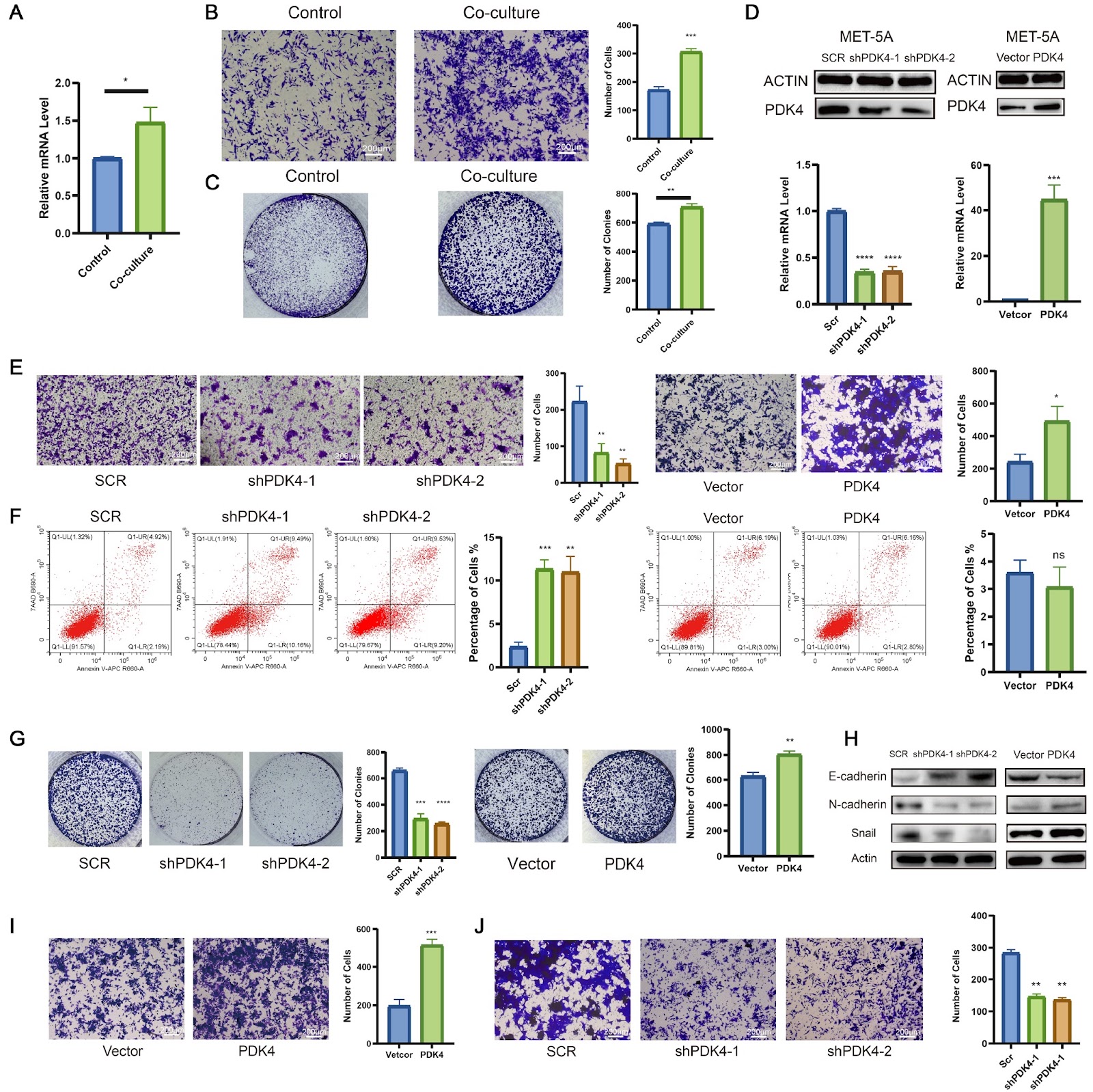
Mesothelial cells play an important role in colorectal cancer peritoneal metastasis (CRC-PM), where they support tumor growth and invasion. In this study, we investigate the molecular mechanisms by which mesothelial cells contribute to CRC metastasis. Using single-cell RNA sequencing (scRNA-seq) on tissue samples from 12 CRC patients with peritoneal metastasis, we identify PDK4 as a key gene in mesothelial cells during metastasis. The expression of PDK4 is significantly greater in mesothelial cells undergoing mesothelial-to-mesenchymal transition (MMT) compared to normal peritoneal cells, suggesting its involvement in mesothelial cell reprogramming during peritoneal metastasis. In vitro experiments show that coculturing mesothelial cells with CRC cells leads to increased PDK4 expression, which in turn enhances mesothelial cell migration and invasion. Knockdown of PDK4 reduces mesothelial cell invasion, while overexpression of PDK4 increases invasive ability, highlighting its critical role in mesothelial cell invasion. Additionally, PDK4 promotes metabolic changes, specifically increasing fatty acid oxidation (FAO), which is necessary for mesothelial cell invasion. Blocking FAO reduces the invasive ability of PDK4-overexpressing mesothelial cells, while restoring FAO in PDK4-knockdown cells rescues their invasion potential. Further analysis shows that PDK4 enhances the acetylation of β-catenin, a protein involved in cell movement, and that this modification is crucial for mesothelial cell invasion. Our results suggest that PDK4 regulates mesothelial cell invasion through β-catenin acetylation following metabolic reprogramming, offering a potential target for therapies aimed at inhibiting CRC-PM.

Read Full Abstract10.3724/abbs.2025148
Biochemical and structural studies of the midnolin Catch domain bound with both wild-type and mutant IRF4 peptides reveal the molecular basis for its broad substrate specificityGraphical AbstractVerified
Acta Biochimica et Biophysica Sinica

Biochemical and structural studies of the midnolin Catch domain bound with both wild-type and mutant IRF4 peptides reveal the molecular basis for its broad substrate specificity

The newly discovered midnolin-proteasome pathway is a unique ubiquitin-independent mechanism for degrading nuclear proteins, which is crucial for maintaining cellular protein homeostasis. The Catch domain of midnolin is essential for substrate recognition and binding, yet the underlying mechanism for its broad substrate specificity remains elusive. Transcription factor IRF4, essential for the functions of B and T cells, is a substrate of midnolin. This study presents comprehensive biochemical and structural analyses of the human midnolin Catch domain in complexes with both wild-type and mutant IRF4 peptides. The crystal structure of the Catch-IRF4 complex reveals that the Catch domain creates a substrate-binding groove at the interface of the Catch1 and Catch2 subdomains, recognizing and binding to the 215-QVTGTFYAC-223 sequence motif of IRF4. The binding motif of IRF4 forms a β-strand that is embedded into the substrate-binding groove, resulting in an antiparallel five-stranded β-sheet. The interactions between the IRF4 peptide and the Catch domain are predominantly hydrophobic and exhibit high spatial complementarity. Additionally, the biochemical, modeling and structural data indicate that the V2 and A8 positions of the IRF4 sequence motif can be substituted with other hydrophobic or small polar residues (G/A/V/L/I/M/P/F/Y/C/S/T), but not with large polar and charged residues (D/N/E/Q/H/K/R). The G4 position can be replaced by Ser, while the F6 position can be substituted with Tyr. These results suggest that the Catch domain can recognize and bind to a variety of substrates containing the sequence motif x[G/A/V/L/I/M/P/F/Y/C/S/T]x[G/S]x[F/Y]x[G/A/V/L/I/M/P/F/Y/C/S/T]x or briefly the G/SxF/Y motif (where x represents polar residues) located in an unstructured or loop region on the protein surface, and the hydrophobic interactions and spatial complementarity between the binding motifs of substrates and the Catch domain govern the substrate specificity. Collectively, these findings elucidate the molecular basis for midnolin’s broad substrate specificity.

Read Full Abstract10.3724/abbs.2026002
The EBV-MS Paradigm: Beyond Molecular Mimicry Toward New Therapeutic StrategiesGraphical AbstractVerified
Acta Biochimica et Biophysica Sinica2026

The EBV-MS Paradigm: Beyond Molecular Mimicry Toward New Therapeutic Strategies

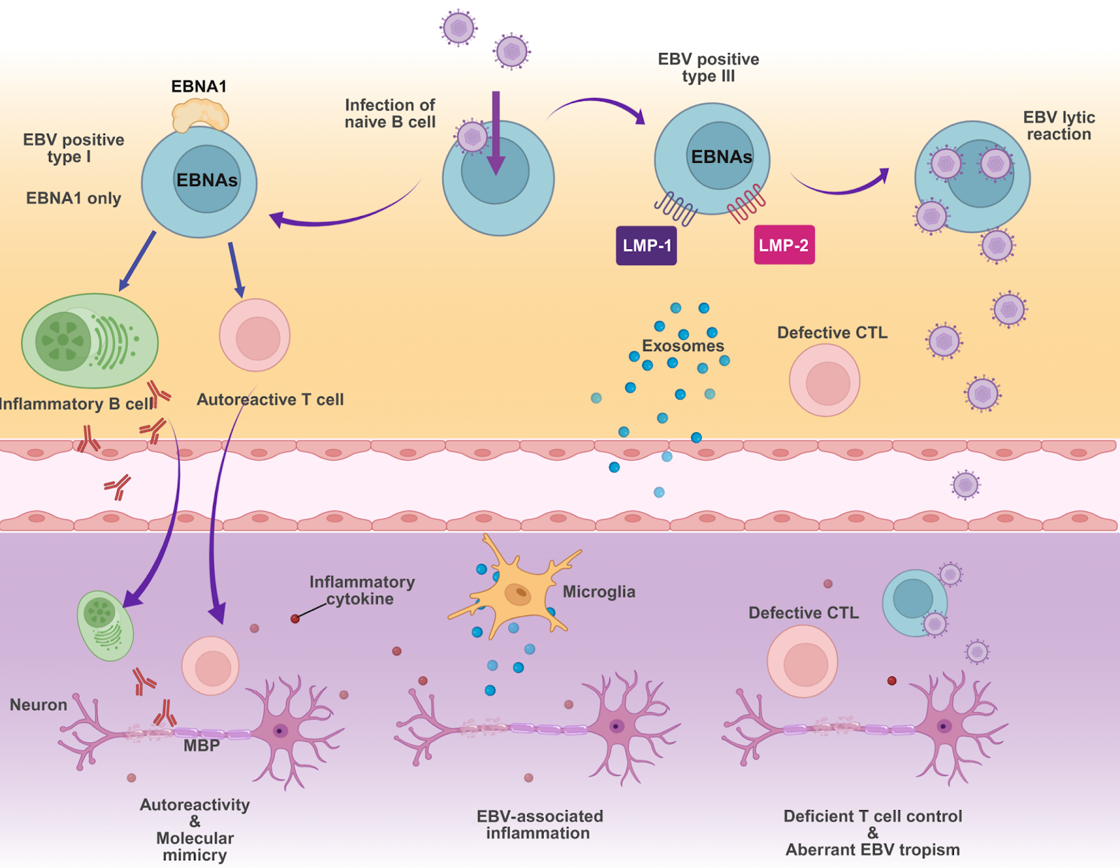
Epstein-Barr virus (EBV) is now recognized as the definitive environmental driver of multiple sclerosis (MS), shifting the conceptual landscape of this autoimmune disorder to an infection-triggered model. In this review, we systematically evaluate the multidimensional evidence linking EBV to MS. This evidence ranges from epidemiological associations and the identification of mimotopes to emerging therapeutic strategies. We also discuss the broader implications of infection-driven immune dysregulation for autoimmune research. This pathogenic link is underpinned by molecular mimicry, where immune responses against the viral protein EBNA1 cross-react with the central nervous system (CNS) protein GlialCAM. Beyond this initial insult, EBV reprograms B cells to survive and proliferate abnormally, creating a compartmentalized viral reservoir within the CNS that sustains chronic neuroinflammation. These mechanistic insights catalyze a transition from broad immunosuppression to precision therapies targeting the EBV-MS axis, including CNS-penetrant kinase inhibitors and EBV-specific CAR-T cells. By integrating etiological discovery with mechanism-based intervention, the EBV-MS paradigm serves as a blueprint for transforming idiopathic autoimmune diseases into mechanistically tractable conditions with actionable therapeutic targets.

Read Full Abstract10.3724/abbs.2026026
Structural basis for the conformational changes of insulin receptor induced by three different hormone ligandsGraphical AbstractVerified
Acta Biochimica et Biophysica Sinica2026

Structural basis for the conformational changes of insulin receptor induced by three different hormone ligands

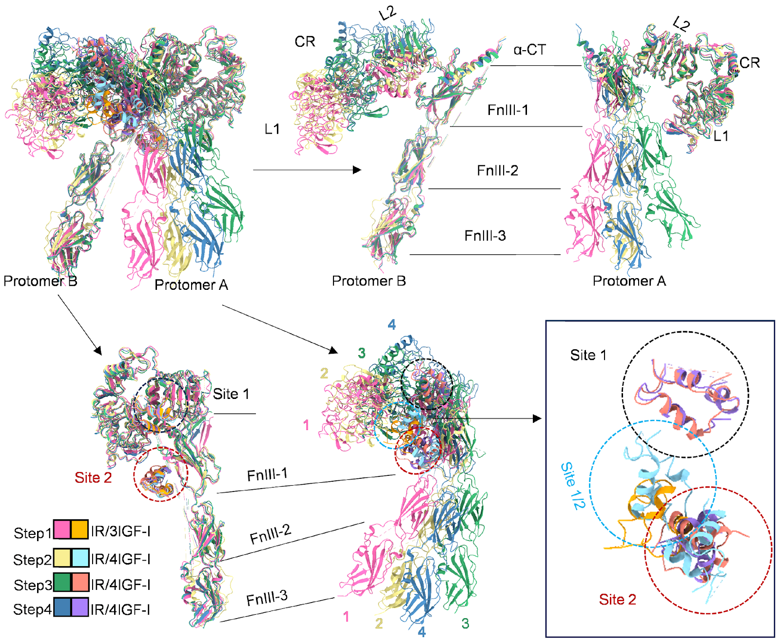
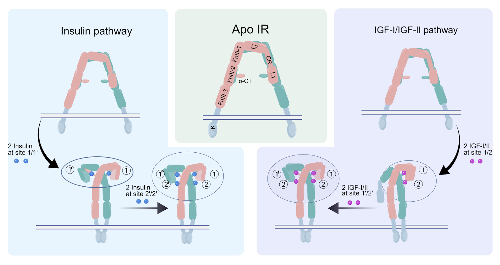
The insulin receptor (IR) is central to the regulation of glucose and lipid metabolism. Although insulin is its primary ligand, insulin-like growth factors I and II (IGF-I and IGF-II) also engage IR, albeit with reduced affinity. The structural basis of cooperative ligand binding, however, has remained poorly understood. Here, we report cryo-Electron Microscopy (cryo-EM) structures of IR in complex with insulin, IGF-I, and IGF-II, revealing that all three ligands engage the receptor at overlapping binding sites and can induce a conserved T-shaped quaternary assembly involving four ligand molecules at site 1/1′ and site 2/2′. Despite this shared overall architecture, distinct ligand-specific conformational changes are observed. Notably, IGF-I and IGF-II adopt different binding sequence at site 1 and site 2 compared to insulin, suggesting unique interaction dynamics. These structural insights highlight divergent mechanisms of ligand recognition and cooperative binding, providing a deeper understanding of hormone-induced conformational modulation of the IR.

Read Full Abstract10.3724/abbs.2026020
Andrographolide prevents necroptosis by suppressing the generation of reactive oxygen speciesGraphical AbstractVerified
Acta Biochimica et Biophysica Sinica

Andrographolide prevents necroptosis by suppressing the generation of reactive oxygen species

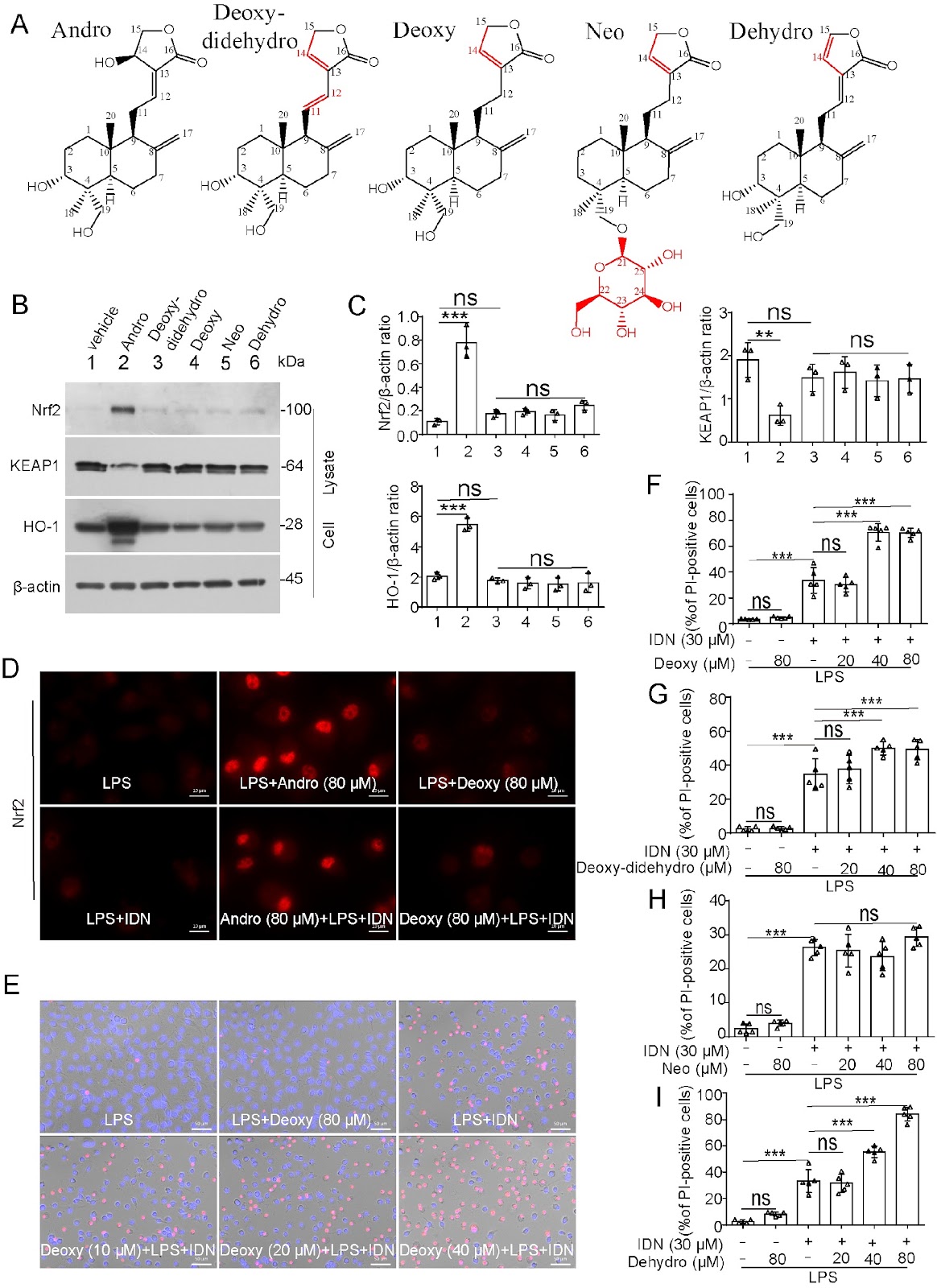
Andrographolide (Andro), a natural product extracted from the Chinese traditional medicine herb Andrographis paniculata, has been applied for the treatment of diverse inflammatory diseases. However, its effects on necroptosis, a lytic form of cell death implicated in various inflammatory diseases, remain uncharacterized. In the present study, we investigate whether Andro and its derivatives can suppress necroptosis. Our results demonstrate that Andro notably inhibits necroptosis in the in vitro cellular models induced by either lipopolysaccharide (LPS) plus IDN-6556 or a combination of TNF-α, LCL-161 (Smac mimetic) and IDN-6556. In these cellular models, Andro inhibits the phosphorylation of receptor-interacting protein kinase 1 (RIPK1), RIPK3, and mixed lineage kinase domain-like pseudokinase (MLKL), as well as the formation of necrosomes. Specifically, Andro reduces the levels of intracellular reactive oxygen species (ROS) and mitochondrial superoxide (mtROS), preserves the mitochondrial membrane potential during necroptotic induction, and activates the antioxidant transcription factor nuclear factor E2-related factor 2 (Nrf2). Upon necroptotic stimulation, some mitochondrial proteins, such as Bcl-2 and Bak, oligomerize and co-localize with RIPK1, RIPK3, and phosphorylated MLKL (p-MLKL) in necrosomes. However, this process of necrosome formation can be prevented by Andro. In contrast, derivatives, including dehydroandrographolide, neoandrographolide, 14-deoxy-11,12-didehydroandrographolide, and 14-deoxyandrographolide, have no anti-necroptotic effects and fail to upregulate Nrf2. Collectively, our findings demonstrate that Andro specifically inhibits the RIPK1/RIPK3/MLKL signaling axis to suppress necroptosis, highlighting its therapeutic potential against necroptosis-related disorders.

Read Full Abstract10.3724/abbs.2025077
The tRNA Landscape in Cancer: From Pathogenesis to Therapeutic InterventionsGraphical AbstractVerified
Acta Biochimica et Biophysica Sinica2026

The tRNA Landscape in Cancer: From Pathogenesis to Therapeutic Interventions

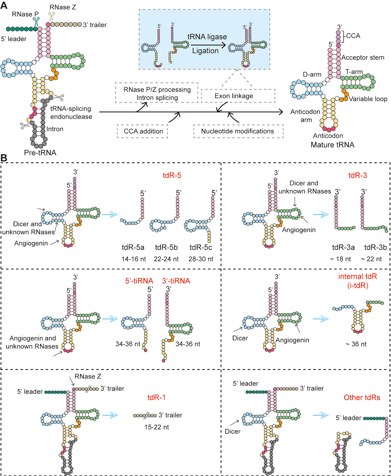
Transfer RNA (tRNA) acts not only as an indispensable adaptor in protein synthesis but also as a key contributor to tumorigenesis when its regulation is disrupted. This review systematically summarizes aberrant tRNA-related mechanisms in cancer, including altered tRNA expression profiles, abnormal post-transcriptional modifications, dysregulated aminoacylation, production of tRNA-derived small RNAs (tdRs), and defects in tRNA trafficking and translational fidelity. Notably, the metabolically abnormal tumor microenvironment actively shapes tRNA reprogramming to facilitate adaptive survival, while dysregulated tRNA elements, such as specific modifications and tdRs, further drive cancer stem cell properties and therapeutic resistance. Collectively, these alterations reprogram the oncoproteome and signaling networks, thereby promoting tumor cell proliferation, metastasis, immune evasion, and drug resistance. Targeting these mechanisms represents a promising strategy for developing novel cancer therapies. Potential approaches include the use of suppressor tRNAs to restore tumor suppressor gene function, the employment of tdRs to modulate oncogenic signaling pathways, or direct inhibition of enzymes involved in tRNA biogenesis. These strategies aim to remodel the dysfunctional tRNA network in cancer and offer new avenues for innovative treatments.

Read Full Abstract10.3724/abbs.2026093
How Distinct Hormones Sculpt a Common Receptor: Ligand-Specific Conformational Pathways of the Insulin ReceptorGraphical AbstractVerified
Acta Biochimica et Biophysica Sinica2026

How Distinct Hormones Sculpt a Common Receptor: Ligand-Specific Conformational Pathways of the Insulin Receptor

The insulin receptor (IR) is a central regulator of metabolism, integrating hormonal cues to coordinate glucose uptake, lipid metabolism, growth, and survival. While insulin is its canonical ligand, IR also responds to insulin-like growth factors (IGF-I and IGF-II) with lower affinity, a cross-reactivity that is physiologically relevant during development and pathologically important in cancer. Despite extensive research, the molecular basis for how distinct hormones engage the same receptor yet elicit different activation modes remained unresolved. In this study, Yan and colleagues determined the first cryo-electron microscopy (cryo-EM) structures of full-length human IR-A bound to IGF-I, alongside those with insulin and IGF-II, under matched experimental conditions. By capturing multiple ligand-occupancy and conformational states, they revealed that insulin and IGFs drive distinct conformational trajectories through a shared architectural framework, reframing IR activation as a ligand-dependent conformational selection process. Insulin rapidly stabilizes the receptor head region, minimizing conformational heterogeneity and promoting synchronized activation, consistent with its fast, concentration-dependent physiological actions. In contrast, IGF-I and IGF-II induce greater conformational plasticity and a pronounced preference for asymmetric intermediate states, favoring sustained and adaptable engagement suited to long-term growth and differentiation. Site-specific analysis showed that while core residues are shared, insulin forms a denser hydrogen-bond network at site 1, whereas IGFs rely more on secondary sites. These findings provide a structural basis for ligand-specific cooperativity and signaling bias, with implications for understanding metabolic diseases and cancer, and for designing selective IR modulators.

Read Full Abstract10.3724/abbs.2026024
Mixed fungal polysaccharides enhance intestinal health, antioxidant capacity, and microbiota diversity in broiler chickensGraphical AbstractVerified
Acta Biochimica et Biophysica Sinica

Mixed fungal polysaccharides enhance intestinal health, antioxidant capacity, and microbiota diversity in broiler chickens

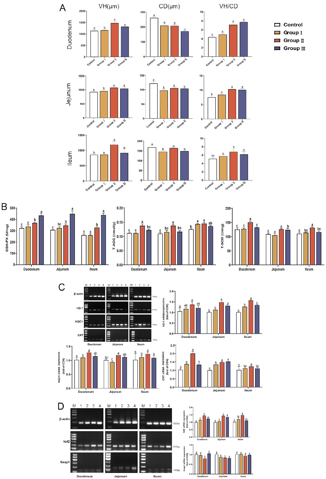
Poultry production faces escalating challenges from intensive farming practices, where stressors, including high stocking density, pathogen exposure, and dietary fluctuations, disrupt intestinal integrity, microbiota balance, and antioxidant defenses. These disruptions impair nutrient absorption, growth performance, and immune function, leading to significant economic losses. Although antibiotics have historically mitigated such issues, growing restrictions due to antimicrobial resistance necessitate natural alternatives. Fungal polysaccharides (FP)—notably lentinan (LNT) from Lentinula edodes and polysaccharide from Ganoderma lucidum (GLP), are promising candidates owing to their immunomodulatory, antioxidant, and prebiotic properties. However, existing research focuses predominantly on individual FP, neglecting potential synergies in blended formulations. Structurally, LNT (β-(1→3)-D-glucan backbone) enhances rumen volatile fatty acid production and fiber degradation, whereas GLP (heterogeneous α/β-glycans) potently activates the Nrf2/HO-1 antioxidant pathway and modulates Th1/Th2 immunity. These divergent mechanisms imply complementary effects when combined. In our previous experiments on broiler feeding, we reported that a combination of GLP (68.32% polysaccharide content, composed of mannose, glucose, arabinose, rhamnose, and galactose at a molar ratio of 1.00:16.37:18.82:1.42:17.42) and LNT (76.52% polysaccharide content, composed of mannose, galacturonic acid, arabinose, galactose, glucose, and rhamnose at a molar ratio of 1.00:15.22:8.23:2.05:1.78:4.26) at a 1:1 ratio maximally promoted broiler growth (unpublished data), but their impacts on intestinal morphology, antioxidant signaling, and the microbiota remain uncharacterized. We therefore hypothesize that mixed FP synergistically may enhance intestinal health by simultaneously improving nutrient absorption, activating antioxidant pathways, and stabilizing microbial ecosystems. To investigate the effects of mixed FP on intestinal development, 240 one-day-old Arbor Acres male broilers were randomly assigned to the 0 mg/kg FP (Control), 200 mg/kg FP (Group I), 400 mg/kg FP (Group II), and 600 mg/kg FP (Group III) groups. Broilers were housed in three-tier battery cages (0.7 m × 0.7 m × 0.4 m; 12 broilers/cage), with five replicate cages per experimental group maintained under identical conditions. The experiments were approved by the College of Animal Science and Technology in Anhui Agricultural University (approval number: SYXK 2016-007). All the cages were subjected to a 16 h light: 8 h dark cycle with ad libitum access to water and twice-daily feeding (09:00/16:00) of basal diets (Supplementary Table S1). On day 42, the duodenum, jejunum, and ileum segments were collected, fixed in 4% paraformaldehyde, sectioned at 5 μm, and stained with hematoxylin-eosin. Villus height (VH), crypt depth (CD), and VH/CD ratios were measured via Case Viewer software. The results revealed that Group II significantly increased VH and VH/CD across all the intestinal segments while reducing CD (Figure 1A; P < 0.05 vs the control); these findings suggest enhanced nutrient absorption capacity and intestinal health. To evaluate antioxidant capacity and signaling pathway activation, intestinal tissues were homogenized in PBS (1:9, w/v). The total antioxidant capacity (T-AOC), total superoxide dismutase (T-SOD), and glutathione peroxidase (GSH-Px) activities were determined via commercial kits (Nanjing Jiancheng Bioengineering Institute, Nanjing, China)). For gene expression analysis, total RNA was extracted and reverse-transcribed. The qPCR was performed via specific primers for HO-1, NQO1, CAT, Nrf2, and Keap1, with β-actin used as the reference gene (primer sequences and product sizes are listed in Supplementary Table S2). The results demonstrated that Group II significantly elevated antioxidant enzyme activities (P < 0.05), upregulated HO-1, NQO1, CAT, and Nrf2, and
Read Full Abstract10.3724/abbs.2025222
RNF126 writes a non-canonical ubiquitin code on midnolin to tune protein stabilityGraphical AbstractVerified
Acta Biochimica et Biophysica Sinica2026

RNF126 writes a non-canonical ubiquitin code on midnolin to tune protein stability

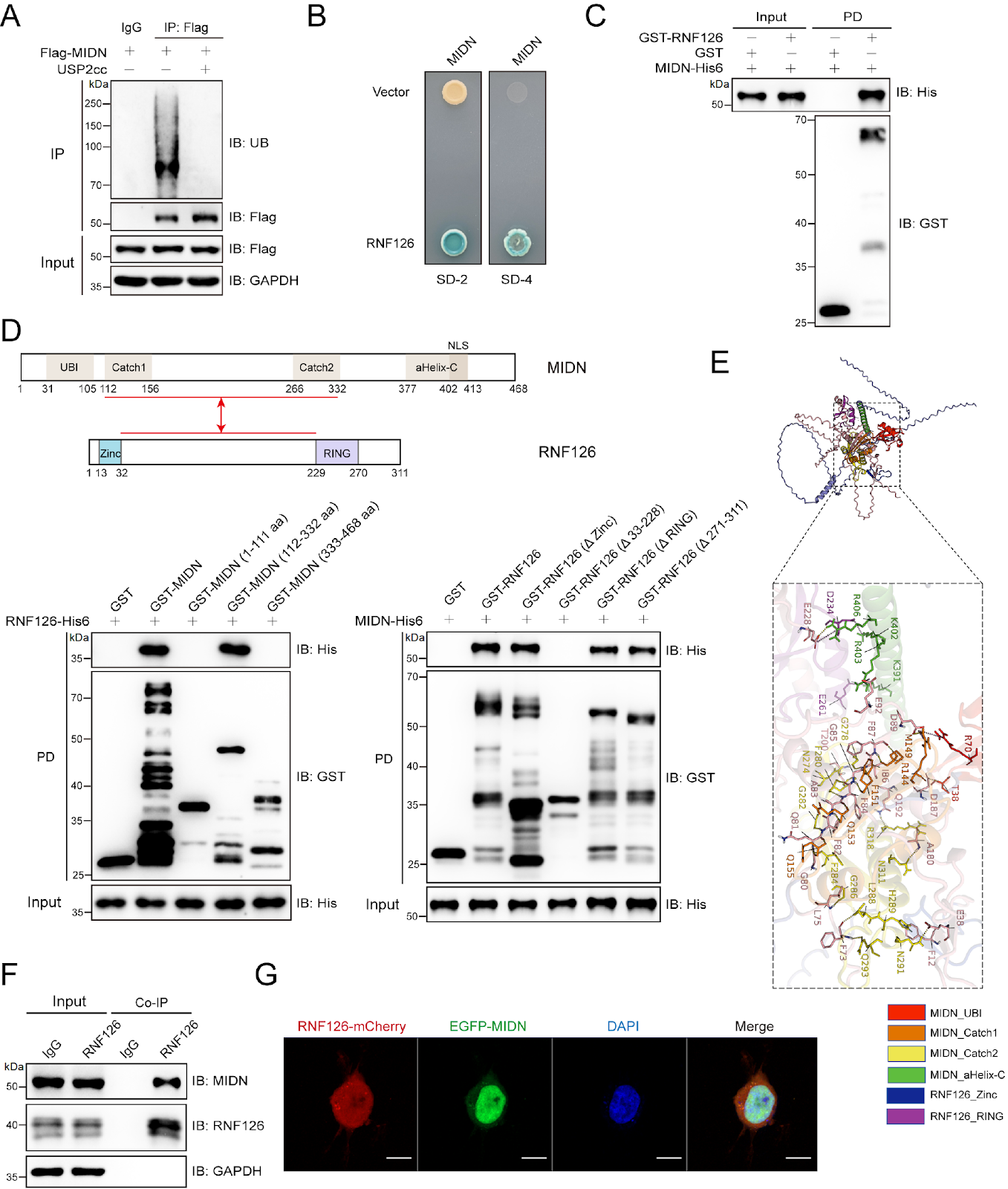
Midnolin (MIDN) is a newly recognized master regulator that drives ubiquitin-independent proteasomal degradation, yet the mechanisms governing its own turnover remain enigmatic. Here, we demonstrate that MIDN is ubiquitinated and identify RNF126 as the cognate E3 ligase. RNF126 physically associates with MIDN and catalyzes its ubiquitination, and mass spectrometry mapping reveals that this process occurs primarily at non-canonical cysteine, serine, and threonine residues (C230, C236, S237, T239, and S241) rather than at lysine residues. This non-classical ubiquitination targets MIDN for 26S-proteasomal degradation. In vivo dissection of the RNF126-MIDN axis shows that it governs EGR1 abundance and, consequently, the tumor-suppressor proteins PTEN and p53, thereby restraining the progression of testicular germ-cell tumors (TGCTs). Our findings reveal an unappreciated layer of MIDN regulation and identify the RNF126-MIDN ubiquitination cascade as a potential therapeutic vulnerability in TGCTs and related malignancies.

Read Full Abstract10.3724/abbs.2025232
Resident CD24+LCN2+ LPCs aggravate fibrosis and inflammatory progression via the recruitment of TPPP3+COL10A1+ macrophages in NASHGraphical AbstractVerified
Acta Biochimica et Biophysica Sinica

Resident CD24+LCN2+ LPCs aggravate fibrosis and inflammatory progression via the recruitment of TPPP3+COL10A1+ macrophages in NASH

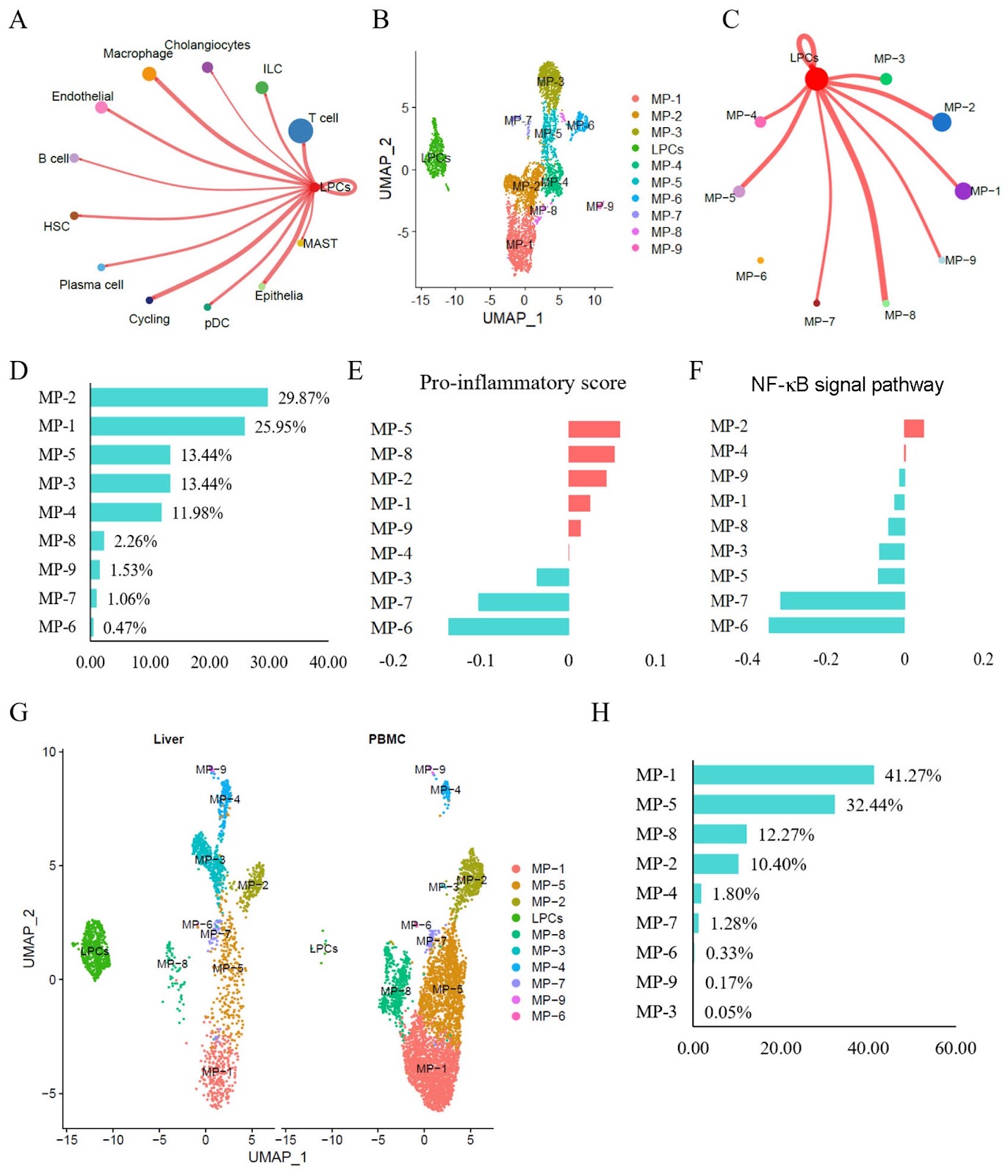
Resident CD24+LCN2+ liver progenitor cells (LPCs) reportedly contribute to the expanding ductular reaction and macrophage-mediated inflammation associated with chronic liver damage. Both ductular reactions and macrophage-driven inflammation are associated with liver fibrosis and injury in various mouse liver disorders. This study aims to investigate the molecular phenotypes of LPCs and their regulatory mechanisms in humans with non-alcoholic steatohepatitis (NASH). Single-cell RNA sequencing (scRNA-seq) datasets are used to characterize the status and molecular phenotypes of LPCs in clinical NASH samples. To elucidate the regulatory mechanisms of LPCs, CellChat and NicheNet are employed to assess cell-cell communication between LPCs and other cell types. The findings are validated using RNA sequencing datasets associated with NASH progression, NASH mouse models (CDAHFD and HFD), and human NASH liver samples. Results show that resident CD24+LCN2+ LPCs are identified and found to be significantly enriched in NASH patients. Cell communication analyses predict strong interactions between LPCs and proinflammatory macrophage subtypes. Additionally, in NASH, the liver recruits peripheral blood mononuclear cell (PBMC)-derived macrophages and polarizes them into proinflammatory subtypes. The macrophage subtype MP-2 is identified as the primary recipient of LPC-derived signals, exhibiting marked hyperactivation of the NF-κB pathway and a strong association with liver fibrosis. Finally, the MP-2 markers COL10A1 and TPPP3 are characterized and validated. In summary, this study reveals that resident CD24+LCN2+ LPCs are activated in NASH and contribute to fibrosis progression by promoting the activation of the proinflammatory COL10A1+TPPP3+ macrophage subtype.

Read Full Abstract10.3724/abbs.2025081
Cardiac PTN-SIRT1 axis alleviates oxidative stress and promotes mitochondrial energy reprogramming to mitigate doxorubicin-induced cardiotoxicity through AMPK/PGC1α signalingGraphical AbstractVerified
Acta Biochimica et Biophysica Sinica

Cardiac PTN-SIRT1 axis alleviates oxidative stress and promotes mitochondrial energy reprogramming to mitigate doxorubicin-induced cardiotoxicity through AMPK/PGC1α signaling

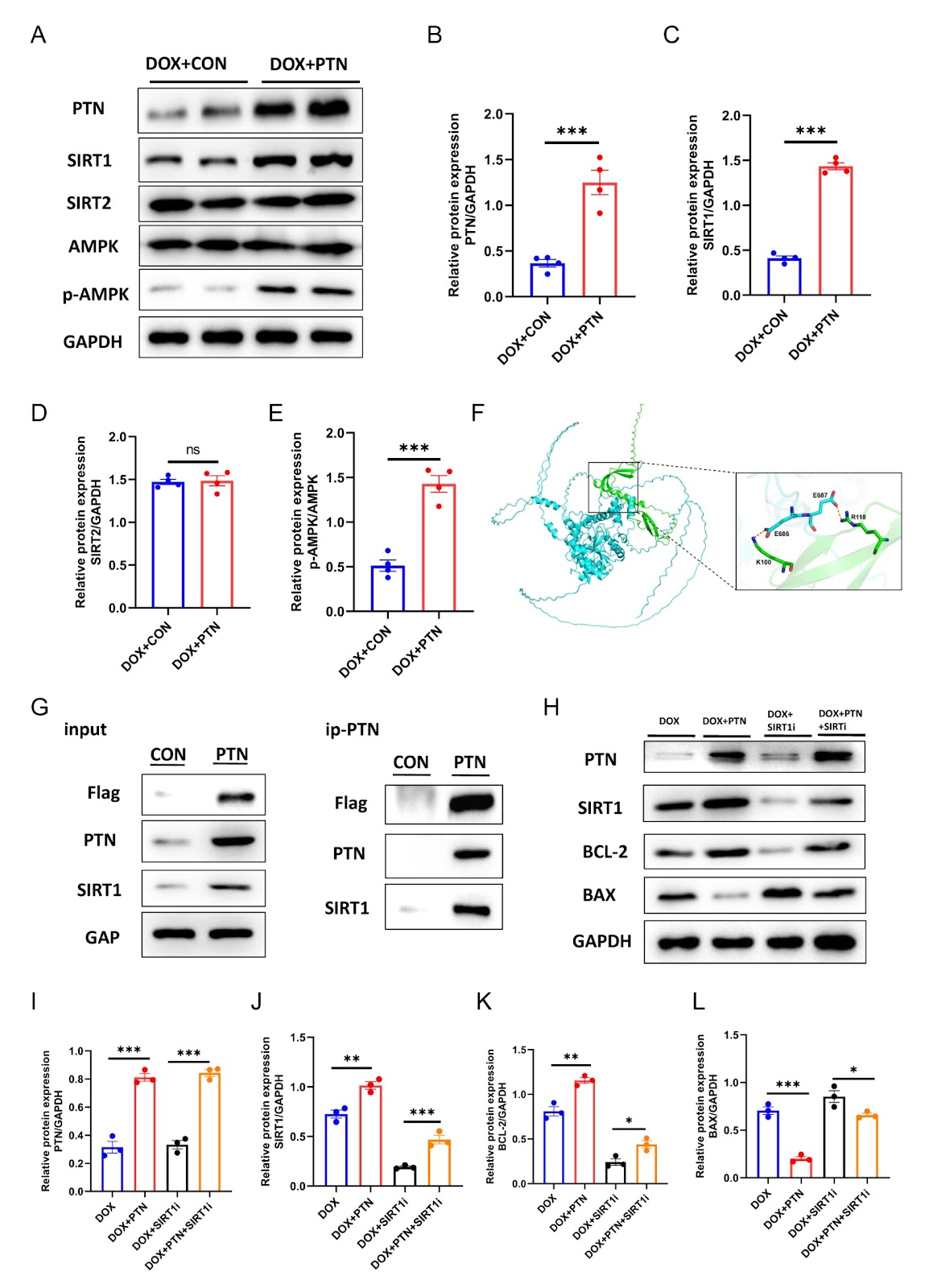
Doxorubicin (DOX) remains a cornerstone chemotherapeutic agent for malignancies, yet its clinical utility is severely limited by dose-dependent cardiotoxicity, which can lead to progressive left ventricular dysfunction and heart failure. Pleiotrophin (PTN), a heparin-binding growth factor with diverse physiological functions, regulates glucose and lipid metabolism and promotes oxidative energy pathways. However, whether PTN exerts protective effects against DOX-induced cardiotoxicity (DIC) remains unclear. In this study, we establish cellular and animal models of DIC. DOX administration induces pronounced myocardial injury in both models, characterized by impaired ventricular contractility, increased fibrotic remodeling, and reduced cell viability. Concurrently, PTN protein expression is significantly downregulated in cardiomyocytes under DOX treatment. Overexpression of PTN substantially alleviates these pathological changes. In vitro, PTN reduces mitochondrial oxidative stress and apoptosis while restoring energy production and cell viability. In vivo, PTN improves mitochondrial ultrastructure, decreases cardiomyocyte apoptosis, and enhances cardiac function. Mechanistically, PTN directly binds to SIRT1 and activates AMPK phosphorylation at Thr172, triggering a downstream cascade through the AMPK-PGC1α axis that reprograms mitochondrial energy metabolism and attenuates cardiotoxicity. In conclusion, the PTN-SIRT1 axis protects against DIC by reducing oxidative stress and promoting mitochondrial energy homeostasis via the AMPK/PGC1α pathway, highlighting its potential as a novel therapeutic target for preventing chemotherapy-related cardiac injury.

Read Full Abstract10.3724/abbs.2026018
Characterization of the mechanisms underlying sulfasalazine-induced ferroptotic cell death: role of protein disulfide isomerase-mediated NOS activation and NO accumulationGraphical AbstractVerified
Acta Biochimica et Biophysica Sinica

Characterization of the mechanisms underlying sulfasalazine-induced ferroptotic cell death: role of protein disulfide isomerase-mediated NOS activation and NO accumulation

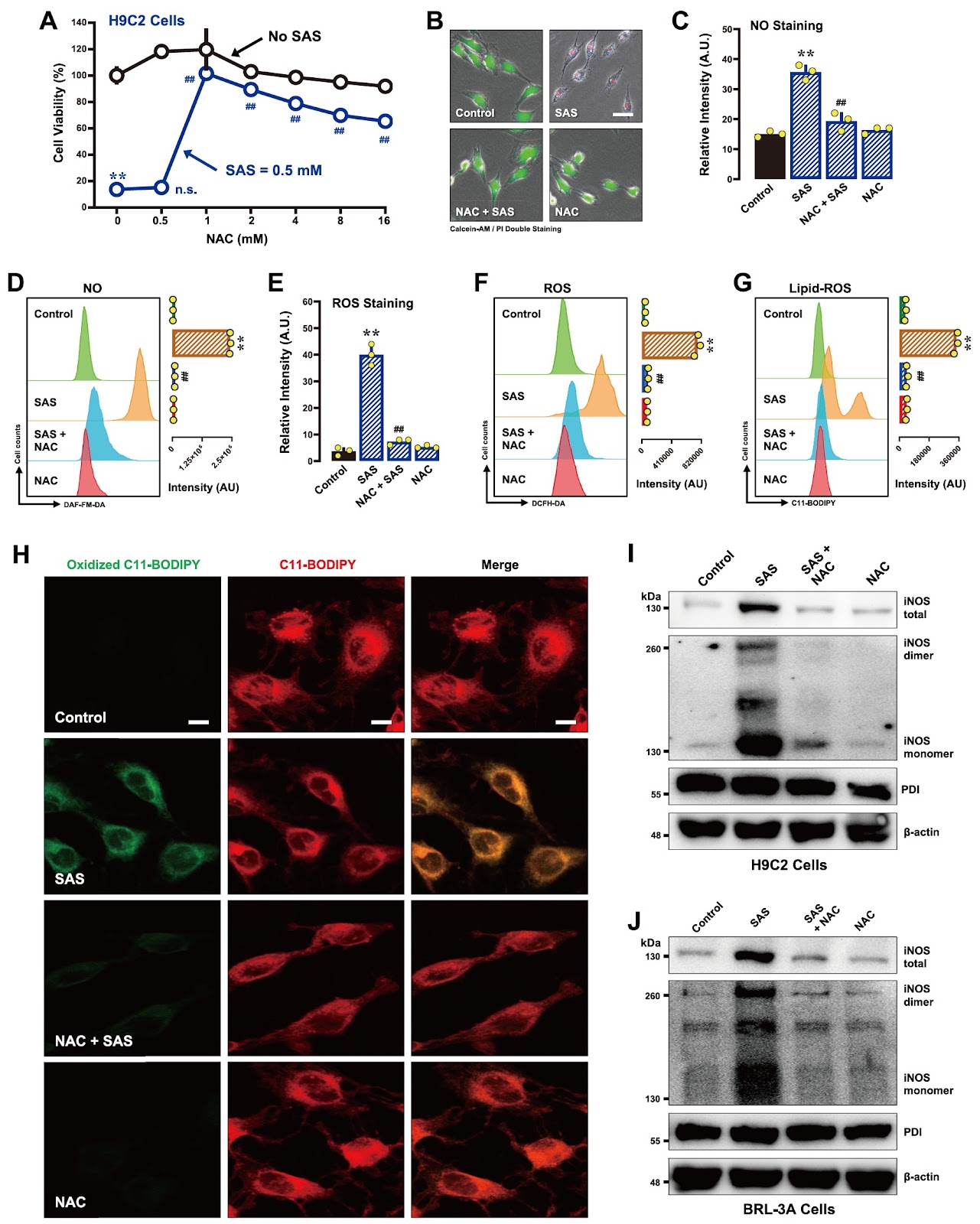
Sulfasalazine (SAS), a clinically utilized anti-inflammatory drug, has been shown to induce ferroptosis by inhibiting system Xc− activity, thereby causing cellular glutathione depletion. Recently, protein disulfide isomerase (PDI) was shown to be an upstream mediator of the oxidative cell death (oxytosis/ferroptosis) induced by glutamate, erastin, RSL3 and SAS. The present study aims to further characterize the detailed biochemical and cellular mechanisms of SAS-induced ferroptosis in two cell lines, i.e., H9C2 rat cardiomyocytes and BRL-3A rat hepatocytes, with a focus on elucidating the critical role of PDI in mediating SAS-induced toxicity. We find that SAS can induce ferroptosis in H9C2 and BRL-3A cells, which is accompanied by a sequential increase in the buildup of cellular nitric oxide (NO), reactive oxygen species (ROS) and lipid-ROS. SAS activates PDI-mediated dimerization of inducible NO synthase (iNOS) and cellular accumulation of NO, and these effects are followed by ROS and lipid-ROS accumulation. Furthermore, SAS markedly upregulates the iNOS protein levels in these cells. Knockdown of PDI or pharmacological inhibition of PDI catalytic activity effectively suppresses SAS-induced iNOS dimerization, abrogates SAS-induced accumulation of NO, ROS and lipid-ROS, and prevents ferroptosis. On the other hand, PDI activation through the use of TrxR1 inhibitors sensitizes these cells to SAS-induced ferroptosis. These findings provide further experimental support for a pivotal role of PDI in SAS-induced cytotoxicity through the activation of the PDI-NOS-NO axis, which then leads to the accumulation of cellular ROS and lipid-ROS and ultimately the induction of oxidative cell death.

Read Full Abstract10.3724/abbs.2025100
Rhamnose alleviates the proinflammatory response during endotoxemia via the CEACAM1/LGALS9-p38 axisGraphical AbstractVerified
Acta Biochimica et Biophysica Sinica

Rhamnose alleviates the proinflammatory response during endotoxemia via the CEACAM1/LGALS9-p38 axis

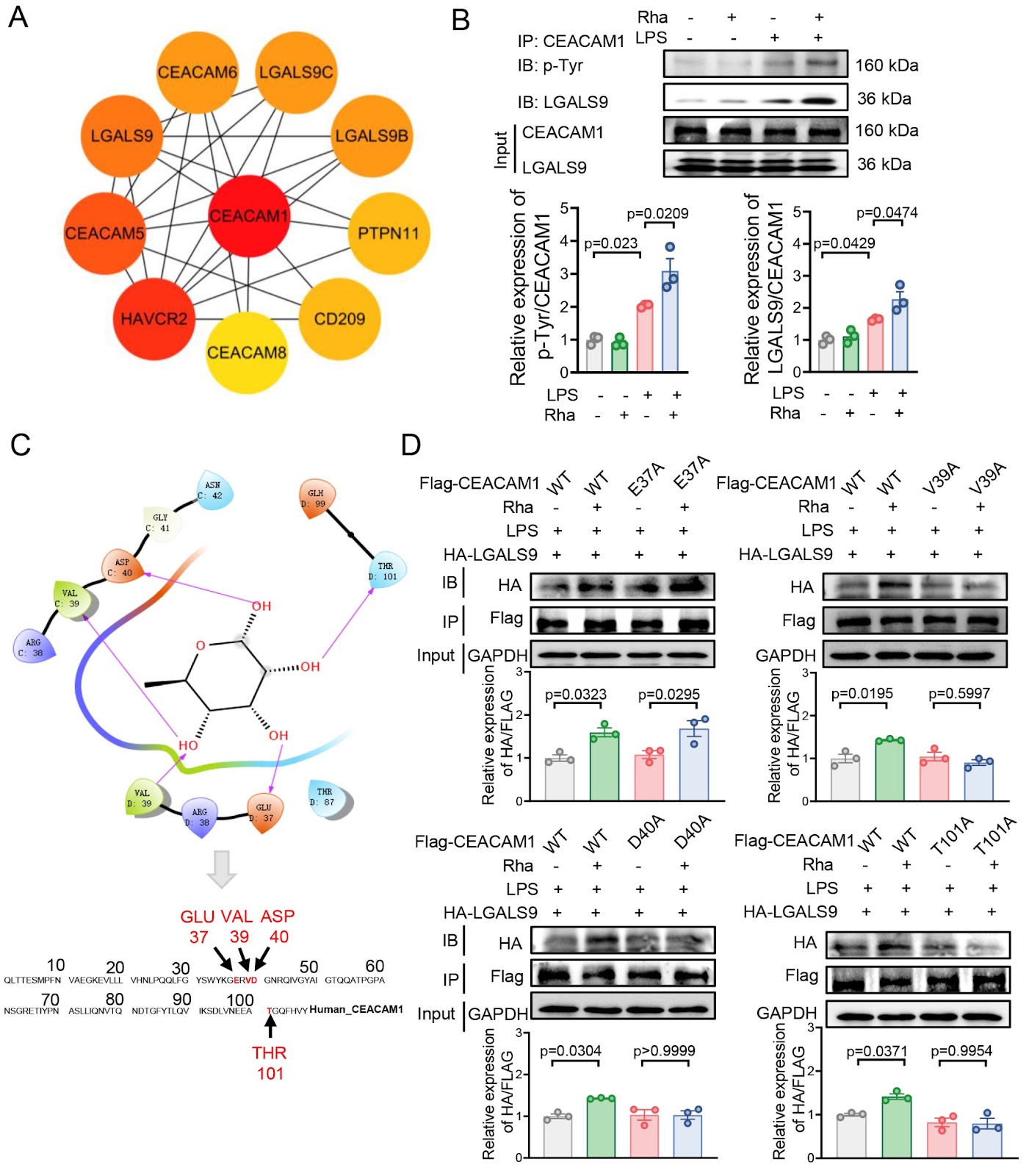
Gut microbiota plays an important role in orchestrating the host immune response. We previously reported that gut microbiota-derived rhamnose enhances the phagocytosis of macrophages, upon which we further asked whether rhamnose has modulatory effects on inflammation. Here, we show that, in an LPS-induced endotoxic mouse model, plasma rhamnose levels are increased. This bacteria-derived sugar alone does not impact inflammatory cytokine homeostasis or cause organ damage. In contrast, it is able to alleviate endotoxin-induced systemic inflammation and organ damage. Mechanistically, in macrophages in vitro, rhamnose binds to the V39, D40, and T101 sites of carcinoembryonic antigen-associated cell adhesion molecule 1 (CEACAM1), subsequently promoting the interaction between CEACAM1 and galectin 9 (LGALS9), which increases the protein level of dual-specificity protein phosphatase 1 (DUSP1). This inhibits p38 phosphorylation and thus attenuates the LPS-triggered expressions of proinflammatory factors. Collectively, our results suggest that rhamnose signals via the CEACAM1/LGALS9-p38 axis, which suppresses endotoxemia-associated inflammation, and that rhamnose is a candidate anti-inflammatory agent for the control of infection-induced organ damage.

Read Full Abstract10.3724/abbs.2025109
Angptl4 is upregulated by microenvironmental factors during the wound healing process and promotes epidermal stem cell proliferation via PRL8a6Graphical AbstractVerified
Acta Biochimica et Biophysica Sinica

Angptl4 is upregulated by microenvironmental factors during the wound healing process and promotes epidermal stem cell proliferation via PRL8a6

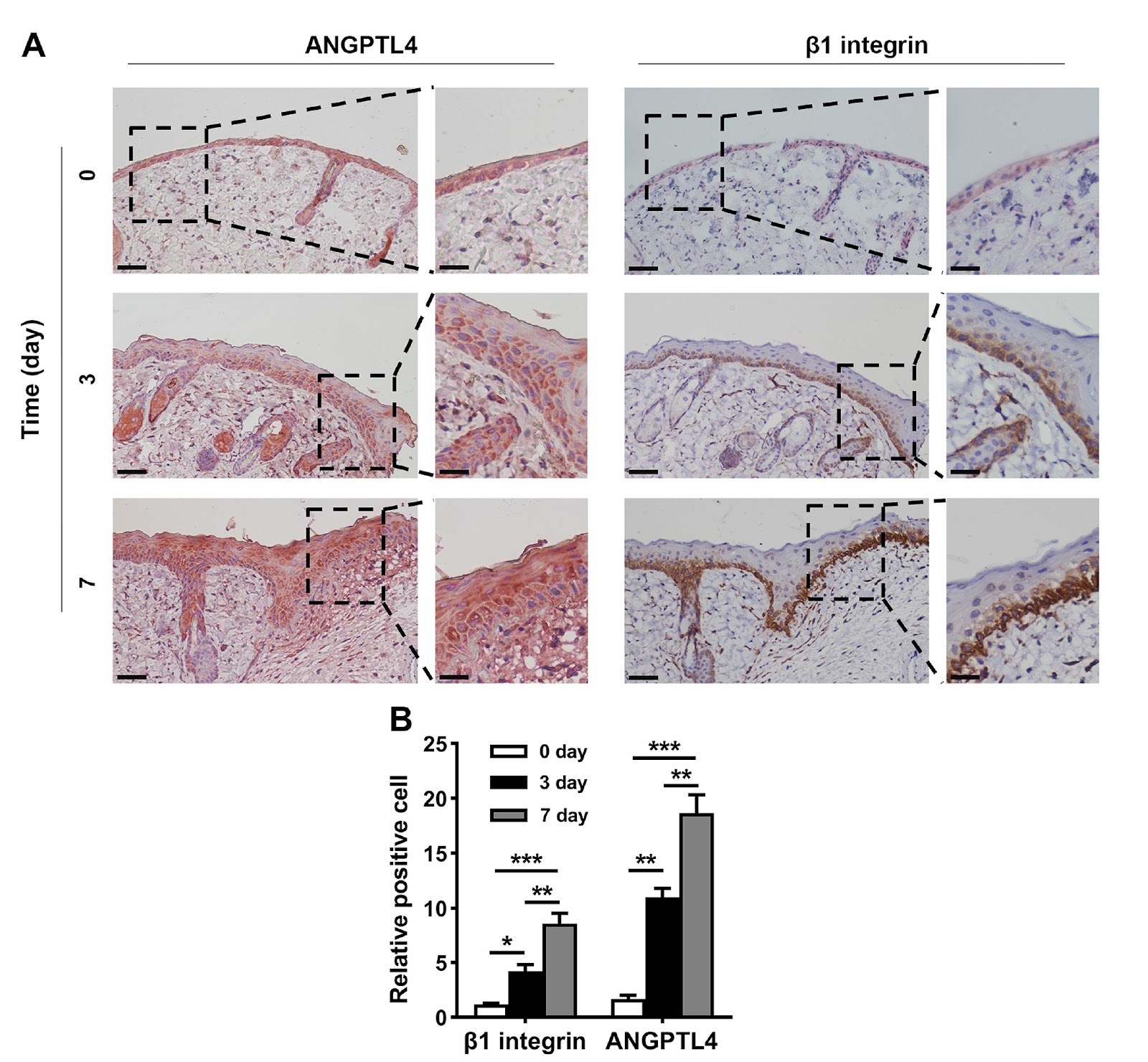
Angiopoietin-like 4 (ANGPTL4) expression is increased in wound tissue and contributes to wound healing. However, the underlying mechanisms are not fully understood. Here, we demonstrate that ANGPTL4 expression is significantly increased in epidermal stem cells (EpSCs) in the periwound epidermis during wound healing in mice. Increased Angptl4 expression is positively correlated with increased expressions of tumor growth factor-α, interleukin-1β, epidermal growth factor, nerve growth factor, fibroblast growth factor 7, and transforming growth factor-β1. Each of these molecules induces Angptl4 expression in mouse EpSCs. RNA sequencing of EpSCs derived from wild-type and Angptl4 knockout (Angptl4–/–) mice reveals altered expressions of genes involved in the cell cycle and cell proliferation in Angptl4–/– EpSCs, including a decrease in cyclin E2/A2/B1 and cyclin-dependent kinase 1 (Cdk1) expression; an increase in Cdk inhibitor 2a (Cdkn2a) and Cdkn2b expression; and a decrease in the prolactin (PRL) family members Prl2a1, Prl8a1, Prl8a9, and Prl8a6. Mechanistic studies reveal that ANGPTL4 stimulates EpSC proliferation via PRL8a6-mediated upregulation of cyclins A2/E2/B1 and Cdk1, downregulation of Cdkn2a, and acceleration of cell cycle progression from the G1 to the S and G2 phases. In vivo studies demonstrate that Prl8a6 mRNA is upregulated by ANGPTL4 in mouse periwound tissue during skin wound healing. Knockdown of Angptl4 or Prl8a6 in periwound skin tissue impairs EpSC proliferation and delays wound re-epithelialization. In conclusion, our study demonstrates that, after skin injury, elevated levels of proinflammatory cytokines and growth factors in periwound tissue stimulate Angptl4 expression in EpSCs and that ANGPTL4 promotes EpSC proliferation by increasing Prl8a6 expression, thereby accelerating wound re-epithelialization.

Read Full Abstract10.3724/abbs.2025145
Integration of the hammerhead ribozyme into structured RNAs to measure ligand-binding events for riboswitch candidates and aptamersGraphical AbstractVerified
Acta Biochimica et Biophysica Sinica

Integration of the hammerhead ribozyme into structured RNAs to measure ligand-binding events for riboswitch candidates and aptamers

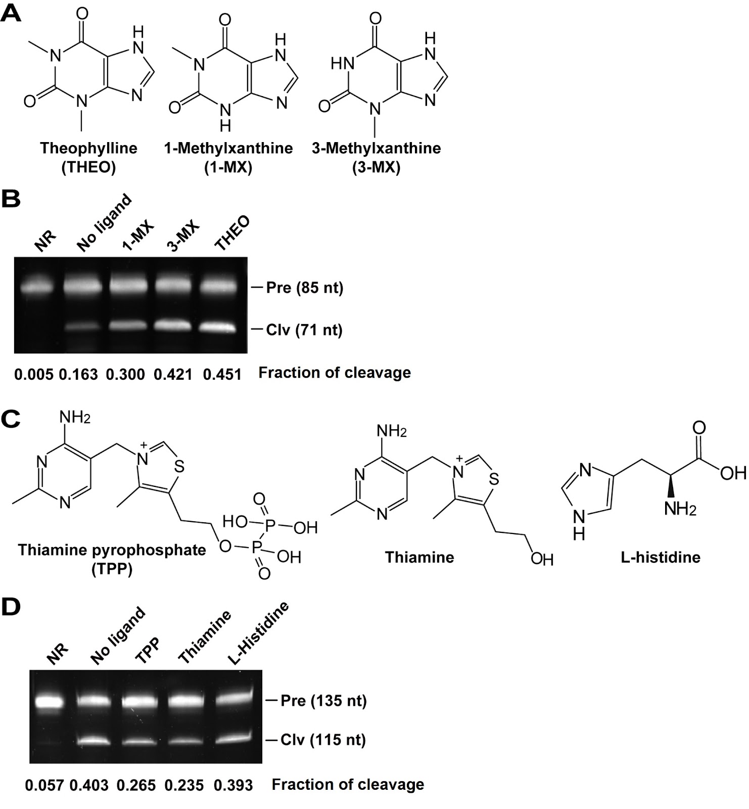
Some structured RNAs, such as riboswitches and aptamers, can bind to their cognate ligands and have been used in biosensors and gene expression control elements. However, current methods for detecting ligand binding to structured RNAs are either severely limited or inconvenient. In this study, we design a multibase pair bridge to integrate a hammerhead ribozyme into structured RNAs to detect ligand binding events. The experimental results demonstrate that the length of the bridge has a significant effect on the cleavage of the ribozyme; optimal cleavage can be achieved with three to six base pairs in the bridge. The dissociation constant (KD) values obtained through this method are in agreement with those determined by in-line probing techniques, and 1 pmol of allosteric ribozyme RNA is sufficient for measurement. We apply this method to evaluate the binding affinity of the riboswitch candidate Motif_9307. Our findings indicate that this motif has no binding affinity for S-adenosylmethionine or several other tested ligands, which is consistent with the results of the in-line probing experiments. Notably, our method reveals an increase in cleavage activity when yeast extract is added as a mixture of ligands, suggesting that the ligand of Motif_9307 is present in the extract. In conclusion, we develop an alternative approach for measuring ligand binding events associated with riboswitch candidates and aptamers.

Read Full Abstract10.3724/abbs.2025097