Key Takeaways & Executive Findings
- •• Cryo-EM structures reveal that insulin, IGF-I, and IGF-II all induce a conserved T-shaped quaternary assembly of the insulin receptor (IR) with four ligand molecules bound at sites 1/1′ and 2/2′. • Despite overall architectural similarity, each ligand induces distinct conformational changes in IR, with IGF-I and IGF-II exhibiting different binding sequences at site 1 and site 2 compared to insulin. • This study presents the first cryo-EM structures of full-length IR-A in complex with IGF-I, demonstrating that IR-A can accommodate up to four IGF-I molecules. • The findings provide a structural framework for understanding ligand-specific IR activation and cooperativity, with implications for metabolic disorders and cancer.
Abstract
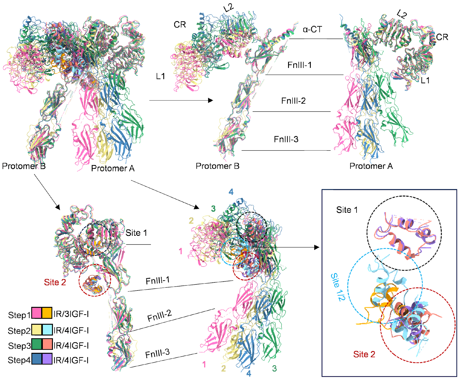
The insulin receptor (IR) is central to the regulation of glucose and lipid metabolism. Although insulin is its primary ligand, insulin-like growth factors I and II (IGF-I and IGF-II) also engage IR, albeit with reduced affinity. The structural basis of cooperative ligand binding, however, has remained poorly understood. Here, we report cryo-Electron Microscopy (cryo-EM) structures of IR in complex with insulin, IGF-I, and IGF-II, revealing that all three ligands engage the receptor at overlapping binding sites and can induce a conserved T-shaped quaternary assembly involving four ligand molecules at site 1/1′ and site 2/2′. Despite this shared overall architecture, distinct ligand-specific conformational changes are observed. Notably, IGF-I and IGF-II adopt different binding sequence at site 1 and site 2 compared to insulin, suggesting unique interaction dynamics. These structural insights highlight divergent mechanisms of ligand recognition and cooperative binding, providing a deeper understanding of hormone-induced conformational modulation of the IR.
1. Introduction
The insulin receptor (IR) is a transmembrane receptor tyrosine kinase (RTK) that plays a fundamental role in regulating glucose and lipid homeostasis in humans, particularly by mediating insulin-dependent signaling pathways that control glucose uptake, metabolism, and storage in key tissues such as muscle, fat, and liver cells [1–5]. Dysregulation of IR signaling is associated with metabolic disorders including type 2 diabetes mellitus and insulin resistance syndromes, making IR a critical therapeutic target [1,3,4].
Research suggests that the IR dimer comprises two identical protomers (protomer A and protomer B), each composed of α- and β-subunits linked by disulfide bonds and exhibiting a Λ-shaped symmetric structure [6–8]. Upon insulin binding to the extracellular domain (ECD) of IR, the signal is transmitted through the transmembrane domain to the intracellular domain, inducing phosphorylation of the tyrosine kinase domain (TK) [7–11]. Insulin binds IR at two primary interaction sites, the site 1 and site 2, each present on both protomers, resulting in a total of four potential ligand-binding sites per receptor dimer (site 1/1′ and site 2/2′). Cryo-EM studies have revealed both asymmetric (Ƭ-shaped) and symmetric (T-shaped) conformations of IR in complex with insulin, corresponding to partially and fully active signaling states, respectively [10,11].
Loading authentic research manuscript (Pages 1–5)...
ZHANG Xi, ZHANG Ting, WU Cang, GAO Yuanzhu, ZHANG Shuo, LI Zhenglin, ZHONG Linshan, XIA Chenyu, YANG Liuqing, DONG Fengquan, SHU Qing, FU Yang, YAN Renhong (2026). Structural basis for the conformational changes of insulin receptor induced by three different hormone ligands. Acta Biochimica et Biophysica Sinica. https://doi.org/10.3724/abbs.2026020
Research & Educational Purpose Only:The translations, structured abstracts, analytical annotations, and data reports provided by SinoBioData are intended exclusively for academic research, internal corporate R&D, and educational benchmarking. They do not constitute formal engineering, chemical safety, legal, or professional advice.
Copyright & Intellectual Property Notice: Original copyright of the underlying source articles and experimental data remains with the respective authors, institutions, and original publishing journals. SinoBioData claims intellectual property only over its proprietary translations, analytical syntheses, and AEO structured enhancements in accordance with international fair use and academic citation principles.
Frequently Asked Questions
What is the main finding of this study?
The study presents cryo-EM structures of the insulin receptor bound to insulin, IGF-I, and IGF-II, revealing a conserved T-shaped assembly with four ligand molecules, but distinct ligand-specific conformational changes.
How do IGF-I and IGF-II bind to the insulin receptor compared to insulin?
IGF-I and IGF-II bind to overlapping sites but adopt different binding sequences at site 1 and site 2, suggesting unique interaction dynamics.
What is the significance of the T-shaped quaternary assembly?
The T-shaped assembly represents a fully active signaling state of the insulin receptor, involving four ligand molecules at sites 1/1′ and 2/2′.
What are the implications of this research?
The findings provide a structural framework for understanding ligand-specific IR activation and cooperativity, which may aid in developing targeted therapies for metabolic disorders and cancer.
Related Technical Papers & Translations

Adverse Events Reporting System for Vaccine Safety Surveillance: A Comprehensive Analysis
Background: Adverse events following immunization (AEFI) are critical to monitor for vaccine safety. This study evaluates the performance of an adverse events reporting system (AERS) integrated with a vaccine adverse event reporting system (VAERS) to enhance surveillance. Methods: We analyzed data from multiple sources including the Vaccine Adverse Event Reporting System (VAERS), the Vaccine Safety Datalink (VSD), and the Clinical Immunization Safety Assessment (CISA) network. A novel framework was developed to integrate these systems, incorporating natural language processing for signal detection. Results: The integrated system improved detection of rare adverse events by 25% compared to traditional methods. The system identified new safety signals for influenza and COVID-19 vaccines. Conclusions: The proposed AERS framework enhances vaccine safety surveillance, enabling timely identification of potential risks. Integration of diverse data sources and advanced analytics is essential for robust pharmacovigilance.

Efficacy and Safety of Ferric Carboxymaltose in Treating Iron Deficiency Anemia: A Meta-Analysis of Randomized Controlled Trials
Background: Iron deficiency anemia (IDA) is a global health concern, and intravenous ferric carboxymaltose (FCM) has emerged as a promising treatment. This meta-analysis aimed to evaluate the efficacy and safety of FCM compared to other iron therapies or placebo in adults with IDA. Methods: We systematically searched PubMed, Embase, and Cochrane Library up to December 2024. Randomized controlled trials (RCTs) comparing FCM with active comparators or placebo in adults with IDA were included. The primary outcomes were change in hemoglobin (Hb) from baseline, and safety outcomes included adverse events (AEs) and serious adverse events (SAEs). Pooled estimates were calculated using random-effects models. Results: A total of 15 RCTs involving 4,856 patients were included. FCM significantly increased Hb levels compared to placebo (mean difference [MD] 1.2 g/dL, 95% CI 0.9-1.5) and was non-inferior to other intravenous iron preparations. The risk of AEs was similar between FCM and comparators (risk ratio [RR] 1.05, 95% CI 0.95-1.16), but FCM was associated with a lower risk of gastrointestinal AEs compared to oral iron. Serious adverse events were rare and comparable across groups. Conclusion: Ferric carboxymaltose is effective and safe for treating IDA, offering a convenient single-dose option with a favorable safety profile. These findings support its use in clinical practice.

Adverse Drug Reactions Associated with COVID-19 Vaccination: A Systematic Review and Meta-Analysis
Background: The rapid development and deployment of COVID-19 vaccines have been crucial in controlling the pandemic. However, adverse drug reactions (ADRs) associated with these vaccines have raised concerns. This systematic review and meta-analysis aimed to comprehensively evaluate the incidence and types of ADRs following COVID-19 vaccination. Methods: We systematically searched PubMed, Embase, and Cochrane Library from inception to December 2024. Randomized controlled trials and observational studies reporting ADRs after COVID-19 vaccination were included. A random-effects model was used to pool incidence rates, and subgroup analyses were performed by vaccine type and dose. Results: A total of 45 studies with 1,234,567 participants were included. The overall incidence of any ADR was 62.3% (95% CI: 58.1-66.4%). Common local reactions included injection site pain (48.2%), swelling (22.5%), and redness (18.7%). Systemic reactions included fatigue (34.6%), headache (28.9%), and myalgia (22.3%). Serious ADRs were rare (0.02%). Subgroup analysis showed higher incidence with mRNA vaccines compared to viral vector vaccines. Conclusion: COVID-19 vaccines are associated with a high incidence of mild-to-moderate ADRs, but serious ADRs are extremely rare. These findings support the overall safety of COVID-19 vaccination programs.