🧬 SinoBioData Academic Portal
📚 Peer-Reviewed Translated Literature

All Biomedical & Clinical Articles (Page 62)

Browse complete peer-reviewed translations from top Chinese biomedical, oncology, and genomics journals. Read verified previews and download full authentic clinical reports.

Published Research Papers

Showing 24 of 1542 peer-reviewed translated articles (Page 62 of 65)

AlphaFold2 assists in providing novel mechanistic insights into the interactions among the LUBAC subunitsGraphical AbstractVerified
Acta Biochimica et Biophysica Sinica

AlphaFold2 assists in providing novel mechanistic insights into the interactions among the LUBAC subunits

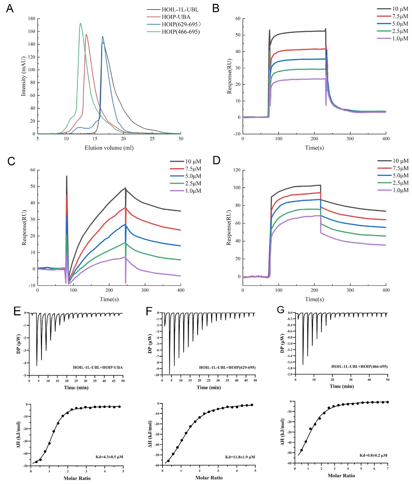
The linear ubiquitin chain assembly complex (LUBAC) is the only known E3 ligase complex in which the ubiquitin-like (UBL) domains of SHARPIN and HOIL-1L interact with HOIP to determine the structural stability of LUBAC. The interactions between subunits within LUBAC have been a topic of extensive research. However, the impact of the LTM motif on the interaction between the UBL domains of SHARPIN and HOIL-1L with HOIP remains unclear. Here, we discover that the absence of the LTM motif in the AlphaFold2-predicted LUBAC structure alters the HOIP-UBA structure. We employ GeoPPI to calculate the changes in binding free energy (ΔG) caused by single-point mutations between subunits, simulating their protein-protein interactions. The results reveal that the presence of the LTM motif decreases the interaction between the UBL domains of SHARPIN and HOIL-1L with HOIP, leading to a decrease in the structural stability of LUBAC. Furthermore, using the AlphaFold2-predicted results, we find that HOIP (629‒695) and HOIP-UBA bind to both sides of HOIL-1L-UBL, respectively. The experiments of Gromacs molecular dynamics simulations, SPR and ITC demonstrate that the elongated domain formed by HOIP (629‒695) and HOIP-UBA, hereafter referred to as the HOIP (466‒695) structure, interacts with HOIL-1L-UBL to form a structurally stable complex. These findings illustrate the collaborative interaction between HOIP-UBA and HOIP (629‒695) with HOIL-1L-UBL, which influences the structural stability of LUBAC.

Read Full Abstract10.3724/abbs.2024047
DLPC induces ferroptosis in cancer cellsGraphical AbstractVerified
Acta Biochimica et Biophysica Sinica

DLPC induces ferroptosis in cancer cells

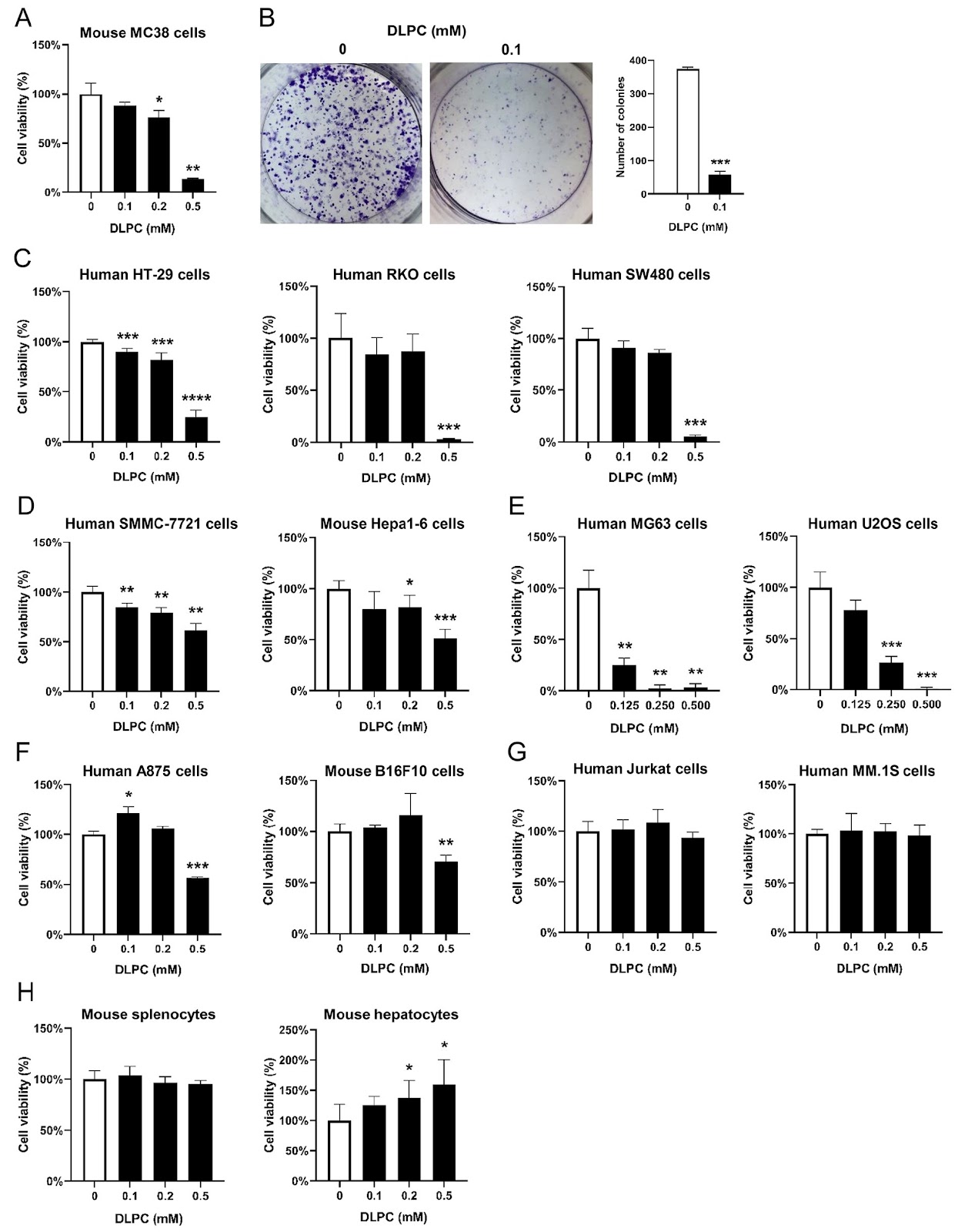
Phosphatidylcholine (PC) is the most abundant phospholipid in mammalian cells, accounting for approximately 50% of all phospholipids and serving as a main component of cellular and subcellular membranes. PC is a mixture of many species with distinct functions, and its levels are altered in cancer. Previous studies have shown contradictory roles of PC in cancer development. Here, we investigated the effects of PC and its main component, 1,2-dilinoleoyl-sn-glycero-3-phosphocholine (DLPC), on mouse colon cancer MC38 cells. PC dose-dependently decreased cell viability, and DLPC was identified as the active component. DLPC inhibited MC38 cell growth more effectively than PC, while structurally similar PCs with different acyl chain lengths or unsaturation degrees did not. This suggests that the specific structure of DLPC is crucial for its activity. Further mechanistic studies revealed that DLPC induces ferroptosis, a form of regulated cell death, in cancer cells. These findings highlight DLPC as a potential therapeutic agent for cancer treatment and underscore the importance of studying individual PC species.

Read Full Abstract10.3724/abbs.2024097
Construction of a cell-based aggregation and seeding model for the Tau proteinGraphical AbstractVerified
Acta Biochimica et Biophysica Sinica

Construction of a cell-based aggregation and seeding model for the Tau protein

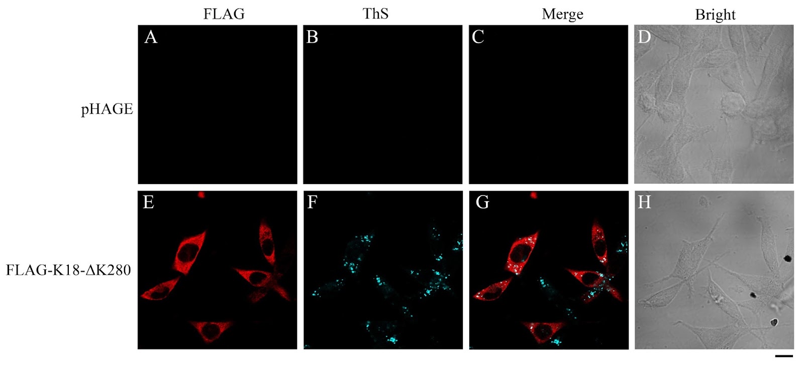
A pathological hallmark of Alzheimer’s disease (AD), the most common neurodegenerative disease in elderly people, is the formation of neurofibrillary tangles (NFTs), which are mainly composed of bundles of amyloid fibrils formed by abnormal deposition of hyperphosphorylated full-length human Tau protein [1–3]. Recent studies have shown that AD-related cognitive decline and brain atrophy are closely correlated with Tau PET signal, further supporting the link between Tau pathology and AD symptomatology [4,5]. Despite the high incidence and severe burden to patients, caregivers, and health systems caused by AD, there are few disease-modifying therapies available. Normal functional human Tau protein binds to tubulin heterodimers through its microtubule-binding repeats and stabilizes microtubules. Hyperphosphorylated Tau detaches from microtubules and exposes the microtubule-binding domain, thereby leading to Tau self-oligomerization and aggregation [6]. Accumulating evidence suggests that filamentous Tau inclusions form first in a small number of brain cells, from which they are released and taken up by neighboring cells via endocytosis; these filamentous Tau inclusions act as templates for their own replication through monomeric Tau addition and propagate to other regions of the cells [7,8]. Propagation of neuropathology is called prion-like, which refers to the capacity of an abnormally assembled protein to induce the same pathological conformation in the same protein, initiating a self-amplifying cascade. Transcellular propagation and prion-like phenomena are thought to contribute to the progression of pathology in AD, suggesting that inhibiting Tau aggregation and seeding could slow disease progression [7,8]. Accordingly, different experimental aggregation models for the Tau protein have been developed. A cell-based model offers a physiological assay environment with controllable costs and reproducible results, making it the most widely used model for the development of Tau-targeted therapies. In most cellular models, self-assembly of naive monomeric Tau is promoted by the addition of an exogenous ‘seed’ template of synthetic or patient-derived pre-aggregated Tau [9]. Pre-prepared fibrillar seeds of Tau are added to the cell culture medium, taken up by cells, and act as templates to induce the aggregation of monomeric Tau. In addition to homotypic seeding, heterotypic seeding has also been demonstrated for Tau. Direct cross-seeding between pre-aggregated Aβ and Tau is supported by direct binding between Aβ peptides and Tau and direct induction of Tau fibrillization by pre-aggregated Aβ seeds [10]. Human-derived seeds are the most relevant source of pathological Tau protein; however, clinical material is not straightforward to obtain and work with, and it is difficult to guarantee the quality and stability of aggregated seeds. In addition, aggregate seeds could damage the integrity of the cell membrane and lead to cytotoxicity. In the present study, we reported the construction of a cell-based model for the aggregation of endogenous Tau protein in cells without pre-prepared seeds. By introducing an aggregation-driven pathological mutant, ΔK280, to the aggregation-prone truncated core fragment of Tau (Tau244‒372, K18), we constructed a stable cell line over-expressing K18-ΔK280. Over-expressed K18-ΔK280 spontaneously aggregated in SH-SY5Y cells, forming amyloid fibrils positive for thioflavin S (ThS) (Figure 1), a fluorescent dye with β-sheet binding properties, which is widely employed to observe amyloid plaque accumulation [10]. Based on the present cellular model, the properties of Tau aggregation after seeding can be further observed. The aggregates formed by K18-ΔK280 induce co-aggregation and phosphorylation of endogenous Tau in SH-SY5Y cells, which can be recognized by AT8 (phosphorylation at Ser202/Thr205) and pS396 (phosphorylation at Ser396) (Figures 2 and 3) because phosphorylation at Ser202, Thr205, and Ser396 occurred in endogenous Tau but not at K18-ΔK280. This model is easy to use and avoids the potential cytotoxicity caused by fibrillar seeds.

Read Full Abstract10.3724/abbs.2024057
ATIP/ATIP1 regulates prostate cancer metastasis through mitochondrial dynamic-dependent signalingGraphical AbstractVerified
Acta Biochimica et Biophysica Sinica

ATIP/ATIP1 regulates prostate cancer metastasis through mitochondrial dynamic-dependent signaling

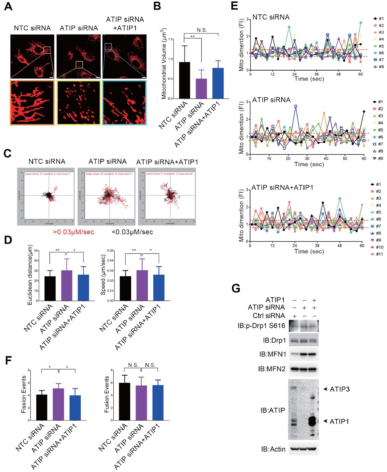
Mitochondria play a fundamental role in cell survival and motility. Abnormalities in mitochondria are associated with carcinogenesis, especially with tumor metastasis. In this study, we explore the biological function of ATIP1, which is a mitochondrial-located isoform of angiotensin II AT2 receptor interacting proteins (ATIPs) in prostate cancer cells. The results showed that ATIP is downregulated in prostate cancer tissues and is negatively correlated with the disease-free survival rate of prostate cancer patients. Silencing of ATIP promotes mitochondrial fission and enhances tumor cell migration and invasion. Reconstitution of ATIP1 in ATIP-deficient cells significantly attenuates mitochondrial trafficking and tumor cell movement. Therefore, ATIP1 is a negative regulator of mitochondrial dynamics and tumor cell motility and is also a potential biomarker for predicting prostate cancer malignancy.

Read Full Abstract10.3724/abbs.2024006
CCL2 promotes EGFR-TKIs resistance in non-small cell lung cancer via the AKT-EMT pathwayGraphical AbstractVerified
Acta Biochimica et Biophysica Sinica

CCL2 promotes EGFR-TKIs resistance in non-small cell lung cancer via the AKT-EMT pathway

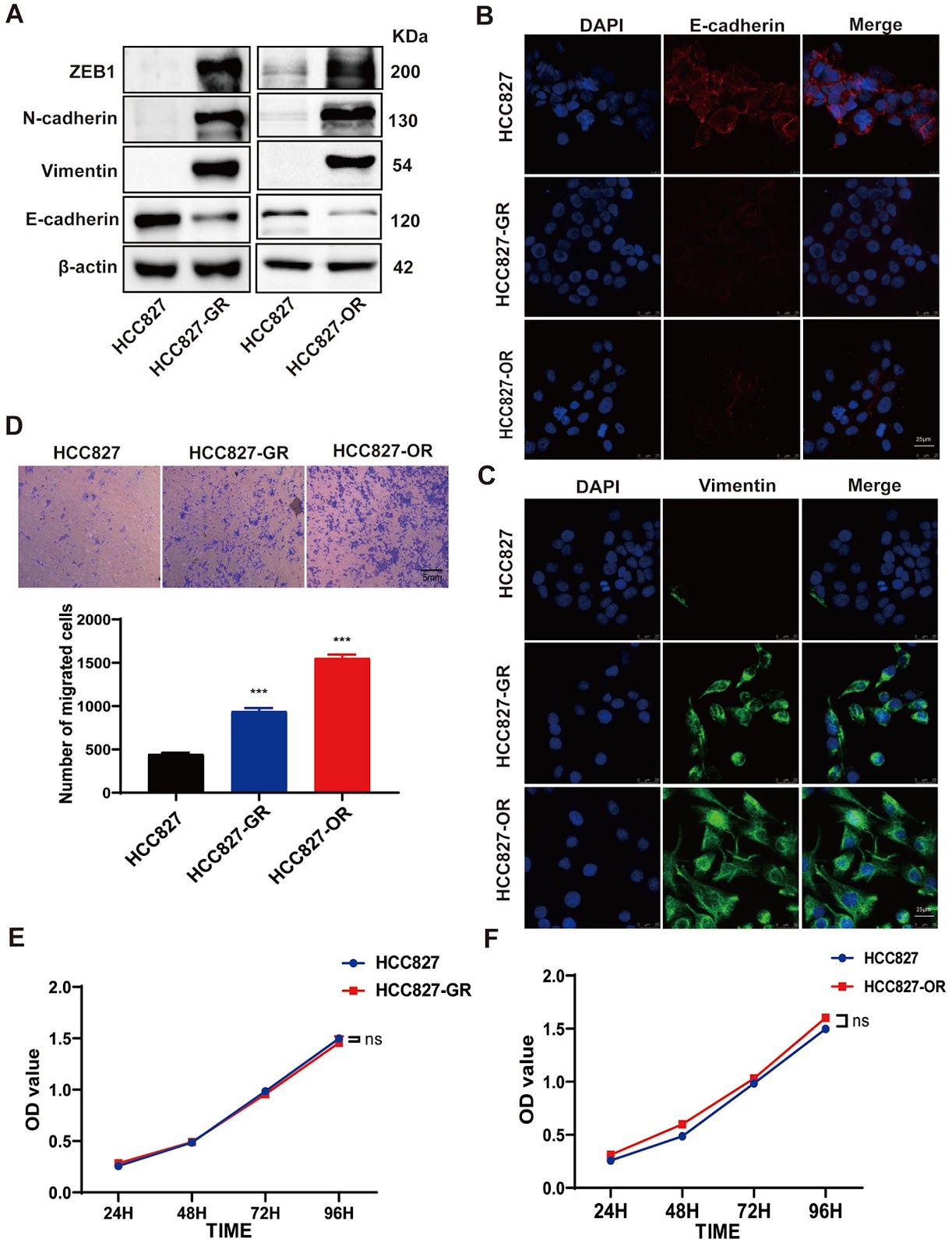
Acquired resistance to EGFR tyrosine kinase inhibitors (EGFR-TKIs) represents a primary cause of treatment failure in non-small cell lung cancer (NSCLC) patients. Chemokine (C-C motif) ligand 2 (CCL2) is recently found to play a pivotal role in determining anti-cancer treatment response. However, the role and mechanism of CCL2 in the development of EGFR-TKIs resistance have not been fully elucidated. In the present study, we focus on the function of CCL2 in the development of acquired resistance to EGFR-TKIs in NSCLC cells. Our results show that CCL2 is aberrantly upregulated in EGFR-TKIs-resistant NSCLC cells and that CCL2 overexpression significantly diminishes sensitivity to EGFR-TKIs. Conversely, CCL2 suppression by CCL2 synthesis inhibitor, bindarit, or CCL2 knockdown can reverse this resistance. CCL2 upregulation can also lead to enhanced migration and increased expressions of epithelial-mesenchymal transition (EMT) markers in EGFR-TKI-resistant NSCLC cells, which could also be rescued by CCL2 knockdown or inhibition. Furthermore, our findings suggest that CCL2-dependent EGFR-TKIs resistance involves the AKT-EMT signaling pathway; inhibition of this pathway effectively attenuates CCL2-induced cell migration and EMT marker expression. In summary, CCL2 promotes the development of acquired EGFR-TKIs resistance and EMT while activating AKT signaling in NSCLC. These insights suggest a promising avenue for the development of CCL2-targeted therapies that prevent EGFR-TKIs resistance in NSCLC.

Read Full Abstract10.3724/abbs.2024106
lncRNA H19 facilitates vascular neointima formation by targeting miR-125a-3p/FLT1 axisGraphical AbstractVerified
Acta Biochimica et Biophysica Sinica

lncRNA H19 facilitates vascular neointima formation by targeting miR-125a-3p/FLT1 axis

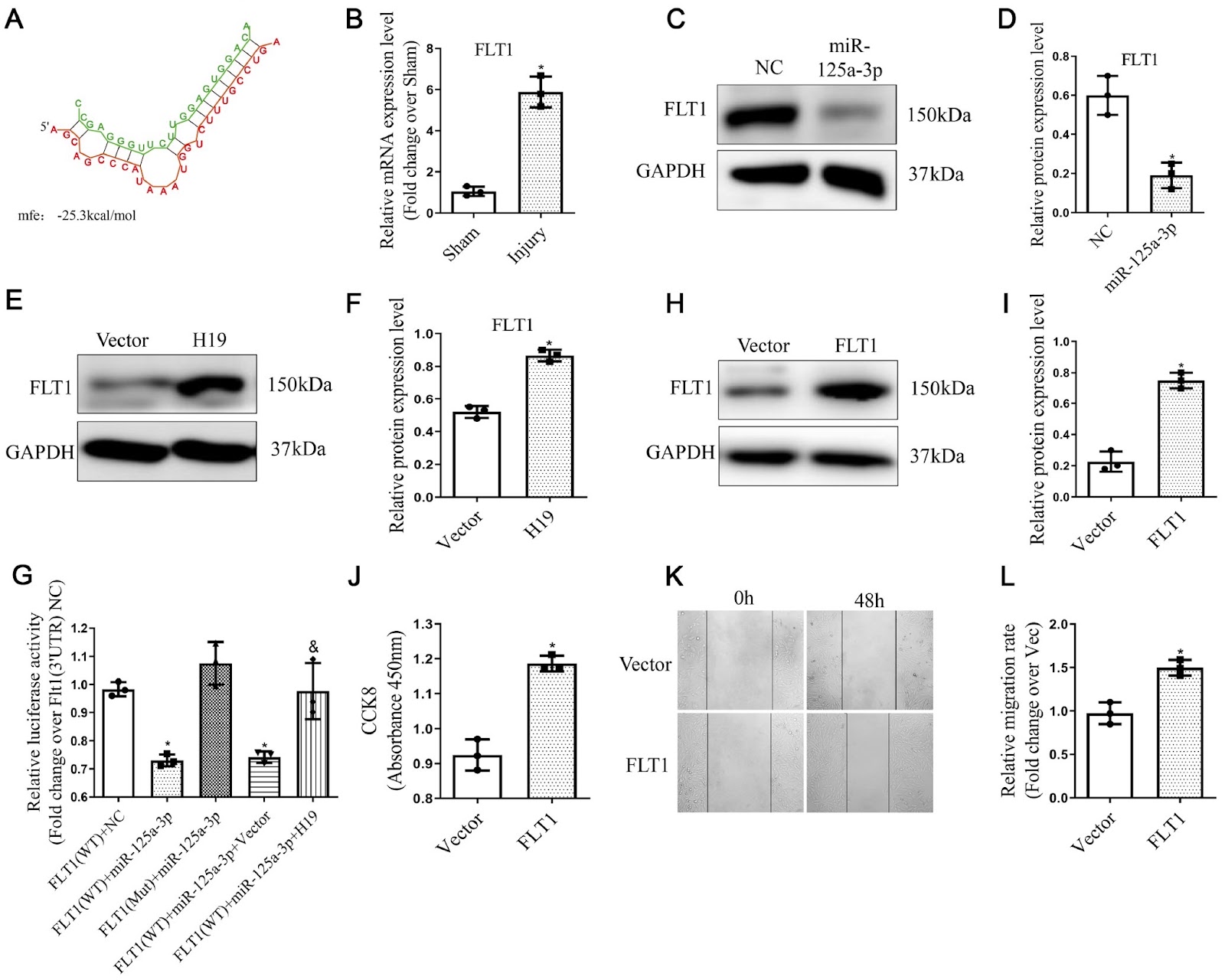
The aberrant proliferation and migration of vascular smooth muscle cells (VSMCs) contribute to the development of neointima formation in vascular restenosis. This study aims to explore the function of the long noncoding RNA H19 in neointima formation. A mouse carotid ligation model was established, and human vascular smooth muscle cells (VSMCs) were used as a cell model. lncRNA H19 overexpression promoted VSMC proliferation and migration. Moreover, miR-125a-3p potentially bound to lncRNA H19, and Fms-like tyrosine kinase-1 (FLT1) might be a direct target of miR-125a-3p in VSMCs. Upregulation of miR-125a-3p alleviated lncRNA H19-enhanced VSMC proliferation and migration. Furthermore, rescue experiments showed that enhanced expression of miR-125a-3p attenuated lncRNA H19-induced FLT1 expression in VSMCs. In addition, the overexpression of lncRNA H19 significantly exacerbated neointima formation in a mouse carotid ligation model. In summary, lncRNA H19 stimulates VSMC proliferation and migration by acting as a competing endogenous RNA (ceRNA) of miR-125a-3p. lncRNA H19 may be a therapeutic target for restenosis.

Read Full Abstract10.3724/abbs.2024087
miR-194-3p regulates epithelial-mesenchymal transition in embryonic epicardial cells via p120/β-catenin signalingGraphical AbstractVerified
Acta Biochimica et Biophysica Sinica

miR-194-3p regulates epithelial-mesenchymal transition in embryonic epicardial cells via p120/β-catenin signaling

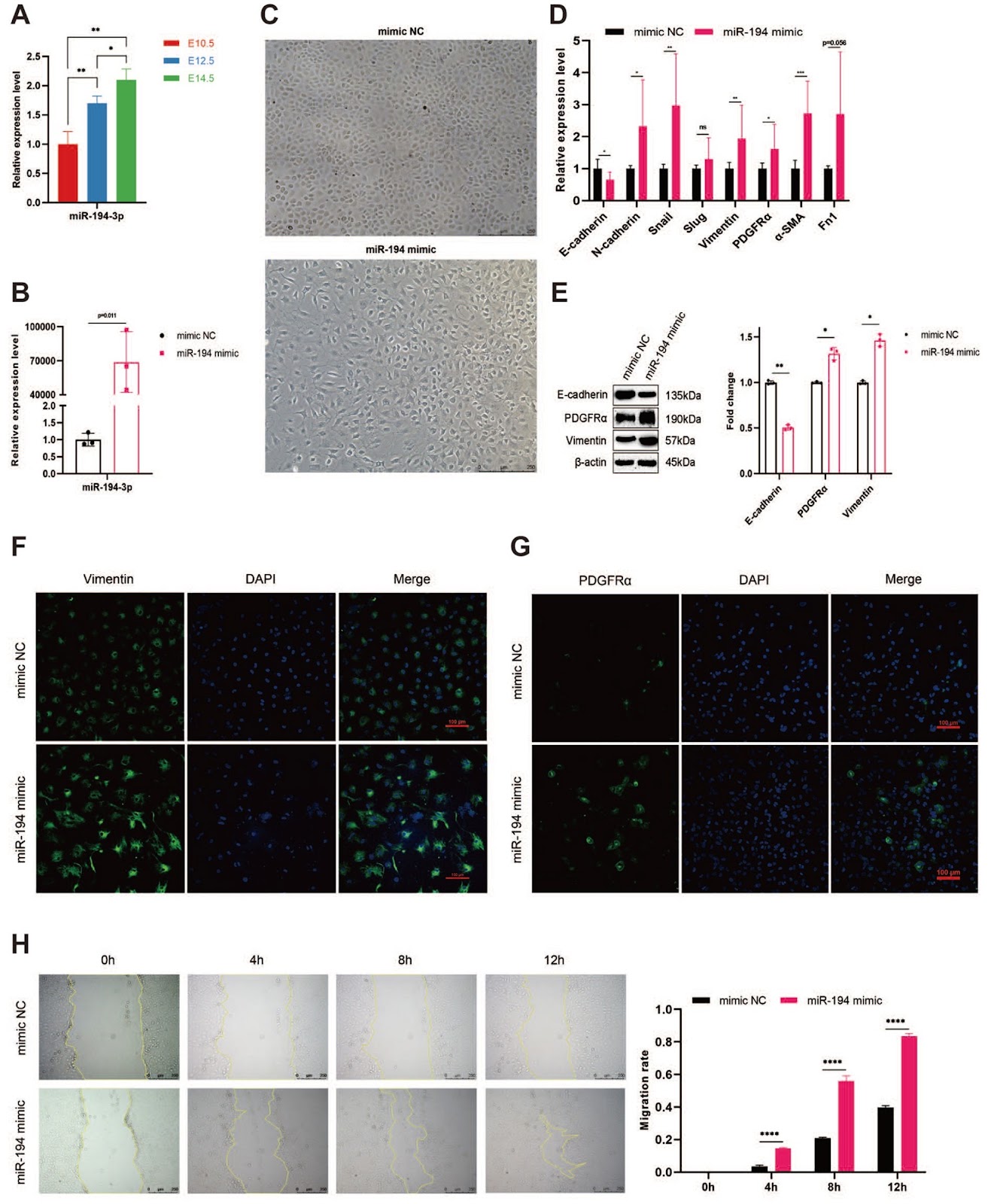
The epicardium is integral to cardiac development and facilitates endogenous heart regeneration and repair. While miR-194-3p is associated with cellular migration and invasion, its impact on epicardial cells remains uncharted. In this work we use gain-of-function and loss-of-function methodologies to investigate the function of miR-194-3p in cardiac development. We culture embryonic epicardial cells in vitro and subject them to transforming growth factor β (TGF-β) treatment to induce epithelial-mesenchymal transition (EMT) and monitor miR-194-3p expression. In addition, the effects of miR-194-3p mimics and inhibitors on epicardial cell development and changes in EMT are investigated. To validate the binding targets of miR-194-3p and its ability to recover the target gene-phenotype, we produce a mutant vector p120-catenin-3′UTR-MUT. In epicardial cells, TGF-β-induced EMT results in a notable overexpression of miR-194-3p. The administration of miR-194-3p mimics promotes EMT, which is correlated with elevated levels of mesenchymal markers. Conversely, miR-194-3p inhibitor attenuates EMT. Further investigations reveal a negative correlation between miR-194-3p and p120-catenin, which influences β-catenin level in the cell adhesion pathway. The suppression of EMT caused by the miR-194-3p inhibitor is balanced by silencing of p120-catenin. In conclusion, miR-194-3p directly targets p120-catenin and modulates its expression, which in turn alters β-catenin expression, critically influencing the EMT process in the embryonic epicardial cells via the cell adhesion mechanism.

Read Full Abstract10.3724/abbs.2024051
Unveiling the cytotoxicity of a new gold(I) complex towards hepatocellular carcinoma by inhibiting TrxR activityGraphical AbstractVerified
Acta Biochimica et Biophysica Sinica

Unveiling the cytotoxicity of a new gold(I) complex towards hepatocellular carcinoma by inhibiting TrxR activity

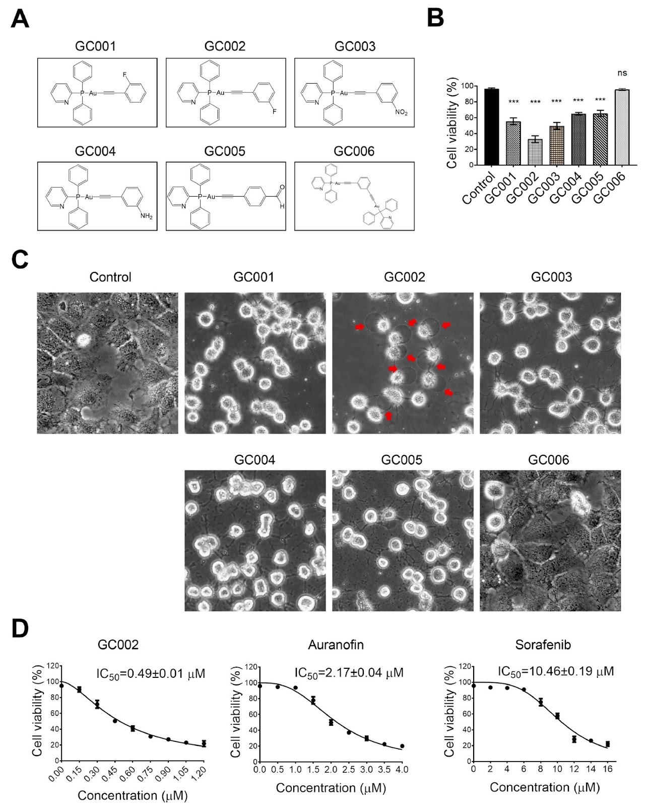
Hepatocellular carcinoma (HCC), the predominant type of liver cancer, is an aggressive malignancy with limited therapeutic options. In this study, we assess a collection of newly designed gold(I) phosphine complexes. Remarkably, the compound GC002 exhibits the greatest toxicity to HCC cells and outperforms established medications, such as sorafenib and auranofin, in terms of antitumor efficacy. GC002 triggers irreversible necroptosis in HCC cells by increasing the intracellular accumulation of reactive oxygen species (ROS). Mechanistically, GC002 significantly suppresses the activity of thioredoxin reductase (TrxR), which plays a crucial role in regulating redox homeostasis and is often overexpressed in HCC by binding directly to the enzyme. Our in vivo xenograft study confirms that GC002 possesses remarkable antitumor activity against HCC without severe side effects. These findings not only highlight the novel mechanism of controlling necroptosis via TrxR and ROS but also identify GC002 as a promising candidate for the further development of antitumor agents targeting HCC.

Read Full Abstract10.3724/abbs.2024155
Glycosylation in the tumor immune response: the bitter side of sweetnessGraphical AbstractVerified
Acta Biochimica et Biophysica Sinica

Glycosylation in the tumor immune response: the bitter side of sweetness

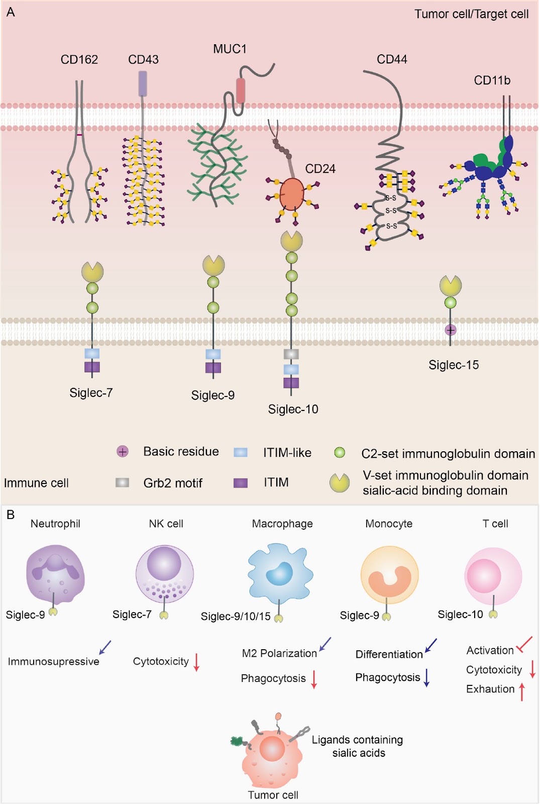
Glycosylation is the most structurally diverse form of post-translational modification (PTM) of proteins that affects a myriad of cellular processes. As a pivotal regulator of protein homeostasis, glycosylation notably impacts the function of proteins, spanning from protein localization and stability to protein-protein interactions. Aberrant glycosylation is a hallmark of cancer, and extensive studies have revealed the multifaceted roles of glycosylation in tumor growth, migration, invasion and immune escape. Over the past decade, glycosylation has emerged as an immune regulator in the tumor microenvironment (TME). Here, we summarize the intricate interplay between glycosylation and the immune system documented in recent literature, which orchestrates the regulation of the tumor immune response through endogenous lectins, immune checkpoints and the extracellular matrix (ECM) in the TME. In addition, we discuss the latest progress in glycan-based cancer immunotherapy. This review provides a basic understanding of glycosylation in the tumor immune response and a theoretical framework for tumor immunotherapy.

Read Full Abstract10.3724/abbs.2024107
Targeting the TRAF3-ULK1-NLRP3 regulatory axis to control alveolar macrophage pyroptosis in acute lung injuryGraphical AbstractVerified
Acta Biochimica et Biophysica Sinica

Targeting the TRAF3-ULK1-NLRP3 regulatory axis to control alveolar macrophage pyroptosis in acute lung injury

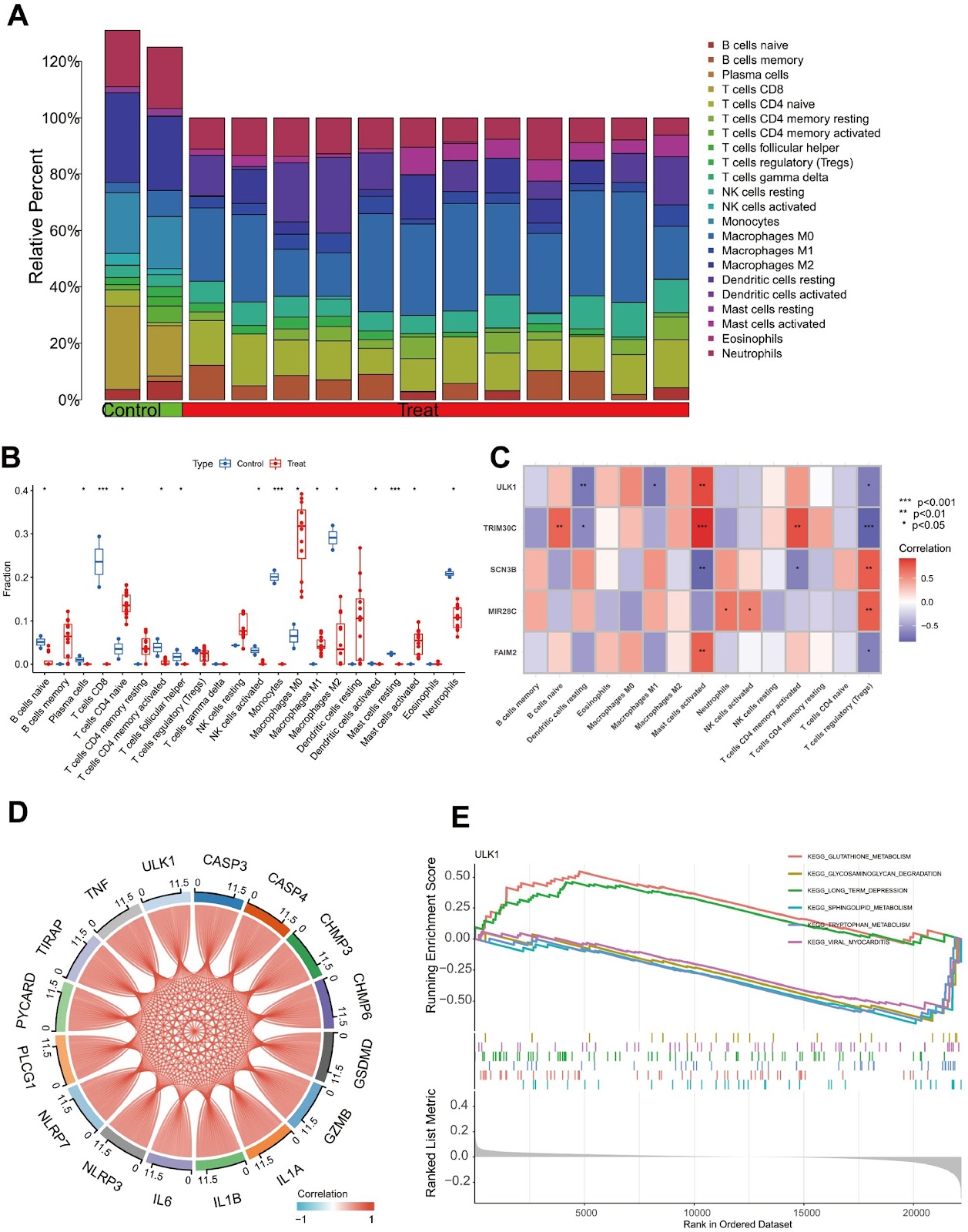
Acute lung injury (ALI) is a serious condition characterized by damage to the lungs. Recent research has revealed that activation of the NLRP3 inflammasome in alveolar macrophages, a type of immune cell in the lungs, plays a key role in the development of ALI. This process, known as pyroptosis, contributes significantly to ALI pathogenesis. Researchers have conducted comprehensive bioinformatics analyses and identified 15 key genes associated with alveolar macrophage pyroptosis in ALI. Among these, NLRP3 has emerged as a crucial regulator. This study further reveal that the ULK1 protein diminishes the expression of NLRP3, thereby reducing the immune response of alveolar macrophages and mitigating ALI. Conversely, TRAF3, another protein, is found to inhibit ULK1 through a process called ubiquitination, leading to increased activation of the NLRP3 inflammasome and exacerbation of ALI. This TRAF3-mediated suppression of ULK1 and subsequent activation of NLRP3 are confirmed through various in vitro and in vivo experiments. The presence of abundant M0 and M1 alveolar macrophages in the ALI tissue samples further support these findings. This research highlights the TRAF3-ULK1-NLRP3 regulatory axis as a pivotal pathway in ALI development and suggests that targeting this axis could be an effective therapeutic strategy for ALI treatment.

Read Full Abstract10.3724/abbs.2024035
RNA modifications: emerging players in the regulation of reproduction and developmentGraphical AbstractVerified
Acta Biochimica et Biophysica Sinica

RNA modifications: emerging players in the regulation of reproduction and development

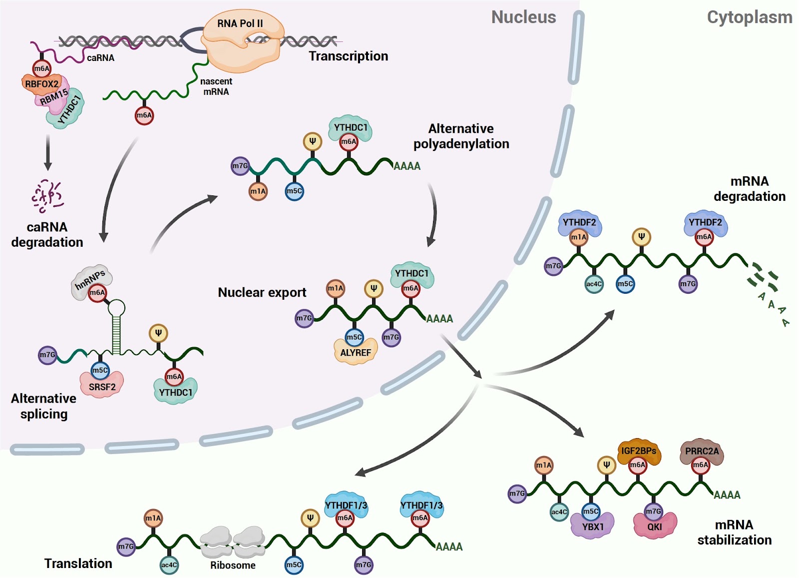
The intricate world of RNA modifications, collectively termed the epitranscriptome, covers over 170 identified modifications and impacts RNA metabolism and, consequently, almost all biological processes. In this review, we focus on the regulatory roles and biological functions of a panel of dominant RNA modifications (including m6A, m5C, Ψ, ac4C, m1A, and m7G) on three RNA types—mRNA, tRNA, and rRNA—in mammalian development, particularly in the context of reproduction as well as embryonic development. We discuss in detail how those modifications, along with their regulatory proteins, affect RNA processing, structure, localization, stability, and translation efficiency. We also highlight the associations among dysfunctions in RNA modification-related proteins, abnormal modification deposition and various diseases, emphasizing the roles of RNA modifications in critical developmental processes such as stem cell self-renewal and cell fate transition. Elucidating the molecular mechanisms by which RNA modifications influence diverse developmental processes holds promise for developing innovative strategies to manage developmental disorders. Finally, we outline several unexplored areas in the field of RNA modification that warrant further investigation.

Read Full Abstract10.3724/abbs.2024201
Nuclear mRNA exportGraphical AbstractVerified
Acta Biochimica et Biophysica Sinica

Nuclear mRNA export

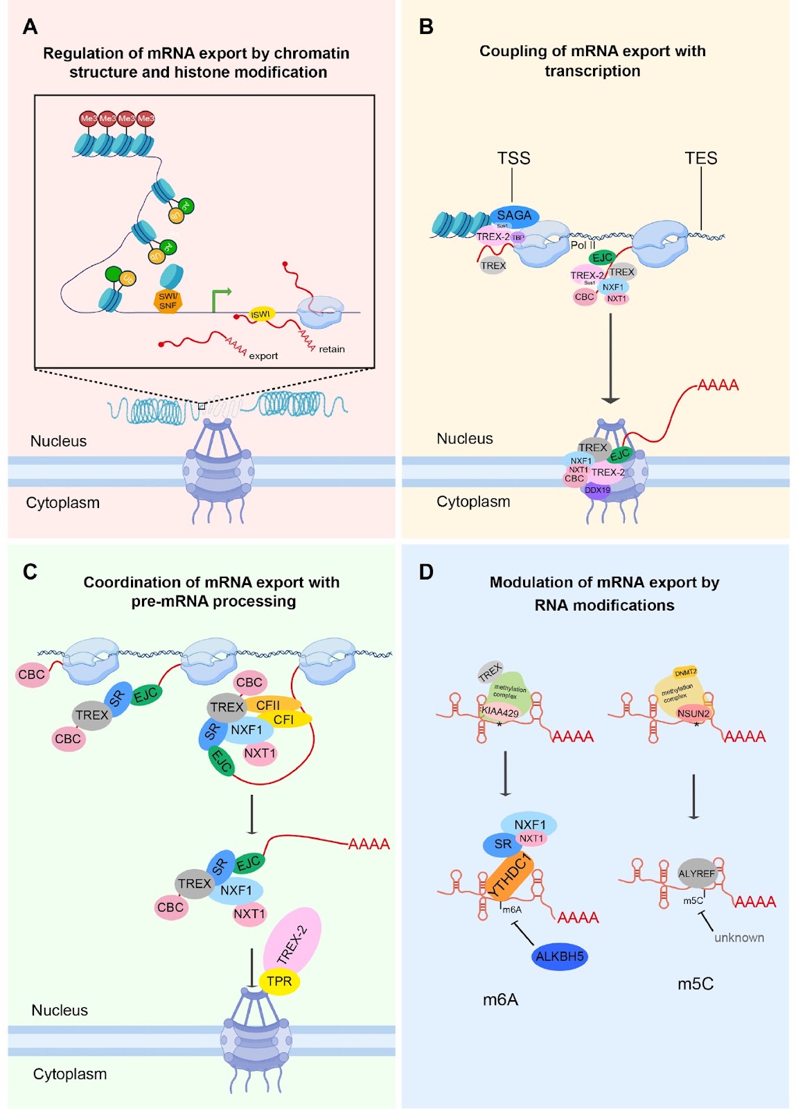
In eukaryotic cells, gene expression begins with transcription in the nucleus, followed by the maturation of messenger RNAs (mRNAs). These mRNA molecules are then exported to the cytoplasm through the nuclear pore complex (NPC), a process that serves as a critical regulatory phase of gene expression. The export of mRNA is intricately linked to precursor mRNA (pre-mRNA) processing, ensuring that only properly processed mRNA reaches the cytoplasm. This coordination is essential, as recent studies have revealed that mRNA export factors not only assist in transport but also influence upstream processing steps, adding a layer of complexity to gene regulation. Furthermore, the export process competes with RNA processing and degradation pathways, maintaining a delicate balance vital for accurate gene expression. While these mechanisms are generally conserved across eukaryotes, significant differences exist between yeast and higher eukaryotic cells, particularly due to the more genome complexity of the latter. This review delves into the current research on mRNA export in higher eukaryotic cells, focusing on its role in the broader context of gene expression regulation and highlighting how it interacts with other gene expression processes to ensure precise and efficient gene functionality in complex organisms.

Read Full Abstract10.3724/abbs.2024145
A subunit vaccine based on Brucella rBP26 induces Th1 immune responses and M1 macrophage activationGraphical AbstractVerified
Acta Biochimica et Biophysica Sinica

A subunit vaccine based on Brucella rBP26 induces Th1 immune responses and M1 macrophage activation

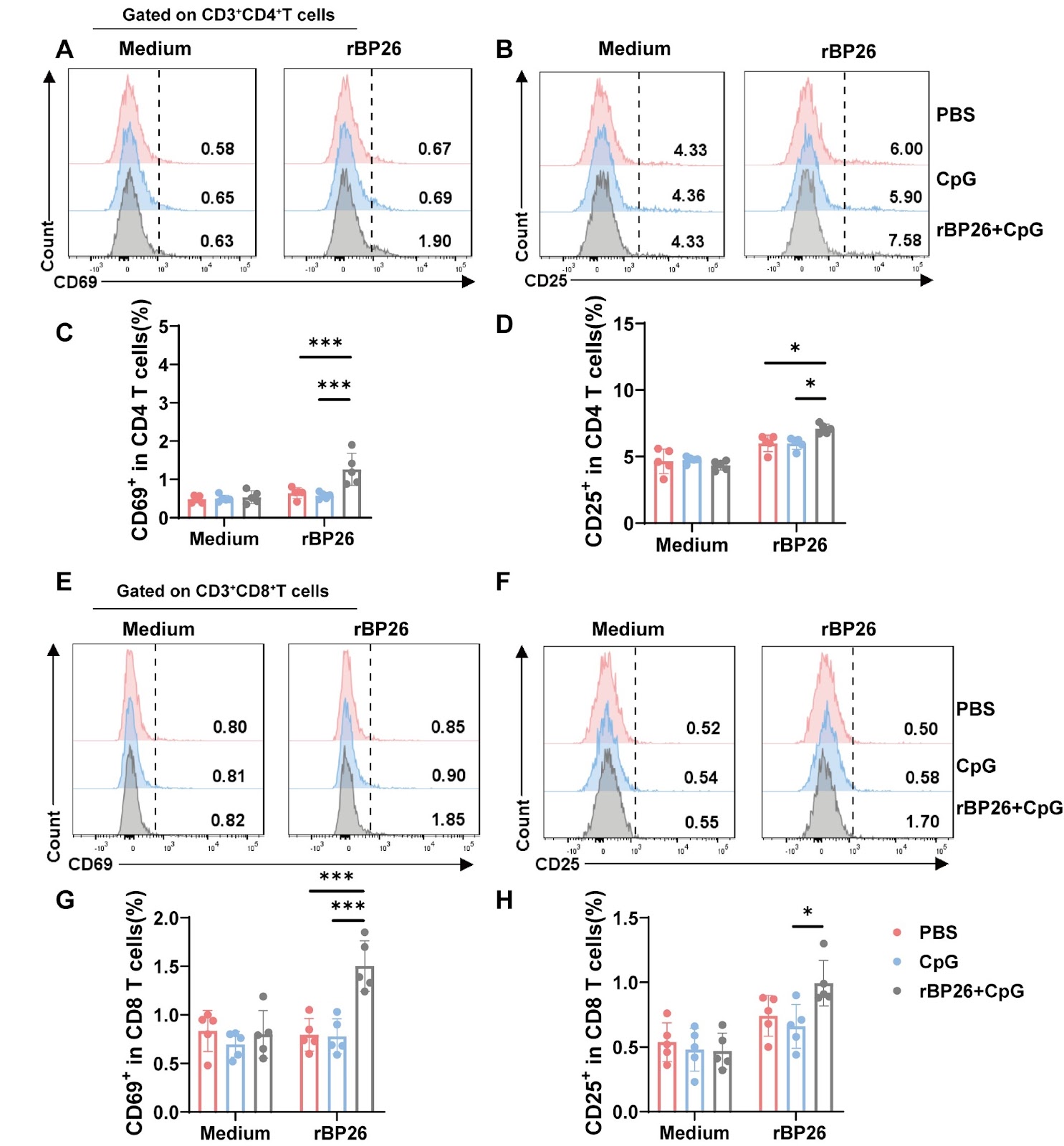
Brucellosis is a global zoonotic infection caused by Brucella bacteria, which poses a significant burden on society. While transmission prevention is currently the most effective method, the absence of a licenced vaccine for humans necessitates the urgent development of a safe and effective vaccine. Recombinant protein-based subunit vaccines are considered promising options, and in this study, the Brucella BP26 protein is expressed using prokaryotic expression systems. The immune responses are evaluated using the well-established adjuvant CpG-ODN. The results demonstrate that rBP26 supplemented with a CpG adjuvant induces M1 macrophage polarization and stimulates cellular immune responses mediated by Th1 cells and CD8+ T cells. Additionally, it generates high levels of rBP26-specific antibodies in immunized mice. Furthermore, rBP26 immunization activates, proliferates, and produces cytokines in T lymphocytes while also maintaining immune memory for an extended period of time. These findings shed light on the potential biological function of rBP26, which is crucial for understanding brucellosis pathogenesis. Moreover, rBP26 holds promise as an effective subunit vaccine candidate for use in endemic areas.

Read Full Abstract10.3724/abbs.2024023
Integrated 16S rRNA sequencing and metabolomic analysis reveals the potential protective mechanism of Germacrone on diabetic nephropathy in miceGraphical AbstractVerified
Acta Biochimica et Biophysica Sinica

Integrated 16S rRNA sequencing and metabolomic analysis reveals the potential protective mechanism of Germacrone on diabetic nephropathy in mice

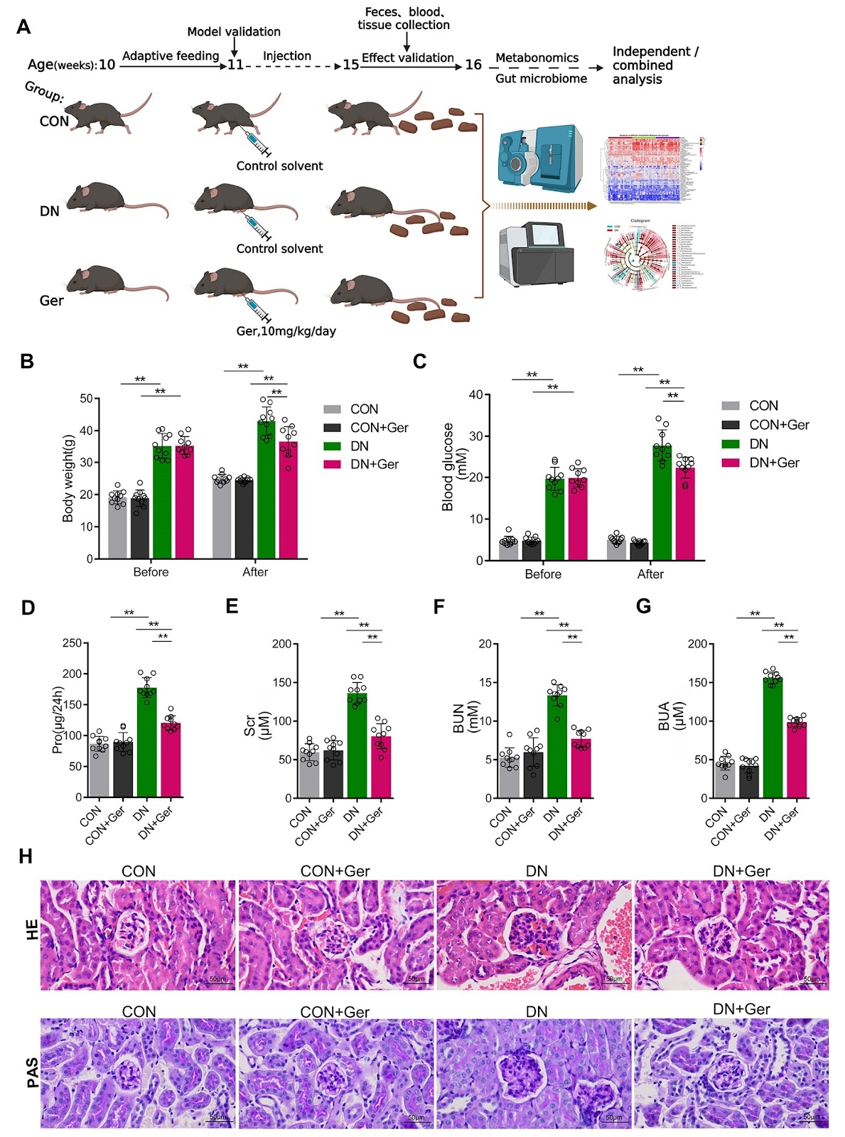
Diabetic nephropathy (DN) is a severe complication of diabetes and the leading cause of end-stage renal disease and death. Germacrone (Ger) possesses anti-inflammatory, antioxidant and anti-DN properties. However, it is unclear whether the improvement in kidney damage caused by Ger in DN mice is related to abnormal compositions and metabolites of the gut microbiota. This study generates a mouse model of DN to explore the potent therapeutic ability and mechanism of Ger in renal function by 16S rRNA sequencing and untargeted fecal metabolomics. Although there is no significant change in microbiota diversity, the structure of the gut microbiota in the DN group is quite different. Serratia_marcescens and Lactobacillus_iners are elevated in the model group but significantly decreased after Ger intervention (P<0.05). Under the treatment of Ger, no significant differences in the diversity and richness of the gut microbiota are observed. An imbalance in the intestinal flora leads to the dysregulation of metabolites, and non-targeted metabolomics data indicate high expression of stearic acid in the DN group, and oleic acid could serve as a potential marker of the therapeutic role of Ger in the DN model. Overall, Ger improves kidney injury in diabetic mice, in part potentially by reducing the abundance of Serratia_marcescens and Lactobacillus_iners, as well as regulating the associated increase in metabolites such as oleic acid, lithocholic acid and the decrease in stearic acid. Our research expands the understanding of the relationship between the gut microbiota and metabolites in Ger-treated DN. This contributes to the usage of natural products as a therapeutic approach for the treatment of DN via microbiota regulation.

Read Full Abstract10.3724/abbs.2024021
Novel FGF21 analogues through structure-based optimization for therapeutic developmentGraphical AbstractVerified
Acta Biochimica et Biophysica Sinica

Novel FGF21 analogues through structure-based optimization for therapeutic development

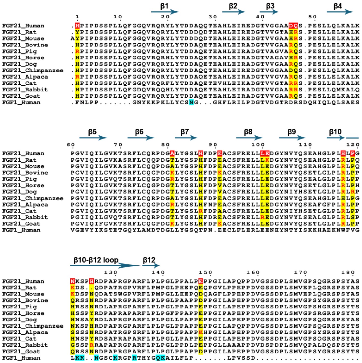
Fibroblast growth factor 21 (FGF21) plays a pivotal role in regulating metabolic processes and energy homeostasis, making it a promising therapeutic avenue for various obesity-related conditions. However, its therapeutic efficacy faces challenges due to its suboptimal pharmacokinetics and bioactivity. To overcome these limitations, we adapt a strategy in which key amino acid residues responsible for enhanced activity are pinpointed through sequence alignment and comparative analysis to develop long-acting FGF21 analogs. The mutant FGF21 analogs are fused with the Fc fragment. Here, we report the design, identification, and characterization of two distinct Fc-fused FGF21 analogs, Fc-FGF21(P119R) and Fc-FGF21(H125R), with significantly augmented potency. These findings hold promise for clinical applications, offering potential interventions for obesity-related metabolic disorders.

Read Full Abstract10.3724/abbs.2024227
Melatonin protects TEGDMA-induced preodontoblast mitochondrial apoptosis via the JNK/MAPK signaling pathwayGraphical AbstractVerified
Acta Biochimica et Biophysica Sinica

Melatonin protects TEGDMA-induced preodontoblast mitochondrial apoptosis via the JNK/MAPK signaling pathway

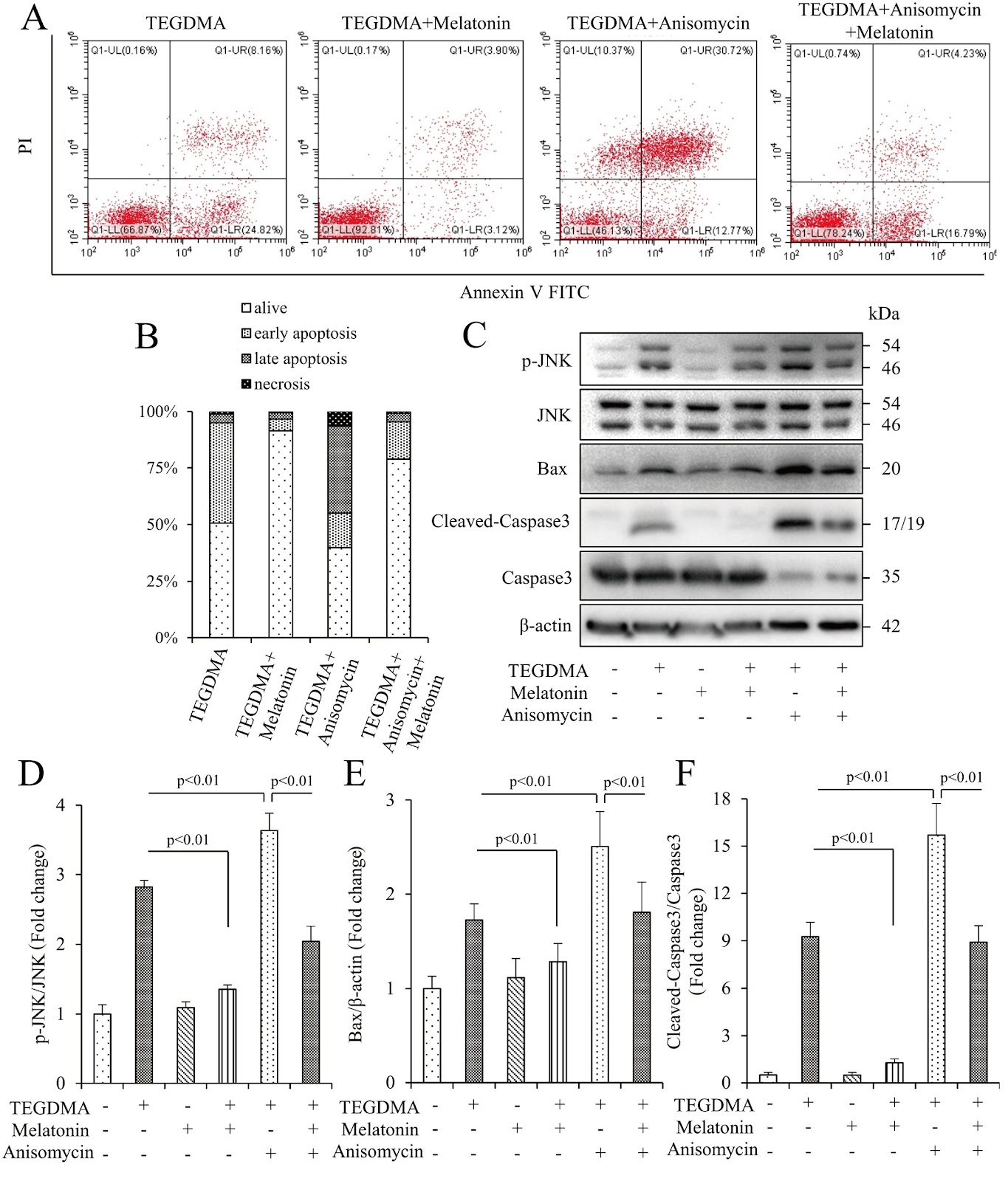
Resin monomer-induced dental pulp injury presents a pathology related to mitochondrial dysfunction. Melatonin has been regarded as a strong mitochondrial protective bioactive compound from the pineal gland. However, it remains unknown whether melatonin can prevent dental pulp from resin monomer-induced injury. The aim of this study is to investigate the effects of melatonin on apoptosis of mouse preodontoblast cells (mDPC6T) induced by triethylene glycol dimethacrylate (TEGDMA), a major component in dental resin, and to determine whether the JNK/MAPK signaling pathway mediates the protective effect of melatonin. A well-established TEGDMA-induced mDPC6T apoptosis model is adopted to investigate the preventive function of melatonin by detecting cell viability, apoptosis rate, expressions of apoptosis-related proteins, mitochondrial ROS (mtROS) production, mitochondrial membrane potential (MMP) and adenosine triphosphate (ATP) level. Inhibitors of MAPKs are used to explore which pathway is involved in TEGDMA-induced apoptosis. Finally, the role of the JNK/MAPK pathway is verified using JNK agonists and antagonists. Our results show that melatonin attenuates TEGDMA-induced mDPC6T apoptosis by reducing mtROS production and rescuing MMP and ATP levels. Furthermore, mitochondrial dysfunction and apoptosis are alleviated only by the JNK/MAPK inhibitor SP600125 but not by other MAPK inhibitors. Additionally, melatonin downregulates the expression of phosphorylated JNK and counteractes the activating effects of anisomycin on the JNK/MAPK pathway, mimicking the effects of SP600125. Our findings demonstrate that melatonin protects mDPC6T cells against TEGDMA-induced apoptosis partly through JNK/MAPK and the maintenance of mitochondrial function, offering a novel therapeutic strategy for the prevention of resin monomer-induced dental pulp injury.

Read Full Abstract10.3724/abbs.2023263
Agrimol B alleviates cisplatin-induced acute kidney injury by activating the Sirt1/Nrf2 signaling pathway in miceGraphical AbstractVerified
Acta Biochimica et Biophysica Sinica

Agrimol B alleviates cisplatin-induced acute kidney injury by activating the Sirt1/Nrf2 signaling pathway in mice

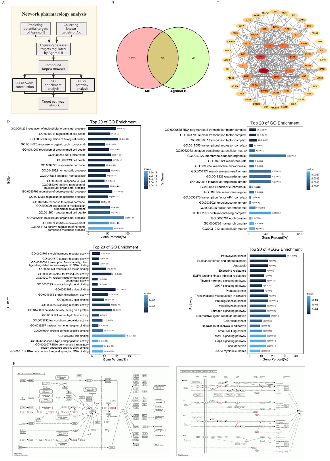
Cisplatin (CDDP) is a widely used chemotherapeutic agent that has remarkable antineoplastic effects. However, CDDP can cause severe acute kidney injury (AKI), which limits its clinical application. Agrimol B is the main active ingredient found in Agrimonia pilosa Ledeb and has a variety of pharmacological activities. The effect of agrimol B on CDDP-induced renal toxicity has not been determined. To investigate whether agrimol B has a protective effect against CDDP-induced AKI, we first identify Sirtuin 1 (Sirt1) as a critical target protein of agrimol B in regulating AKI through network pharmacology analysis. Subsequently, the AKI mouse model is induced by administering a single dose of CDDP via intraperitoneal injection. By detecting the serum urea nitrogen and creatinine levels, as well as the histopathological changes, we confirm that agrimol B effectively reduces CDDP-induced AKI. In addition, treatment with agrimol B counteracts the increase in renal malondialdehyde level and the decrease in superoxide dismutase (SOD), catalase and glutathione levels induced by CDDP. Moreover, western blot results reveal that agrimol B upregulates the expressions of Sirt1, SOD2, nuclear factor erythroid2-related factor 2, and downstream molecules, including heme oxygenase 1 and NAD(P)H quinone dehydrogenase 1. However, administration of the Sirt1 inhibitor EX527 abolishes the effects of agrimol B. Finally, we establish a tumor-bearing mouse model and find that agrimol B has a synergistic antitumor effect with CDDP. Overall, agrimol B attenuates CDDP-induced AKI by activating the Sirt1/Nrf2 signaling pathway to counteract oxidative stress, suggesting that this compound is a potential therapeutic agent for the treatment of CDDP-induced AKI.

Read Full Abstract10.3724/abbs.2023285
Unveiling a novel GJB2 dominant K22T mutation in a Chinese family with hearing lossGraphical AbstractVerified
Acta Biochimica et Biophysica Sinica

Unveiling a novel GJB2 dominant K22T mutation in a Chinese family with hearing loss

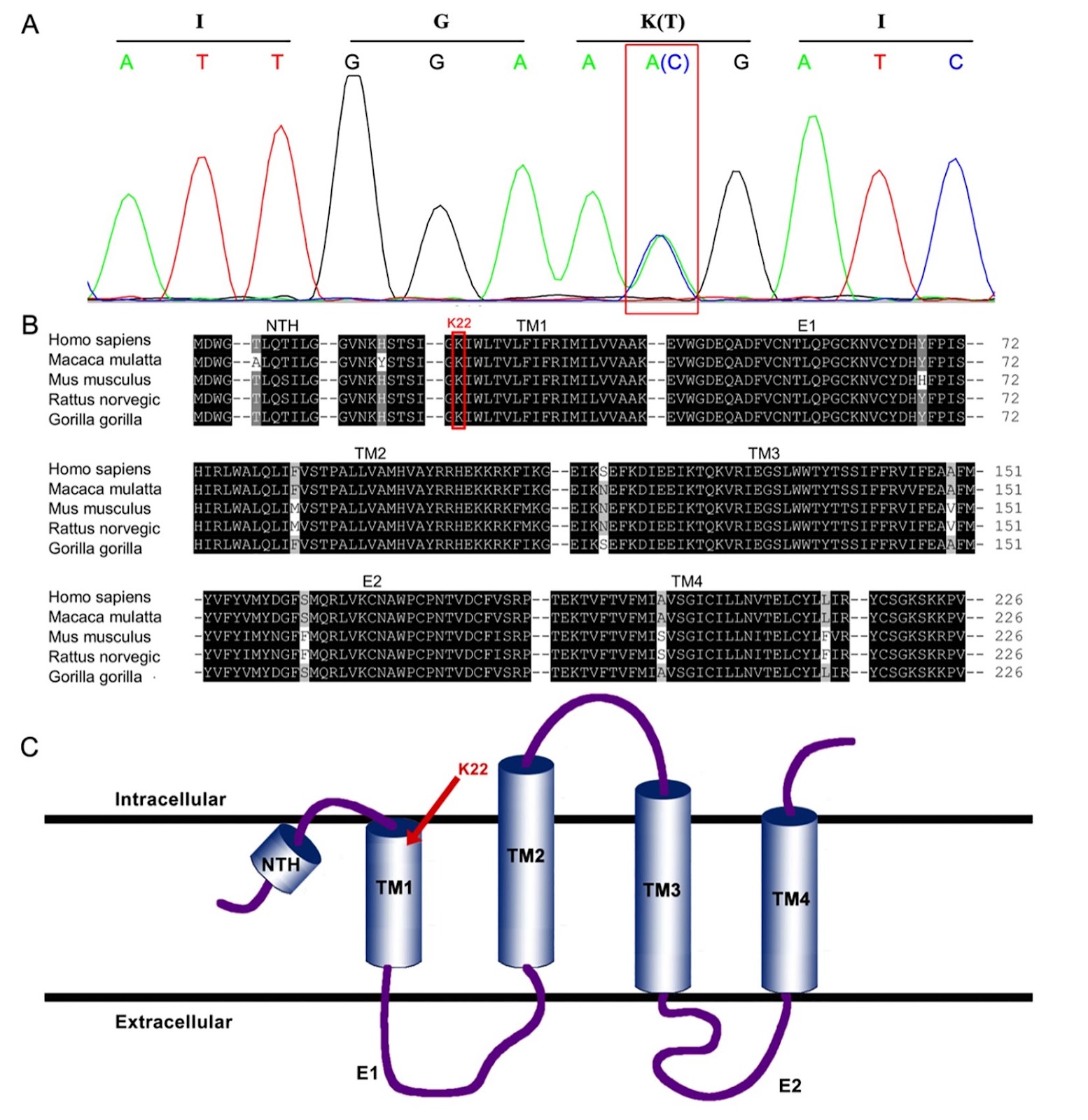
Hearing loss constitutes one of the most prevalent conditions within the field of otolaryngology. Recent investigations have revealed that mutations in deafness-associated genes, including point mutations and variations in DNA sequences, can cause hearing impairments. With the ethology of deafness remaining unclear for a substantial portion of the affected population, further screenings for pathogenic mutations are imperative to unveil the underlying mechanisms. On this study, by using next-generation sequencing, we examine 129 commonly implicated deafness-related genes in a Chinese family with hearing loss, revealing a novel heterozygous dominant mutation in the GJB2 gene (GJB2: c.65T>G: p. Lys22Thr). This mutation consistently occurs in affected family members but is not detected in unaffected individuals, strongly suggesting its causative role in hearing loss. Structural analysis indicates potential disruption to the Cx26 gap junction channel’s hydrogen bond and electrostatic interactions, aligning with predictions from the PolyPhen and SIFT algorithms. In conclusion, our study provides conclusive evidence that the identified heterozygous GJB2 mutation (GJB2: c.65T>G: p. Lys22Thr), specifically the K22T alteration, is the primary determinant of the family’s deafness. This contribution enhances our understanding of the interplay between common deafness-associated genes and hearing loss, offering valuable insights for diagnostic guidance and the formulation of therapeutic strategies for this condition.

Read Full Abstract10.3724/abbs.2024064
Structure-based design of covalent nanobody binders for a thermostable green fluorescence proteinGraphical AbstractVerified
Acta Biochimica et Biophysica Sinica

Structure-based design of covalent nanobody binders for a thermostable green fluorescence protein

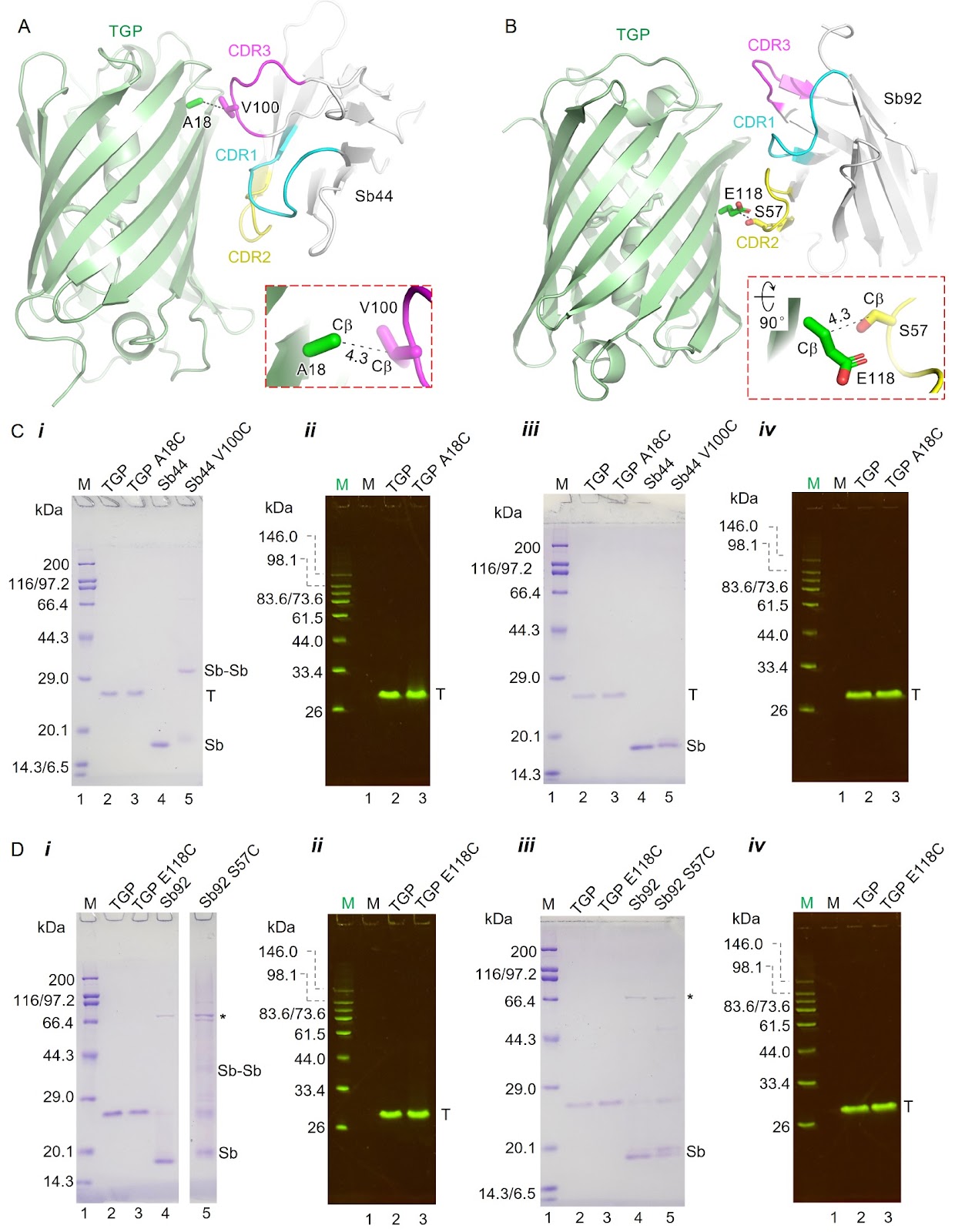
The use of green fluorescence protein (GFP) has advanced numerous areas of life sciences. An ultra-thermostable GFP (TGP), engineered from a coral GFP, offers potential advantages over traditional jellyfish-derived GFP because of its high stability. However, owing to its later discovery, TGP lacks the extensive toolsets available for GFP, such as heavy chain-only antibody binders known as nanobodies. In this study, we report the crystal structure of TGP in complex with Sb92, a synthetic nanobody identified from a previous in vitro screening, revealing Sb92’s precise three-dimensional epitope. This structural insight, alongside the previously characterized Sb44-TGP complex, allows us to rationally design disulfide bonds between the antigen and the antibody for tighter interactions. Using biochemical analysis, we identify two bridged complexes (TGP A18C-Sb44 V100C and TGP E118C-Sb92 S57C), with the TGP-Sb92 disulfide pair showing high resistance to reducing agents. Our study expands the toolkit available for TGP and should encourage its wider applications.

Read Full Abstract10.3724/abbs.2024233
Retraction: miR-129-5p suppresses proliferation, migration, and induces apoptosis in pancreatic cancer cells by targeting PBX3Graphical AbstractVerified
Acta Biochimica et Biophysica Sinica

Retraction: miR-129-5p suppresses proliferation, migration, and induces apoptosis in pancreatic cancer cells by targeting PBX3

This article has been retracted by the authors and the Editor of Acta Biochimica et Biophysica Sinica. The experimental results of the original paper could not be repeated, leading to inaccurate results and conclusions. The authors apologize for any inconvenience caused.

Read Full Abstract10.3724/abbs.2024209
Evolutionary analysis of paired box gene family and biological function exploration of Lr.Pax7 in lamprey (Lethenteron reissneri)Graphical AbstractVerified
Acta Biochimica et Biophysica Sinica

Evolutionary analysis of paired box gene family and biological function exploration of Lr.Pax7 in lamprey (Lethenteron reissneri)

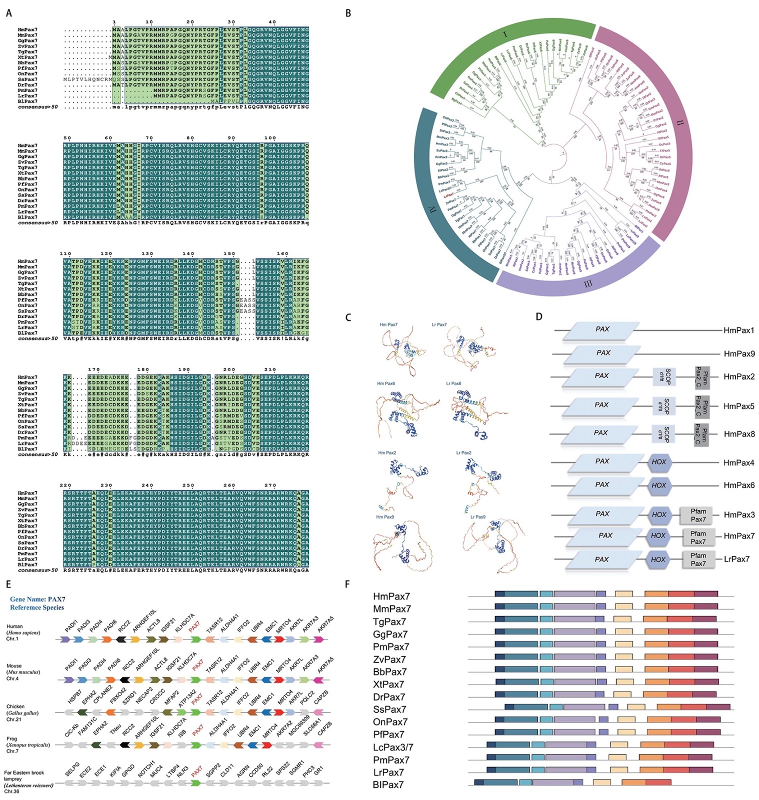
Gene regulation refers to the precise regulation of gene expression in an organism, and transcription factors are proteins that bind to DNA and regulate gene expression by promoting or inhibiting the expressions of target genes. Since the late 1980s [1], scientists have studied special genes called Pax genes that control how genes function in organisms as they grow. There are nine Pax genes found in animals such as mice, zebrafish, and humans [2]. Based on the composition domain and homology of the sequence, the Pax family is divided into four subfamilies: Pax1/9, Pax2/5/8, Pax4/6, and Pax3/7 [3]. Pax7 plays a pivotal role in the implementation, protection, and repair of skeletal muscle. Pax7 helps to control the balance between self-renewal and differentiation of satellite cells, ensuring that they can proliferate when needed to generate new muscle cells and differentiate into mature muscle fibers when necessary for muscle development and repair. The expression of the Pax7 gene in nerve cells is critical for dorsal root and sensory ganglia development. The Pax7 gene serves as a primary controlling factor for skeletal muscle development while influencing different biological processes; however, its exact role in jawless vertebrates such as lamprey remains unclear, and extensive research is needed to elucidate the intricate underlying mechanisms involved. Given the unique status of lamprey as an ancient jawless fish, possessing an ancient lineage and distinctive biological features, it is rare to explore gene function across hundreds of millions of years of vertebrate evolution. The use of lamprey as a model system for gene function research represents an innovative approach in the fields of evolutionary and comparative genomics. In this study, we investigated the regulatory mechanism of Pax7 in lamprey via gene cloning, gene expression analysis, gene silencing and transcriptome data analysis. We also explored the interactions between genes with significant differences. Identification of Lr.Pax7 in lamprey tissues began with the retrieval of protein sequences that are similar to those of human Pax family members in sea lamprey (Petromyzon marinus) or zebrafish (Danio rerio) from the NCBI protein database (Supplementary Table S1) and the use of BLAST to identify corresponding homologs (Supplementary Table S2). Subsequently, we extracted the Pax sequences from our library. Lethenteron reissneri specimens were dissected to isolate various tissues. Primers targeting the pax domains were designed based on the Pax7 nucleotide sequence in the Lampreys cDNA library, and the aim was to verify the effectiveness of the Lampreys cDNA as a template for validation (Supplementary Table S3). Lr.Pax7 was successfully amplified via PCR in muscle tissue. Here, a variety of methods were used for bioinformatics analysis. The results showed that the amino acid sequence of Pax7 is highly similar among animals (Figure 1A), with a decreasing trend from higher to lower organisms, as revealed by sequence alignment. It can be observed from the evolutionary tree (Figure 1B) that Pax genes for each subfamily are present in ancestral chordate and that Pax genes are present in amphioxus. Petromyzon marinus, Lethenteron camtschaticum, and Lethenteron reissneri constitute a sister group and have become good models for the study of jawless vertebrates. Pax9, Pax2, Pax6, and Pax7 show high similarity to those of other higher vertebrates. Therefore, these genes were named Lr.Pax9, Lr.Pax2, Lr.Pax7, and Lr.Pax6. The results indicate that the Pax7 gene is significantly preserved across various species, from higher to lower. This suggests that the DNA sequence of the gene is remarkably similar among different species. Lr.Pax7 is positioned between vertebrates and invertebrates and is most closely related to P. marinus Pax7. This finding provides more insight into the original evolutionary position of the lamprey. Crystal structure prediction analysis revealed that Lr.Paxs and Hm.Paxs have highly homologous structures (Figure 1C). The Pax gene has a similar structure (Figure 1D), including a conserved DNA-binding structure called the pair-box domain. This structure contains approximately 128 amino acids and is responsible for binding with specific DNA sequences, regulating gene expression, and interacting with other proteins. To further investigate the evolutionary history of Pax7 in vertebrates, we compared the genetic environment of Pax7 with that of other vertebrates (Figure 1E). In addition, many Pax gene members also contain DNA-binding structures called homeodomains, which play important roles in development.
Read Full Abstract10.3724/abbs.2024121
Excessive ER-phagy mediated by FAM134B contributes to trophoblast cell mitochondrial dysfunction in preeclampsiaGraphical AbstractVerified
Acta Biochimica et Biophysica Sinica

Excessive ER-phagy mediated by FAM134B contributes to trophoblast cell mitochondrial dysfunction in preeclampsia

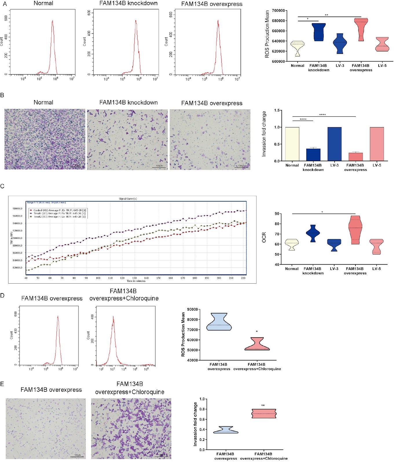
Autophagy dysregulation and Ca2+-induced mitochondrial dysfunction in trophoblast cells are proposed to contribute to preeclampsia (PE) development. FAM134B is identified as a receptor associated with endoplasmic reticulum autophagy (ER-phagy). In this study, the placentas of normal pregnant women and PE patients are collected and analyzed by immunohistochemistry, quantitative real-time PCR, and western blot analysis. The effects of ER-phagy are investigated in HTR8/SVneo cells. Significantly increased levels of FAM134B, inositol-1,4,5-triphosphate receptor type 1 (IP3R), calnexin, cleaved caspase 3 and cytochrome C are detected in the PE placenta and sodium nitroprusside (SNP)-treated HTR-8/SVneo cells. Overexpression of FAM134B in HTR-8/SVneo cells results in increased apoptosis, impaired invasion capacity, and diminished mitochondrial function, while an autophagy inhibitor improves mitochondrial performance. Excessive ER-phagy is also associated with an increased concentration of gamma linolenic acid. Our findings suggest that FAM134B contributes to trophoblast apoptosis by mediating ER-mitochondria Ca2+ transfer through mitochondria-associated endoplasmic reticulum membranes (MAMs) and subsequent mitochondrial function, further enhancing our understanding of PE etiology.

Read Full Abstract10.3724/abbs.2024065
Human umbilical cord mesenchymal stem cells enhance liver regeneration and decrease collagen content in fibrosis mice after partial hepatectomy by activating Wnt/β-catenin signalingGraphical AbstractVerified
Acta Biochimica et Biophysica Sinica

Human umbilical cord mesenchymal stem cells enhance liver regeneration and decrease collagen content in fibrosis mice after partial hepatectomy by activating Wnt/β-catenin signaling

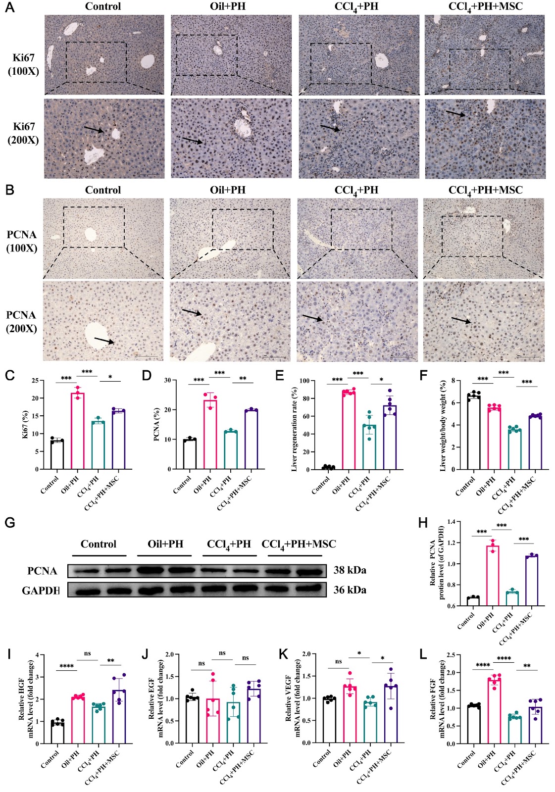
Liver fibrosis is a critical stage in the progression of various chronic liver diseases to cirrhosis and liver cancer. Early inhibition of liver fibrosis is crucial for the treatment of liver disease. Hepatectomy, a common treatment for liver-related diseases, promotes liver regeneration. However, in the context of liver fibrosis, liver regeneration is hindered. Many studies have shown that mesenchymal stem cells (MSCs) can promote liver regeneration after partial hepatectomy (PH). However, there are few reports on the impact of MSC therapy on liver regeneration post-PH in the context of hepatic fibrosis. The objective of this study is to examine the impact of MSCs on liver regeneration following PH in the fibrotic liver and uncover the related molecular mechanisms. This study reveals that MSC therapy significantly enhances liver function and mitigates liver inflammation after PH in the context of hepatic fibrosis. MSCs also significantly promote liver regeneration and alleviate liver fibrosis. In addition, this study identifies the role of MSCs in promoting liver regeneration and alleviating liver fibrosis via the activation of Wnt/β-catenin signaling. The combination of MSCs with hepatectomy may offer a novel approach for the treatment of liver fibrotic diseases.

Read Full Abstract10.3724/abbs.2024207
TIPE1 suppresses the invasion and migration of breast cancer cells and inhibits epithelial-to-mesenchymal transition primarily via the ERK signaling pathwayGraphical AbstractVerified
Acta Biochimica et Biophysica Sinica

TIPE1 suppresses the invasion and migration of breast cancer cells and inhibits epithelial-to-mesenchymal transition primarily via the ERK signaling pathway

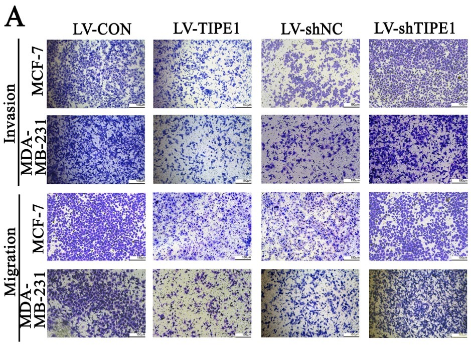
This is a corrigendum to the original article published in Acta Biochimica et Biophysica Sinica 2019, 51(10): 1008–1015. The original article reported that TIPE1 suppresses the invasion and migration of breast cancer cells and inhibits epithelial-to-mesenchymal transition primarily via the ERK signaling pathway. In the original version, an error was found in Figure 2A and Figure 3B respectively. The correct figures are shown in this corrigendum. The authors apologize for the error.

Read Full Abstract10.3724/abbs.2024177