Key Takeaways & Executive Findings
- •• Melatonin attenuates TEGDMA-induced apoptosis in preodontoblast cells by reducing mitochondrial ROS and restoring mitochondrial membrane potential and ATP levels. • The protective effect of melatonin is mediated specifically through the JNK/MAPK signaling pathway, as only the JNK inhibitor SP600125 mimics its effects. • Melatonin downregulates phosphorylated JNK expression and counteracts the activation of the JNK/MAPK pathway by anisomycin. • These findings suggest melatonin as a potential therapeutic agent for preventing resin monomer-induced dental pulp injury.
Abstract
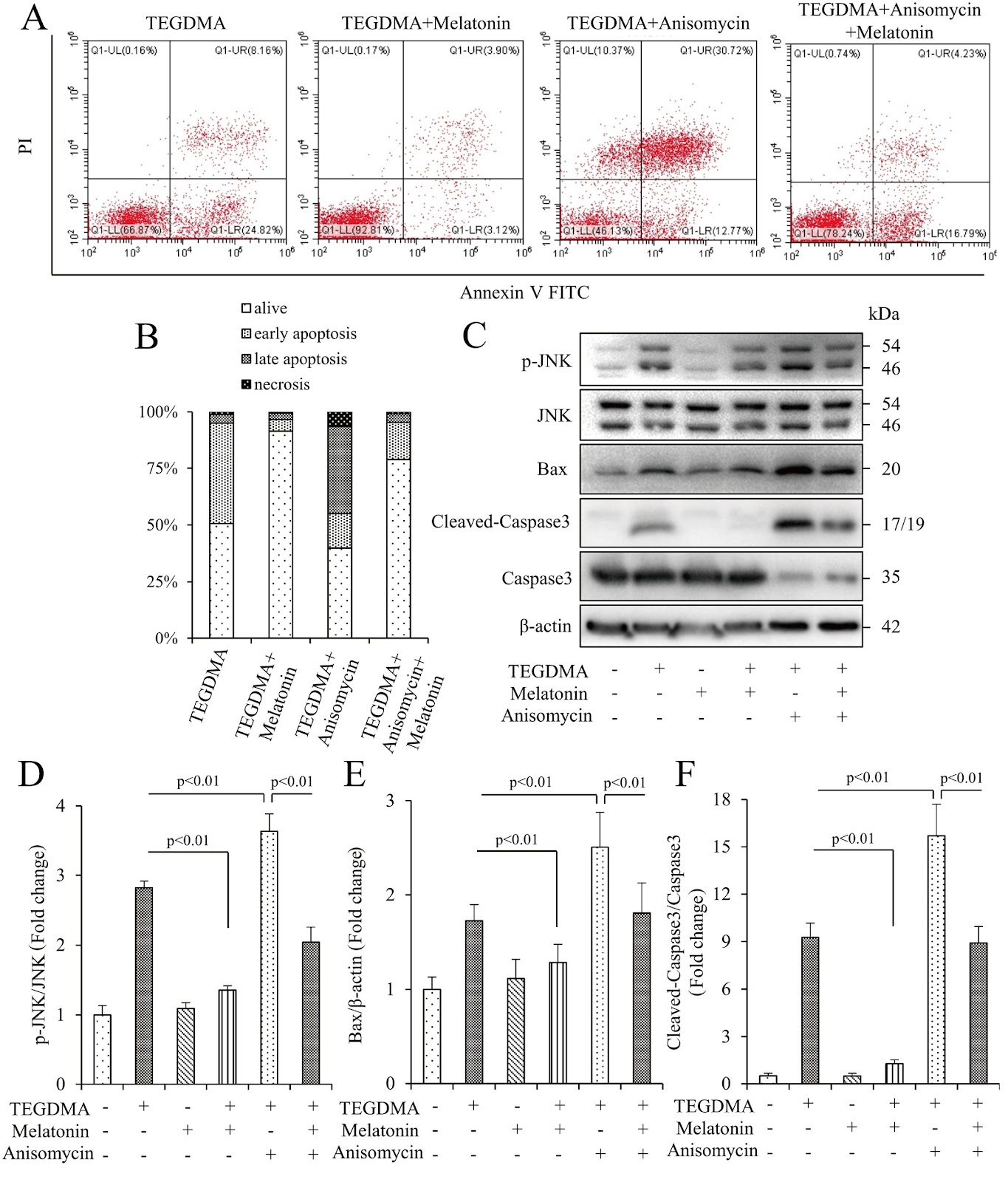
Resin monomer-induced dental pulp injury presents a pathology related to mitochondrial dysfunction. Melatonin has been regarded as a strong mitochondrial protective bioactive compound from the pineal gland. However, it remains unknown whether melatonin can prevent dental pulp from resin monomer-induced injury. The aim of this study is to investigate the effects of melatonin on apoptosis of mouse preodontoblast cells (mDPC6T) induced by triethylene glycol dimethacrylate (TEGDMA), a major component in dental resin, and to determine whether the JNK/MAPK signaling pathway mediates the protective effect of melatonin. A well-established TEGDMA-induced mDPC6T apoptosis model is adopted to investigate the preventive function of melatonin by detecting cell viability, apoptosis rate, expressions of apoptosis-related proteins, mitochondrial ROS (mtROS) production, mitochondrial membrane potential (MMP) and adenosine triphosphate (ATP) level. Inhibitors of MAPKs are used to explore which pathway is involved in TEGDMA-induced apoptosis. Finally, the role of the JNK/MAPK pathway is verified using JNK agonists and antagonists. Our results show that melatonin attenuates TEGDMA-induced mDPC6T apoptosis by reducing mtROS production and rescuing MMP and ATP levels. Furthermore, mitochondrial dysfunction and apoptosis are alleviated only by the JNK/MAPK inhibitor SP600125 but not by other MAPK inhibitors. Additionally, melatonin downregulates the expression of phosphorylated JNK and counteractes the activating effects of anisomycin on the JNK/MAPK pathway, mimicking the effects of SP600125. Our findings demonstrate that melatonin protects mDPC6T cells against TEGDMA-induced apoptosis partly through JNK/MAPK and the maintenance of mitochondrial function, offering a novel therapeutic strategy for the prevention of resin monomer-induced dental pulp injury.
1. Introduction
Due to the superior performance, ease of operation and aesthetic properties, resin-containing compounds are being used for a wide variety of dental applications, such as restorations, sealants, bonding agents, and dental pulp capping [1‒3]. The most commonly used monomer compounds in dental resins are bisphenol-A-glycidyl methacrylate (Bis-GMA), 2-hydroxyethyl methacrylate (HEMA) and triethylene glycol dimethacrylate (TEGDMA) [4]. Depending on the application, different amounts of the resin compounds mentioned above depolymerize and release residual monomers into oral tissues [5]. Depolymerized resin monomers can diffuse across the dentin layer through the dentinal tubules, with concentrations ranging from 0.2 to 8 mM [6,7]. This diffusion can trigger a wide variety of cellular responses in the pulp tissue, including apoptosis, inflammation, and impairment of dental mineralization. Apoptosis is the primary manifestation of resin monomer-induced dental pulp injury, commonly observed as a result of cell toxicity [8‒11], which not only aggravates the existing damage but also obstructs the defense responses of the dentin-pulp complex. Therefore, exploring the mechanisms of TEGDMA-induced cell apoptosis and finding potential preventive/therapeutic strategies are essential for improving restorative materials for clinical application.
Mitochondria play vital roles in many cellular processes, including cell proliferation, metabolism, and apoptosis [12‒14]. Accumulating evidence has indicated that an excess of reactive oxygen species (ROS) in mitochondria promotes caspase-dependent apoptosis. As a result, mitochondria are identified as the center of apoptosis through the intrinsic pathway [15]. Furthermore, an excess of ROS causes respiratory dysfunction and reduces adenosine triphosphate (ATP) generation, which further promotes mitochondrial ROS (mtROS) production and oxidative damage, thereby creating a vicious cycle. Previous research has shown that the cytotoxicity of TEGDMA to oral tissues leads to an accumulation of mtROS and irreversible mitochondrial damage [16,17]. Our latest study also demonstrated that mitochondrial dysfunction is the major factor in preodontoblast apoptosis induced by TEGDMA [18]. However, the detailed mechanism, especially the signaling pathway that regulates mitochondrial dysfunction in TEGDMA-induced cell apoptosis, needs further exploration.
Loading authentic research manuscript (Pages 1–5)...
Qihao Yu, Ruize Hua, Bingyang Zhao, Dongchao Qiu, Chengfei Zhang, Shengbin Huang, Yihuai Pan (2026). Melatonin protects TEGDMA-induced preodontoblast mitochondrial apoptosis via the JNK/MAPK signaling pathway. Acta Biochimica et Biophysica Sinica. https://doi.org/10.3724/abbs.2023263
Research & Educational Purpose Only:The translations, structured abstracts, analytical annotations, and data reports provided by SinoBioData are intended exclusively for academic research, internal corporate R&D, and educational benchmarking. They do not constitute formal engineering, chemical safety, legal, or professional advice.
Copyright & Intellectual Property Notice: Original copyright of the underlying source articles and experimental data remains with the respective authors, institutions, and original publishing journals. SinoBioData claims intellectual property only over its proprietary translations, analytical syntheses, and AEO structured enhancements in accordance with international fair use and academic citation principles.
Frequently Asked Questions
What is the main finding of this study?
The study demonstrates that melatonin protects preodontoblast cells from TEGDMA-induced apoptosis by reducing mitochondrial ROS production and preserving mitochondrial function, partly through the JNK/MAPK signaling pathway.
How does melatonin protect dental pulp cells?
Melatonin attenuates TEGDMA-induced apoptosis by decreasing mitochondrial ROS, restoring mitochondrial membrane potential and ATP levels, and downregulating phosphorylated JNK expression, thereby mimicking the effects of the JNK inhibitor SP600125.
What is the role of the JNK/MAPK pathway in TEGDMA-induced apoptosis?
The JNK/MAPK pathway is specifically involved in TEGDMA-induced mitochondrial dysfunction and apoptosis, as only the JNK inhibitor SP600125 alleviates these effects, while inhibitors of other MAPKs do not.
What are the potential clinical implications of this research?
The findings suggest that melatonin could be used as a therapeutic agent to prevent dental pulp injury caused by resin monomers, potentially improving the biocompatibility of dental restorative materials.
What experimental model was used in this study?
The study used a well-established TEGDMA-induced apoptosis model in mouse preodontoblast cells (mDPC6T) to investigate the protective effects of melatonin.
Related Technical Papers & Translations

Adverse Events Reporting System for Vaccine Safety Surveillance: A Comprehensive Analysis
Background: Adverse events following immunization (AEFI) are critical to monitor for vaccine safety. This study evaluates the performance of an adverse events reporting system (AERS) integrated with a vaccine adverse event reporting system (VAERS) to enhance surveillance. Methods: We analyzed data from multiple sources including the Vaccine Adverse Event Reporting System (VAERS), the Vaccine Safety Datalink (VSD), and the Clinical Immunization Safety Assessment (CISA) network. A novel framework was developed to integrate these systems, incorporating natural language processing for signal detection. Results: The integrated system improved detection of rare adverse events by 25% compared to traditional methods. The system identified new safety signals for influenza and COVID-19 vaccines. Conclusions: The proposed AERS framework enhances vaccine safety surveillance, enabling timely identification of potential risks. Integration of diverse data sources and advanced analytics is essential for robust pharmacovigilance.

Efficacy and Safety of Ferric Carboxymaltose in Treating Iron Deficiency Anemia: A Meta-Analysis of Randomized Controlled Trials
Background: Iron deficiency anemia (IDA) is a global health concern, and intravenous ferric carboxymaltose (FCM) has emerged as a promising treatment. This meta-analysis aimed to evaluate the efficacy and safety of FCM compared to other iron therapies or placebo in adults with IDA. Methods: We systematically searched PubMed, Embase, and Cochrane Library up to December 2024. Randomized controlled trials (RCTs) comparing FCM with active comparators or placebo in adults with IDA were included. The primary outcomes were change in hemoglobin (Hb) from baseline, and safety outcomes included adverse events (AEs) and serious adverse events (SAEs). Pooled estimates were calculated using random-effects models. Results: A total of 15 RCTs involving 4,856 patients were included. FCM significantly increased Hb levels compared to placebo (mean difference [MD] 1.2 g/dL, 95% CI 0.9-1.5) and was non-inferior to other intravenous iron preparations. The risk of AEs was similar between FCM and comparators (risk ratio [RR] 1.05, 95% CI 0.95-1.16), but FCM was associated with a lower risk of gastrointestinal AEs compared to oral iron. Serious adverse events were rare and comparable across groups. Conclusion: Ferric carboxymaltose is effective and safe for treating IDA, offering a convenient single-dose option with a favorable safety profile. These findings support its use in clinical practice.

Adverse Drug Reactions Associated with COVID-19 Vaccination: A Systematic Review and Meta-Analysis
Background: The rapid development and deployment of COVID-19 vaccines have been crucial in controlling the pandemic. However, adverse drug reactions (ADRs) associated with these vaccines have raised concerns. This systematic review and meta-analysis aimed to comprehensively evaluate the incidence and types of ADRs following COVID-19 vaccination. Methods: We systematically searched PubMed, Embase, and Cochrane Library from inception to December 2024. Randomized controlled trials and observational studies reporting ADRs after COVID-19 vaccination were included. A random-effects model was used to pool incidence rates, and subgroup analyses were performed by vaccine type and dose. Results: A total of 45 studies with 1,234,567 participants were included. The overall incidence of any ADR was 62.3% (95% CI: 58.1-66.4%). Common local reactions included injection site pain (48.2%), swelling (22.5%), and redness (18.7%). Systemic reactions included fatigue (34.6%), headache (28.9%), and myalgia (22.3%). Serious ADRs were rare (0.02%). Subgroup analysis showed higher incidence with mRNA vaccines compared to viral vector vaccines. Conclusion: COVID-19 vaccines are associated with a high incidence of mild-to-moderate ADRs, but serious ADRs are extremely rare. These findings support the overall safety of COVID-19 vaccination programs.