🧬 SinoBioData Academic Portal
šŸ“š Peer-Reviewed Translated Literature

All Biomedical & Clinical Articles (Page 63)

Browse complete peer-reviewed translations from top Chinese biomedical, oncology, and genomics journals. Read verified previews and download full authentic clinical reports.

Published Research Papers

Showing 24 of 1542 peer-reviewed translated articles (Page 63 of 65)

Small molecules enhance the high-efficiency generation of pancreatic ductal organoidsGraphical AbstractVerified
Acta Biochimica et Biophysica Sinica

Small molecules enhance the high-efficiency generation of pancreatic ductal organoids

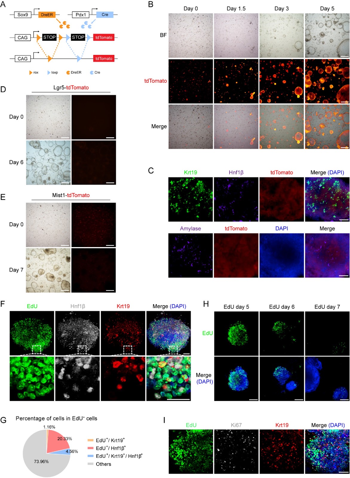
Advancements in three-dimensional (3D) organoid cultures have created more physiologically relevant models for pancreatic disease research, but efficiently generating mature pancreatic ductal cells remains challenging. In this study, we develop a novel protocol to generate pancreatic ductal organoids (PDOs) with high initiation efficiency and an enrichment of pancreatic ductal cells. By utilizing a cocktail of small molecules, we optimize the culture conditions to improve organoid formation. Our findings demonstrate that this protocol facilitates the formation and expansion of PDOs derived from Sox9-positive ductal cells, including heterogeneous ductal cells and acinar cells. These organoid cultures exhibit remarkable stability, supporting long-term expansion. This system provides an efficient model with potential applications in high-throughput drug screening. Moreover, these organoids recapitulate the exocrine cell composition and may reflect the cellular plasticity between ductal and acinar cells, providing a valuable platform for investigating pancreatic diseases such as pancreatic ductal adenocarcinoma (PDAC). The model presents a promising tool for future research aimed at understanding disease mechanisms and potentially helping drug development for pancreatic disorders.

Read Full Abstract10.3724/abbs.2024218
Brucella secretory protein VceA promotes FOXO1 entry into the nucleus to shift host cell metabolism toward glycolysisGraphical AbstractVerified
Acta Biochimica et Biophysica Sinica

Brucella secretory protein VceA promotes FOXO1 entry into the nucleus to shift host cell metabolism toward glycolysis

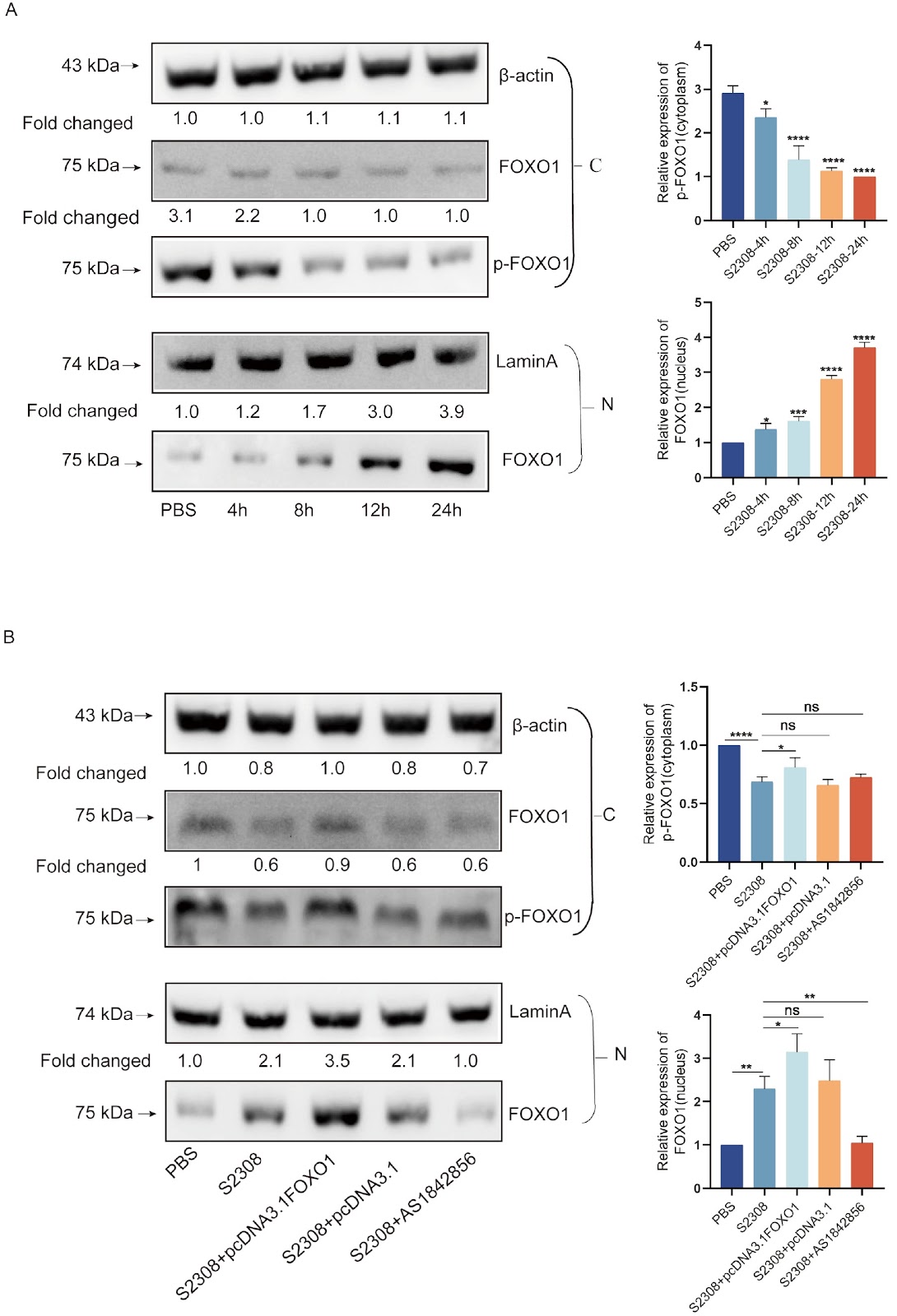
Increased glycolytic metabolism is a key step in the reproduction of Brucella and the induction of brucellosis, however, little is known about how this process is regulated during infection. Forkhead box protein O1 (FOXO1) is a transcription factor that regulates energy metabolism. In this study, we employ the yeast two-hybrid system (Y2H) and immunoprecipitation (Co-IP) to reverse screen for the FOXO1 for the first time and identify interactions between FOXO1 and the Brucella secretory protein VceA. Our findings reveal that the Brucella secretory protein VceA colocalizes with FOXO1 in the cytoplasm. Additionally, we observe that infection of macrophages with Brucella abortus 2308 (S2308) promotes FOXO1 entry into the nucleus, leading to a significant upregulation of glycolysis level in macrophage. Conversely, in a VceA mutant strain (S2308-ΔVceA), we note a significant reduction in the ability of FOXO1 to enter the nucleus, accompanied by a decrease in glycolysis level. Furthermore, Brucella interacts with FOXO1 through the secreted protein VceA, promoting the entry of FOXO1 into the nucleus and thereby altering host metabolic patterns. This study provides insights into the mechanisms by which Brucella invades host macrophages and induces unique metabolic changes. These insights may offer a novel rationale for developing metabolic therapeutic strategies for the treatment and prevention of related diseases.

Read Full Abstract10.3724/abbs.2024203
HADHA promotes esophageal cancer progression by activating mTOR signaling and the SP1/MDM2 axisGraphical AbstractVerified
Acta Biochimica et Biophysica Sinica

HADHA promotes esophageal cancer progression by activating mTOR signaling and the SP1/MDM2 axis

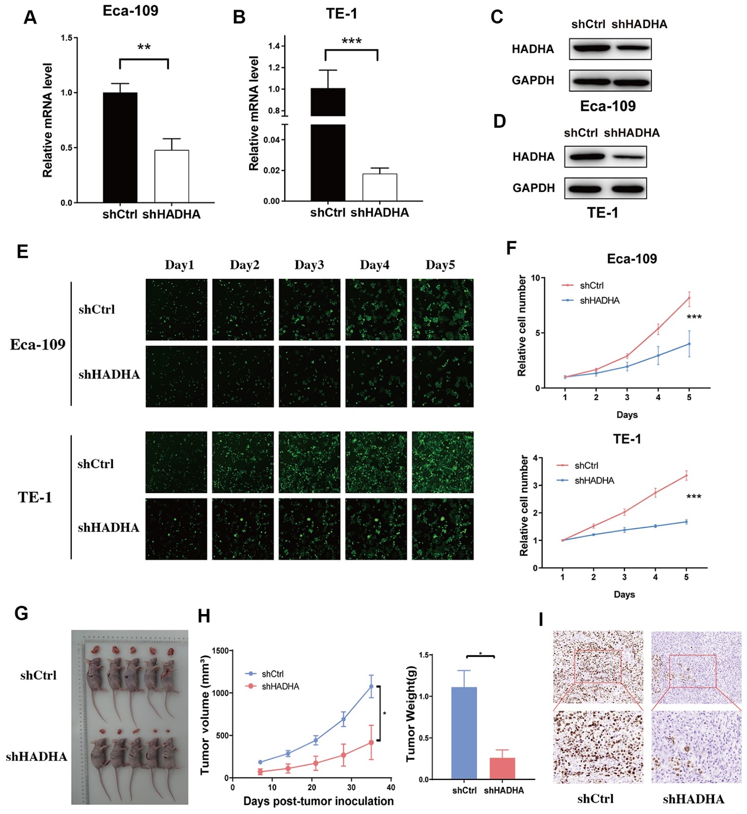
Esophageal cancer (EC) is one of the most recalcitrant cancers, with a 5-year survival rate of < 30%. The hydroxyacyl-CoA dehydrogenase alpha subunit (HADHA) plays an essential role in long-chain fatty acid metabolism, and dysregulation of HADHA has been demonstrated to be involved in a series of metabolic diseases and cancers. However, its role in cancers remains controversial. HADHA has seldom been investigated in EC, and little is known about how HADHA regulates the malignant progression of EC. In this study, we find that HADHA is significantly upregulated in EC tissues and is correlated with poor survival. HADHA knockdown markedly inhibits EC cell proliferation both in vitro and in vivo. The loss of HADHA also induces EC cell apoptosis, causes cell cycle arrest and inhibits cell migration. Additionally, RNA profiling reveals that mTOR signaling is significantly suppressed after HADHA knockdown. Mechanistically, HADHA interacts with SP1 and induces MDM2 expression. In conclusion, both mTOR signaling and the SP1-MDM2 axis participate in the HADHA-induced malignant behavior of EC cells.

Read Full Abstract10.3724/abbs.2024139
Significant biomarkers for predicting 1-month changes in IGF-1 in growth hormone-deficient children following r-hGH therapyGraphical AbstractVerified
Acta Biochimica et Biophysica Sinica

Significant biomarkers for predicting 1-month changes in IGF-1 in growth hormone-deficient children following r-hGH therapy

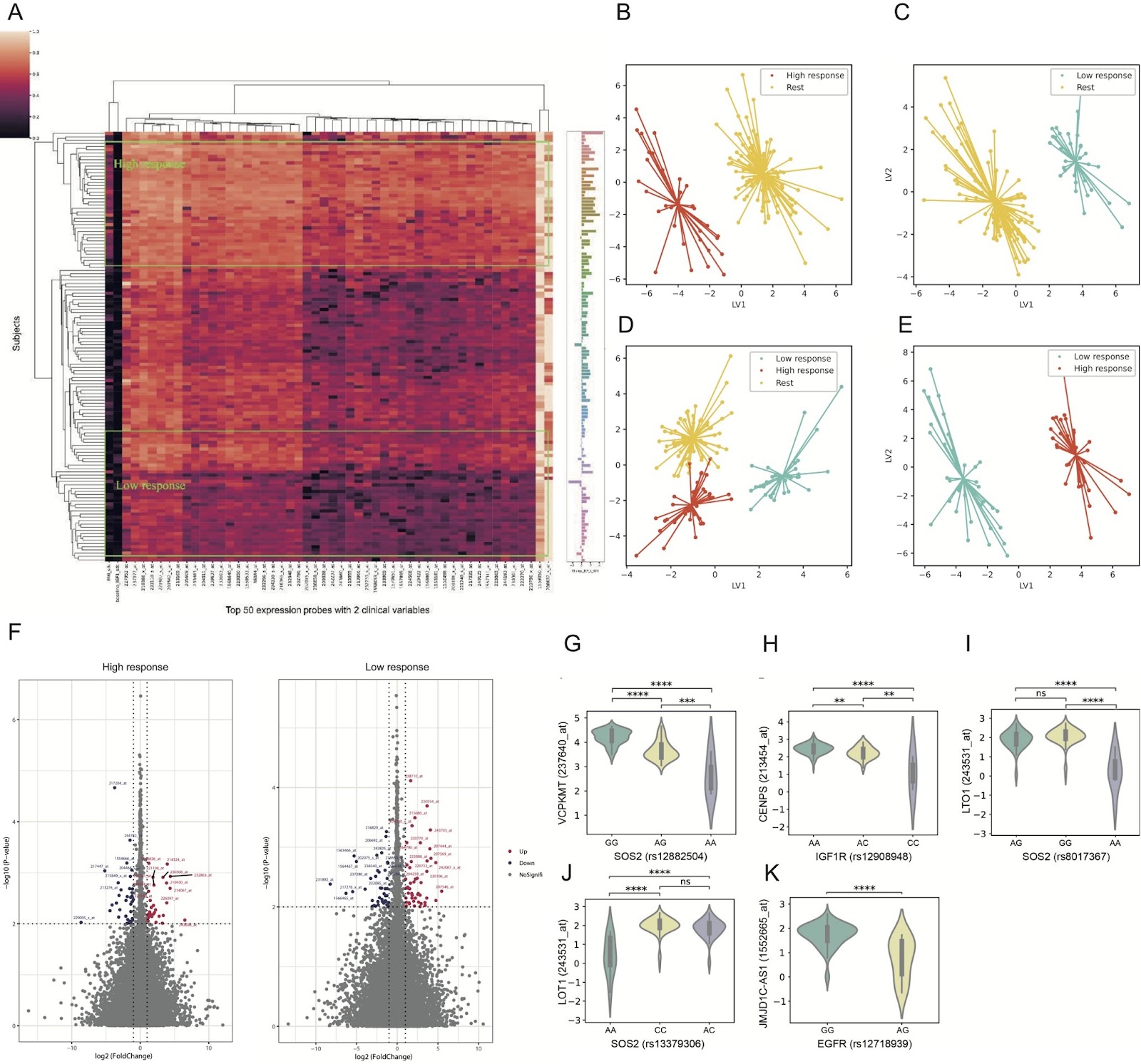
Growth hormone deficiency (GHD) is the most common pituitary hormone deficiency and is clinically characterized by short stature, delayed bone age and central distribution of body fat, and it has also been proven to be mildly heritable. Treatment with recombinant human growth hormone (r-hGH) is primary and safe for GHD children, and a dose of 0.15‒0.20 mg/kg each week results in a considerable increase in height velocity, with noteworthy growth during the first year of therapy [1]. Previous studies have shown that serum IGF-1 is strongly correlated with the growth response [2]. Therefore, IGF-1 can serve as a clinical indicator for monitoring compliance, efficacy and safety. However, the response to GH therapy shows significant individual variation, which is strongly associated with genetic factors. The prevalence rate of severe childhood GHD-related short stature varies from 1:4000 to 1:10,000 [3], while approximately 3%‒4% of the population in China suffers from short stature with an increasing trend. Therefore, an open-label, prospective, multicentric, noncomparative, nonrandomized phase IV interventional study (NCT01187550, Merck Serono Study 27709) was conducted to investigate the relationship between the prospective biomarkers of GHD patients and the individual variation in the primary therapeutic response following 4 weeks of r-hGH therapy. Given the significance of predicting GHD treatment response and the gaps in previous research, we sought to adopt a comprehensive strategy to accurately predict the therapeutic response utilizing the transcriptome, single nucleotide polymorphisms (SNPs) and clinical factors. We employed continuous variables and standard deviation scores of differences in serum IGF-1 levels after 4 weeks of r-hGH therapy (Ī”IGF-1) as targets to filter possible influencing variables. Furthermore, we compared several potential machine learning techniques, validated by PCA and PLS-DA, and ultimately applied the elastic net algorithm to determine the optimized predictive factors with consistent effect sizes. Additionally, expression quantitative trait locus (eQTL) analysis and differentially expressed gene (DEG) analysis were conducted to identify significant biomarkers for GHD treatment.

Read Full Abstract10.3724/abbs.2024089
Oxypeucedanin hydrate alleviates rheumatoid arthritis by inhibiting the TLR4-MD2/NF-ĪŗB/MAPK signaling axisGraphical AbstractVerified
Acta Biochimica et Biophysica Sinica

Oxypeucedanin hydrate alleviates rheumatoid arthritis by inhibiting the TLR4-MD2/NF-ĪŗB/MAPK signaling axis

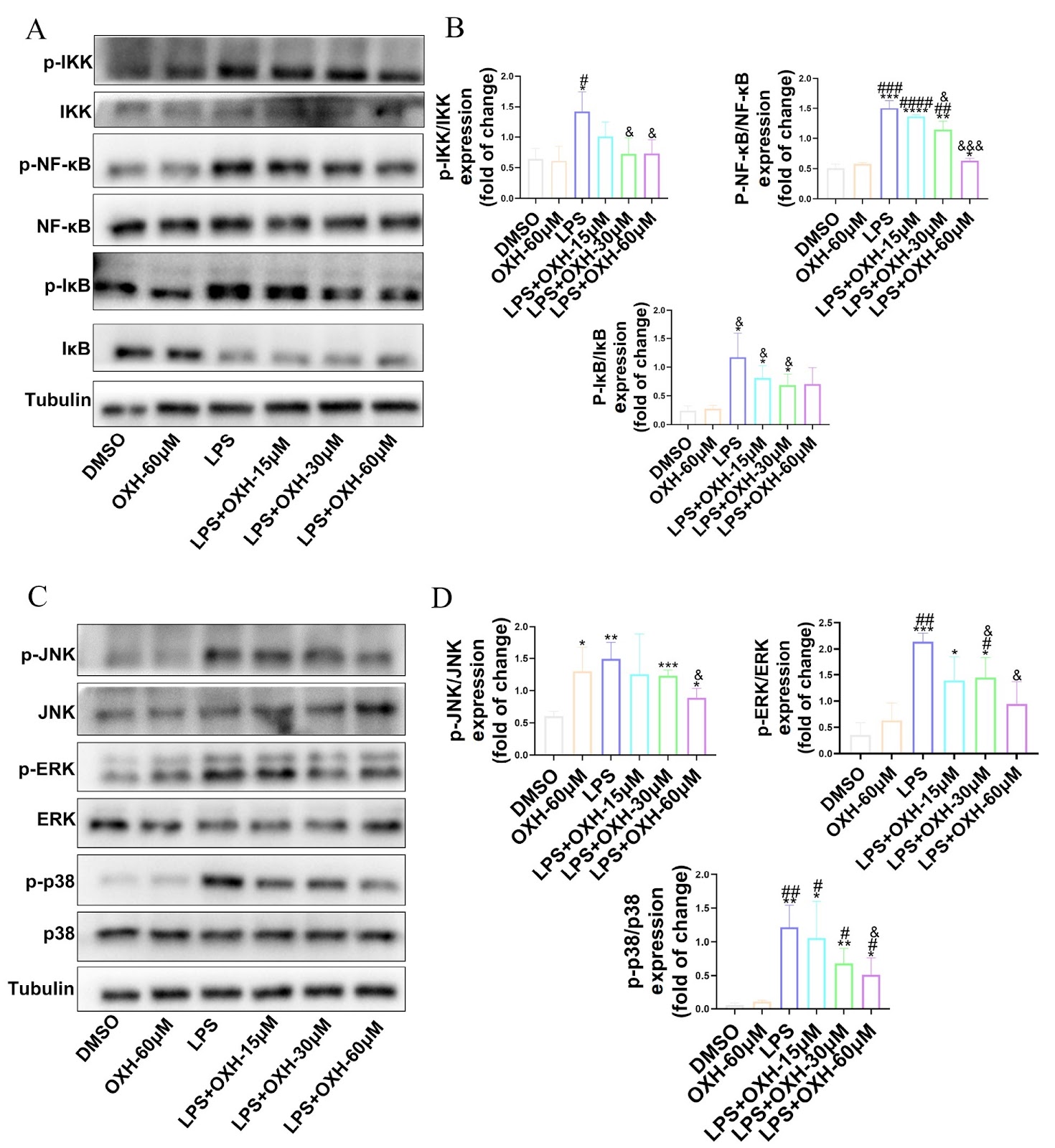
Rheumatoid arthritis (RA) is an idiopathic and chronic autoimmune disease for which there are currently no effective treatments. Oxypeucedanin hydrate (OXH) is a natural coumarin known for its potent anti-inflammatory properties. However, further investigations are needed to determine its therapeutic efficacy in treating RA. In this study, we evaluate the anti-inflammatory activity of OXH by treating LPS-induced RAW264.7 macrophages. Our results show that OXH treatment reverses the changes in iNOS, COX-2, IL-1β, IL-6, and TNF-α levels. Additionally, OXH reduces ROS production. Further analysis reveals that OXH suppresses the activation of the NF-κB/MAPK pathway. CETSA results show that OXH competes with LPS for binding to the TLR4/MD2 complex. MST experiments demonstrate the specific affinity of OXH for the TLR4/MD2 complex, with a Kd value of 33.7 μM. Molecular docking analysis suggests that OXH binds to the pocket of the TLR4/MD2 complex and interacts with specific amino acids, such as GLY-343, LYS-388, and PHE-345. Molecular dynamics simulations further confirm this conclusion. Finally, we investigate the potential of OXH in treating RA using a collagen-induced arthritis (CIA) model in rats. OXH effectively ameliorates the symptoms of CIA, including improving body weight, reducing swelling and redness, increasing talus volume, and decreasing bone erosion. OXH also decreases the mRNA levels of pro-inflammatory factors in synovial tissue. Transcriptome enrichment analysis and western blot analysis confirm that OXH suppresses the NF-κB/MAPK pathway, which is consistent with our in vitro findings.

Read Full Abstract10.3724/abbs.2024076
Lung metastases formed by disseminated tumor cells exhibit different proliferation statesGraphical AbstractVerified
Acta Biochimica et Biophysica Sinica

Lung metastases formed by disseminated tumor cells exhibit different proliferation states

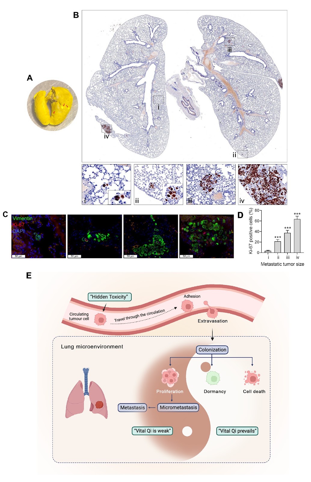
Lung cancer is the leading cause of cancer mortality in China and worldwide, and metastasis is the main cause of patient death. Cancer cells invade and migrate from the primary tumor, enter the circulation system through intravasation, and become circulating tumor cells (CTCs). CTCs that survive in blood vessels extravasate and invade target organs to become disseminated tumor cells (DTCs). DTCs proliferate in target organs to metastasize to distant organs. The previous view was that metastasis is the final stage of cancer progression. Normal cells first transform into tumor cells and then into invasive cancer cells; thus, metastasis occurs. Therefore, the possibility of metastasis is closely related to the size of the primary tumor. This is reflected in the TNM stage (T, tumor size; N, extent of spread to regional lymph nodes; M, metastasis to distant organs), which is often referenced in clinical diagnosis. However, an increasing number of studies are currently challenging this view. A previous study showed that the metastasis of malignant tumors occurs in the early stages of cancer. When patients are diagnosed with primary cancer, dissemination occurs. CTCs already exist in the blood vessels of early-stage lung cancer patients, and in early-stage lung cancer patients, DTCs are likely to be the main source of late-stage metastasis in some cancers; they do not proliferate in target organs, so they cannot be eliminated by surgery, radiotherapy or chemotherapy. As a result, even if the lesions are removed through surgery in these patients, metastasis is still found months or years later, which affects the patient’s quality of life and reduces the patient’s survival period. These observations prompt scientists in the field of metastasis to pay more attention to the prevention and treatment of DTCs when formulating metastasis prevention strategies. To determine whether DTCs exist in different states after entering the target organ, we used a mouse lung cancer metastasis model to generate CTC-TJH-01 cells, which are circulating tumor cells derived from the peripheral blood of early-stage lung adenocarcinoma patients who extravasate into target organs and become DTCs. Then, we observed the distribution and proliferation of DTCs in the lungs. Combined with traditional Chinese medicine theory, our findings can improve clinical medication regimens and promote innovations in metastasis prevention and treatment strategies. We observed the potential distribution and proliferation status of DTCs in the lungs in a mouse lung cancer metastasis model. A lung colonization assay was performed by injecting 5 Ɨ 105 CTC-TJH-01 cells into the lateral tail vein of NOD/SCID mice, and vimentin was used as a lung tumor marker. Immunofluorescence staining was performed, and CTC-TJH-01 cells that reached the lungs through the peripheral circulation were evenly spread over 24 h. This finding showed that cancer cells can move to distant sites through the circulation, especially the lungs, which are rich in blood vessels, and stay there in the form of DTCs. However, after 12 weeks, there were only a few visible metastases in the lungs, and many tumor cells in the visible metastases were Ki67-positive. Immunohistochemistry revealed other Vimentin-positive tumor cells in the lungs, but as the number of cells decreased, the Ki67 positivity rate also decreased, and a single tumor cell was negative for Ki67. This finding shows that an unsuitable microenvironment induces DTC apoptosis, and only a very small number of DTCs mediate the formation of an immunosuppressive microenvironment and then proliferate to form metastatic lesions. In addition, DTCs that survive have different proliferation rates; some proliferate to form metastases, while others remain dormant somewhere as individuals. These metastases of different sizes that coexist in the lungs may also have different responses to radiotherapy and chemotherapy due to their different proliferation rates. This may also explain why early-stage lung cancer patients still develop metastasis after standard clinical treatment. Before disseminated tumor cells proliferate and form visible metastases, they generally cannot be detected clinically through conventional diagnostic methods or tumor biomarkers, and patients at this stage often have no clinically significant symptoms; this stage can be called the ā€œmetastasis subclinical stageā€. Tian et al. proposed the pathogenesis theory of ā€œhidden toxicity due to vital Qi deficiencyā€ for this stage of lung cancer metastasis. According to this theory, DTCs in a dormant state are already present in the metastatic target organs of patients with early-stage lung cancer after surgery. Immunosenescence or stress mediates immune dysfunction, leading to the activation and proliferation of dormant DTCs, which in turn leads to the occurrence of clinical metastasis.

Read Full Abstract10.3724/abbs.2024118
RNA structure in alternative splicing regulation: from mechanism to therapyGraphical AbstractVerified
Acta Biochimica et Biophysica Sinica

RNA structure in alternative splicing regulation: from mechanism to therapy

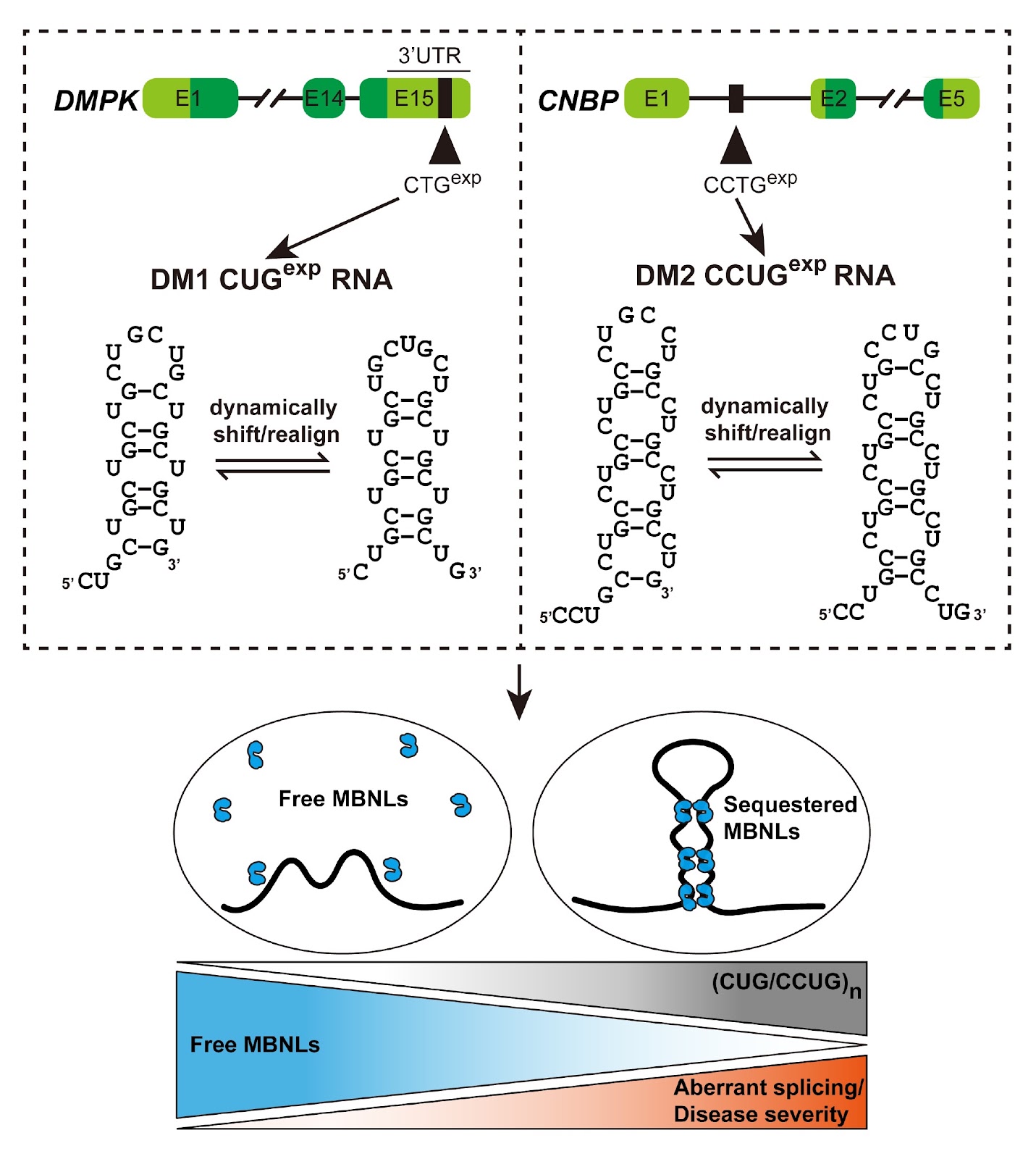
Alternative splicing is a highly intricate process that plays a crucial role in post-transcriptional regulation and significantly expands the functional proteome of a limited number of coding genes in eukaryotes. Its regulation is multifactorial, with RNA structure exerting a significant impact. Aberrant RNA conformations lead to dysregulation of splicing patterns, which directly affects the manifestation of disease symptoms. In this review, the molecular mechanisms of RNA secondary structure-mediated splicing regulation are summarized, with a focus on the complex interplay between aberrant RNA conformations and disease phenotypes resulted from splicing defects. This study also explores additional factors that reshape structural conformations, enriching our understanding of the mechanistic network underlying structure-mediated splicing regulation. In addition, an emphasis has been placed on the clinical role of targeting aberrant splicing corrections in human diseases. The principal mechanisms of action behind this phenomenon are described, followed by a discussion of prospective development strategies and pertinent challenges.

Read Full Abstract10.3724/abbs.2024119
Three-dimensional reconstruction of rat sperm using volume electron microscopyGraphical AbstractVerified
Acta Biochimica et Biophysica Sinica

Three-dimensional reconstruction of rat sperm using volume electron microscopy

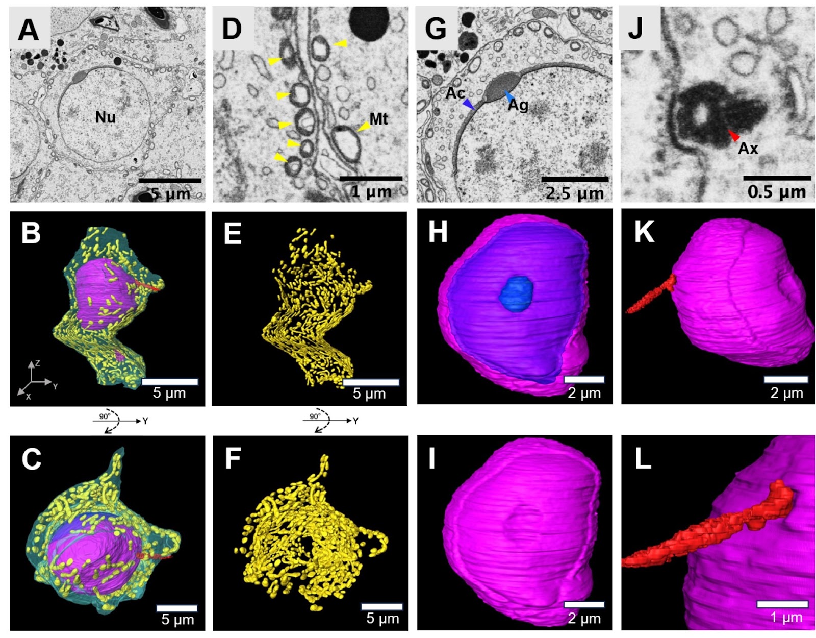
Three-dimensional (3D) reconstruction serves as a crucial instrument for the analysis of biological structures. In particular, a comprehensive and accurate 3D ultrastructural examination of rat sperm is vital for understanding and diagnosing male fertility issues and the underlying causes of infertility. In this study, we utilize the automated tape-collecting ultramicrotome scanning electron microscopy (ATUM-SEM) imaging technique, which is a highly effective method for 3D cellular ultrastructural analysis. Our findings reveal that during spermiogenesis, the volume of the nucleus significantly decreases, shrinking to just 10% of its original size. The acrosomal vesicles derived from the Golgi apparatus converge and elongate along the spermatid nucleus. These vesicles then attach to the nucleus via a cap-like structure, thereby defining the head side of the spermatozoa. In the initial stages of spermiogenesis, the mitochondria in spermatids are distributed beneath the cell membrane. As the process progresses, these mitochondria gradually migrate to the sperm tail, where they form the mitochondrial sheath. This sheath plays a crucial role in providing the energy required for the movement of the sperm. In addition, we reconstruct the mRNA-stroring structure-chromatoid body in sperm cells, which are cloud-like or net-like structures in the cytoplasm. The precise and comprehensive nature of 3D ultrastructural examination allows for a deeper understanding of the morphological process of spermiogenesis, thereby contributing to our knowledge of male fertility and the causes of infertility. Our research has significantly advanced the understanding of the 3D ultrastructure of sperm more comprehensively than ever before.

Read Full Abstract10.3724/abbs.2024144
RHBDF1 promotes PERK expression through the JNK/FoxO3 pathway in breast cancer cellsGraphical AbstractVerified
Acta Biochimica et Biophysica Sinica

RHBDF1 promotes PERK expression through the JNK/FoxO3 pathway in breast cancer cells

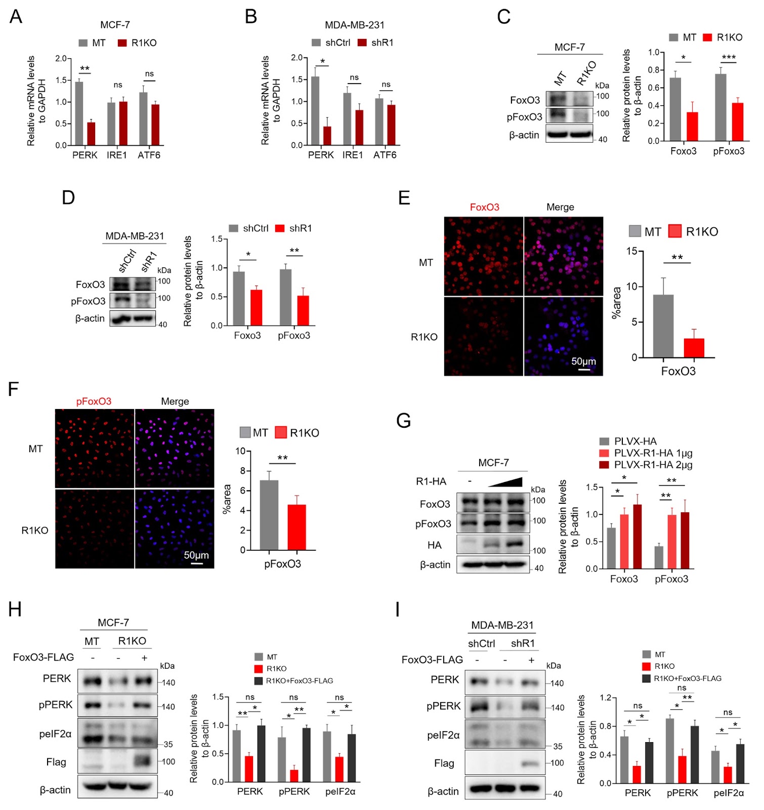
Human rhomboid family-1 (RHBDF1) gene is recognized as an oncogene involved in breast cancer development. Previous studies have indicated that RHBDF1 contributes significantly to endoplasmic reticulum (ER) protein homeostasis by stabilizing the binding immunoglobulin protein (BiP) and promoting the unfolded protein response (UPR). Here, we report a relationship between RHBDF1 and the ER stress sensors PERK, IRE1, and ATF6. We show that RHBDF1 deficiency in breast cancer cells results in decreased levels of PERK, pPERK, and peIF2α. These protein levels can be restored in RHBDF1-deficient breast cancer cells by artificial overexpression of RHBDF1 but not IRE1 or ATF6. Additionally, we show that the transcription factor FoxO3 is essential for the RHBDF1-mediated production of PERK. Subsequent analysis reveals that RHBDF1 activates JNK, which causes FoxO3 to translocate into the cell nucleus. These findings demonstrate that RHBDF1 supports the UPR by upregulating the PERK/peIF2α pathway via the JNK/FoxO3 axis and that the functions of RHBDF1 are essential for preserving the homeostasis of ER proteins.

Read Full Abstract10.3724/abbs.2024163
Bronchial thermoplasty decreases airway remodeling by inhibiting autophagy via the AMPK/mTOR signaling pathwayGraphical AbstractVerified
Acta Biochimica et Biophysica Sinica

Bronchial thermoplasty decreases airway remodeling by inhibiting autophagy via the AMPK/mTOR signaling pathway

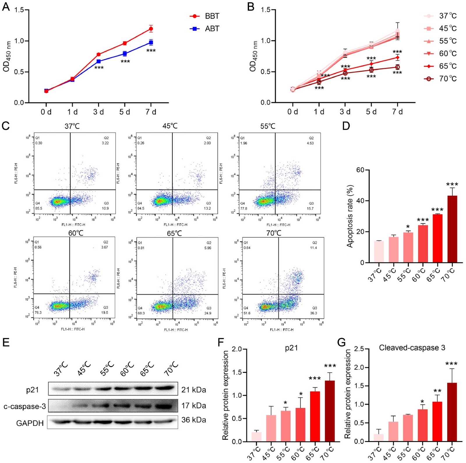
Bronchial thermoplasty (BT), an effective treatment for severe asthma, requires heat to reach the airway to reduce the mass of airway smooth muscle cells (ASMCs). Autophagy is involved in the pathological process of airway remodeling in patients with asthma. However, it remains unclear whether autophagy participates in controlling airway remodeling induced by BT. In this study, we aim to elucidate the autophagy-mediated molecular mechanisms in BT. Our study reveal that the number of autophagosomes and the level of alpha-smooth muscle actin (α-SMA) fluorescence are significantly decreased in airway biopsy tissues after BT. As the temperature increased, BT causes a decrease in cell proliferation and a concomitant increase in the apoptosis of human airway smooth muscle cells (HASMCs). Furthermore, increase in temperature significantly downregulates cellular autophagy, autophagosome accumulation, the LC3II/LC3I ratio, and Beclin-1 expression, upregulates p62 expression, and inhibits the AMPK/mTOR pathway. Furthermore, cotreatment with AICAR (an AMPK agonist) or RAPA (an mTOR antagonist) abolishes the inhibition of autophagy and attenuates the increase in the apoptosis rate of HASMCs induced by the thermal effect. Therefore, we conclude that BT decreases airway remodeling by blocking autophagy induced by the AMPK/mTOR signaling pathway in HASMCs.

Read Full Abstract10.3724/abbs.2024028
Integrated network pharmacology and experimental verification to explore the potential mechanism of San Ying decoction for treating triple-negative breast cancerGraphical AbstractVerified
Acta Biochimica et Biophysica Sinica

Integrated network pharmacology and experimental verification to explore the potential mechanism of San Ying decoction for treating triple-negative breast cancer

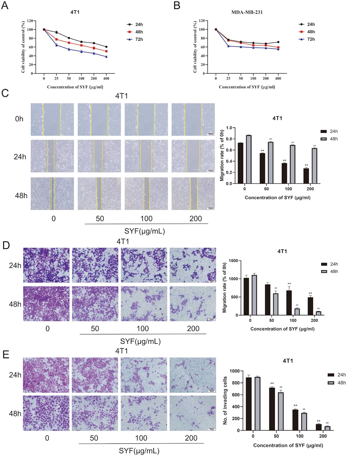
Traditional Chinese medicine (TCM) has been used to treat triple-negative breast cancer (TNBC), a breast cancer subtype with poor prognosis. Clinical studies have verified that the Sanyingfang formula (SYF), a TCM prescription, has obvious effects on inhibiting breast cancer recurrence and metastasis, prolonging patient survival, and reducing clinical symptoms. However, its active ingredients and molecular mechanisms are still unclear. In this study, the active ingredients of each herbal medicine composing SYF and their target proteins are obtained from the Traditional Chinese Medicine Systems Pharmacology database. Breast cancer-related genes are obtained from the GeneCards database. Major targets and pathways related to SYF treatment in breast cancer are identified by analyzing the above data. By conducting molecular docking analysis, we find that the active ingredients quercetin and luteolin bind well to the key targets KDR1, PPARG, SOD1, and VCAM1. In vitro experiments verify that SYF can reduce the proliferation, migration, and invasion ability of TNBC cells. Using a TNBC xenograft mouse model, we show that SYF could delay tumor growth and effectively inhibit the occurrence of breast cancer lung metastasis in vivo. PPARG, SOD1, KDR1, and VCAM1 are all regulated by SYF and may play important roles in SYF-mediated inhibition of TNBC recurrence and metastasis.

Read Full Abstract10.3724/abbs.2024015
Construction and efficacy testing of DNA vaccines containing HLA-A*02:01-restricted SARS-CoV-2 T-cell epitopes predicted by immunoinformaticsGraphical AbstractVerified
Acta Biochimica et Biophysica Sinica

Construction and efficacy testing of DNA vaccines containing HLA-A*02:01-restricted SARS-CoV-2 T-cell epitopes predicted by immunoinformatics

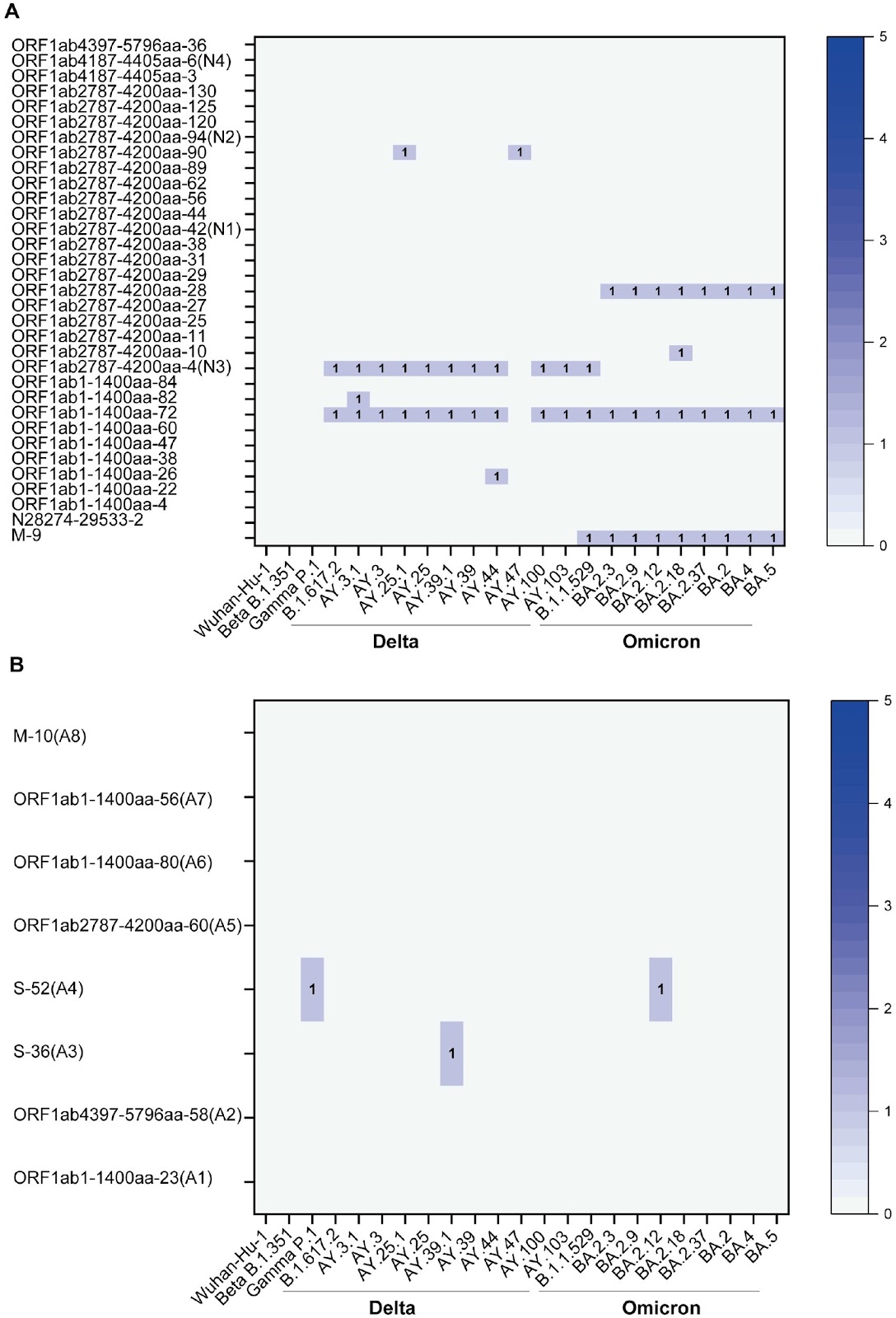
Vaccines play essential roles in the fight against the COVID-19 pandemic. The development and assessment of COVID-19 vaccines have generally focused on the induction and boosting of neutralizing antibodies targeting the SARS-CoV-2 spike (S) protein. Due to rapid and continuous variation in the S protein, such vaccines need to be regularly updated to match newly emerged dominant variants. T-cell vaccines that target MHC I- or II-restricted epitopes in both structural and non-structural viral proteins have the potential to induce broadly cross-protective and long-lasting responses. In this work, the entire proteome encoded by SARS-CoV-2 (Wuhan-hu-1) is subjected to immunoinformatics-based prediction of HLA-A*02:01-restricted epitopes. The immunogenicity of the predicted epitopes is evaluated using peripheral blood mononuclear cells from convalescent Wuhan-hu-1-infected patients. Furthermore, predicted epitopes that are conserved across major SARS-CoV-2 lineages and variants are used to construct DNA vaccines expressing multi-epitope polypeptides. Most importantly, two DNA vaccine constructs induce epitope-specific CD8+ T-cell responses in a mouse model of HLA-A*02:01 restriction and protect immunized mice from challenge with Wuhan-hu-1 virus after hACE2 transduction. These data provide candidate T-cell epitopes useful for the development of T-cell vaccines against SARS-CoV-2 and demonstrate a strategy for quick T-cell vaccine candidate development applicable to other emerging pathogens.

Read Full Abstract10.3724/abbs.2024039
Ferroptosis: a potential target for the treatment of atherosclerosisGraphical AbstractVerified
Acta Biochimica et Biophysica Sinica

Ferroptosis: a potential target for the treatment of atherosclerosis

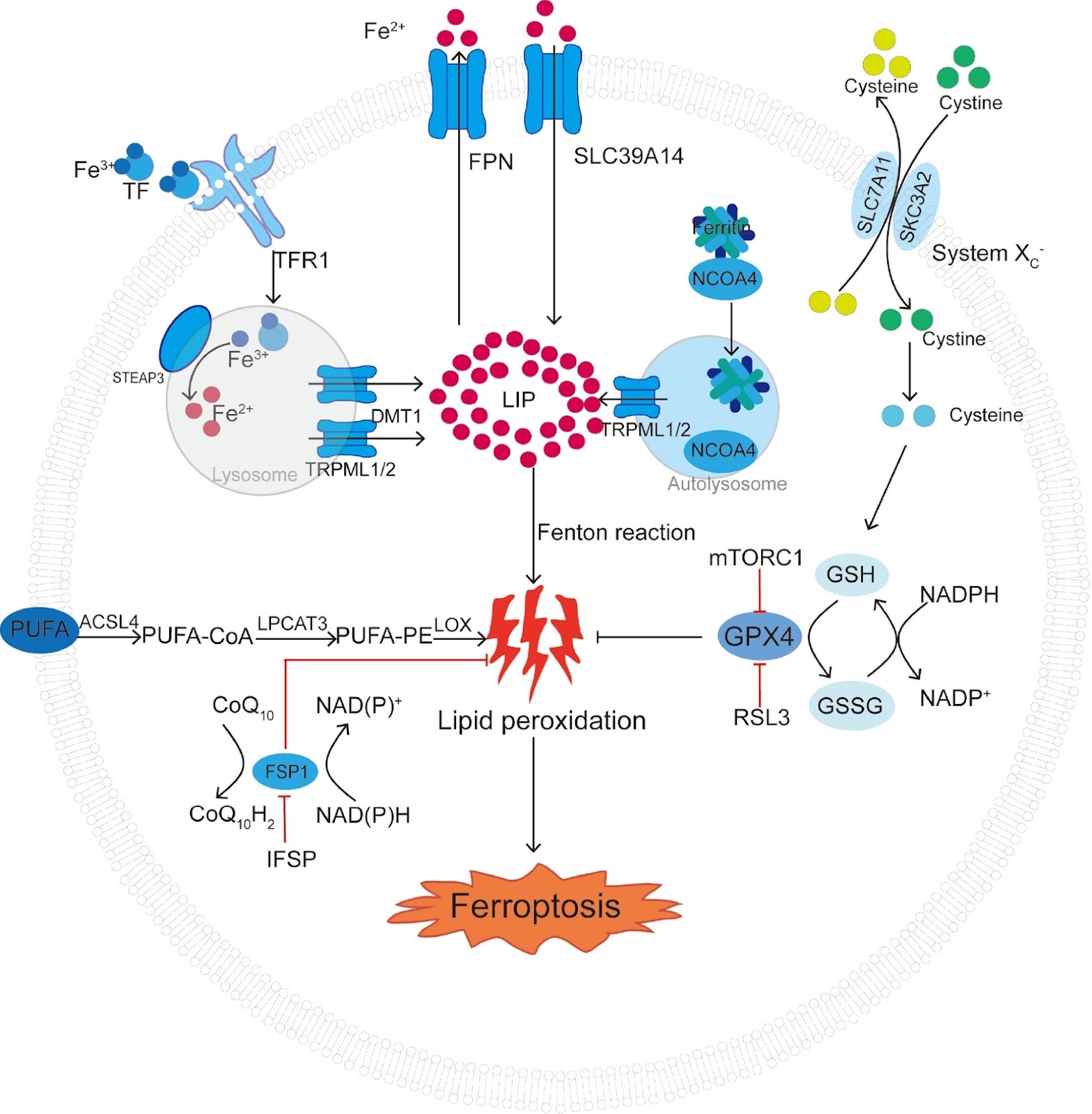
Atherosclerosis (AS), the main contributor to acute cardiovascular events, such as myocardial infarction and ischemic stroke, is characterized by necrotic core formation and plaque instability induced by cell death. The mechanisms of cell death in AS have recently been identified and elucidated. Ferroptosis, a novel iron-dependent form of cell death, has been proven to participate in atherosclerotic progression by increasing endothelial reactive oxygen species (ROS) levels and lipid peroxidation. Furthermore, accumulated intracellular iron activates various signaling pathways or risk factors for AS, such as abnormal lipid metabolism, oxidative stress, and inflammation, which can eventually lead to the disordered function of macrophages, vascular smooth muscle cells, and vascular endothelial cells. However, the molecular pathways through which ferroptosis affects AS development and progression are not entirely understood. This review systematically summarizes the interactions between AS and ferroptosis and provides a feasible approach for inhibiting AS progression from the perspective of ferroptosis.

Read Full Abstract10.3724/abbs.2024016
lncRNA CYTOR promotes lung adenocarcinoma gemcitabine resistance and epithelial-mesenchymal transition by sponging miR-125a-5p and upregulating ANLN and RRM2Graphical AbstractVerified
Acta Biochimica et Biophysica Sinica

lncRNA CYTOR promotes lung adenocarcinoma gemcitabine resistance and epithelial-mesenchymal transition by sponging miR-125a-5p and upregulating ANLN and RRM2

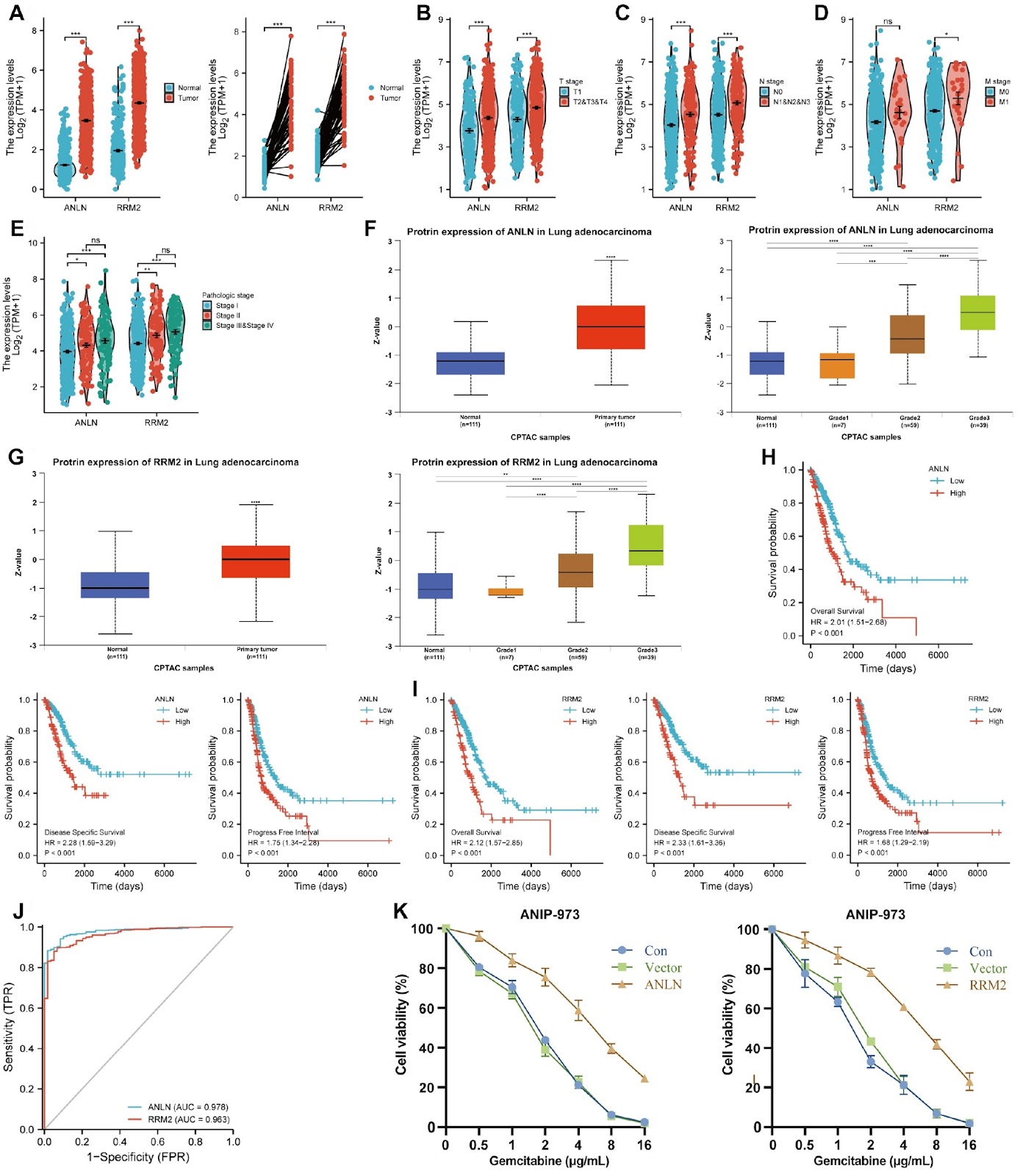
Lung adenocarcinoma (LUAD) is one of the most aggressive types of lung cancer. The prognosis of LUAD patients remains poor, and the overall efficacy of gemcitabine-based chemotherapy is still unsatisfactory. Long noncoding RNAs (lncRNAs) play important roles in several cancer types by interacting with multiple proteins, RNA, and DNA. However, the relationship between lncRNA dysregulation and gemcitabine resistance in LUAD has not been fully elucidated. In this study, lncRNA CYTOR expression and its association with the prognosis of LUAD patients are assessed by quantitative RT-PCR and Kaplan-Meier survival analysis. In vitro and in vivo functional studies are conducted to evaluate the biological functions of CYTOR in LUAD. The underlying mechanism regarding the tumor-promoting effects of CYTOR is explored using RNA immunoprecipitation, biotin-labelled RNA pulldown, luciferase reporter assays, and western blot analysis. We identify that CYTOR is an oncogenic lncRNA and is apparently upregulated in LUAD by analysing TCGA-LUAD data. High CYTOR expression is a poor prognostic factor for LUAD. Functional studies reveal that CYTOR confers LUAD cells with stronger resistance to gemcitabine treatment and upregulates the expression levels of epithelial-mesenchymal transition (EMT)-related proteins. Mechanically, CYTOR acts as a competitive endogenous RNA (ceRNA) to absorb miR-125a-5p, weakens the antitumor function of miR-125a-5p, and ultimately upregulates ANLN and RRM2 expressions. Taken together, this study explains the mechanism of lncRNA in the gemcitabine resistance of LUAD and formulates a theoretical framework for the in depth study of LUAD.

Read Full Abstract10.3724/abbs.2023287
R-loop formation contributes to mTORC1 activation-dependent DNA replication stress induced by p53 deficiencyGraphical AbstractVerified
Acta Biochimica et Biophysica Sinica

R-loop formation contributes to mTORC1 activation-dependent DNA replication stress induced by p53 deficiency

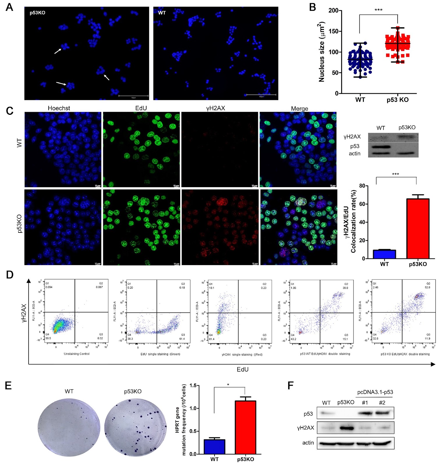
DNA replication stress is a significant contributor to spontaneous DNA damage and genome instability. While the impact of p53 deficiency on increasing DNA replication stress is known, the specific molecular mechanism underlying this phenomenon remains poorly understood. This study explores how p53 deficiency induces DNA replication stress by activating mTORC1 through R-loop formation, which is facilitated by the upregulation of RNR. Research has shown that p53 deficiency results in increased γH2AX expression and a higher mutation rate in the HPRT gene. Interestingly, these effects can be alleviated by rapamycin, an mTORC1 inhibitor. Additionally, rapamycin reduces the abundance of R-loop structures in p53KO cells, which is linked to mTORC1’s regulation of ribonucleotide reductase (RNR) level. These findings suggest that p53 deficiency-induced DNA replication stress relies on mTORC1 activation, with the upregulation of RNR expression and R-loop formation. Overall, this study underscores the importance of R-loops in mTORC1 activation-dependent DNA replication stress triggered by p53 deficiency.

Read Full Abstract10.3724/abbs.2024188
Diacylglycerol kinase γ facilitates the proliferation and migration of neural stem cells in the developing neural tubeGraphical AbstractVerified
Acta Biochimica et Biophysica Sinica

Diacylglycerol kinase γ facilitates the proliferation and migration of neural stem cells in the developing neural tube

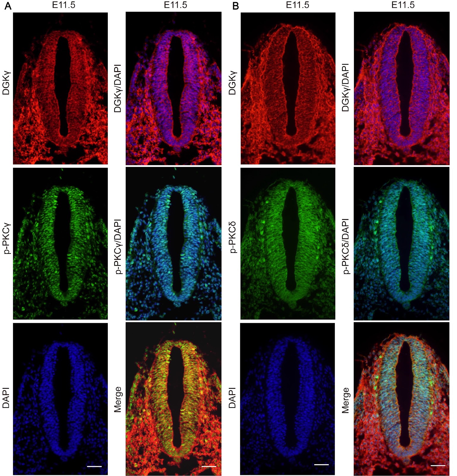
In this study, we aim to investigate diacylglycerol kinase (DGK) γ expression in developing neural tubes (NTs) and its effects on neural stem cell (NSC) proliferation and migration. Whole-mount in situ hybridization (WMISH) and immunohistochemistry are performed to explore DGKγ localization in developing NTs in vivo. NSCs are treated with sh-DGKγ, R59949, or PMA in vitro. Cell counting kit-8 (CCK-8) assay, 5-ethynyl-2′-deoxyuridine (EdU) assay and neurosphere formation assay are utilized to evaluate NSC proliferation. Neurosphere migration assay and a trans-well chamber assay are used to assess NSC migration. The diacylglycerol (DAG) content is detected via enzyme-linked immunosorbent assay (ELISA). The mRNA expression of DGKγ is detected via quantitative real-time polymerase chain reaction (qRT-PCR). The protein expression levels of DGKγ, protein kinase C (PKC) and phosphorylated PKC (p-PKC) are detected via western blot analysis. The results show that DGKγ mRNA is expressed predominantly in developing NTs. The neuroepithelium in developing NTs is positive for NSC markers, including Nestin, glial fibrillary acidic protein (GFAP), and DGKγ. DGKγ is expressed in the cytoplasm and nucleus of the neuroepithelium and is coexpressed with p-PKCγ and p-PKCĪ“. The proliferation of NSCs, the number of EdU-positive NSCs, and the number of neurospheres are decreased by sh-DGKγ and R59949 but increased by PMA. There is a shorter migration distance of NSCs and fewer migrated NSCs in the sh-DGKγ, R59949 and PMA groups. DAG content and the p-PKCĪ“/PKCĪ“ ratio are increased by sh-DGKγ, R59949 and PMA, whereas the p-PKCγ/PKCγ ratio is decreased by PMA. Taken together, our findings indicate that DGKγ facilitates NSC proliferation and migration, which is responsible for the participation of DGK in NT development. DGKγ facilitates NSC migration via the DAG/PKCĪ“ pathway.

Read Full Abstract10.3724/abbs.2024156
Cardioprotective effect of Saussurea involucrata injection against Doxorubicin-induced cardiotoxicity by network pharmacology analysis and experimental verificationGraphical AbstractVerified
Acta Biochimica et Biophysica Sinica

Cardioprotective effect of Saussurea involucrata injection against Doxorubicin-induced cardiotoxicity by network pharmacology analysis and experimental verification

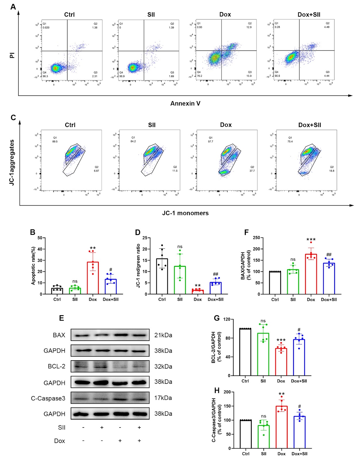
Doxorubicin (Dox) is widely utilized in the clinical treatment of various cancers. Despite its efficacy, Dox induces numerous adverse effects in humans with significant cardiotoxicity, posing a major limitation to its use. Saussurea involucrata injection (SII), derived from Saussurea involucrata, exhibits notable anti-inflammatory and anti-oxidative stress properties. However, its potential protective effects against Dox-induced cardiotoxicity (DIC) remain unexplored. In this study, we investigate the ability of SII to mitigate DIC and elucidate the underlying mechanisms through experimental research and network pharmacology analysis. Results from both in vitro and in vivo experiments reveal that SII treatment significantly improves Dox-induced cardiac dysfunction, reducing pathological alterations and fibrosis in cardiomyocytes. Moreover, SII has cardioprotective effects by diminishing the inflammation, oxidative stress, and apoptosis triggered by Dox. Network pharmacological analysis further shows that SII downregulates P53 protein expression by activating the AKT/MDM2 signaling pathway, thus attenuating DIC. In conclusion, this study confirms that SII mitigates DIC through downregulation of the AKT/MDM2/P53 signaling pathway, suggesting a promising therapeutic strategy for alleviating DIC.

Read Full Abstract10.3724/abbs.2024170
Co-profiling of translatome and transcriptome reveals the regulation of dynamic gene expression during Drosophila embryogenesisGraphical AbstractVerified
Acta Biochimica et Biophysica Sinica

Co-profiling of translatome and transcriptome reveals the regulation of dynamic gene expression during Drosophila embryogenesis

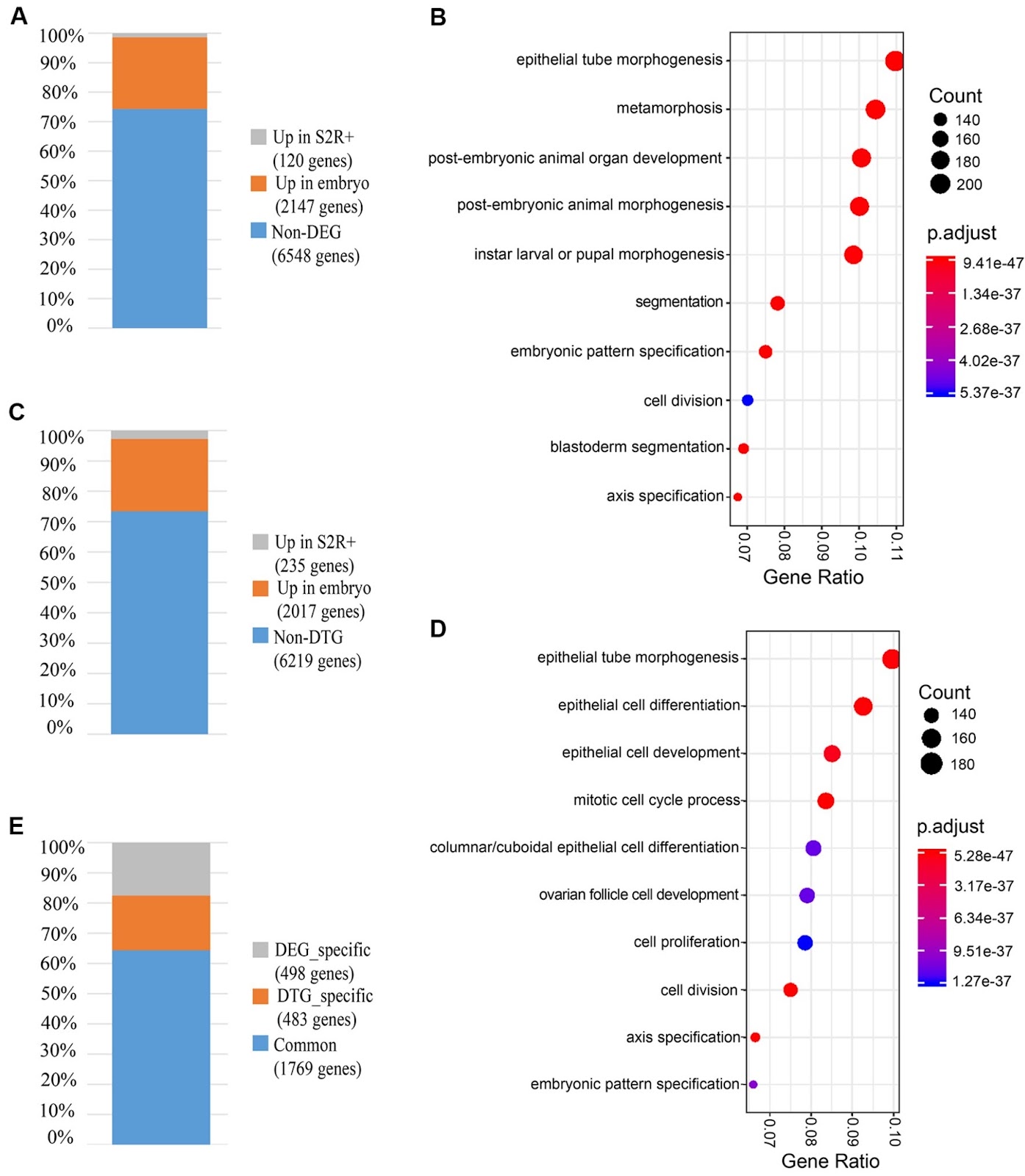
Eukaryotic gene expression is regulated at multiple levels, aiding in maintaining normal phenotypes and environmental adaptability. Transcriptional regulation complexity has been extensively studied using high-throughput sequencing, and previous studies have shown that different transcript isoforms can be produced through complex regulatory mechanisms via large-scale RNA sequencing. Additionally, translational regulation, which significantly influences gene expression, is controlled by complex mechanisms [1]. The untranslated regions (UTRs) of eukaryotic mRNA, encompassing the 5′ UTR, 3′ UTR and polyadenylation tail (polyA), are pivotal for translational regulation, with distinct cis-regulatory elements in the 5′ UTR and 3′ UTR of various transcript isoforms, leading to substantial variations in translational regulation across transcripts. To shed light on translational regulation, previous studies have performed isolation of ribosome-associated poly-adenylated RNAs (i.e., translatome) and deep sequencing for mRNA translation [2]. Polysome profiling is the most common method used to study translatome, which can enable the isolation of full-length translated mRNAs, thereby facilitating the identification of UTRs, assessment of selective translation, and comprehension of the regulatory mechanisms underlying gene expression [2]. Drosophila embryonic development progresses very rapidly and requires precise regulation of the transcription and translation of a large number of genes to ensure normal gene expression. Although Drosophila has been extensively studied as a model organism, the specific interplay between transcription and translation during embryonic development stages is not yet fully understood. To investigate the dynamic regulation of gene expression during Drosophila embryogenesis, we conducted transcriptome and translatome co-profiling on early (0‒4 h) embryos and S2R+ cells, a cell line derived from late embryonic stages of Drosophila melanogaster [3], to compare the differences in translational regulation at the gene and transcript isoform levels. S2R+ cell culture and early (0–4 h) embryo collection were performed (see Supplementary Methods) to compare transcriptome and translatome profiling, as shown in Supplementary Figure S1. Cytosolic RNA and ribosome-associated RNA were isolated from embryos [4] and S2R+ cells, which were used for constructing RNA-Seq libraries. Four libraries were generated for RNA-seq (see Supplementary Methods), consisting of two cytosolic RNA libraries and two ribosome-associated RNA libraries (Supplementary Figure S1A,B). The strand-specific RNA-seq libraries were prepared using the Illumina TruSeq Stranded mRNA Sample Preparation Kit (Illumina, San Diego, USA). The library was sequenced on the Illumina HiSeq X Ten System. We employed Trimmomatic [5] to remove low-quality reads, which resulted in approximately 89 million, 76 million, 72 million, and 56 million clean reads for the transcriptome and translatome of the early embryos and S2R+ cells, respectively. These reads were then mapped to the Drosophila genome (UCSC dm6) using HISAT2 [6]. The unique mapped reads ratio ranges from 94% to 85% and reads mapped to rRNA were less than 6% (Supplementary Table S1), indicating the high quality of the four RNA-seq libraries. Using StringTie [7], 33,470 transcripts were assembled for four mapping sequencing libraries, which revealed an average of 1.9 transcribed transcripts and 1.8 translated transcripts per gene (Supplementary Table S1), suggesting the usage of transcript isoforms widely existed in both transcription and translation of Drosophila embryos. To explore the divergence of the transcriptome during Drosophila development, we compared the transcriptome of the early embryos and S2R+ cells to identify genes with |log2(fold change)| ≄1, FPKM ≄1 in at least one condition, and adjusted P value ≤0.001. In total, we identified 2267 differentially expressed genes (DEGs) from 8815 genes. Among these DEGs, 2147 genes showed higher expression levels in the embryos, while 120 genes showed higher expression levels in S2R+ cells (Figure 1A and Supplementary Figure S2A). To investigate the underlying functional mechanism, we performed enrichment analysis to identify DEG-enriched pathways (Supplementary Table S2). Interestingly, the top 10 enriched pathways are related to morphogenesis an

Read Full Abstract10.3724/abbs.2024146
Tim-1-mediated extracellular matrix promotes the development of hepatocellular carcinomaGraphical AbstractVerified
Acta Biochimica et Biophysica Sinica

Tim-1-mediated extracellular matrix promotes the development of hepatocellular carcinoma

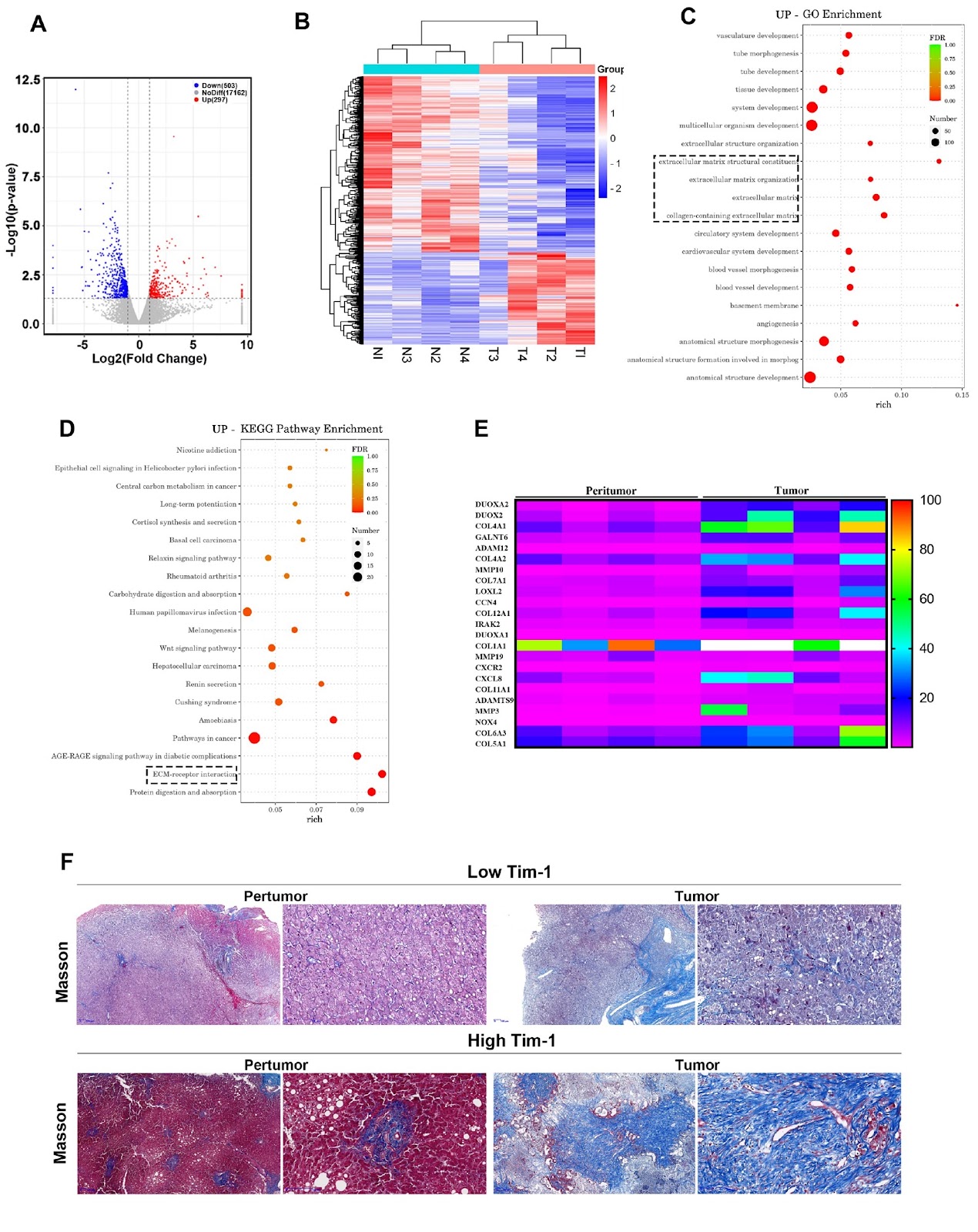
Tim-1 (T-cell immunoglobulin and mucin domain 1), also known as Kim-1 (kidney injury molecule 1) or hepatitis A virus cellular receptor 1 (HAVCR1), is a transmembrane protein expressed on various immune and epithelial cells. It plays a role in modulating inflammatory and immune responses. In this study, we find that Tim-1 is overexpressed in hepatocellular carcinoma (HCC) samples and that its expression is significantly correlated with postoperative survival. Bulk RNA sequencing reveals a general upregulation of extracellular matrix-related genes in HCC tissues with Tim-1 overexpression. The results of the cell and in vivo experiments reveal that Tim-1 in HCC not only affects biological processes such as the proliferation, migration, and invasion of HCC cells but also broadly promotes extracellular matrix processes by influencing cytokine secretion. Further studies demonstrate that Tim-1 mediates the activation of hepatic stellate cells and upregulates Th1 and Th2 cytokines, thereby promoting HCC progression. Thus, Tim-1 may represent a novel target for future interventions in HCC and liver fibrosis.

Read Full Abstract10.3724/abbs.2024191
Buzhong Yiqi Decoction accelerates skeletal muscle regenerationGraphical AbstractVerified
Acta Biochimica et Biophysica Sinica

Buzhong Yiqi Decoction accelerates skeletal muscle regeneration

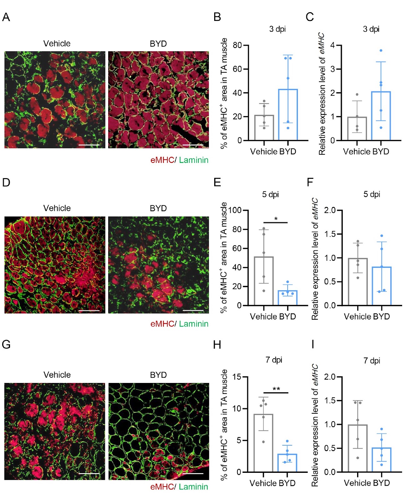
Adult skeletal muscle possesses an exceptional regenerative capacity, fundamentally reliant on adult muscle stem cells, known as satellite cells, which reside beneath the basal lamina of myofibers [1]. In their resting state, satellite cells remain quiescent; however, they activate, proliferate, differentiate, and fuse in response to pathological stress or injury, ultimately contributing to the repair and restoration of damaged myofibers [2]. Aging and the onset of skeletal muscle degenerative diseases significantly impair this regenerative ability, leading to a marked reduction in muscle mass and strength, which culminates in progressive muscle weakness and dysfunction [3,4]. Two notable examples of such degenerative conditions are age-related sarcopenia and muscular dystrophy, both of which present considerable public health challenges due to their increasing global prevalence. Currently, these diseases lack definitive therapeutic interventions, underscoring the urgent need for innovative treatments. Restoring the regenerative capacity of skeletal muscle may offer a promising therapeutic approach to halt or even reverse the progression of these muscular degenerative disorders. Buzhong Yiqi Decoction (BYD), a traditional Chinese medicine formula known for its qi-supplementing properties, comprises several key herbs, including Huangqi (Astragalus membranaceus), Baizhu (Atractylodes atractylodes), Chenpi (Pericarpium citri reticulatae), Shengma (Rhizoma cimicifugae), Chaihu (Radix bupleuri), Rensheng (Ginseng), Gancao (Liquo rice), and Danggui (Radix Angelicae Sinensis). Clinically, BYD is utilized to treat conditions such as allergic rhinitis, gut microbiota disorders, and chronic obstructive pulmonary disease. Notably, BYD is frequently prescribed for myasthenia gravis, a condition characterized by partial or systemic skeletal muscle weakness and fatigue. Modified BYD treatments have been shown to alleviate fatigue and muscle weakness while improving the quality of life for patients with myasthenia gravis [5,6]. Numerous clinical observations indicate that combining BYD with Western medicine is more effective than Western medicine alone in managing myasthenia gravis [7,8]. A randomized controlled trial conducted by Hu et al. [9] demonstrated the efficacy of BYD in addressing cancer-related fatigue and weakness in patients with cervical carcinoma. The effectiveness of BYD in alleviating myasthenia gravis and mitigating cancer-related fatigue suggests its potential role in regulating skeletal muscle homeostasis and function. The maintenance of skeletal muscle homeostasis is primarily achieved through effective muscle regeneration in response to injury or pathological stress. However, no experimental evidence indicates whether BYD can enhance skeletal muscle regeneration. To explore the potential role of BYD in regulating skeletal muscle regeneration, we established a cardiotoxin (CTX)-induced muscle injury and regeneration model in mice. All animal procedures were approved by the Animal Ethics Committee of Peking Union Medical College (ACUC-A01-2019-012). The tibialis anterior (TA) muscle of 8-week-old male C57BL/6j mice was injured via intramuscular injection of CTX (20 μL of 10 μM), followed by daily intragastric administration of BYD (15 μL/g body weight) (Figure 1A). Mice receiving daily intragastric administration of double-distilled water (ddH2O) served as vehicle controls (Figure 1A). Muscle regeneration was assessed at 3, 5, 7 and 14 days post-injury (dpi) through hematoxylin and eosin (H&E) staining (Figure 1B) and by quantifying the size of regenerating myofibers (Figure 1C,D). The H&E-stained cross-section of the TA muscle revealed a significant infiltration of immune cells in the injured muscle at the early time point of 3 dpi (Figure 1B). Notably, we observed a reduction in immune cell presence at 5 dpi in the BYD-treated group compared to the vehicle control (Figure 1B), indicating that BYD promotes the subsidence of inflammation during acute muscle injury and regeneration. Both H&E staining and quantification data demonstrated that regenerating myofibers, characterized by centralized myonuclei, were significantly larger at 7 dpi (Figure 1B,C) and 14 dpi (Figure 1B,D) in the BYD-treated group compared to controls, suggesting that BYD accelerates skeletal muscle regeneration.
Read Full Abstract10.3724/abbs.2024223
SEC61 translocon gamma subunit is correlated with glycolytic activity, epithelial mesenchymal transition and the immune suppressive phenotype of lung adenocarcinomaGraphical AbstractVerified
Acta Biochimica et Biophysica Sinica

SEC61 translocon gamma subunit is correlated with glycolytic activity, epithelial mesenchymal transition and the immune suppressive phenotype of lung adenocarcinoma

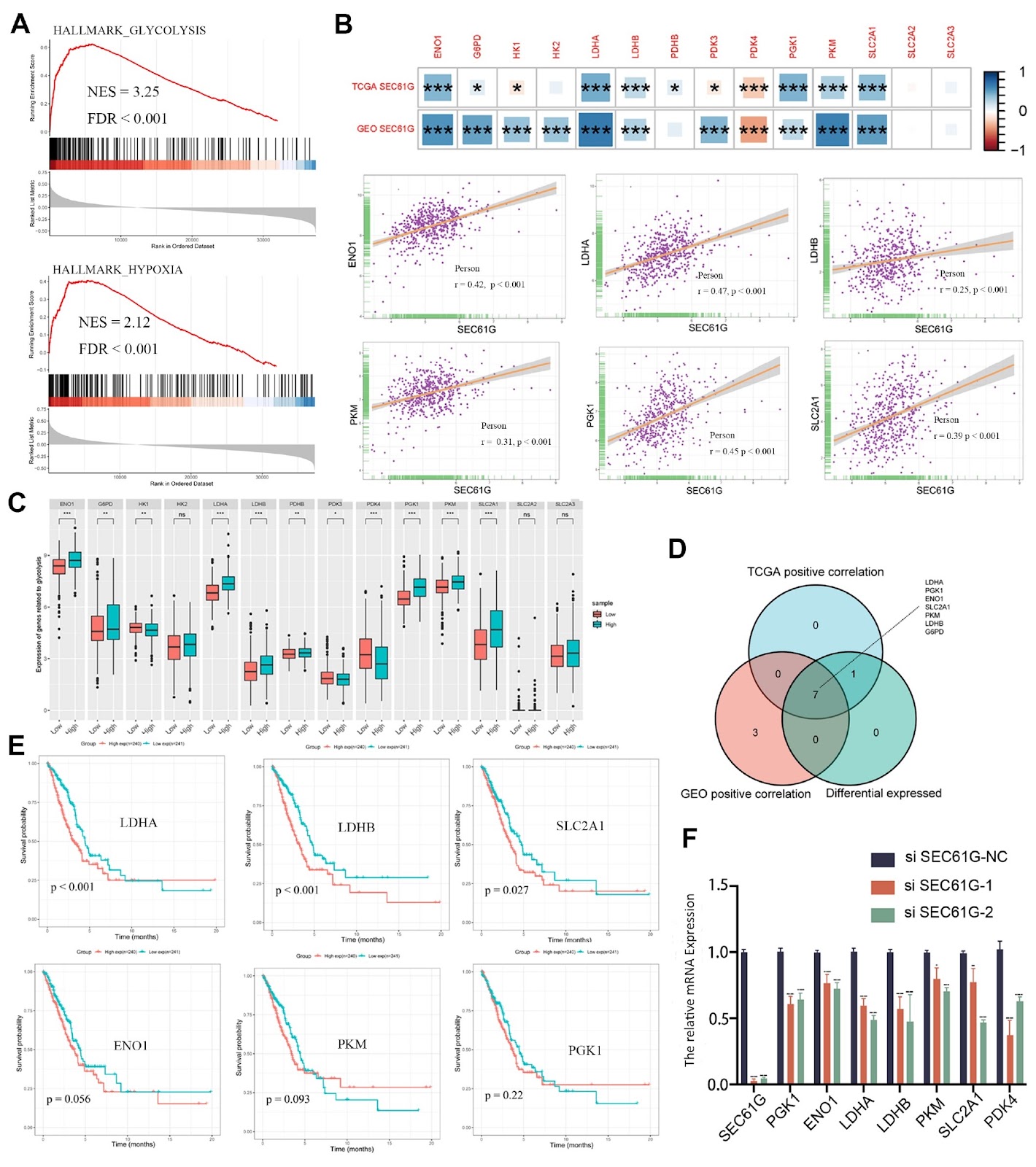
Lung adenocarcinoma (LUAD) remains a predominant cause of cancer-related mortality globally, underscoring the urgency for targeted therapeutic strategies. The specific role and impact of the SEC61 translocon gamma subunit (SEC61G) in LUAD progression and metastasis remain largely unexplored. In this study, we use a multifaceted approach, combining bioinformatics analysis with experimental validation, to elucidate the pivotal role of SEC61G and its associated molecular mechanisms in LUAD. Our integrated analyses reveal a significant positive correlation between SEC61G expression and the glycolytic activity of LUAD, as evidenced by increased fluorodeoxyglucose (FDG) uptake on positron emission tomography (PET)/CT scans. Further investigations show the potential influence of SEC61G on metabolic reprogramming, which contributes to the immunosuppressive tumor microenvironment (TME). Remarkably, we identify a negative association between SEC61G expression levels and the infiltration of critical immune cell populations within the TME, along with correlations with immune checkpoint gene expression and tumor heterogeneity scores in LUAD. Functional studies demonstrate that SEC61G knockdown markedly inhibits the migration of A549 and H2030 LUAD cells. This inhibitory effect is accompanied by a significant down-regulation of key regulators of tumor progression, including hypoxia-inducible factor-1 alpha (HIF-1α), lactate dehydrogenase A, and genes involved in the epithelial-mesenchymal transition pathway. In conclusion, our comprehensive analyses position SEC61G as a potential prognostic biomarker intricately linked to glycolytic metabolism, the EMT pathway, and the establishment of an immune-suppressive phenotype in LUAD. These findings underscore the potential of SEC61G as a therapeutic target and predictive marker for immunotherapeutic responses in LUAD patients.

Read Full Abstract10.3724/abbs.2024109
SinoBioData ResearchChinese Journal of Pathophysiology
Chinese Journal of Pathophysiology2026

Enterococcus faecalis Promotes Chemoresistance in Colorectal Cancer via Lactate-Mediated MOB3B Down-Regulation

AIM: To investigate the role of Enterococcus faecalis (E. faecalis) in colorectal cancer (CRC) chemoresistance and elucidate the underlying molecular mechanisms. METHODS: Conditioned media (CM) were collected from cultures of E. faecalis treated with oxaliplatin or 5-fluorouracil (5-FU). The effects of these media on CRC chemoresistance were evaluated using in vitro functional assays and in vivo xenograft models in nude mice. Bioinformatics analysis was conducted to identify candidate genes associated with E. faecalis-induced chemoresistance. Gain- and loss-of-function experiments were performed to assess the role of MOB3B in regulating CRC cell proliferation and drug sensitivity. RT-qPCR, Western blot, and immunohistochemistry were used to validate the molecular mechanisms involved. Metabolomic profiling identified key metabolites in E. faecalis-oxaliplatin CM, and their roles in drug resistance were also confirmed. RESULTS: Compared with oxaliplatin treatment alone, E. faecalis-oxaliplatin CM significantly promoted CRC cell growth and chemoresistance in vitro (P<0.01). Tumors treated with E. faecalis-oxaliplatin CM exhibited significantly larger volumes and faster growth in vivo (P<0.01). Mechanistically, down-regulation of MOB3B mediated the chemoresistance-promoting effects of E. faecalis-oxaliplatin CM (P<0.01). Overexpression of MOB3B inhibited CRC cell proliferation and enhanced chemosensitivity, whereas MOB3B knockdown produced the opposite effect (P<0.01). Metabolomic analysis revealed elevated lactate levels in the E. faecalis-oxaliplatin CM (P<0.01). Lactate inhibition significantly reduced CRC cell proliferation, reversed chemoresistance, and restored MOB3B expression (P<0.01). CONCLUSION: E. faecalis promotes chemoresistance in CRC through lactate-mediated down-regulation of MOB3B, highlighting MOB3B as a potential therapeutic target for overcoming CRC chemoresistance.

Read Full Abstract10.3969/j.issn.1000-4718.2026.06.013
Butyrate attenuates sympathetic activation in rats with chronic heart failure by inhibiting microglial inflammation in the paraventricular nucleusGraphical AbstractVerified
Acta Biochimica et Biophysica Sinica

Butyrate attenuates sympathetic activation in rats with chronic heart failure by inhibiting microglial inflammation in the paraventricular nucleus

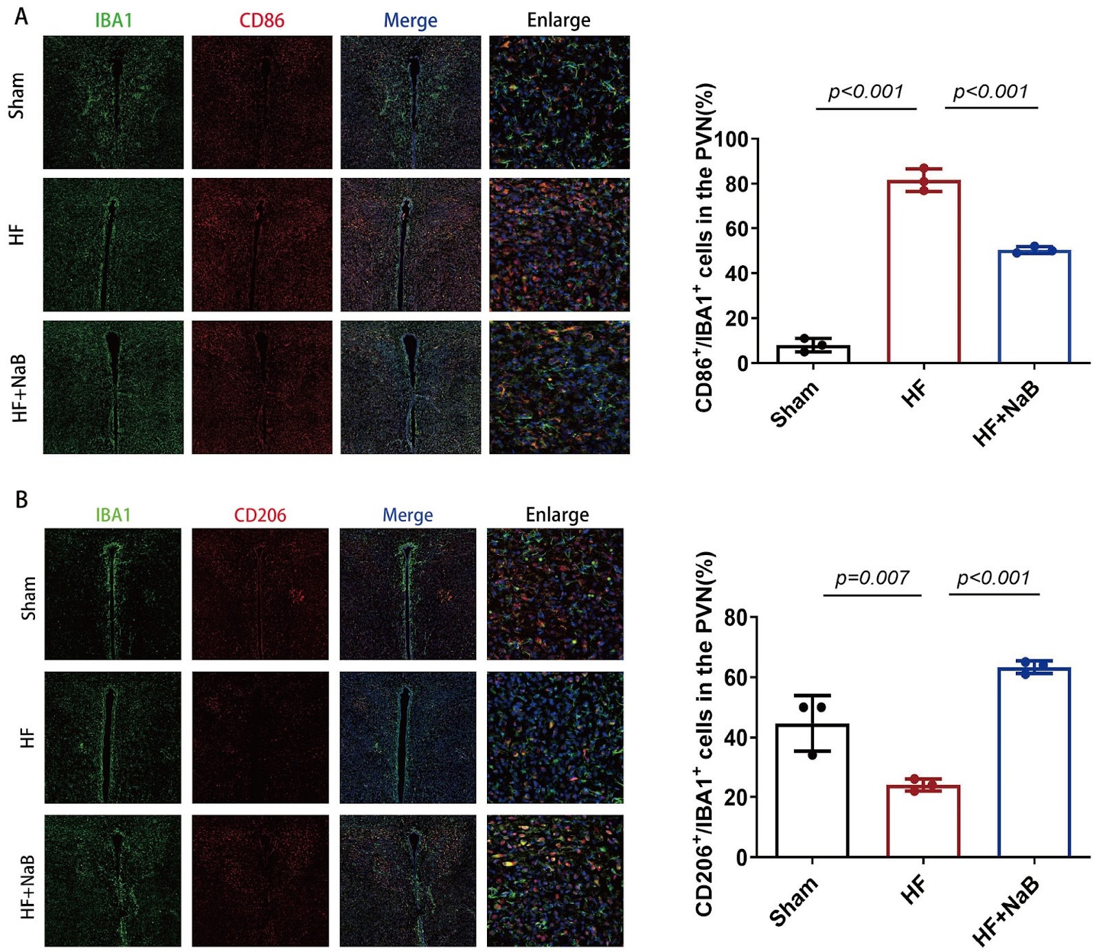
Sympathetic activation is a hallmark of heart failure and the underlying mechanism remains elusive. Butyrate is generated by gut microbiota and influences numerous physiological and pathological processes in the host. The present study aims to investigate whether the intestinal metabolite butyrate reduces sympathetic activation in rats with heart failure (HF) and the underlying mechanisms involved. Sprague-Dawley rats (220‒250 g) are anaesthetized with isoflurane, and the left anterior descending artery is ligated to model HF. Then, the rats are treated with or without butyrate sodium (NaB, a donor of butyrate, 10 g/L in water) for 8 weeks. Blood pressure and renal sympathetic nerve activity (RSNA) are recorded to assess sympathetic outflow. Cardiac function is improved (mean ejection fraction, 22.6%±4.8% vs 38.3%±5.3%; P<0.05), and sympathetic activation is decreased (RSNA, 36.3%±7.9% vs 23.9%±7.6%; P<0.05) in HF rats treated with NaB compared with untreated HF rats. The plasma and cerebrospinal fluid levels of norepinephrine are decreased in HF rats treated with NaB. The infusion of N-methyl-D-aspartic acid (NMDA) into the paraventricular nucleus (PVN) of the hypothalamus of HF model rats increases sympathetic nervous activity by upregulating the NMDA receptor. Microglia polarized to the M2 phenotype and inflammation are markedly attenuated in the PVN of HF model rats after NaB administration. In addition, HF model rats treated with NaB exhibit enhanced intestinal barrier function and increased levels of GPR109A, zona occludens-1 and occludin, but decreased levels of lipopolysaccharide-binding protein and zonulin. In conclusion, butyrate attenuates sympathetic activation and improves cardiac function in rats with HF. The improvements in intestinal barrier function, reductions in microglia-mediated inflammation and decreases in NMDA receptor 1 expression in the PVN are all due to the protective effects of NaB.

Read Full Abstract10.3724/abbs.2024092
CARF regulates the alternative splicing and piwi/piRNA complexes during mouse spermatogenesis through PABPC1Graphical AbstractVerified
Acta Biochimica et Biophysica Sinica

CARF regulates the alternative splicing and piwi/piRNA complexes during mouse spermatogenesis through PABPC1

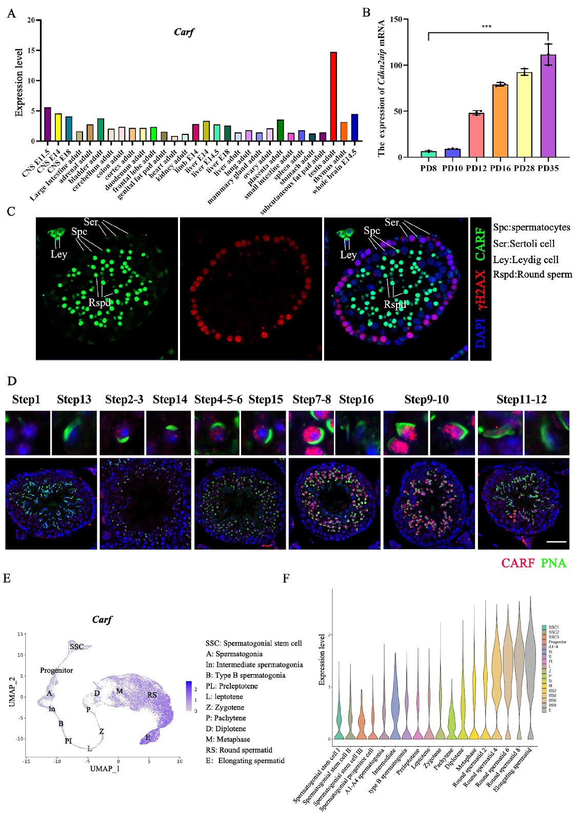
ADP-ribosylation factor collaborator (CARF), which is also known as CDKN2AIP, was first recognized as an ADP-ribosylation factor-interacting protein that participates in the activation of the ARF-p53-p21 (WAF1) signaling pathway under different conditions, such as oxidative and oncogenic stresses. The activation of this pathway often leads to cell growth arrest and apoptosis as well as senescence. Previous studies revealed that CARF, an RNA-binding protein, is critical for maintaining stem cell pluripotency and somatic differentiation. Nevertheless, its involvement in spermatogenesis has not been well examined. In this study, we show that male mice deficient in Carf expression present impaired spermatogenesis and fertility. IP-MS and RNA-seq analyses reveal that CARF/Carf interacts with multiple key splicing factors, such as PABPC1, and directly targets 356 different types of mRNAs in spermatocytes. Carf-associated mRNAs display aberrant splicing patterns when Carf expression is deficient. In addition, our results demonstrate that PIWIL1 expression and localization are altered in the Carf-/- mouse model through the downregulation of PABPC1, which further affects the ratio of pachytene-piRNA. Our study suggests that CARF is critical for regulating alternative splicing in mammalian spermatogenesis and determining infertility in male mice.

Read Full Abstract10.3724/abbs.2024224