🧬 SinoBioData Academic Portal
🏛️ Indexed Academic JournalOriginal: 干细胞研究与转化

Stem Cell Research & Therapy

Premier Chinese Biomedical Journal indexed in SinoBioData: Stem Cell Research & Therapy (干细胞研究与转化).

Total Research Papers: 200
Access: 100% Free Open Access
Browse by Publication Year & VolumeReset All Filters ✕
Select Specific Issue:

Published Research PapersFiltered: Year 2024 • 15 • 1

Showing 21 of 200 peer-reviewed papers with full Graphical Abstracts.

Original ResearchVol. 15, Issue 1 • pp. 135DOI: 10.1186/s13287-024-03745-w

Osteoinductive micro-nano guided bone regeneration membrane for in situ bone defect repair

Authors: Bingqian Wang, Xinfang Xie, Wenbin Jiang, Yichen Zhan, Yifan Zhang, Yaqi Guo, Zhenxing Wang, Nengqiang Guo, Ke Guo, Jiaming Sun

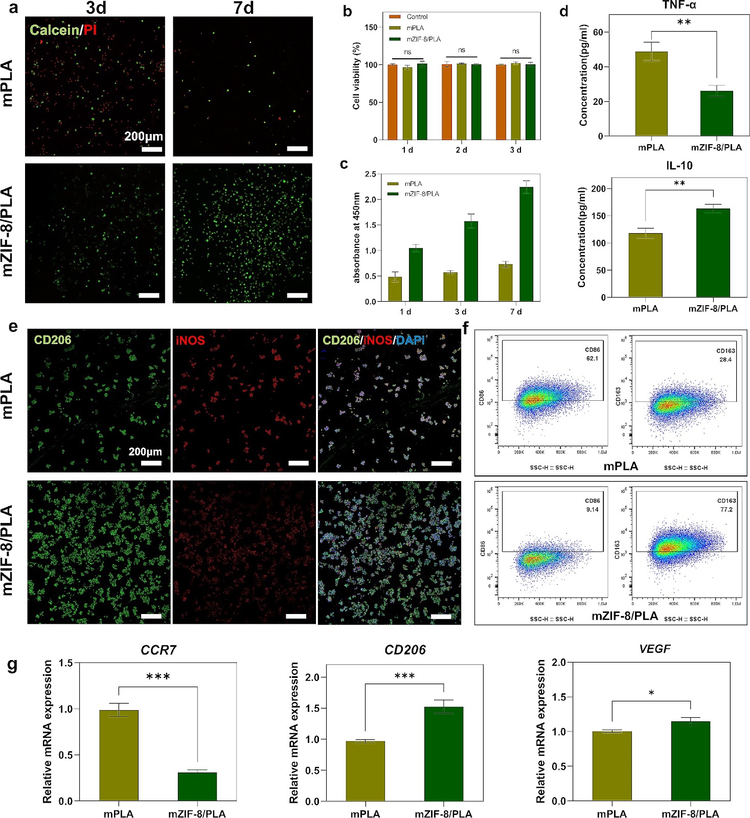
Background Biomaterials used in bone tissue engineering must fulfill the requirements of osteoconduction, osteoinduction, and osseointegration. However, biomaterials with good osteoconductive properties face several challenges, including inadequate vascularization, limited osteoinduction and barrier ability, as well as the potential to trigger immune and inflammatory responses. Therefore, there is an urgent need to develop guided bone regeneration membranes as a crucial component of tissue engineering strategies for repairing bone defects. Methods The mZIF-8/PLA membrane was prepared using electrospinning technology and simulated body fluid external mineralization method. Its ability to induce biomimetic mineralization was evaluated through TEM, EDS, XRD, FT-IR, zeta potential, and wettability techniques. The biocompatibility, osteoinduction properties, and osteo-immunomodulatory effects of the mZIF-8/PLA membrane were comprehensively evaluated by examining cell behaviors of surface-seeded BMSCs and macrophages, as well as the regulation of cellular genes and protein levels using PCR and WB. In vivo, the mZIF-8/PLA membrane's potential to promote bone regeneration and angiogenesis was assessed through Micro-CT and immunohistochemical staining. Results The mineralized deposition enhances hydrophilicity and cell compatibility of mZIF-8/PLA membrane. mZIF-8/PLA membrane promotes up-regulation of osteogenesis and angiogenesis related factors in BMSCs. Moreover, it induces the polarization of macrophages towards the M2 phenotype and modulates the local immune microenvironment. After 4-weeks of implantation, the mZIF-8/PLA membrane successfully bridges critical bone defects and almost completely repairs the defect area after 12-weeks, while significantly improving the strength and vascularization of new bone. Conclusions The mZIF-8/PLA membrane with dual osteoconductive and immunomodulatory abilities could pave new research paths for bone tissue engineering.
Osteoinductive micro-nano guided bone regeneration membrane for in situ bone defect repair
Graphical Abstract
Original ResearchVol. 15, Issue 1 • pp. 344DOI: 10.1186/s13287-024-03945-4

Optimized administration of human embryonic stem cell-derived immunity-and-matrix regulatory cells for mouse lung injury and fibrosis

Authors: Dingyun Song, Zhongwen Li, Faguo Sun, Kaiwei Wu, Kan Zhang, Wenjing Liu, Kaidi Liu, Bin An, Zai Wang, Tiemei Zhao, Huaiyong Chen, Li Xiao, Liu Wang, Lixin Xie, Wei Li, Liang Peng, Jie Hao, Jun Wu, Huaping Dai

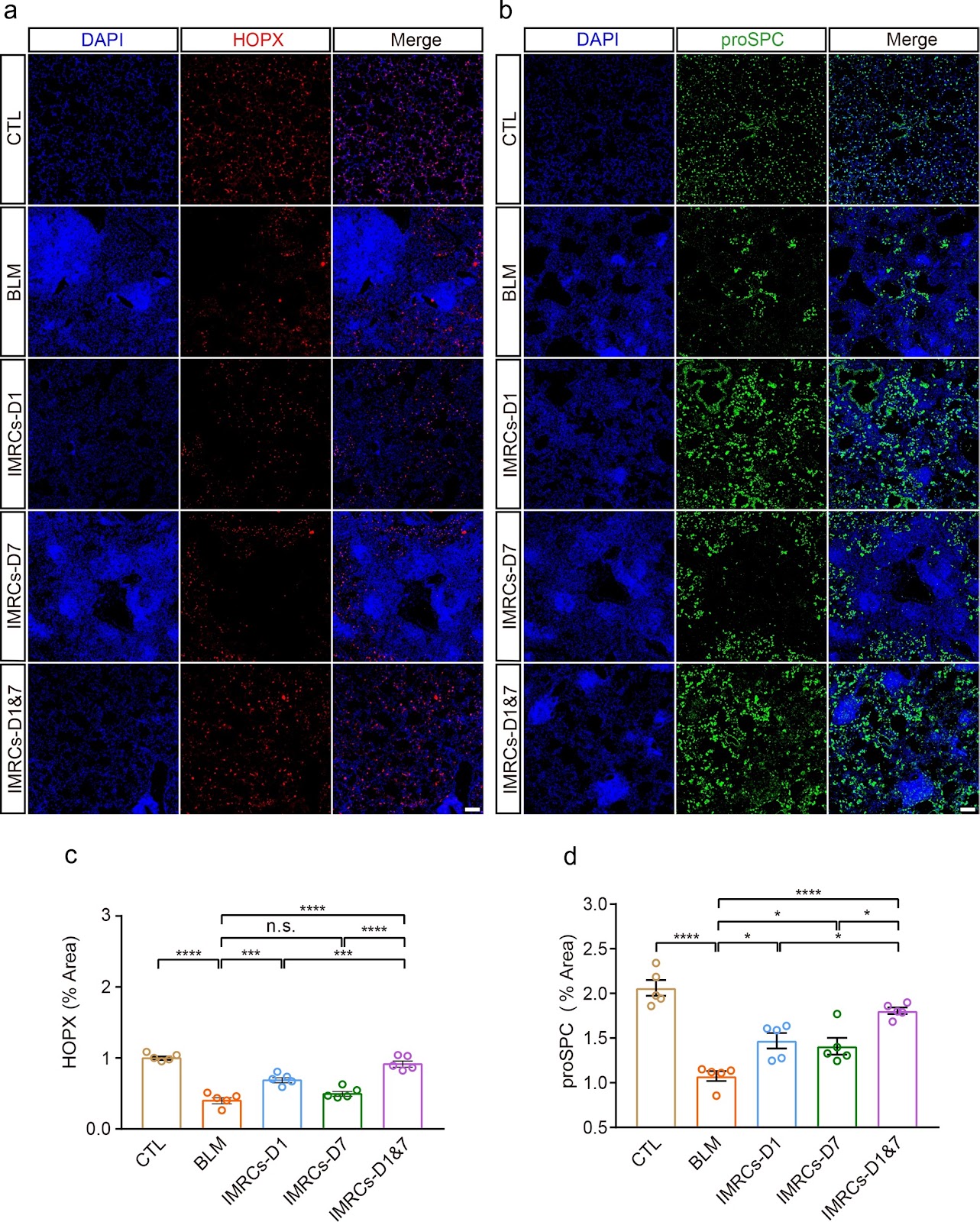
Background Lung injury and pulmonary fibrosis (PF), frequently arising as sequelae of severe and acute lung disease, currently face a dearth of effective therapeutic potions. Mesenchymal stem cells (MSCs) with immunomodulatory and tissue repair functions have immense potential to treat lung injury and PF. However, the optimal route of administration, timing, and frequency of dosing remain elusive. Human embryonic stem cell-derived immunity-and-matrix-regulatory cells (IMRCs) have shown therapeutic potential for lung injury and PF. Methods To ascertain the optimal therapeutic regimen for IMRCs in PF, we conducted an experimental study. Utilizing a mouse model of PF induced by bleomycin (BLM), IMRCs were administered via either a single or double intravenous (IV) or intratracheal (IT) injection on the first and seventh days post-BLM induction. Results Our findings revealed that IV infusion of IMRCs surpassed IT infusion in enhancing survival rates, facilitating body weight recovery, and optimizing Ashcroft and Szapiel scores among the model mice. Notably, IV administration exhibited a more profound ability to mitigate lung inflammation and fibrosis. Moreover, earlier and more frequent administrations of IMRCs were found to be advantageous in enhancing their therapeutic effects. Specifically, early administration with two IV infusions significantly improved body weight, lung organ coefficient, pulmonary ventilation and diffusion functions, and PF. This was accompanied by an increase in alveolar type I and II epithelial cells and a suppression of macrophage infiltration via CD24. Conclusion Collectively, these results suggested that IMRCs infusion ameliorated lung injury by promoting lung regeneration and inhibiting macrophage infiltration in a route, time, and frequency-dependent manner.

Optimized administration of human embryonic stem cell-derived immunity-and-matrix regulatory cells for mouse lung injury and fibrosis
Graphical Abstract
Original ResearchVol. 15, Issue 1 • pp. 33DOI: 10.1186/s13287-024-03639-x

Osteogenic human MSC-derived extracellular vesicles regulate MSC activity and osteogenic differentiation and promote bone regeneration in a rat calvarial defect model

Authors: Niyaz Al-Sharabi, Samih Mohamed-Ahmed, Siddharth Shanbhag, Carina Kampleitner, Rammah Elnour, Shuntaro Yamada, Neha Rana, Even Birkeland, Stefan Tangl, Reinhard Gruber, Kamal Mustafa

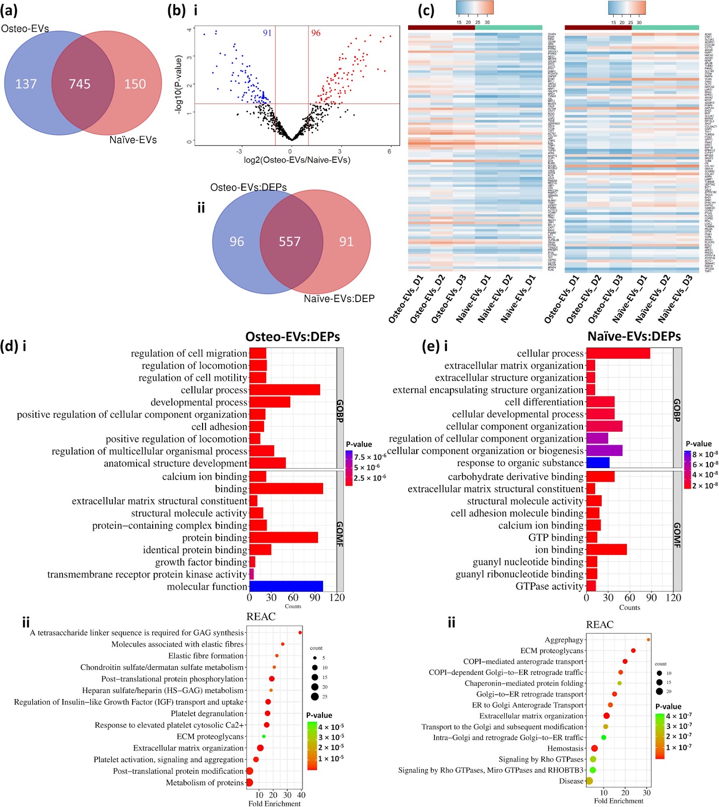
Background There is growing evidence that extracellular vesicles (EVs) play a crucial role in the paracrine mechanisms of transplanted human mesenchymal stem cells (hMSCs). Little is known, however, about the influence of microenvironmental stimuli on the osteogenic effects of EVs. This study aimed to investigate the properties and functions of EVs derived from undifferentiated hMSC (Naïve-EVs) and hMSC during the early stage of osteogenesis (Osteo-EVs). A further aim was to assess the osteoinductive potential of Osteo-EVs for bone regeneration in rat calvarial defects. Methods EVs from both groups were isolated using size-exclusion chromatography and characterized by size distribution, morphology, flow cytometry analysis and proteome profiling. The effects of EVs (10 µg/ml) on the proliferation, migration, and osteogenic differentiation of cultured hMSC were evaluated. Osteo-EVs (50 µg) or serum-free medium (SFM, control) were combined with collagen membrane scaffold (MEM) to repair critical-sized calvarial bone defects in male Lewis rats and the efficacy was assessed using µCT, histology and histomorphometry. Results Although Osteo- and Naïve-EVs have similar characteristics, proteomic analysis revealed an enrichment of bone-related proteins in Osteo-EVs. Both groups enhance cultured hMSC proliferation and migration, but Osteo-EVs demonstrate greater efficacy in promoting in vitro osteogenic differentiation, as evidenced by increased expression of osteogenesis-related genes, and higher calcium deposition. In rat calvarial defects, MEM with Osteo-EVs led to greater and more consistent bone regeneration than MEM loaded with SFM. Conclusions This study discloses differences in the protein profile and functional effects of EVs obtained from naïve hMSC and hMSC during the early stage of osteogenesis, using different methods. The significant protein profile and cellular function of EVs derived from hMSC during the early stage of osteogenesis were further verified

Osteogenic human MSC-derived extracellular vesicles regulate MSC activity and osteogenic differentiation and promote bone regeneration in a rat calvarial defect model
Graphical Abstract
Original ResearchVol. 15, Issue 1 • pp. 166DOI: 10.1186/s13287-024-03776-3

Inhibition and reversal of a TGF‑β1 induced myofibroblast phenotype by adipose tissue‑derived paracrine factors

Authors: S. Higginbotham, V. L. Workman, A-V. Giblin, N. H. Green, D. W. Lambert, V. Hearnden

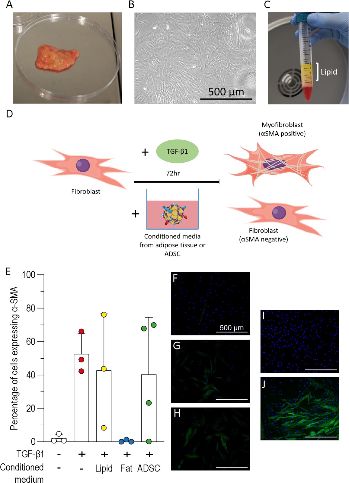
Background Hypertrophic scarring results from myofibroblast differentiation and persistence during wound healing. Currently no effective treatment for hypertrophic scarring exists however, autologous fat grafting has been shown to improve scar elasticity, appearance, and function. The aim of this study was to understand how paracrine factors from adipose tissues and adipose-derived stromal cells (ADSC) affect fibroblast to myofibroblast differentiation. Methods The transforming growth factor-β1 (TGF-β1) induced model of myofibroblast differentiation was used to test the effect of conditioned media from adipose tissue, ADSC or lipid on the proportion of fibroblasts and myofibroblasts. Results Adipose tissue conditioned media inhibited the differentiation of fibroblasts to myofibroblasts but this inhibition was not observed following treatment with ADSC or lipid conditioned media. Hepatocyte growth factor (HGF) was readily detected in the conditioned medium from adipose tissue but not ADSC. Cells treated with HGF, or fortinib to block HGF, demonstrated that HGF was not responsible for the inhibition of myofibroblast differentiation. Conditioned media from adipose tissue was shown to reduce the proportion of myofibroblasts when added to fibroblasts previously treated with TGF-β1, however, conditioned media treatment was unable to significantly reduce the proportion of myofibroblasts in cell populations isolated from scar tissue. Conclusions Cultured ADSC or adipocytes have been the focus of most studies, however, this work highlights the importance of considering whole adipose tissue to further our understanding of fat grafting. This study supports the use of autologous fat grafts for scar treatment and highlights the need for further investigation to determine the mechanism.

Inhibition and reversal of a TGF‑β1 induced myofibroblast phenotype by adipose tissue‑derived paracrine factors
Graphical Abstract
Original ResearchVol. 15, Issue 1 • pp. 23DOI: 10.1186/s13287-024-03636-0

Current status of stem cell therapy for type 1 diabetes: a critique and a prospective consideration

Authors: Mohamed A. Ghoneim, Mahmoud M. Gabr, Sawsan M. El-Halawani, Ayman F. Refaie

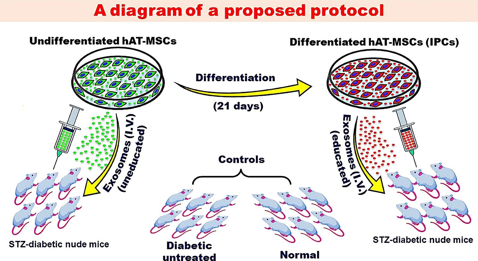
Over the past decade, there had been progress in the development of cell therapy for insulin-dependent diabetes. Nevertheless, important hurdles that need to be overcome still remain. Protocols for the differentiation of pluripotent stem cells into pancreatic progenitors or fully differentiated β-cells have been developed. The resulting insulin-producing cells can control chemically induced diabetes in rodents and were the subject of several clinical trials. However, these cells are immunogenic and possibly teratogenic for their transplantation, and an immunoisolation device and/or immunosuppression is needed. A growing number of studies have utilized genetic manipulations to produce immune evasive cells. Evidence must be provided that in addition to the expected benefit, gene manipulations should not lead to any unforeseen complications. Mesenchymal stem/stromal cells (MSCs) can provide a viable alternative. MSCs are widely available from many tissues. They can form insulin-producing cells by directed differentiation. Experimentally, evidence has shown that the transplantation of allogenic insulin-producing cells derived from MSCs is associated with a muted allogeneic response that does not interfere with their functionality. This can be explained by the immunomodulatory functions of the MSC subpopulation that did not differentiate into insulin-producing cells. Recently, exosomes derived from naive MSCs have been used in the experimental domain to treat diabetes in rodents with varying degrees of success. Several mechanisms for their beneficial functions were proposed including a reduction in insulin resistance, the promotion of autophagy, and an increase in the T regulatory population. However, euglycemia was not achieved in any of these experiments. We suggest that exosomes derived from β-cells or insulin-producing cells (educated) can provide a better therapeutic effect than those derived from undifferentiated cells.

Current status of stem cell therapy for type 1 diabetes: a critique and a prospective consideration
Graphical Abstract
Original ResearchVol. 15, Issue 1 • pp. 427DOI: 10.1186/s13287-024-04025-3

Astilbin improves the therapeutic effects of mesenchymal stem cells in AKI-CKD mice by regulating macrophage polarization through PTGS2-mediated pathway

Authors: Xiaodong Geng, Zhangning Fu, Guangrui Geng, Kun Chi, Chao Liu, Haijuan Hong, Guangyan Cai, Xiangmei Chen, Quan Hong

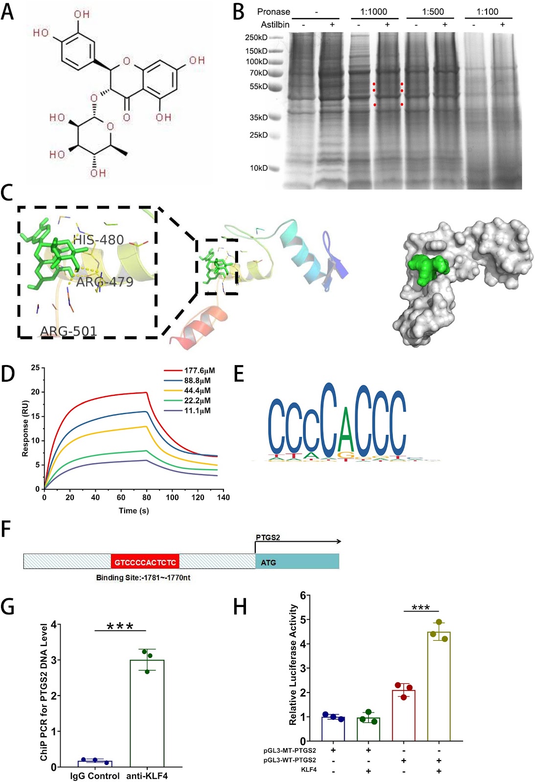
Background Although mesenchymal stem cells (MSCs) have been proven to be appropriate candidates for the treatment of AKI-CKD, their efficacy is limited and variable. Astilbin (AST) had a protective effect on MSCs from oxidative stress via ROS-scavenging, however, whether it can improve MSCs’ renoprotection and the underlying mechanism need to be elucidated. Methods AST-pretreated MSCs were administered intravenously into the ischemia–reperfusion injury mice models and the renal function, pathological changes and inflammation. Were evaluated. In addition, DARTS, molecular docking, surface plasma resonance(SPR), dual-luciferase reporter gene assay and the ChIP-PCR were utilized to explore the potential signaling pathways through which AST exert renal protective effects on MSCs. Results AST-pretreated MSCs markedly improved kidney function, reduced kidney pathological injury and inflammation in AKI and AKI-CKD mice. RNA-seq results showed that PTGS2 related pathway was significantly up-regulated in MSCs after AST pretreatment. DARTS assay, molecular docking and SPR assay revealed that AST could bind with the transcriptional factor of Kruppel-Like Factor 4(KLF4) protein. The promoter of PTGS2 had the binding and transcriptional activation by KLF4. Furthermore, AST pretreatment promoted the secretion of PGE2 in MSCs. And then the westren blot results showed that the protein levels of CD163 and CD206 were upregulated after coculture in AST-pretreated MSCs, indicating that the polarization of RAW264.7 cells towards M2-like macrophages was induced. Knockdown of PTGS2 reversed the ability of AST-pretreated MSCs in converting macrophages to M2 phenotype and reducing their therapeutic effects on AKI-CKD mice. Conclusion AST pretreatment enhances the efficacy of MSCs on AKI and AKI-CKD mice by inducing of M2-like phenotype polarization in macrophages through the PTGS2-mediated pathway. This approach not only provides a novel strategy to strengthen the capability of MSCs but also helps elucidate the beneficial effects of the Chinese herbal medicine AST.

Astilbin improves the therapeutic effects of mesenchymal stem cells in AKI-CKD mice by regulating macrophage polarization through PTGS2-mediated pathway
Graphical Abstract
Original ResearchVol. 15, Issue 1 • pp. 87DOI: 10.1186/s13287-024-03689-1

Identification of potential biomarkers for aging diagnosis of mesenchymal stem cells derived from the aged donors

Authors: Miao Hao, Hongyu Jiang, Yuan Zhao, Chunyi Li, Jinlan Jiang

Background The clinical application of human bone-marrow derived mesenchymal stem cells (MSCs) for the treatment of refractory diseases has achieved remarkable results. However, there is a need for a systematic evaluation of the quality and safety of MSCs sourced from donors. In this study, we sought to assess one potential factor that might impact quality, namely the age of the donor. Methods We downloaded two data sets from each of two Gene Expression Omnibus (GEO), GSE39035 and GSE97311 databases, namely samples form young (<65 years of age) and old (>65) donor groups. Through, bioinformatics analysis and experimental validation to these retrieved data, we found that MSCs derived from aged donors can lead to differential expression of gene profiles compared with those from young donors, and potentially affect the function of MSCs, and may even induce malignant tumors. Results We identified a total of 337 differentially expressed genes (DEGs), including two upregulated and eight downregulated genes from the databases of both GSE39035 and GSE97311. We further identified 13 hub genes. Six of them, TBX15, IGF1, GATA2, PITX2, SNAI1 and VCAN, were highly expressed in many human malignancies in Human Protein Atlas database. In the MSCs in vitro senescent cell model, qPCR analysis validated that all six hub genes were highly expressed in senescent MSCs. Our findings confirm that aged donors of MSCs have a significant effect on gene expression profiles. The MSCs from old donors have the potential to cause a variety of malignancies. These TBX15, IGF1, GATA2, PITX2, SNAI1, VCAN genes could be used as potential biomarkers to diagnosis aging state of donor MSCs, and evaluate whether MSCs derived from an aged donor could be used for therapy in the clinic. Our findings provide a diagnostic basis for the clinical use of MSCs to treat a variety of diseases. Conclusions Therefore, our findings not only provide guidance for the safe and standardized use of MSCs in the clinic for the treatment of various diseases, but also provide insights into the use of cell regeneration approaches to reverse aging and support rejuvenation.

Identification of potential biomarkers for aging diagnosis of mesenchymal stem cells derived from the aged donors
Graphical Abstract
Original ResearchVol. 15, Issue 1 • pp. 63DOI: 10.1186/s13287-024-03676-6

Embryonic stem cells overexpressing high molecular weight FGF2 isoform enhance recovery of pre-ganglionic spinal root lesion in combination with fibrin biopolymer mediated root repair

Authors: B. H. M. Lima, L. P. Cartarozzi, S. Kyrylenko, R. S. Ferreira Jr., B. Barraviera, Alexandre L. R. Oliveira

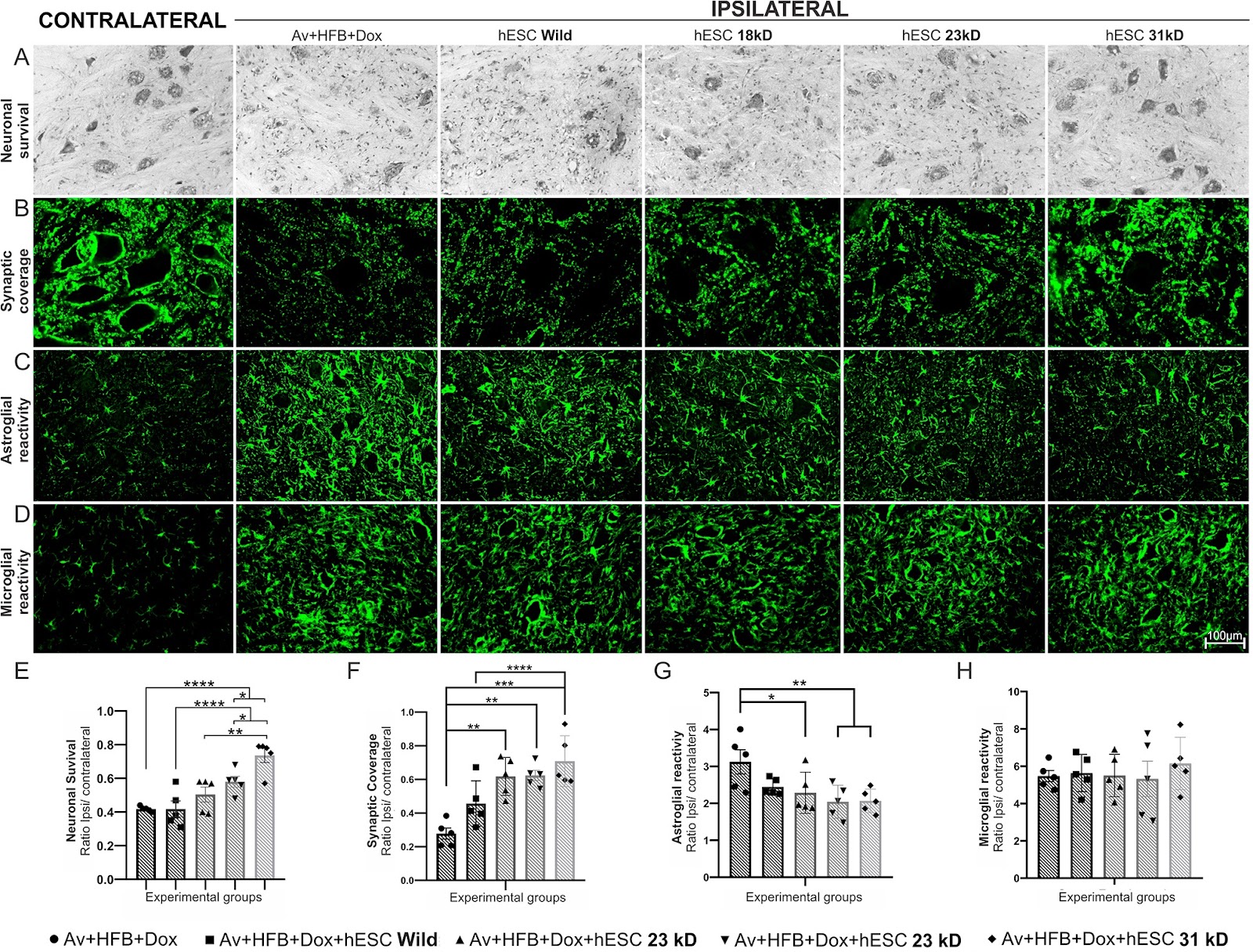
Background Spinal ventral root avulsion results in massive motoneuron degeneration with poor prognosis and high costs. In this study, we compared different isoforms of basic fibroblast growth factor 2 (FGF2), overexpressed in stably transfected Human embryonic stem cells (hESCs), following motor root avulsion and repair with a heterologous fibrin biopolymer (HFB). Methods In the present work, hESCs bioengineered to overexpress 18, 23, and 31 kD isoforms of FGF2, were used in combination with reimplantation of the avulsed roots using HFB. Statistical analysis was conducted using GraphPad Prism software with one-way or two-way ANOVA, followed by Tukey’s or Dunnett’s multiple comparison tests. Significance was set at *p < 0.05, **p < 0.01, ***p < 0.001, and ****p < 0.0001. Results For the first set of experiments, rats underwent avulsion of the ventral roots with local administration of HFB and engraftment of hESCs expressing the above-mentioned FGF2 isoforms. Analysis of motoneuron survival, glial reaction, and synaptic coverage, two weeks after the lesion, indicated that therapy with hESCs overexpressing 31 kD FGF2 was the most effective. Consequently, the second set of experiments was performed with that isoform, so that ventral root avulsion was followed by direct spinal cord reimplantation. Motoneuron survival, glial reaction, synaptic coverage, and gene expression were analyzed 2 weeks post-lesion; while the functional recovery was evaluated by the walking track test and von Frey test for 12 weeks. We showed that engraftment of hESCs led to significant neuroprotection, coupled with immunomodulation, attenuation of astrogliosis, and preservation of inputs to the rescued motoneurons. Behaviorally, the 31 kD FGF2 - hESC therapy enhanced both motor and sensory recovery. Conclusion Transgenic hESCs were an effective delivery platform for neurotrophic factors, rescuing axotomized motoneurons and modulating glial response after proximal spinal cord root injury, while the 31 kD isoform of FGF2 showed superior regenerative properties over other isoforms in addition to the significant functional recovery.

Embryonic stem cells overexpressing high molecular weight FGF2 isoform enhance recovery of pre-ganglionic spinal root lesion in combination with fibrin biopolymer mediated root repair
Graphical Abstract
Original ResearchVol. 15, Issue 1 • pp. 9DOI: 10.1186/s13287-023-03577-0

Pan PPAR agonist stimulation of induced MSCs produces extracellular vesicles with enhanced renoprotective effect for acute kidney injury

Authors: Hongduk Kim, Seul Ki Lee, Sungok Hong, Tae Sub Park, Jimin Kim, Soo Kim, Tae Min Kim

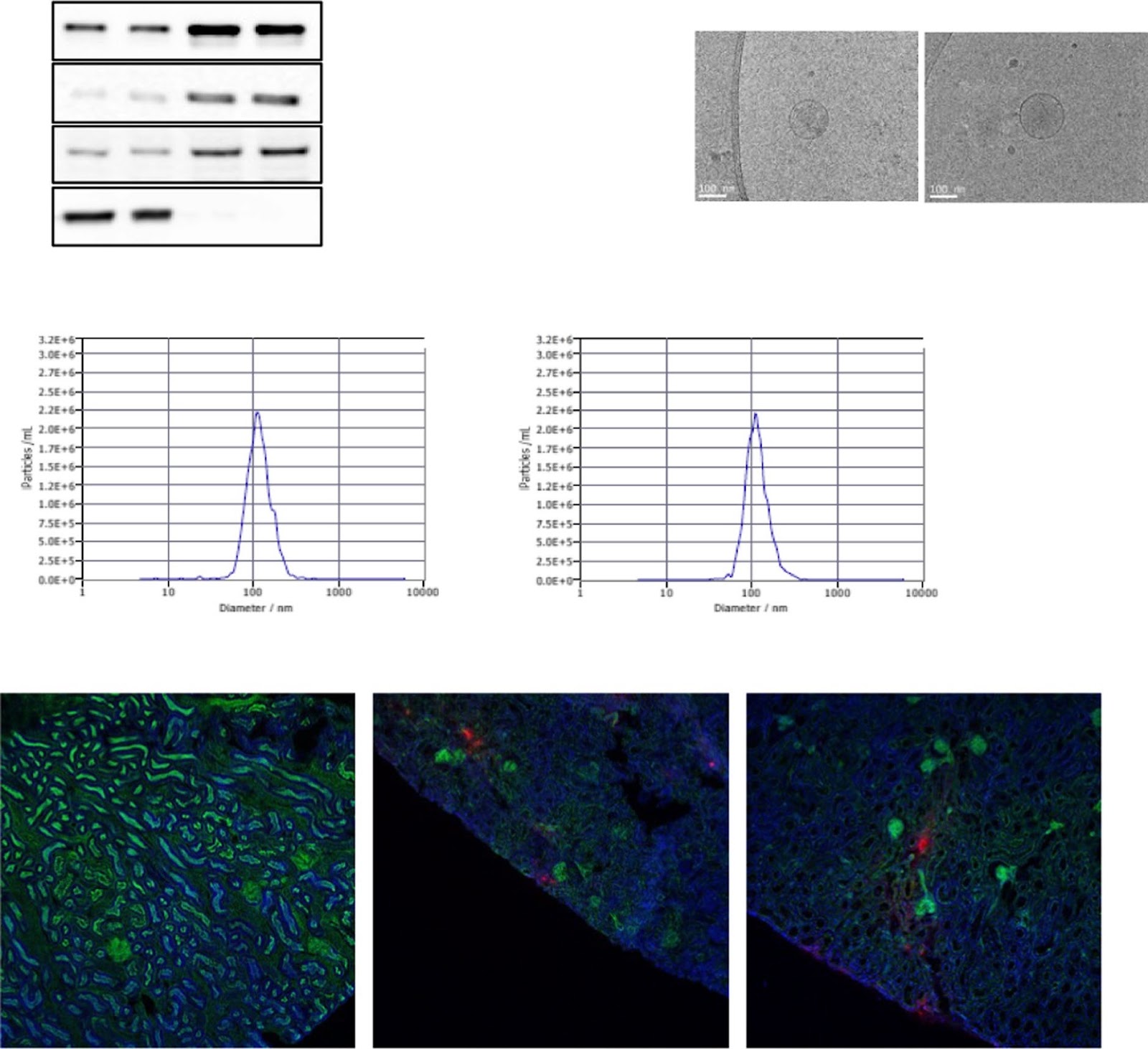
Background Acute kidney injury (AKI) has a complex pathophysiology and imposes serious health concerns worldwide. Extracellular vesicles (EVs) derived from induced mesenchymal stem cells (iMSCs) have been recognized as novel cell-free therapeutics for various inflammatory and degenerative disorders. In this study, we investigated whether iMSCs stimulated with a pan-peroxisome proliferator-activated receptor (PPAR) agonist could enhance the therapeutic efficacy of EVs against AKI. Methods Human iMSCs were primed with or without lanifibranor, a PPAR agonist for 24 h, and EVs were collected after an additional 24 h. The basic characteristics of EVs were evaluated using cryo-transmission electron microscopy imaging, immunoblot detection of EV markers, nanoparticle tracking analysis, and localization in AKI kidneys. In vitro, the potential of the EVs to promote the growth and survival of HK-2 cells undergoing cisplatin-induced apoptosis and anti-inflammatory effects in M1-polarized THP-1 was compared. Subsequently, AKI was induced in BALB/c mice using cisplatin. After 8 and 24 h of cisplatin treatment, iMSC-EVs or pan-PPAR-iMSC-EVs were injected intravascularly. At 96 h after cisplatin administration, the renoprotective effects of iMSC-EVs or pan-PPAR-iMSC-EVs in inhibiting inflammation and apoptosis were compared using serum biochemistry, histology, immunohistochemistry, and gene expression analysis by qPCR. Results Both EV types expressed EV markers and had typical EV morphology, and their localization in the renal tissue was confirmed. The proliferation and survival of HK-2 cells were higher in pan-PPAR-iMSC-EVs than those in iMSC-EVs. In M1-polarized THP-1 cells, the reduction in the mRNA expression of inflammatory cytokines was more significant in pan-PPAR-iMSC-EVs than that in iMSC-EVs. In the mouse model of cisplatin-induced AKI, pan-PPAR-iMSC-EVs markedly enhanced renoprotective effects compared to iMSC-EVs. Specifically, pan-PPAR-iMSC-EVs reduced tissue inflammation, immune cell infiltration, and apoptosis. Pan-PPAR-iMSC-EVs also increased renal capillary density. Conclusion Priming iMSCs with a PPAR agonist significantly improved the therapeutic potential of EVs by reducing inflammation and apoptosis. The reported strategy may contribute to the development of a novel cell-free option for AKI treatment. Trial registration: Not applicable.

Pan PPAR agonist stimulation of induced MSCs produces extracellular vesicles with enhanced renoprotective effect for acute kidney injury
Graphical Abstract
Original ResearchVol. 15, Issue 1 • pp. 73DOI: 10.1186/s13287-024-03690-8

Pre-clinical evaluation of the efficacy and safety of human induced pluripotent stem cell-derived cardiomyocyte patch

Authors: Shigeru Miyagawa, Takuji Kawamura, Emiko Ito, Maki Takeda, Hiroko Iseoka, Junya Yokoyama, Akima Harada, Noriko Mochizuki-Oda, Yukiko Imanishi-Ochi, Junjun Li, Masao Sasai, Fumiyo Kitaoka, Masaki Nomura, Naoki Amano, Tomoko Takahashi, Hiromi Dohi, Eiichi Morii, Yoshiki Sawa

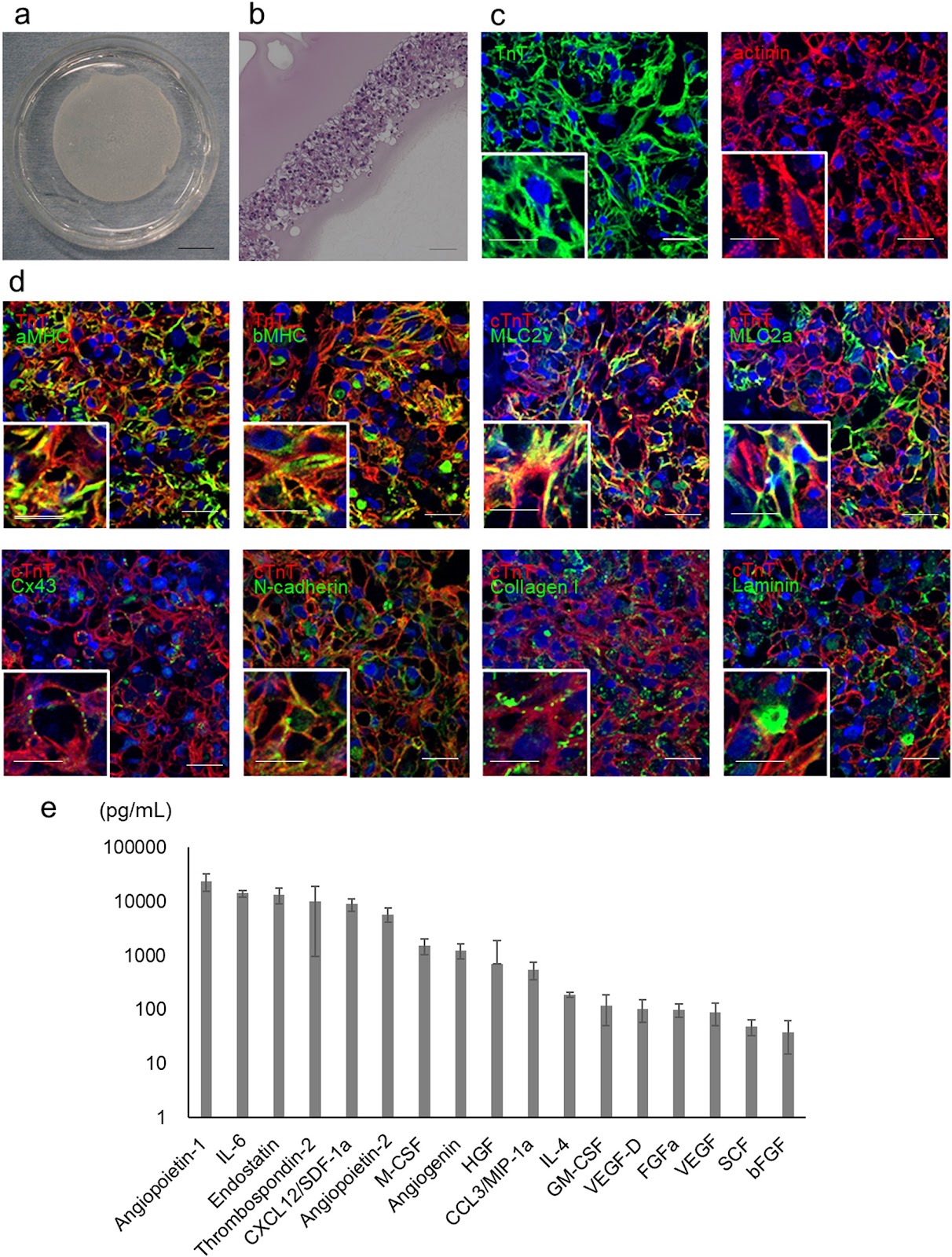
Background Cell- or tissue-based regenerative therapy is an attractive approach to treat heart failure. A tissue patch that can safely and effectively repair damaged heart muscle would greatly improve outcomes for patients with heart failure. In this study, we conducted a preclinical proof-of-concept analysis of the efficacy and safety of clinical-grade human induced pluripotent stem cell-derived cardiomyocyte (hiPSC-CM) patches. Methods A clinical-grade hiPSC line was established using peripheral blood mononuclear cells from a healthy volunteer that was homozygous for human leukocyte antigens. The hiPSCs were differentiated into cardiomyocytes. The obtained hiPSC-CMs were cultured on temperature-responsive culture dishes for patch fabrication. The cellular characteristics, safety, and efficacy of hiPSCs, hiPSC-CMs, and hiPSC-CM patches were analyzed. Results The hiPSC-CMs expressed cardiomyocyte-specific genes and proteins, and electrophysiological analyses revealed that hiPSC-CMs exhibit similar properties to human primary myocardial cells. In vitro and in vivo safety studies indicated that tumorigenic cells were absent. Moreover, whole-genome and exome sequencing revealed no genomic mutations. General toxicity tests also showed no adverse events posttransplantation. A porcine model of myocardial infarction demonstrated significantly improved cardiac function and angiogenesis in response to cytokine secretion from hiPSC-CM patches. No lethal arrhythmias were observed. Conclusions hiPSC-CM patches are promising for future translational research and may have clinical application potential for the treatment of heart failure.

Pre-clinical evaluation of the efficacy and safety of human induced pluripotent stem cell-derived cardiomyocyte patch
Graphical Abstract
Original ResearchVol. 15, Issue 1 • pp. 22DOI: 10.1186/s13287-023-03572-5

Dysregulated lncRNAs regulate human umbilical cord mesenchymal stem cell differentiation into insulin-producing cells by forming a regulatory network with mRNAs

Authors: Tianqin Xie, Qiming Huang, Qiulan Huang, Yanting Huang, Shuang Liu, Haixia Zeng, Jianping Liu

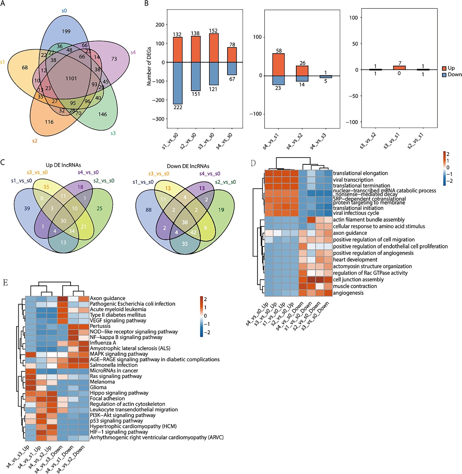
Objective In recent years, cell therapy has emerged as a new research direction in the treatment of diabetes. However, the underlying molecular mechanisms of mesenchymal stem cell (MSC) differentiation necessary to form such treatment have not been clarified. Methods In this study, human umbilical cord mesenchymal stem cells (HUC-MSCs) isolated from newborns were progressively induced into insulin-producing cells (IPCs) using small molecules. HUC-MSC (S0) and four induced stage (S1–S4) samples were prepared. We then performed transcriptome sequencing experiments to obtain the dynamic expression profiles of both mRNAs and long noncoding RNAs (lncRNAs). Results We found that the number of differentially expressed lncRNAs and mRNAs trended downwards during differentiation. Gene Ontology (GO) analysis showed that the target genes of differentially expressed lncRNAs were associated with translation, cell adhesion, and cell connection. Kyoto Encyclopedia of Genes and Genomes (KEGG) analysis revealed that the NF-KB signalling pathway, MAPK signalling pathway, HIPPO signalling pathway, PI3K–Akt signalling pathway, and p53 signalling pathway were enriched in these differentially expressed lncRNA-targeting genes. We also found that the coexpression of the lncRNA CTBP1-AS2 with PROX1 and the lncRNAs AC009014.3 and GS1-72M22.1 with JARID2 mRNA was related to the development of pancreatic beta cells. Moreover, the coexpression of the lncRNAs: XLOC_ 050969, LINC00883, XLOC_050981, XLOC_050925, MAP3K14- AS1, RP11-148K1.12, and CTD2020K17.3 with p53, regulated insulin secretion by pancreatic beta cells. Conclusion In this study, HUC-MSCs combined with small molecule compounds were successfully induced into IPCs. Differentially expressed lncRNAs may regulate the insulin secretion of pancreatic beta cells by regulating multiple signalling pathways. The lncRNAs AC009014.3, Gs1-72m21.1, and CTBP1-AS2 may be involved in the development of pancreatic beta cells, and the lncRNAs: XLOC_050969, LINC00883, XLOC_050981, XLOC_050925, MAP3K14-AS1, RP11-148K1.12, and CTD2020K17.3 may be involved in regulating the insulin secretion of pancreatic beta cells, thus providing a lncRNA catalogue for future research regarding the mechanism of the transdifferentiation of HUC-MSCs into IPCs. It also provides a new theoretical basis for the transplantation of insulin-producing cells into diabetic patients in the future.

Dysregulated lncRNAs regulate human umbilical cord mesenchymal stem cell differentiation into insulin-producing cells by forming a regulatory network with mRNAs
Graphical Abstract
Original ResearchVol. 15, Issue 1 • pp. 322DOI: 10.1186/s13287-024-03931-w

Dissecting human adipose tissue heterogeneity using single-cell omics technologies

Authors: Giuliana Di Rocco, Angelo Trivisonno, Giovanni Trivisonno, Gabriele Toietta

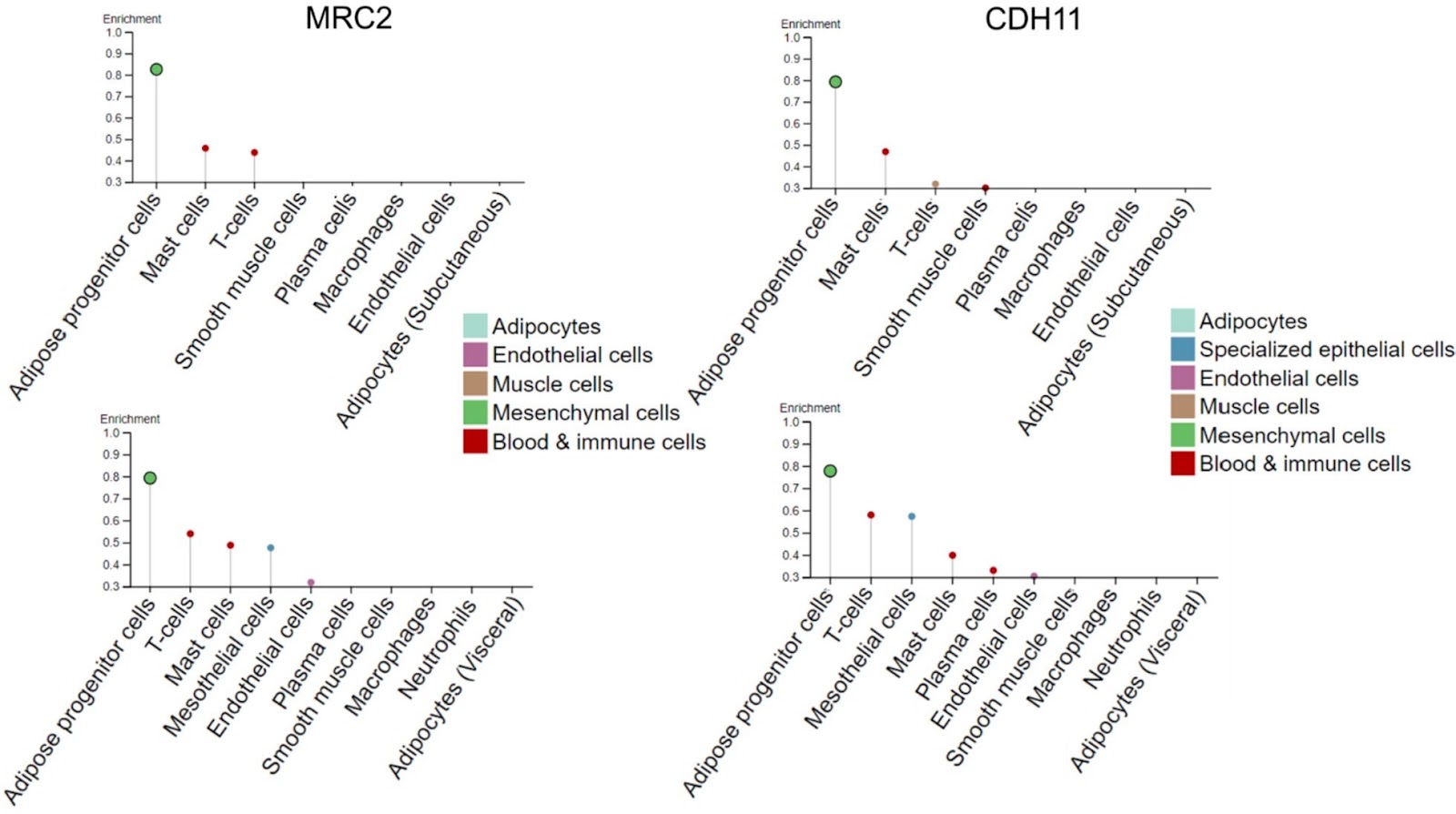
Single-cell omics technologies that profile genes (genomic and epigenomic) and determine the abundance of mRNA (transcriptomic), protein (proteomic and secretomic), lipids (lipidomic), and extracellular matrix (matrisomic) support the dissection of adipose tissue heterogeneity at unprecedented resolution in a temporally and spatially defined manner. In particular, cell omics technologies may provide innovative biomarkers for the identification of rare specific progenitor cell subpopulations, assess transcriptional and proteomic changes affecting cell proliferation and immunomodulatory potential, and accurately define the lineage hierarchy and differentiation status of progenitor cells. Unraveling adipose tissue complexity may also provide for the precise assessment of a dysfunctional state, which has been associated with cancer, as cancer-associated adipocytes play an important role in shaping the tumor microenvironment supporting tumor progression and metastasis, obesity, metabolic syndrome, and type 2 diabetes mellitus. The information collected by single-cell omics has relevant implications for regenerative medicine because adipose tissue is an accessible source of multipotent cells; alternative cell-free approaches, including the use of adipose tissue stromal cell-conditioned medium, extracellular vesicles, or decellularized extracellular matrix, are clinically valid options. Subcutaneous white adipose tissue, which is generally harvested via liposuction, is highly heterogeneous because of intrinsic biological variability and extrinsic inconsistencies in the harvesting and processing procedures. The current limited understanding of adipose tissue heterogeneity impinges on the definition of quality standards appropriate for clinical translation, which requires consistency and uniformity of the administered product. We review the methods used for dissecting adipose tissue heterogeneity and provide an overview of advances in omics technology that may contribute to the exploration of heterogeneity and dynamics of adipose tissue at the single-cell level.

Dissecting human adipose tissue heterogeneity using single-cell omics technologies
Graphical Abstract
Original ResearchVol. 15, Issue 1 • pp. 68DOI: 10.1186/s13287-023-03624-w

Mesenchymal stem/stromal cells from human pluripotent stem cell-derived brain organoid enhance the ex vivo expansion and maintenance of hematopoietic stem/progenitor cells

Authors: Ya Zhou, Xinping Cai, Xiuxiu Zhang, Yong Dong, Xu Pan, Mowen Lai, Yimeng Zhang, Yijin Chen, Xiaohong Li, Xia Li, Jiaxin Liu, Yonggang Zhang, Feng Ma

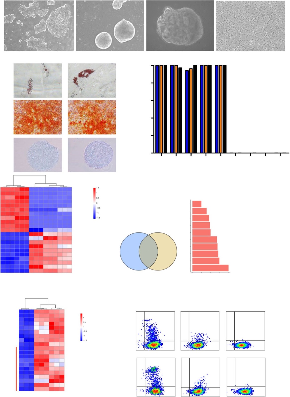
Background Mesenchymal stem/stromal cells (MSCs) are of great therapeutic value due to their role in maintaining the function of hematopoietic stem/progenitor cells (HSPCs). MSCs derived from human pluripotent stem cells represent an ideal alternative because of their unlimited supply. However, the role of MSCs with neural crest origin derived from HPSCs on the maintenance of HSPCs has not been reported. Methods Flow cytometric analysis, RNA sequencing and differentiation ability were applied to detect the characteristics of stromal cells from 3D human brain organoids. Human umbilical cord blood CD34+ (UCB-CD34+) cells were cultured in different coculture conditions composed of stromal cells and umbilical cord MSCs (UC-MSCs) with or without a cytokine cocktail. The hematopoietic stroma capacity of stromal cells was tested in vitro with the LTC-IC assay and in vivo by cotransplantation of cord blood nucleated cells and stroma cells into immunodeficient mice. RNA and proteomic sequencing were used to detect the role of MSCs on HSPCs. Results The stromal cells, derived from both H1-hESCs and human induced pluripotent stem cells forebrain organoids, were capable of differentiating into the classical mesenchymal-derived cells (osteoblasts, chondrocytes, and adipocytes). These cells expressed MSC markers, thus named pluripotent stem cell-derived MSCs (pMSCs). The pMSCs showed neural crest origin with CD271 expression in the early stage. When human UCB-CD34+ HSPCs were cocultured on UC-MSCs or pMSCs, the latter resulted in robust expansion of UCB-CD34+ HSPCs in long-term culture and efficient maintenance of their transplantability. Comparison by RNA sequencing indicated that coculture of human UCB-CD34+ HSPCs with pMSCs provided an improved microenvironment for HSC maintenance. The pMSCs highly expressed the Wnt signaling inhibitors SFRP1 and SFRP2, indicating that they may help to modulate the cell cycle to promote the maintenance of UCB-CD34+ HSPCs by antagonizing Wnt activation.

Mesenchymal stem/stromal cells from human pluripotent stem cell-derived brain organoid enhance the ex vivo expansion and maintenance of hematopoietic stem/progenitor cells
Graphical Abstract
Original ResearchVol. 15, Issue 1 • pp. 423DOI: 10.1186/s13287-024-04032-4

N-CADHERIN+/CD168− subpopulation determines therapeutic variations of UC-MSCs for cardiac repair after myocardial infarction

Authors: Yukang Wu, Jianguo Li, Ke Feng, Ailing Tan, Yingying Gao, Wen Chen, Wenwen Jia, Xudong Guo, Jiuhong Kang

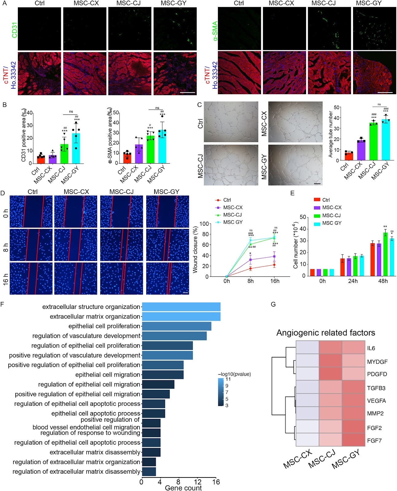
Background The efficiency of mesenchymal stem cells (MSCs) in treating myocardial infarction (MI) remains inconsistent, which limits their therapeutic applications. Therefore, exploring the mechanism for the inconsistent efficacy of MSCs and identification the criteria for screening MSCs are important for improving the efficiency of MSCs. Methods Mouse model after MI was utilized to test the role of MSCs from different donors and the functional subpopulation in improving cardiac function. Heterogeneity of MSCs was identified using single-cell RNA sequencing (scRNA-seq) of MSC-GY. GSEA and Scissor analyses were used to find the functional subpopulations of MSCs that promote angiogenesis. The role of functional subpopulations in promoting angiogenesis was verified by detecting the secretory proteins, the ratio of N-CADHERIN+/CD168− subpopulations in MSCs, and the tube formation, migration, and proliferation of HUVECs after treatment with conditional medium (CM) derived from different MSCs. Results We found that umbilical cord-derived MSCs (UC-MSCs) from different donors have varied therapeutic efficacy in MI mice and UC-MSCs with higher therapeutic effectiveness exhibited the most potent pro-angiogenic effects by secreting elevated levels of angiogenesis-related proteins, such as MYDGF, VEGFA, and FGF2. ScRNA-seq of 10,463 UC-MSCs revealed that the N-CADHERIN+/CD168− subpopulation was closely associated with pro-angiogenic effects, and the ratio of this cell subpopulation was positively correlated with the angiogenic potential of MSCs. We also found that the N-CADHERIN+/CD168− subpopulation was the functional subpopulation of MSCs in improving cardiac function of MI mice. Conclusions Our study identified that the N-CADHERIN+/CD168− subpopulation was the functional subpopulation of MSCs in treating MI, which was essential for the development and utilization of MSCs in MI treatment.

N-CADHERIN+/CD168− subpopulation determines therapeutic variations of UC-MSCs for cardiac repair after myocardial infarction
Graphical Abstract
Original ResearchVol. 15, Issue 1 • pp. 288DOI: 10.1186/s13287-024-03903-0

Urine-derived stem cells serve as a robust platform for generating native or engineered extracellular vesicles

Authors: Anders Toftegaard Boysen, Bradley Whitehead, Anne Louise S. Revenfeld, Dhanu Gupta, Thor Petersen, Peter Nejsum

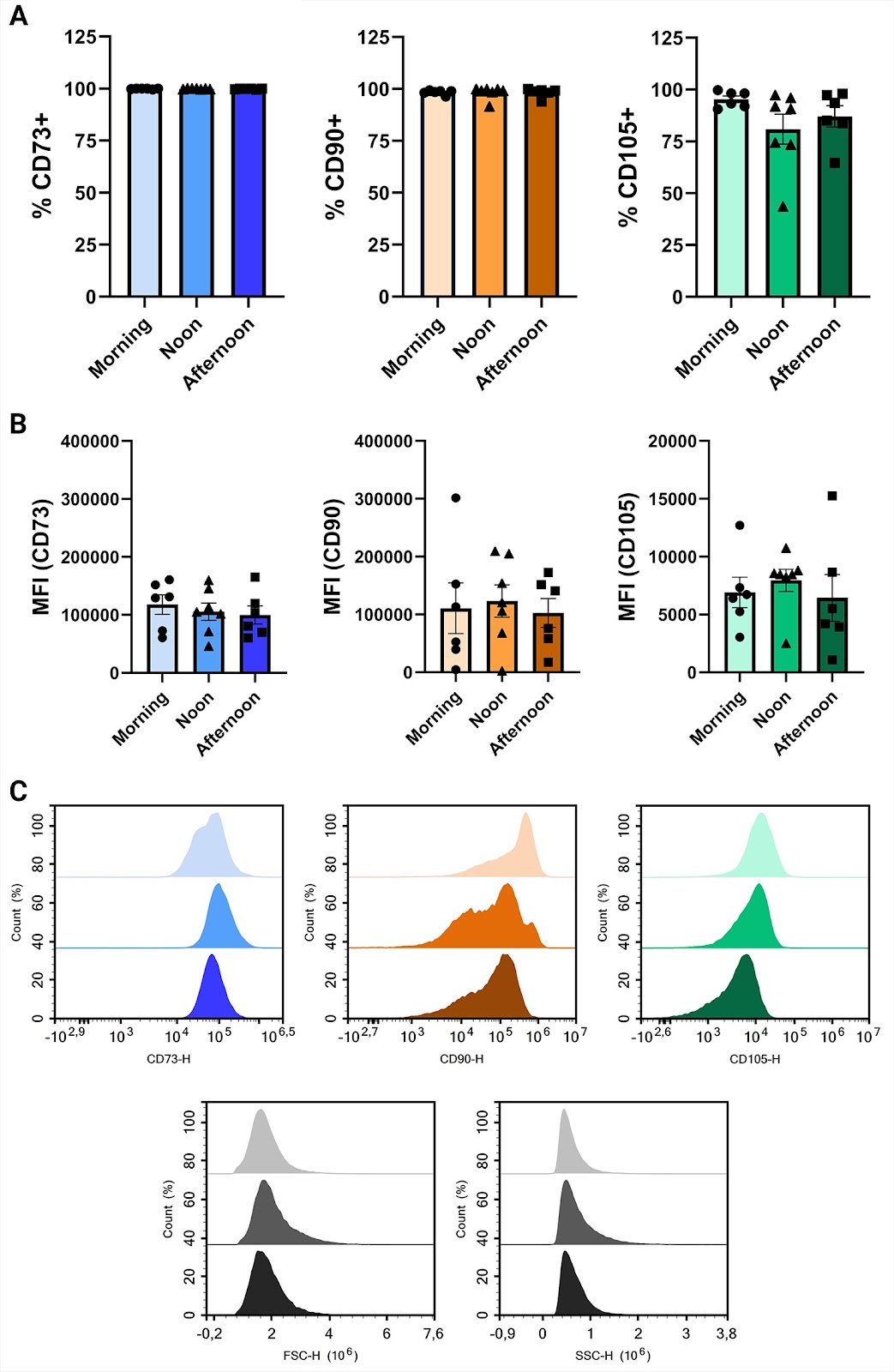
Background Mesenchymal stromal cell (MSC) therapy holds great potential yet efficacy and safety concerns with cell therapy persist. The beneficial effects of MSCs are often attributed to their secretome that includes extracellular vesicles (EVs). EVs carry biologically active molecules, protected by a lipid bilayer. However, several barriers hinder large-scale MSC EV production. A serum-free culturing approach is preferred for producing clinical-grade MSC-derived EVs but this can affect both yield and purity. Consequently, new strategies have been explored, including genetically engineering MSCs to alter EV compositions to enhance potency, increase circulation time or mediate targeting. However, efficient transfection of MSCs is challenging. Typical sources of MSC include adipose tissue and bone marrow, which both require invasive extraction procedures. Here, we investigate the use of urine-derived stem cells (USCs) as a non-invasive and inexhaustible source of MSCs for EV production. Methods We isolated, expanded, and characterized urine-derived stem cells (USCs) harvested from eight healthy donors at three different time points during the day. We evaluated the number of clones per urination, proliferation capacity and conducted flow cytometry to establish expression of surface markers. EVs were produced in chemically defined media and characterized. PEI/DNA transfection was used to genetically engineer USCs using transposon technology. Results There were no differences between time points for clone number, doubling time or viability. USCs showed immunophenotypic characteristics of MSCs, such as expression of CD73, CD90 and CD105, with no difference at the assessed time points, however, male donors had reduced CD73+ cells. Expanded USCs were incubated without growth factors or serum for 72 h without a loss in viability and EVs were isolated. USCs were transfected with high efficiency and after 10 days of selection, pure engineered cell cultures were established. Conclusions Isolation and expansion of MSCs from urine is non-invasive, robust, and without apparent sex-related differences. The sampling time point did not affect any measured markers or USC isolation potential. USCs offer an attractive production platform for EVs, both native and engineered.

Urine-derived stem cells serve as a robust platform for generating native or engineered extracellular vesicles
Graphical Abstract
Original ResearchVol. 15, Issue 1 • pp. 39DOI: 10.1186/s13287-024-03665-9

Correction: Primary explants of the postnatal thymus allow the expansion of clonogenic thymic epithelial cells that constitute thymospheres

Authors: Juan M. Ocampo-Godinez, Jose L. Gonzalez-Quiroz, Hector Cote-Palafox, Elizabeth George, Jael A. Vergara-Lope Nuñez, Guillermo Villagomez-Olea, Febe C. Vazquez-Vazquez, Edgar O. Lopez-Villegas, Gloria Leon-Avila, Maria L. Dominguez-Lopez, Marco A. Alvarez-Perez

The original article contains an erroneous attribution of co-author, Gloria Leon-Avila to affiliation #3. Dr Leon-Avila should instead be affiliated to affiliation #2 as shown in this correction article.

Correction: Primary explants of the postnatal thymus allow the expansion of clonogenic thymic epithelial cells that constitute thymospheres
Graphical Abstract
Original ResearchVol. 15, Issue 1 • pp. 48DOI: 10.1186/s13287-024-03656-w

Kupffer cells abrogate homing and repopulation of allogeneic hepatic progenitors in injured liver site

Authors: Nasir Abbas, Kai You, Anteneh Getachew, Feima Wu, Muzammal Hussain, Xinping Huang, Yan Chen, Tingcai Pan, Yinxiong Li

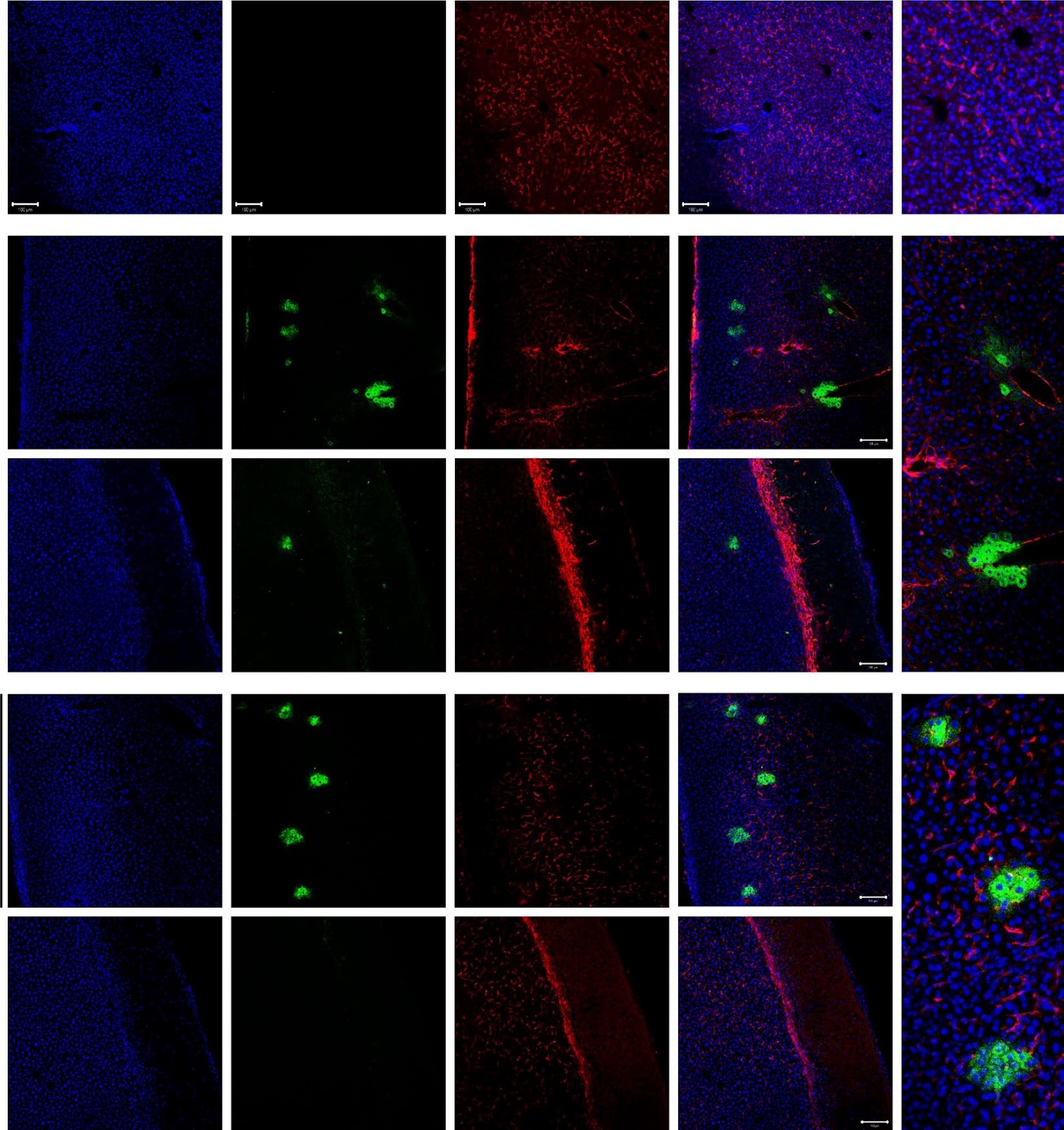
Background Allogeneic hepatocyte transplantation is an emerging approach to treat acute liver defects. However, durable engraftment of the transplanted cells remains a daunting task, as they are actively cleared by the recipient’s immune system. Therefore, a detailed understanding of the innate or adaptive immune cells-derived responses against allogeneic transplanted hepatic cells is the key to rationalize cell-based therapies. Methods Here, we induced an acute inflammatory regenerative niche (3–96 h) on the surface of the liver by the application of cryo-injury (CI) to systematically evaluate the innate immune response against transplanted allogeneic hepatic progenitors in a sustained micro-inflammatory environment. Results The resulting data highlighted that the injured site was significantly repopulated by alternating numbers of innate immune cells, including neutrophils, monocytes and Kupffer cells (KCs), from 3 to 96 h. The transplanted allo-HPs, engrafted 6 h post-injury, were collectively eliminated by the innate immune response within 24 h of transplantation. Selective depletion of the KCs demonstrated a delayed recruitment of monocytes from day 2 to day 6. In addition, the intrasplenic engraftment of the hepatic progenitors 54 h post-transplantation was dismantled by KCs, while a time-dependent better survival and translocation of the transplanted cells into the injured site could be observed in samples devoid of KCs. Conclusion Overall, this study provides evidence that KCs ablation enables a better survival and integration of allo-HPs in a sustained liver inflammatory environment, having implications for rationalizing the cell-based therapeutic interventions against liver defects.

Kupffer cells abrogate homing and repopulation of allogeneic hepatic progenitors in injured liver site
Graphical Abstract
Original ResearchVol. 15, Issue 1 • pp. 158DOI: 10.1186/s13287-024-03753-w

Harnessing three-dimensional porous chitosan microsphere embedded with adipose-derived stem cells to promote nerve regeneration

Authors: Yaqiong Zhu, Dan Yi, Jing Wang, Yongyi Zhang, Molin Li, Jun Ma, Yongjiao Ji, Jiang Peng, Yuexiang Wang, Yukun Luo

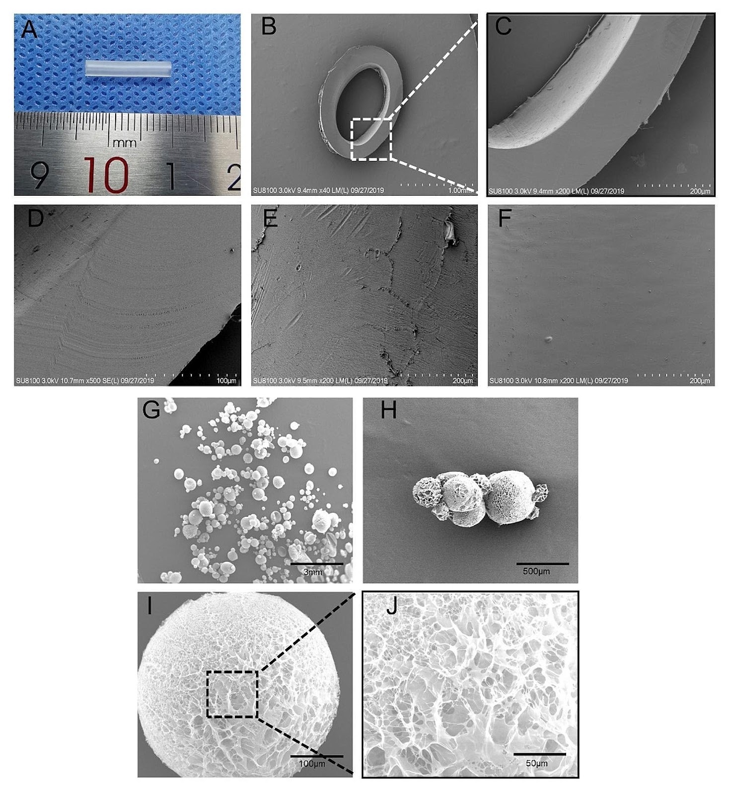
Background Nerve guide conduits are a promising strategy for reconstructing peripheral nerve defects. Improving the survival rate of seed cells in nerve conduits is still a challenge and microcarriers are an excellent three-dimensional (3D) culture scaffold. Here, we investigate the effect of the 3D culture of microcarriers on the biological characteristics of adipose mesenchymal stem cells (ADSCs) and to evaluate the efficacy of chitosan nerve conduits filled with microcarriers loaded with ADSCs in repairing nerve defects. Methods In vitro, we prepared porous chitosan microspheres by a modified emulsion cross-linking method for loading ADSCs and evaluated the growth status and function of ADSCs. In vivo, ADSCs-loaded microcarriers were injected into chitosan nerve conduits to repair a 12 mm sciatic nerve defect in rats. Results Compared to the conventional two-dimensional (2D) culture, the prepared microcarriers were more conducive to the proliferation, migration, and secretion of trophic factors of ADSCs. In addition, gait analysis, neuro-electrophysiology, and histological evaluation of nerves and muscles showed that the ADSC microcarrier-loaded nerve conduits were more effective in improving nerve regeneration. Conclusions The ADSCs-loaded chitosan porous microcarrier prepared in this study has a high cell engraftment rate and good potential for peripheral nerve repair.

Harnessing three-dimensional porous chitosan microsphere embedded with adipose-derived stem cells to promote nerve regeneration
Graphical Abstract
Original ResearchVol. 15, Issue 1 • pp. 11DOI: 10.1186/s13287-023-03626-8

Good manufacturing practice production of human corneal limbus-derived stromal stem cells and in vitro quality screening for therapeutic inhibition of corneal scarring

Authors: Mithun Santra, Moira L. Geary, Elizabeth Rubin, Michael Y. S. Hsu, Martha L. Funderburgh, Christine Chandran, Yiqin Du, Deepinder K. Dhaliwal, Vishal Jhanji, Gary Hin-Fai Yam

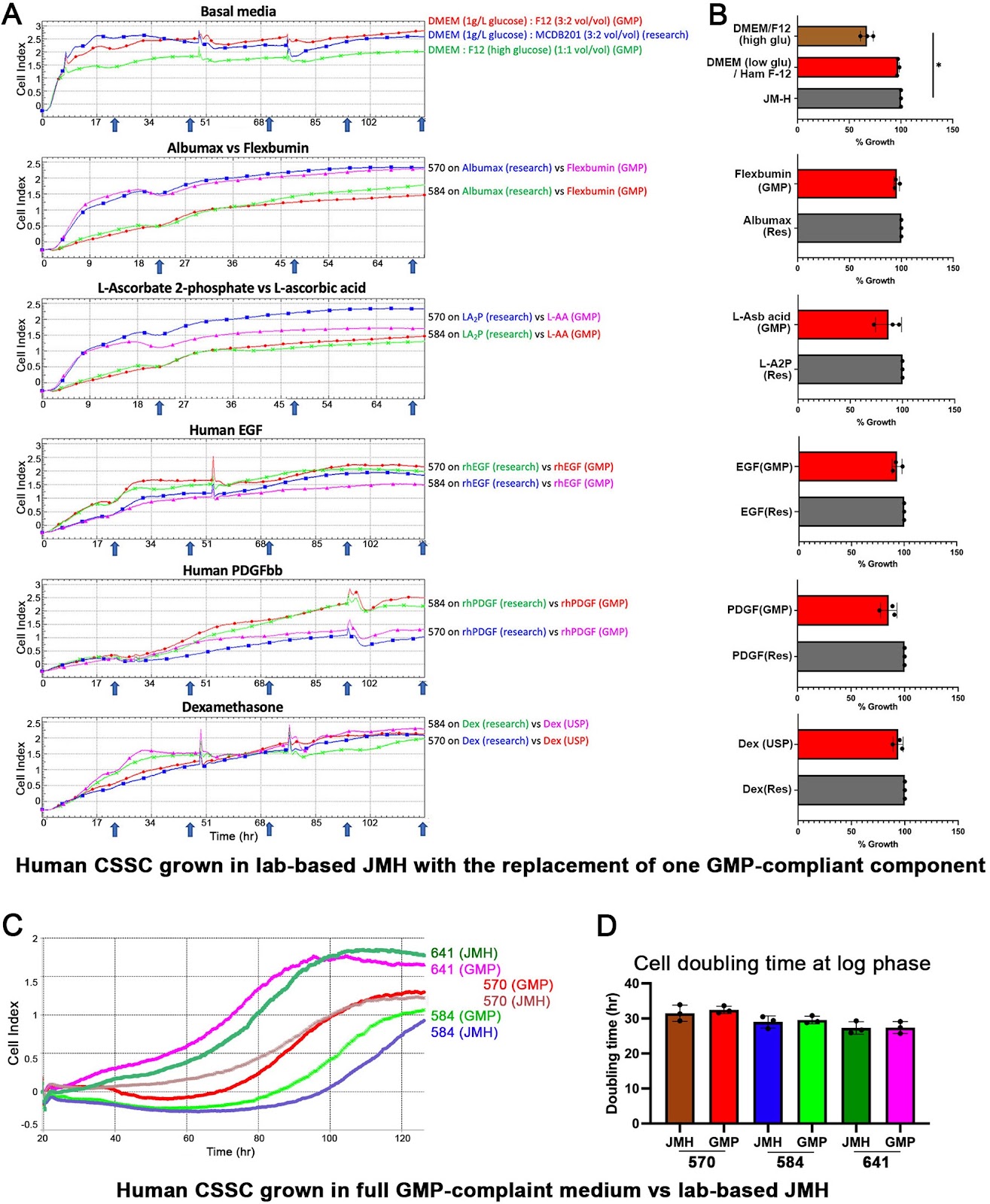
Background Mesenchymal stem cells in the adult corneal stroma (named corneal stromal stem cells, CSSCs) inhibit corneal inflammation and scarring and restore corneal clarity in pre-clinical corneal injury models. This cell therapy could alleviate the heavy reliance on donor materials for corneal transplantation to treat corneal opacities. Herein, we established Good Manufacturing Practice (GMP) protocols for CSSC isolation, propagation, and cryostorage, and developed in vitro quality control (QC) metric for in vivo anti-scarring potency of CSSCs in treating corneal opacities. Methods A total of 24 donor corneal rims with informed consent were used—18 were processed for the GMP optimization of CSSC culture and QC assay development, while CSSCs from the remaining 6 were raised under GMP-optimized conditions and used for QC validation. The cell viability, growth, substrate adhesion, stem cell phenotypes, and differentiation into stromal keratocytes were assayed by monitoring the electric impedance changes using xCELLigence real-time cell analyzer, quantitative PCR, and immunofluorescence. CSSC’s conditioned media were tested for the anti-inflammatory activity using an osteoclastogenesis assay with mouse macrophage RAW264.7 cells. In vivo scar inhibitory outcomes were verified using a mouse model of anterior stromal injury caused by mechanical ablation using an Algerbrush burring. Results By comparatively assessing various GMP-compliant reagents with the corresponding non-GMP research-grade chemicals used in the laboratory-based protocols, we finalized GMP protocols covering donor limbal stromal tissue processing, enzymatic digestion, primary CSSC culture, and cryopreservation. In establishing the in vitro QC metric, two parameters—stemness stability of ABCG2 and nestin and anti-inflammatory ability (rate of inflammation)—were factored into a novel formula to calculate a Scarring Index (SI) for each CSSC batch. Correlating with the in vivo scar inhibitory outcomes, the CSSC batches with SI < 10 had a predicted 50% scar reduction potency, whereas cells with SI > 10 were ineffective to inhibit scarring. Conclusions We established a full GMP-compliant protocol for donor CSSC cultivation, which is essential toward clinical-grade cell manufacturing. A novel in vitro QC–in vivo potency correlation was developed to predict the anti-scarring efficacy of donor CSSCs in treating corneal opacities. This method is applicable to other cell-based therapies and pharmacological treatments.

Good manufacturing practice production of human corneal limbus-derived stromal stem cells and in vitro quality screening for therapeutic inhibition of corneal scarring
Graphical Abstract
Original ResearchVol. 15, Issue 1 • pp. 197DOI: 10.1186/s13287-024-03787-0

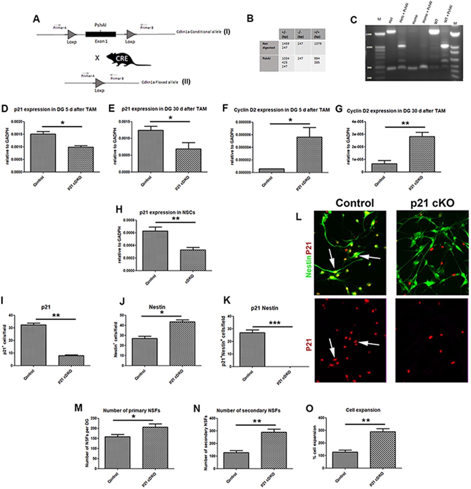
Essential role of p21Waf1/Cip1 in the modulation of post-traumatic hippocampal Neural Stem Cells response

Authors: Francesco Chiani, Valentina Mastrorilli, Nicole Marchetti, Andrea Macioce, Chiara Nappi, Georgios Strimpakos, Miriam Pasquini, Alessia Gambadoro, Jonathan Isacco Battistini, Debora Cutuli, Laura Petrosini, Sara Marinelli, Raffaella Scardigli, Stefano Farioli Vecchioli

Background Traumatic Brain Injury (TBI) represents one of the main causes of brain damage in young people and the elderly population with a very high rate of psycho-physical disability and death. TBI is characterized by extensive cell death, tissue damage and neuro-inflammation with a symptomatology that varies depending on the severity of the trauma from memory loss to a state of irreversible coma and death. Recently, preclinical studies on mouse models have demonstrated that the post-traumatic adult Neural Stem/Progenitor cells response could represent an excellent model to shed light on the neuro-reparative role of adult neurogenesis following damage. The cyclin-dependent kinase inhibitor p21Waf1/Cip1 plays a pivotal role in modulating the quiescence/activation balance of adult Neural Stem Cells (aNSCs) and in restraining the proliferation progression of progenitor cells. Based on these considerations, the aim of this work is to evaluate how the conditional ablation of p21Waf1/Cip1 in the aNSCS can alter the adult hippocampal neurogenesis in physiological and post-traumatic conditions. Methods We designed a novel conditional p21Waf1/Cip1 knock-out mouse model, in which the deletion of p21Waf1/Cip1 (referred as p21) is temporally controlled and occurs in Nestin-positive aNSCs, following administration of Tamoxifen. This mouse model (referred as p21 cKO mice) was subjected to Controlled Cortical Impact to analyze how the deletion of p21 could influence the post-traumatic neurogenic response within the hippocampal niche. Results The data demonstrates that the conditional deletion of p21 in the aNSCs induces a strong increase in activation of aNSCs as well as proliferation and differentiation of neural progenitors in the adult dentate gyrus of the hippocampus, resulting in an enhancement of neurogenesis and the hippocampal-dependent working memory. However, following traumatic brain injury, the increased neurogenic response of aNSCs in p21 cKO mice leads to a fast depletion of the aNSCs pool, followed by declined neurogenesis and impaired hippocampal functionality. Conclusions These data demonstrate for the first time a fundamental role of p21 in modulating the post-traumatic hippocampal neurogenic response, by the regulation of the proliferative and differentiative steps of aNSCs/progenitor populations after brain damage.

Essential role of p21Waf1/Cip1 in the modulation of post-traumatic hippocampal Neural Stem Cells response
Graphical Abstract
Original ResearchVol. 15, Issue 1 • pp. 424DOI: 10.1186/s13287-024-04022-6

Transcriptional landscape of the interaction of human Mesenchymal Stem Cells with Glioblastoma in bioprinted co-cultures

Authors: Lisa Oliver, Yuna Landais, Catherine Gratas, Pierre-François Cartron, François Paris, Dominique Heymann, François M. Vallette, Aurelien Serandour

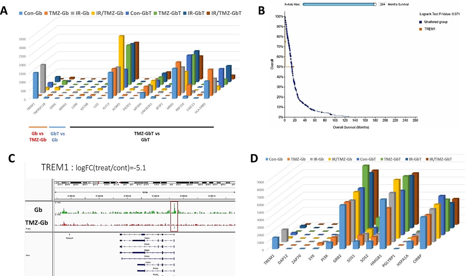
Background The interaction between mesenchymal stem cells (MSC) and Glioblastoma (GBM), although potentially of the highest importance, is ill-understood. This is due, in part, to the lack of relevant experimental models. The similarity between the in vitro situations and the in vivo situation can be improved by 3D co-culture as it reproduces key cell–cell interactions between the tumor microenvironment (TME) and cancer cells. Methods MSC Can acquired characteristics of cancer associated fibroblasts (CAF) by being cultured with conditioned medium from GBM cultures and thus are called MSCCAF. We co Cultured MSCCAF with patient derived GBM in a scaffold 3D bioprinted model. We studied the response to current GBM therapy (e.g. Temozolomide +/Radiation) on the co cultures by bulk transcriptomic (RNA Seq) and epigenetic (ATAC Seq) analyses Results The transcriptomic modifications induced by standard GBM treatment in bioprinted scaffolds of mono- or co-cultures of GBM ± MSC can be analyzed. We found that mitochondrial encoded OXPHOS genes are overexpressed under these conditions and are modified by both co-culture and treatment (chemotherapy ± radiation). We have identified two new markers of MSC/GBM interactions, one epigenetically regulated (i.e. TREM-1) associated with an increased overall survival in GBM patients and another implicated in post-transcriptional regulation (i.e. the long non-coding RNA, miR3681HG), which is associated with a reduced overall survival in GBM patients.

Transcriptional landscape of the interaction of human Mesenchymal Stem Cells with Glioblastoma in bioprinted co-cultures
Graphical Abstract