🧬 SinoBioData Academic Portal
🏛️ Indexed Academic JournalOriginal: 生物化学与生物物理学报

Acta Biochimica et Biophysica Sinica

Premier Chinese Biomedical Journal indexed in SinoBioData: Acta Biochimica et Biophysica Sinica (生物化学与生物物理学报).

Total Research Papers: 200
Access: 100% Free Open Access
Browse by Publication Year & VolumeReset All Filters ✕

Published Research PapersFiltered: Year 2024 • 56

Showing 28 of 200 peer-reviewed papers with full Graphical Abstracts.

Original ResearchVol. 56, Issue 3 • pp. 356-365DOI: 10.3724/abbs.2023246

HSPA8-mediated stability of the CLPP protein regulates mitochondrial autophagy in cisplatin-resistant ovarian cancer cells

Authors: Xinxin Kou, Xiaoxia Yang, Zheng Zhao, Lei Li

This corrigendum corrects the affiliation of the authors in the original article. The affiliation has been changed from 'Cancer Hospital Affiliated to Zhengzhou University' to 'The Affiliated Cancer Hospital of Zhengzhou University & Henan Cancer Hospital'.

HSPA8-mediated stability of the CLPP protein regulates mitochondrial autophagy in cisplatin-resistant ovarian cancer cells
Graphical Abstract
Original ResearchVol. 56, Issue 12 • pp. 1733-1747DOI: 10.3724/abbs.2024130

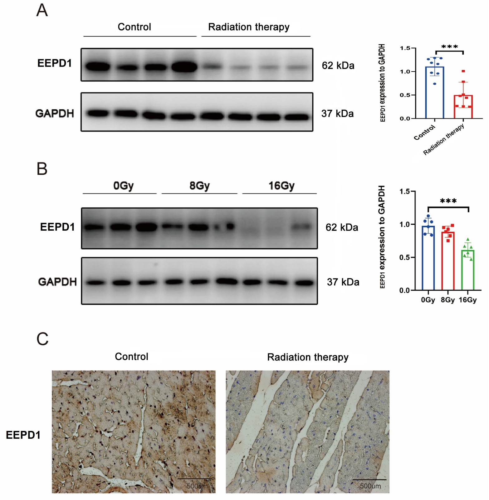
EEPD1 attenuates radiation-induced cardiac hypertrophy and apoptosis by degrading FOXO3A in cardiomyocytes

Authors: Kaiwen Yu, Xi Su, Tongfang Zhou, Xuwei Cai, Min Zhang

Radiation-induced heart disease (RIHD) is a severe delayed complication of thoracic irradiation (IR). Endonuclease/exonuclease/phosphatase family domain-containing 1 (EEPD1) plays an important role in DNA damage repair, but its role in RIHD is less known. In this study, EEPD1 global knockout mice, C57BL/6J mice, and C57BL/6J mice overexpressing EEPD1 are treated with radiation at a total dose of 20 Gy or 0 Gy. After 9 weeks, echocardiography is used to assess cardiac hypertrophy and apoptosis. The results show that EEPD1 deletion exacerbates radiation-induced cardiac hypertrophy and apoptosis, while EEPD1 overexpression has the opposite effect. Further mechanistic investigations reveal that EEPD1 interacts with FOXO3A and destabilizes it by catalyzing its deubiquitination. Inhibition of FOXO3A ameliorates cardiac hypertrophy and apoptosis after EEPD1 knockdown. Thus, EEPD1 protects against radiation-induced cardiac hypertrophy and apoptosis via destabilization of FOXO3A, which may offer new insight into therapeutic strategies for RIHD.

EEPD1 attenuates radiation-induced cardiac hypertrophy and apoptosis by degrading FOXO3A in cardiomyocytes
Graphical Abstract
Original ResearchVol. 56, Issue 12 • pp. 1860-1874DOI: 10.3724/abbs.2024141

Vaccarin suppresses diabetic nephropathy through inhibiting the EGFR/ERK1/2 signaling pathway

Authors: Xuexue Zhu, Xinyu Meng, Xinyao Du, Chenyang Zhao, Xinyu Ma, Yuanyuan Wen, Shijie Zhang, Bao Hou, Weiwei Cai, Bin Du, Zhijun Han, Fei Xu, Liying Qiu, Haijian Sun

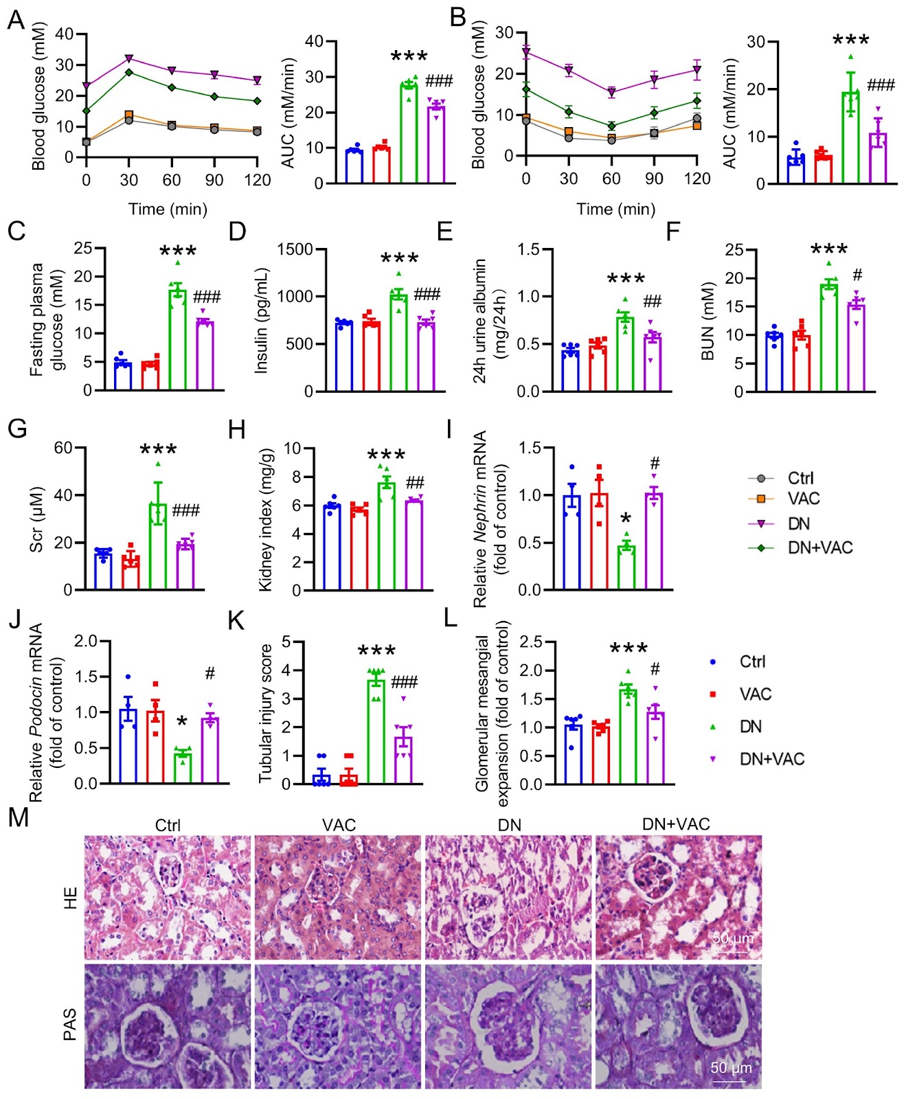
Diabetic nephropathy (DN) is recognized as one of the primary causes of chronic kidney disease and end-stage renal disease. Vaccarin (VAC) confers favorable effects on cardiovascular and metabolic diseases, including type 2 diabetes mellitus (T2DM). Nonetheless, the potential role and mechanism of VAC in the etiology of DN have yet to be completely elucidated. In this study, a classical mouse model of T2DM is experimentally induced via a high-fat diet (HFD)/streptozocin (STZ) regimen. Renal histological changes are assessed via H&E staining. Masson staining and immunohistochemistry (IHC) are employed to assess renal fibrosis. RT-PCR is utilized to quantify the mRNA levels of renal fibrosis, oxidative stress and inflammation markers. The levels of malondialdehyde (MDA) and reactive oxygen species (ROS), as well as the content of glutathione peroxidase (GSH-Px), are measured. The protein expressions of collagen I, TGF-β1, α-SMA, E-cadherin, Nrf2, catalase, SOD3, SOD2, SOD1, p-ERK, p-EGFR (Y845), p-EGFR (Y1173), p-NFκB P65, t-ERK, t-EGFR and t-NFκB P65 are detected by western blot analysis. Our results reveal that VAC has a beneficial effect on DN mice by improving renal function and mitigating histological damage. This is achieved through its inhibition of renal fibrosis, inflammatory cytokine overproduction, and ROS generation. Moreover, VAC treatment effectively suppresses the process of epithelial-mesenchymal transition (EMT), a crucial characteristic of renal fibrosis, in high glucose (HG)-induced HK-2 cells. Network pharmacology analysis and molecular docking identify epidermal growth factor receptor (EGFR) as a potential target for VAC. Amino acid site mutations reveal that Lys-879, Ile-918, and Ala-920 of EGFR may mediate the direct binding of VAC to EGFR. In support of these findings, VAC reduces the phosphorylation levels of both EGFR and its downstream mediator, extracellular signal-regulated kinase 1/2 (ERK1/2), in diabetic kidneys and HG-treated HK-2 cells. Notably, blocking either EGFR or ERK1/2 yields renal benefits similar to those observed with VAC treatment. Therefore, this study reveals that VAC attenuates renal damage via inactivation of the EGFR/ERK1/2 signaling axis in T2DM patients.

Vaccarin suppresses diabetic nephropathy through inhibiting the EGFR/ERK1/2 signaling pathway
Graphical Abstract
Original ResearchVol. 56, Issue 12 • pp. 1886-1890DOI: 10.3724/abbs.2024088

A PCR-independent, annealing-free cloning method for the insertion of short DNA fragments

Authors: Linbo Li, Jin Yan, Yuan Qi, Zhenglong Xiang, Na Jiang, Tongkang Yuan, Zhenyi Wang, Yuan Wang, Huaizhe Zhan, Shiyi Liu, Li Zhao, Jing Xu, Xiaowei Lei, Yuxuan Liu, Gui Wang, Jiayang Xie, Zhenming Guo, Chunhai Cai, Shan Bian

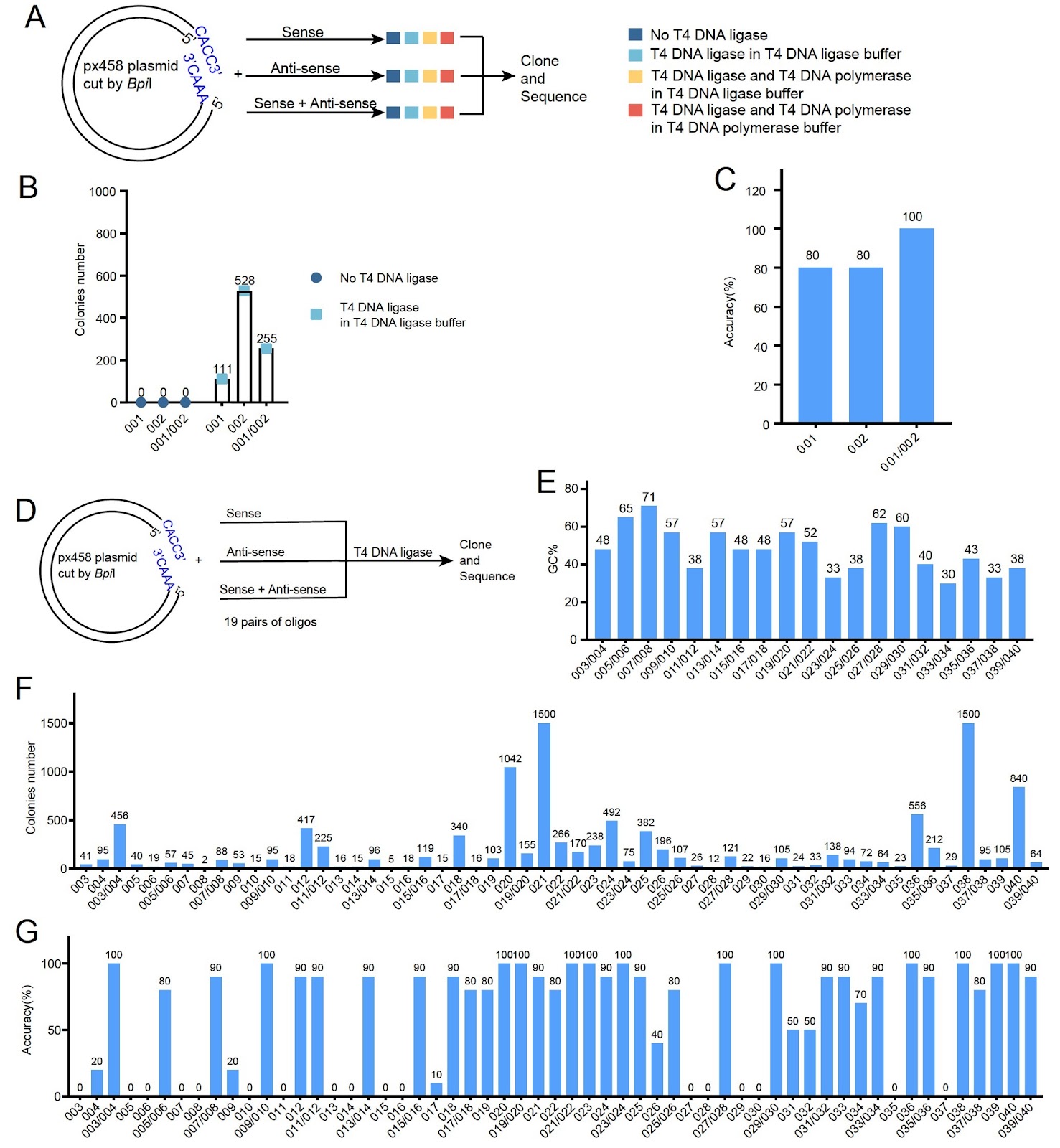
Cloning short DNA fragments, such as shRNA and sgRNA, is a routine but time-consuming task in molecular biology. Traditional methods require annealing of complementary oligos or PCR amplification, which are labor-intensive and time-consuming. Here, we report a novel PCR-independent, annealing-free cloning method that enables the insertion of short DNA fragments using a single oligo. The method relies on T4 DNA ligase for ligation and host cell DNA polymerase for complementary strand synthesis. We demonstrate that adding T4 DNA polymerase and dNTPs to the ligation mixture significantly improves cloning efficiency. This approach simplifies the cloning process, reduces time to less than 1 hour, and is compatible with standard laboratory reagents. Our method provides a rapid and efficient alternative for cloning short DNA fragments, with broad applications in gene knockdown and genome editing.

A PCR-independent, annealing-free cloning method for the insertion of short DNA fragments
Graphical Abstract
Original ResearchVol. 56, Issue 11 • pp. 1573-1583DOI: 10.3724/abbs.2024149

Integrins and NAFLD-associated liver diseases: clinical associations, pathophysiological mechanisms and pharmacological implications

Authors: Yangyue Ni, Mengwen Huang, Shiyang Chen, Shihui Wang, Jianfeng Chen

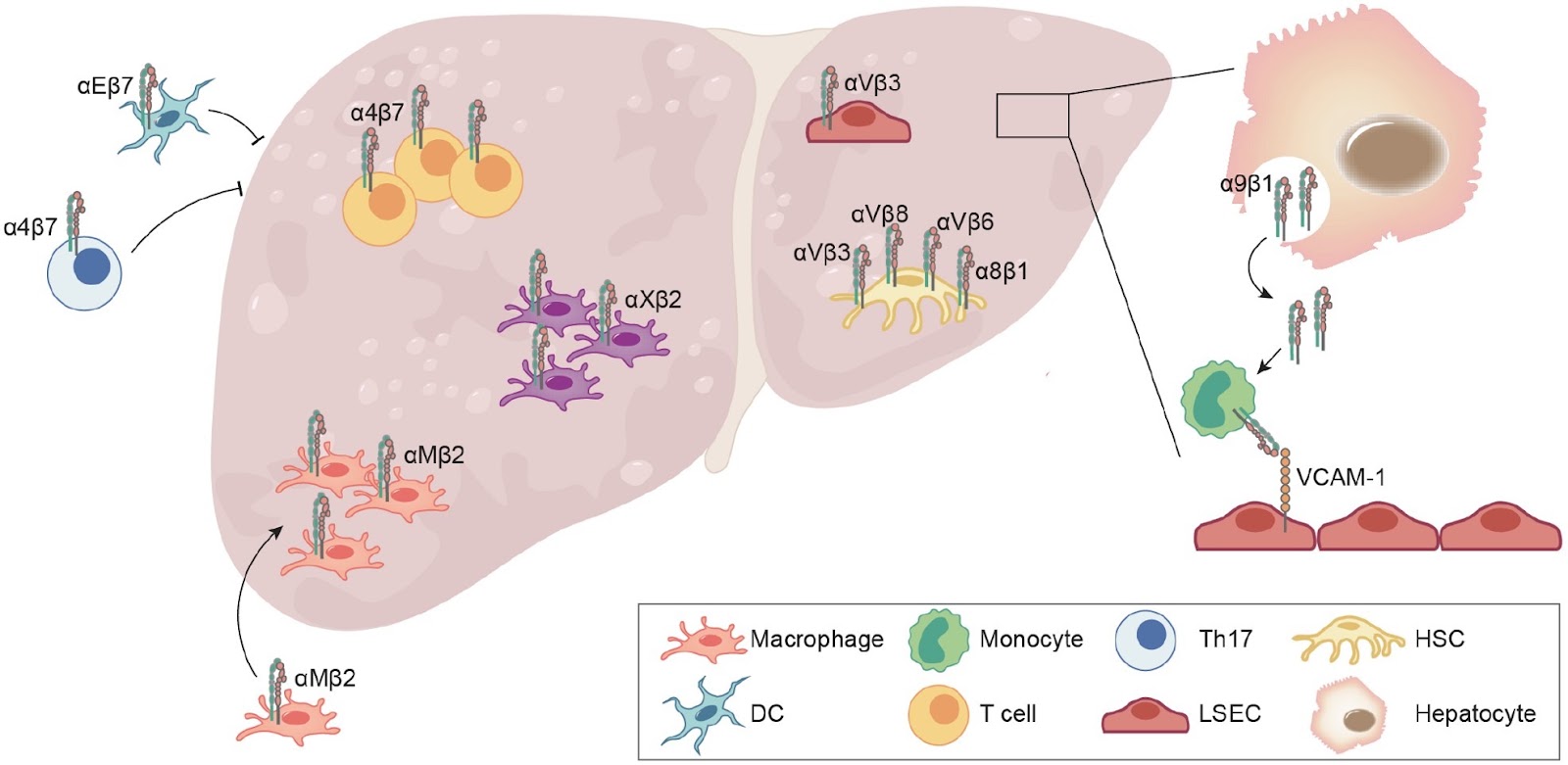
Nonalcoholic fatty liver disease (NAFLD) is a leading cause of chronic liver disease and poses a substantial health burden with increasing incidence globally. NAFLD encompasses a spectrum extending from hepatic steatosis to nonalcoholic steatohepatitis (NASH), with the possibility of progressing to cirrhosis or, in severe instances, hepatocellular carcinoma (HCC). NAFLD extends beyond simple metabolic disruption and involves multiple immune cell-mediated inflammatory processes. Integrins are a family of heterodimeric transmembrane cell adhesion receptors that regulate various aspects of NAFLD onset and progression, including hepatocellular steatosis, hepatic stellate cell (HSC) activation and immune cell infiltration. In this review, we comprehensively summarize the involvement of integrins in NAFLD, as well as the downstream signal transduction mediated by these receptors. Furthermore, we present the latest clinical and preclinical findings on drugs that target integrins for steatosis, inflammation, fibrosis and NAFLD-related HCC treatment.

Integrins and NAFLD-associated liver diseases: clinical associations, pathophysiological mechanisms and pharmacological implications
Graphical Abstract
Original ResearchVol. 56, Issue 11 • pp. 1644-1658DOI: 10.3724/abbs.2024102

Acetyl-11-keto-β-boswellic acid restrains the progression of synovitis in osteoarthritis via the Nrf2/HO-1 pathway

Authors: Jing Zhou, Xueyan Li, Zeyu Han, Yinhua Qian, Lang Bai, Qibin Han, Maofeng Gao, Yi Xue, Dechun Geng, Xing Yang, Yuefeng Hao

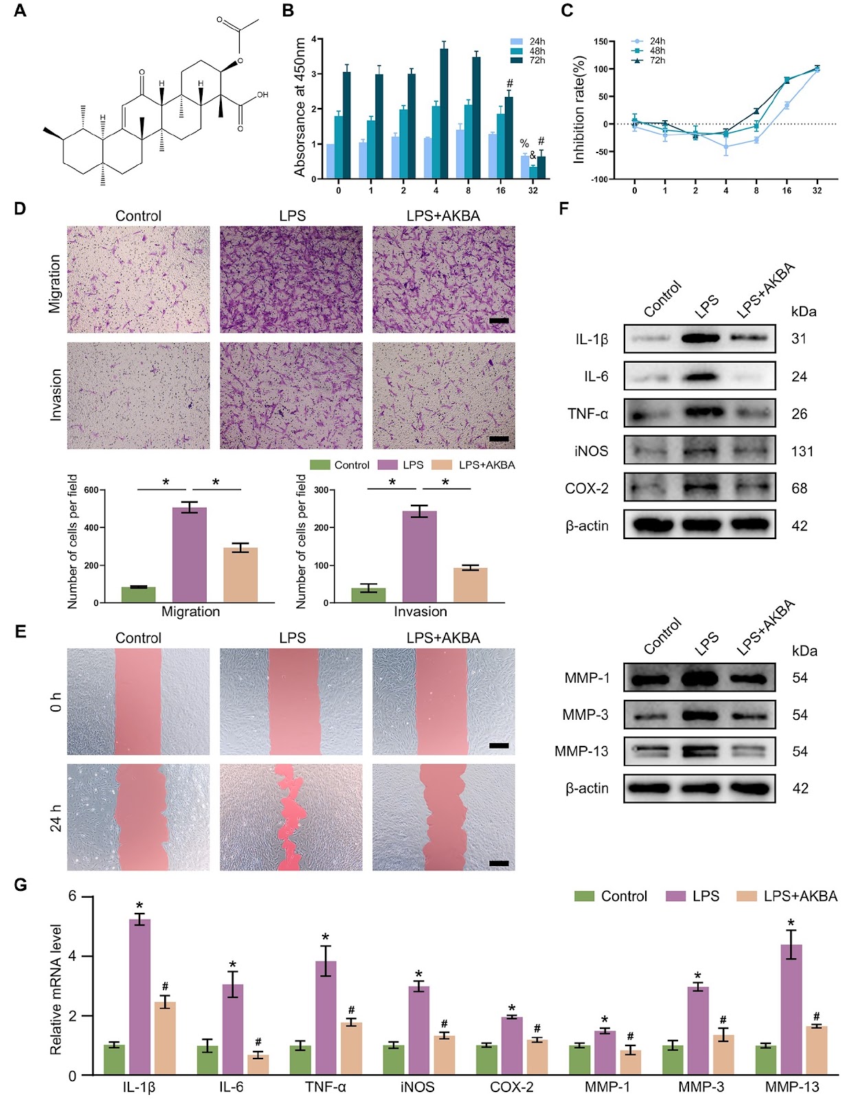
Synovial inflammation plays a key role in osteoarthritis (OA) pathogenesis. Fibroblast-like synoviocytes (FLSs) represent a distinct cell subpopulation within the synovium, and their unique phenotypic alterations are considered significant contributors to inflammation and fibrotic responses. The underlying mechanism by which acetyl-11-keto-β-boswellic acid (AKBA) modulates FLS activation remains unclear. This study aims to assess the beneficial effects of AKBA through both in vitro and in vivo investigations. Network pharmacology evaluation is used to identify potential targets of AKBA in OA. We evaluate the effects of AKBA on FLSs activation in vitro and the regulatory role of AKBA on the Nrf2/HO-1 signaling pathway. ML385 (an Nrf2 inhibitor) is used to verify the binding of AKBA to its target in FLSs. We validate the in vivo efficacy of AKBA in alleviating OA using anterior cruciate ligament transection and destabilization of the medial meniscus (ACLT+DMM) in a rat model. Network pharmacological analysis reveals the potential effect of AKBA on OA. AKBA effectively attenuates lipopolysaccharide (LPS)-induced abnormal migration and invasion and the production of inflammatory mediators, matrix metalloproteinases (MMPs), and reactive oxygen species (ROS) in FLSs, contributing to the restoration of the synovial microenvironment. After treatment with ML385, the effect of AKBA on FLSs is reversed. In vivo studies demonstrate that AKBA mitigates synovial inflammation and fibrotic responses induced by ACLT+DMM in rats via activation of the Nrf2/HO-1 axis. AKBA exhibits theoretical potential for alleviating OA progression through the Nrf2/HO-1 pathway and represents a viable therapeutic candidate for this patient population.

Acetyl-11-keto-β-boswellic acid restrains the progression of synovitis in osteoarthritis via the Nrf2/HO-1 pathway
Graphical Abstract
Original ResearchVol. 56, Issue 11 • pp. 1633-1643DOI: 10.3724/abbs.2024148

UHRF1 knockdown induces cell cycle arrest and apoptosis in breast cancer cells through the ZBTB16/ANXA7/Cyclin B1 axis

Authors: Di Liu, Qin Du, Yuxuan Zhu, Yize Guo, Ya Guo

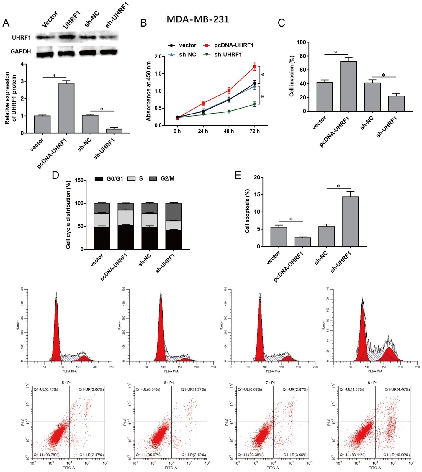
Ubiquitin-like containing PHD and RING finger domains 1 (UHRF1) is involved in tumorigenicity through DNA methylation in various cancers, including breast cancer. This study aims to investigate the regulatory mechanisms of UHRF1 in breast cancer progression. Herein, we show that UHRF1 is upregulated in breast cancer tissues and cell lines as measured by western blot analysis and immunohistochemistry. Breast cancer cells are transfected with a UHRF1 overexpression plasmid (pcDNA-UHRF1) or short hairpin RNA targeting UHRF1 (sh-UHRF1), followed by detection of cell proliferation, invasion, apoptosis, and cell cycle. UHRF1 overexpression promotes proliferation and invasion and attenuates cell cycle arrest and apoptosis in breast cancer cells, while UHRF1 knockdown shows the opposite effect. Moreover, methylation-specific PCR and ChIP assays indicate that UHRF1 inhibits zinc finger and BTB domain containing 16 (ZBTB16) expression by promoting ZBTB16 promoter methylation via the recruitment of DNA methyltransferase 1 (DNMT1). Then, a co-IP assay is used to verify the interaction between ZBTB16 and the annexin A7 (ANXA7) protein. ZBTB16 promotes ANXA7 expression and subsequently inhibits Cyclin B1 expression. Rescue experiments reveal that ZBTB16 knockdown reverses the inhibitory effects of UHRF1 knockdown on breast cancer cell malignancies and that ANXA7 knockdown abolishes the inhibitory effects of ZBTB16 overexpression on breast cancer cell malignancies. Additionally, UHRF1 knockdown significantly inhibits xenograft tumor growth in vivo. In conclusion, UHRF1 knockdown inhibits proliferation and invasion, induces cell cycle arrest and apoptosis in breast cancer cells via the ZBTB16/ANXA7/Cyclin B1 axis, and reduces xenograft tumor growth in vivo.

UHRF1 knockdown induces cell cycle arrest and apoptosis in breast cancer cells through the ZBTB16/ANXA7/Cyclin B1 axis
Graphical Abstract
Original ResearchVol. 56, Issue 11 • pp. 1673-1686DOI: 10.3724/abbs.2024134

SUN5, a testis-specific nuclear membrane protein, participates in recruitment and export of nuclear mRNA in spermatogenesis

Authors: Xiyi He, Yunfei Zhang, Zenghui Mao, Gang Liu, Lihua Huang, Xiaowen Liu, Yuyan Su, Xiaowei Xing

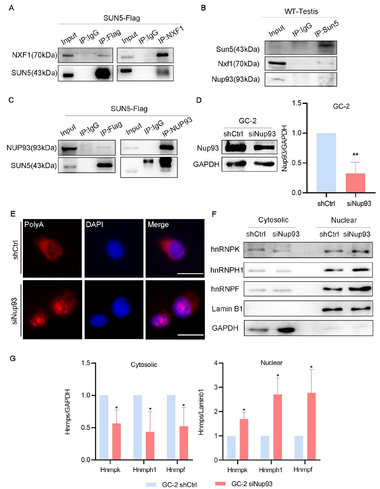
SUN5, a testis-specific gene, is associated with acephalic spermatozoa syndrome (ASS). Here, we demonstrate that SUN5 is involved in mRNA export. In Sun5-knockout mice (Sun5–/–), poly(A)+ RNA accumulates in the nuclei of germ cells, leading to reduced sperm counts, decreased sperm motility and disrupted sperm head-to-tail junctions. Additionally, in the GC-2 germ cell line with RNA interference of Sun5, heterogeneous nuclear ribonucleoproteins (hnRNPs) and poly (A)+ RNA (mainly mRNA) are retained in the nucleus. Further mechanistic studies reveal that SUN5 interacts with Nxf1 (nuclear RNA export factor 1) and nucleoporin 93 (Nup93). Interference with Nup93 inhibits mRNA export. Treatment with leptomycin B to block the CRM1 pathway indicates that Sun5 regulates mRNA export through an Nxf1-dependent pathway. In Sun5–/– mice, the binding of Nxf1 and Nup93 decreases due to loss of Sun5 function, and the process of submitting Nxf1-binding mRNPs to Nup93 is inhibited, resulting in abnormal spermatogenesis. Together, these data may elucidate a novel pathway for mRNA export in male germ cells.

SUN5, a testis-specific nuclear membrane protein, participates in recruitment and export of nuclear mRNA in spermatogenesis
Graphical Abstract
Original ResearchVol. 56, Issue 10 • pp. 1509-1520DOI: 10.3724/abbs.2024098

FTO-mediated m6A demethylation of ULK1 mRNA promotes autophagy and activation of hepatic stellate cells in liver fibrosis

Authors: Tingjuan Huang, Chunhong Zhang, Junjie Ren, Qizhi Shuai, Xiaonan Li, Xuewei Li, Jun Xie, Jun Xu

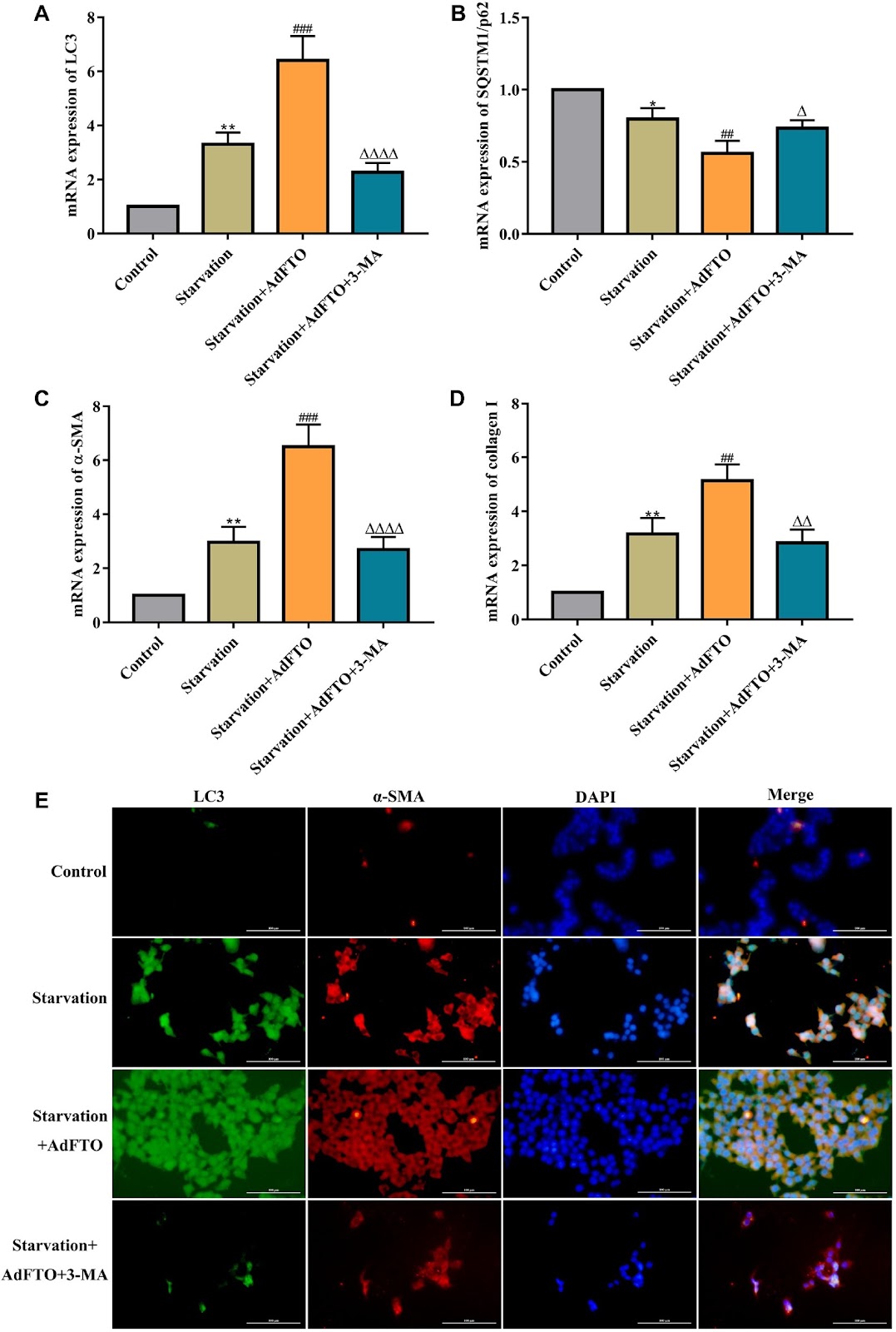
The activation of hepatic stellate cells (HSCs) is central to the occurrence and development of liver fibrosis. Our previous studies showed that autophagy promotes HSC activation and ultimately accelerates liver fibrosis. Unc-51-like autophagy activating kinase 1 (ULK1) is an autophagic initiator in mammals, and N6-methyladenosine (m6A) modification is closely related to autophagy. In this study, we find that the m6A demethylase fat mass and obesity-associated protein (FTO), which is the m6A methylase with the most significant difference in expression, is upregulated during HSC activation and bile duct ligation (BDL)-induced hepatic fibrosis. Importantly, we identify that FTO overexpression aggravates HSC activation and hepatic fibrosis via autophagy. Mechanistically, compared with other autophagy-related genes, ULK1 is a target of FTO because FTO mainly mediates the m6A demethylation of ULK1 and upregulates its expression, thereby enhancing autophagy and the activation of HSCs. Notably, the m6A reader YTH domain-containing protein 2 (YTHDC2) decreases ULK1 mRNA level by recognizing the m6A binding site and ultimately inhibiting autophagy and HSC activation. Taken together, our findings highlight m6A-dependent ULK1 as an essential regulator of HSC autophagy and reveal that ULK1 is a novel potential therapeutic target for hepatic fibrosis treatment.

FTO-mediated m6A demethylation of ULK1 mRNA promotes autophagy and activation of hepatic stellate cells in liver fibrosis
Graphical Abstract
Original ResearchVol. 56, Issue 10 • pp. 1483-1497DOI: 10.3724/abbs.2024078

Up-regulation of miR-10a-5p expression inhibits the proliferation and differentiation of neural stem cells by targeting Chl1

Authors: Juan Zhang, Lihong Yang, Yuqing Sun, Li Zhang, Yufei Wang, Ming Liu, Xiujuan Li, Yuxiang Liang, Hong Zhao, Zhizhen Liu, Zhiyong Qiu, Ting Zhang, Jun Xie

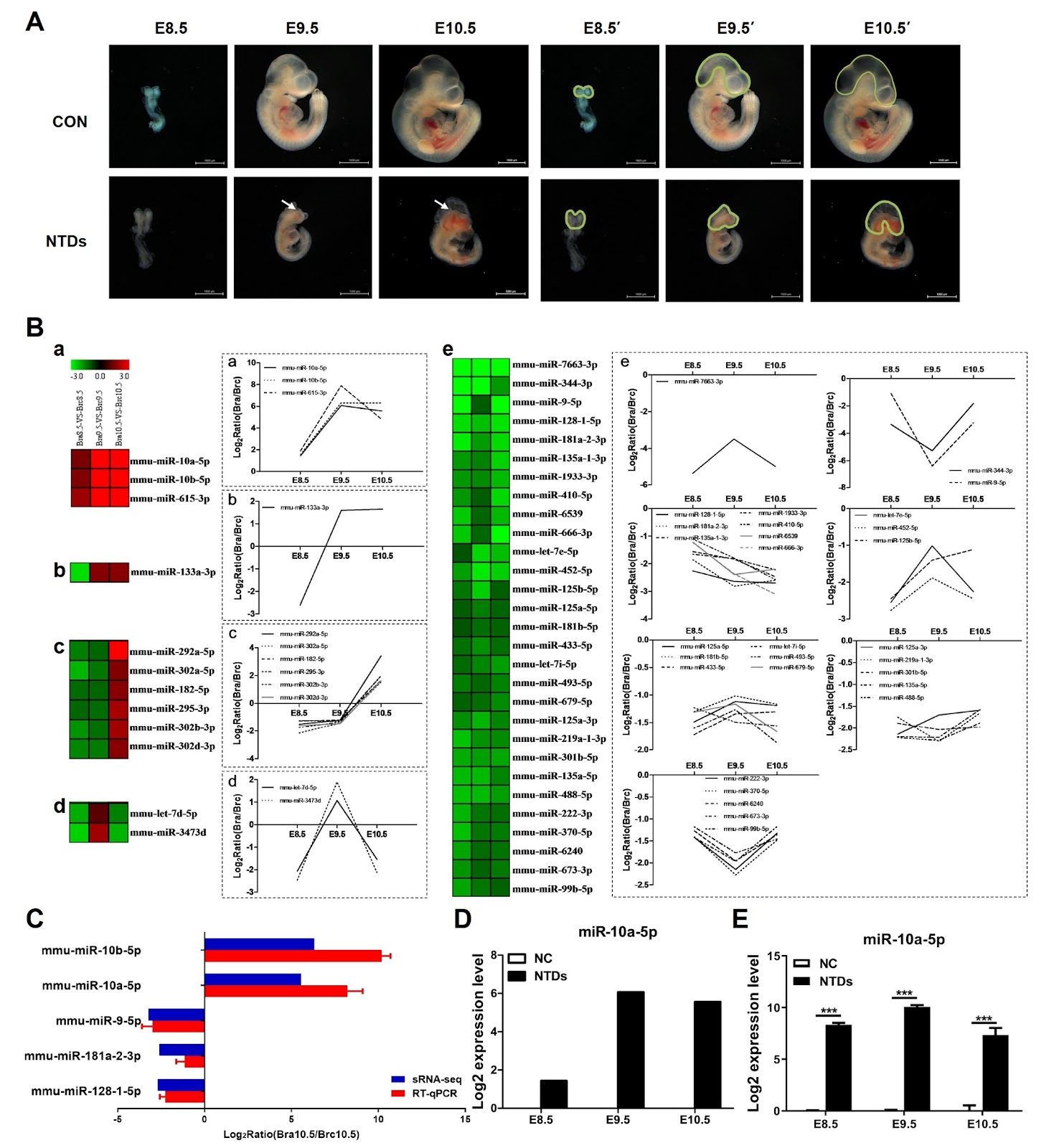
Neural tube defects (NTDs) are characterized by the failure of neural tube closure during embryogenesis and are considered the most common and severe central nervous system anomalies during early development. Recent microRNA (miRNA) expression profiling studies have revealed that the dysregulation of several miRNAs plays an important role in retinoic acid (RA)-induced NTDs. However, the molecular functions of these miRNAs in NTDs remain largely unidentified. Here, we show that miR-10a-5p is significantly upregulated in RA-induced NTDs and results in reduced cell growth due to cell cycle arrest and dysregulation of cell differentiation. Moreover, the cell adhesion molecule L1-like (Chl1) is identified as a direct target of miR-10a-5p in neural stem cells (NSCs) in vitro, and its expression is reduced in RA-induced NTDs. siRNA-mediated knockdown of intracellular Chl1 affects cell proliferation and differentiation similar to those of miR-10a-5p overexpression, which further leads to the inhibition of the expressions of downstream ERK1/2 MAPK signaling pathway proteins. These cellular responses are abrogated by either increased expression of the direct target of miR-10a-5p (Chl1) or an ERK agonist such as honokiol. Overall, our study demonstrates that miR-10a-5p plays a major role in the process of NSC growth and differentiation by directly targeting Chl1, which in turn induces the downregulation of the ERK1/2 cascade, suggesting that miR-10a-5p and Chl1 are critical for NTD formation in the development of embryos.

Up-regulation of miR-10a-5p expression inhibits the proliferation and differentiation of neural stem cells by targeting Chl1
Graphical Abstract
Original ResearchVol. 56, Issue 10 • pp. 1498-1508DOI: 10.3724/abbs.2024090

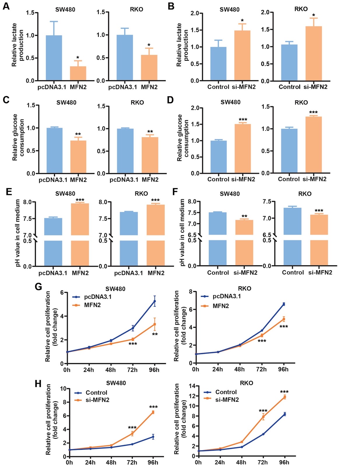
miR-373-3p promotes aerobic glycolysis in colon cancer cells by targeting MFN2

Authors: Yu Wang, Jie Lun, Yuying Zhang, Mengchao Yu, Xingqian Liu, Jing Guo, Hongwei Zhang, Wensheng Qiu, Jing Fang

MicroRNAs (miRNAs) are implicated in the development of cancers and may serve as potential targets for therapy. However, the functions and underlying mechanisms of miRNAs in cancers are not well understood. This work aims to study the role of miR-373-3p in colon cancer cells. We find that the expression of miR-373-3p mimics promotes and the miR-373-3p inhibitor suppresses aerobic glycolysis and proliferation of colon cancer cells. Mechanistically, miR-373-3p inhibits the expression of MFN2, a gene that is known to suppress glycolysis, which leads to the activation of glycolysis and eventually the proliferation of cells. In a nude mouse tumor model, the expression of miR-373-3p in colon cancer cells promotes tumor growth by enhancing lactate formation, which is inhibited by the co-expression of MFN2 in the cells. Administration of the miR-373-3p antagomir blunts in vivo tumor growth by decreasing lactate production. In addition, in human colon cancers, the expression levels of miR-373-3p are increased, while those of MFN2 mRNA are decreased, and the increase of miR-373-3p is associated with the decrease of MFN2 mRNA. Our results reveal a previously unknown function and underlying mechanism of miR-373-3p in the regulation of glycolysis and proliferation in cancer cells and underscore the potential of targeting miR-373-3p for colon cancer treatment.

miR-373-3p promotes aerobic glycolysis in colon cancer cells by targeting MFN2
Graphical Abstract
Original ResearchVol. 56, Issue 10 • pp. 1425-1436DOI: 10.3724/abbs.2024073

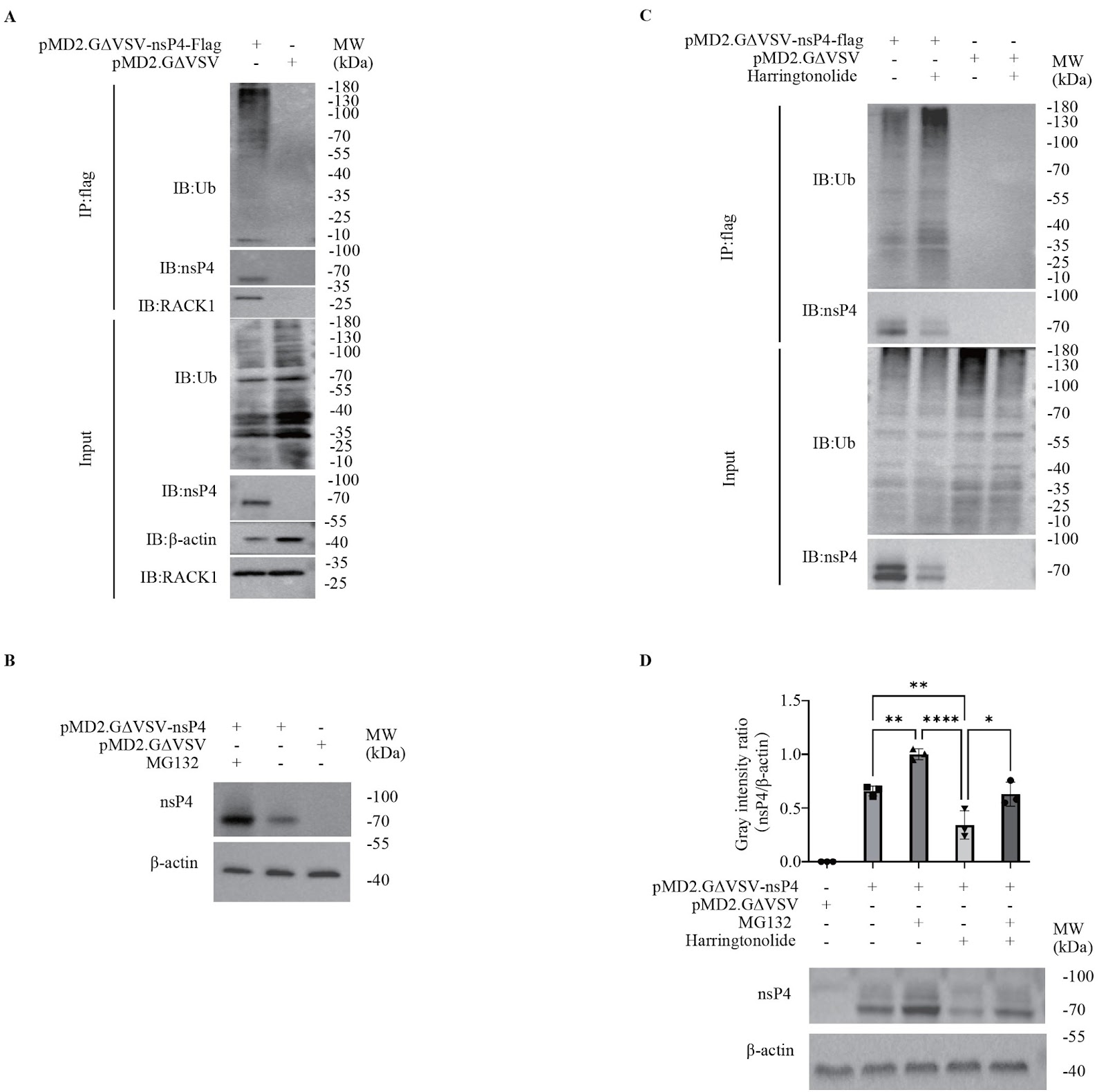
Identification of RACK1 as a novel regulator of non-structural protein 4 of chikungunya virus

Authors: Yao Yan, Fengyuan Zhang, Meng Zou, Hongyu Chen, Jingwen Xu, Shuaiyao Lu, Hongqi Liu

Chikungunya virus (CHIKV) is a neglected arthropod-borne and anthropogenic alphavirus. Over the past two decades, the CHIKV distribution has undergone significant changes worldwide, from the original tropics and subtropics regions to temperate regions, which has attracted global attention. However, the interactions between CHIKV and its host remain insufficiently understood, which dampens the need for the development of an anti-CHIKV strategy. In this study, on the basis of the optimal overexpression of non-structural protein 4 (nsP4), we explore host interactions of CHIKV nsP4 using mass spectrometry-based protein-protein interaction approaches. The results reveal that some cellular proteins that interact with nsP4 are enriched in the ubiquitin-proteasome pathway. Specifically, the scaffold protein receptor for activated C kinase 1 (RACK1) is identified as a novel host interactor and regulator of CHIKV nsP4. The inhibition of the interaction between RACK1 and nsP4 by harringtonolide results in the reduction of nsP4, which is caused by the promotion of degradation but not the inhibition of nsP4 translation. Furthermore, the decrease in nsP4 triggered by the RACK1 inhibitor can be reversed by the proteasome inhibitor MG132, suggesting that RACK1 can protect nsP4 from degradation through the ubiquitin-proteasome pathway. This study reveals a novel mechanism by which the host factor RACK1 regulates CHIKV nsP4, which could be a potential target for developing drugs against CHIKV.

Identification of RACK1 as a novel regulator of non-structural protein 4 of chikungunya virus
Graphical Abstract
Original ResearchVol. 56, Issue 10 • pp. 1566-1570DOI: 10.3724/abbs.2024114

Exploring the mechanism of Panax notoginseng saponin in inhibiting the inflammatory response of microglia in cerebral ischemia based on network pharmacology

Authors: Zhaoda Duan, Wenji Jia, Jianxiang Wang, Dongyao Xu, Yujia Yang, Zhi Qi, Li Yang, Chunyun Wu

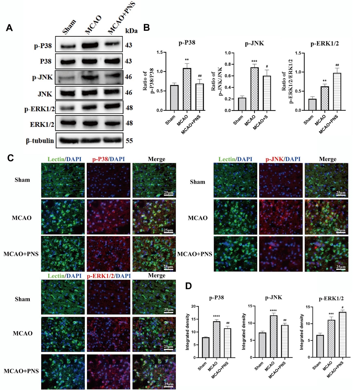
With the increasing global population and aging demographic, the incidence of stroke is rising. Among these, ischemic stroke (IS), also known as cerebral ischemia, constitutes over 80% of all stroke cases. This condition is characterized by an acute cerebrovascular disease caused by the blockage and interruption of the brain's blood supply, resulting in localized tissue ischemia, oxygen, and glucose deficiency, ultimately leading to the death of nerve cells and tissue necrosis [1,2]. "Vascular recanalization and the restoration of cerebral blood flow" are the primary clinical treatment objectives and are achieved through the intravenous administration of drugs such as tissue plasminogen activator or through surgical thrombectomy. These interventions not only restore the delivery of oxygen and glucose to the affected cerebral area but also help prevent the expansion of the infarcted region. However, the restoration of reperfusion cerebral blood flow similarly exposes the infarct area to peripheral immune cells, triggering the activation of the immune response and inflammation-induced injury [3]. Research indicates that IS elicits a robust inflammatory response, with neuroinflammation playing a crucial role in the secondary neurodegeneration process following stroke. Neuroinflammatory responses are initiated and perpetuated through injury cascades that include the release of inflammatory mediators, the migration and recruitment of white blood cells across the blood-brain barrier, and the impairment of endothelial nitric oxide synthase. These mechanisms collectively promote the activation of pro-inflammatory genes, which in turn activate microglia (MG) and exacerbate ischemic damage and neurological dysfunction [4]. MG are resident immune cells of the central nervous system (CNS). Its function is akin to that of macrophages, serving as the first line of defense against injuries within the central nervous system. Under typical conditions, brain microglia participate in immune surveillance and defense against infectious agents. However, in the pathogenesis of neurodegenerative diseases such as IS, MG are activated by various stimuli. Once activated, MG are known to release numerous proinflammatory or cytotoxic factors, such as inducible nitric oxide synthase (iNOS), tumor necrosis factor-α (TNF-α), interleukin-1β (IL-1β), and reactive oxygen species (ROS). These factors initiate the neuroinflammatory response, exacerbate inflammation, intensify damage to brain tissue and neurons, and significantly hinder the repair of brain injuries and neurogenesis [5,6]. Therefore, inhibiting the activation of microglia and reducing the inflammatory response in the central nervous system are crucial for minimizing brain damage caused by IS and are vital for developing effective prevention and treatment strategies. In recent years, certain natural compounds extracted from traditional drug formulations have shown high therapeutic potential in protecting the brain from cerebral ischemic injury. These compounds reduce the neuroinflammatory response and apoptosis following stroke. Traditional Chinese herbal medicine (TCHM) and its constituent herbs feature a multiplicity of components, targets, and pathways owing to their complex formulations and therapeutic principles, making them promising sources for developing effective treatments for IS. Panax notoginseng saponin (PNS), as the principal bioactive component of Panax notoginseng, is extensively utilized in the prevention and treatment of cardiovascular and cerebrovascular diseases. Its pharmacological benefits include dissipating blood stasis, promoting hemostasis, alleviating swelling and pain, regulating energy metabolism disorders, balancing ion metabolism, and reducing and accelerating the clearance of free radicals [7]. Research indicates that PNS mitigates apoptosis by maintaining mitochondrial homeostasis, enhancing the integrity of the blood‒brain barrier (BBB), augmenting cerebral blood supply, and fostering the differentiation of neural stem cells and proliferation of hippocampal neurons. In addition, PNS offers neuroprotection against focal cerebral I/R injury in rats by reducing brain edema, upregulating the expression of the heat shock protein HSP70, and downregulating the expression of transferrin [8,9]. Additionally, PNS has been reported to enhance the recovery of neurogenesis and neurological function in cerebral embolism induced by microspheres and to reduce sepsis-induced acute kidney injury by suppressing inflammation [10]. However, the mechanism by which PNS targets IS has not been fully elucidated. In this study, we investigated the anti-inflammatory effects of PNS on IS and identified potential target pathways that could inhibit microglia-mediated inflammatory response.

Exploring the mechanism of Panax notoginseng saponin in inhibiting the inflammatory response of microglia in cerebral ischemia based on network pharmacology
Graphical Abstract
Original ResearchVol. 56, Issue 10 • pp. 1415-1424DOI: 10.3724/abbs.2024085

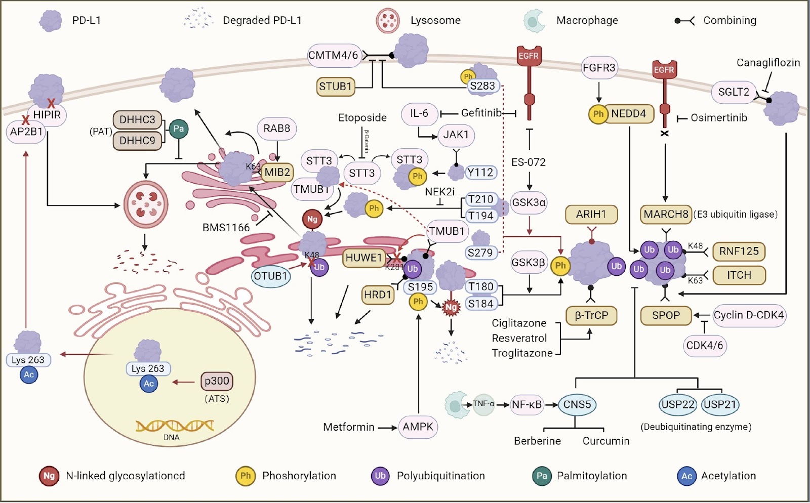
Posttranslational regulatory mechanism of PD-L1 in cancers and associated opportunities for novel small-molecule therapeutics

Authors: Minchen Cai, Mengting Xu, Dianping Yu, Qun Wang, Sanhong Liu

Despite the tremendous progress in cancer research over the past few decades, effective therapeutic strategies are still urgently needed. Accumulating evidence suggests that immune checkpoints are the cause of tumor immune escape. PD-1/PD-L1 are among them. Posttranslational modification is the most critical step for protein function, and the regulation of PD-L1 by small molecules through posttranslational modification is highly valuable. In this review, we discuss the mechanisms of tumor cell immune escape and several posttranslational modifications associated with PD-L1 and describe examples in which small molecules can regulate PD-L1 through posttranslational modifications. Herein, we propose that the use of small molecule compounds that act by inhibiting PD-L1 through posttranslational modifications is a promising therapeutic approach with the potential to improve clinical outcomes for cancer patients.

Posttranslational regulatory mechanism of PD-L1 in cancers and associated opportunities for novel small-molecule therapeutics
Graphical Abstract
Original ResearchVol. 56, Issue 9 • pp. 1256-1266DOI: 10.3724/abbs.2024105

Molecular insight into the potential functional role of pseudoenzyme GFOD1 via interaction with NKIRAS2

Authors: Jiawen Shi, Xinyi Guo, Chan Liu, Yilun Wang, Xiaobao Chen, Guihua Wu, Jianping Ding, Tianlong Zhang

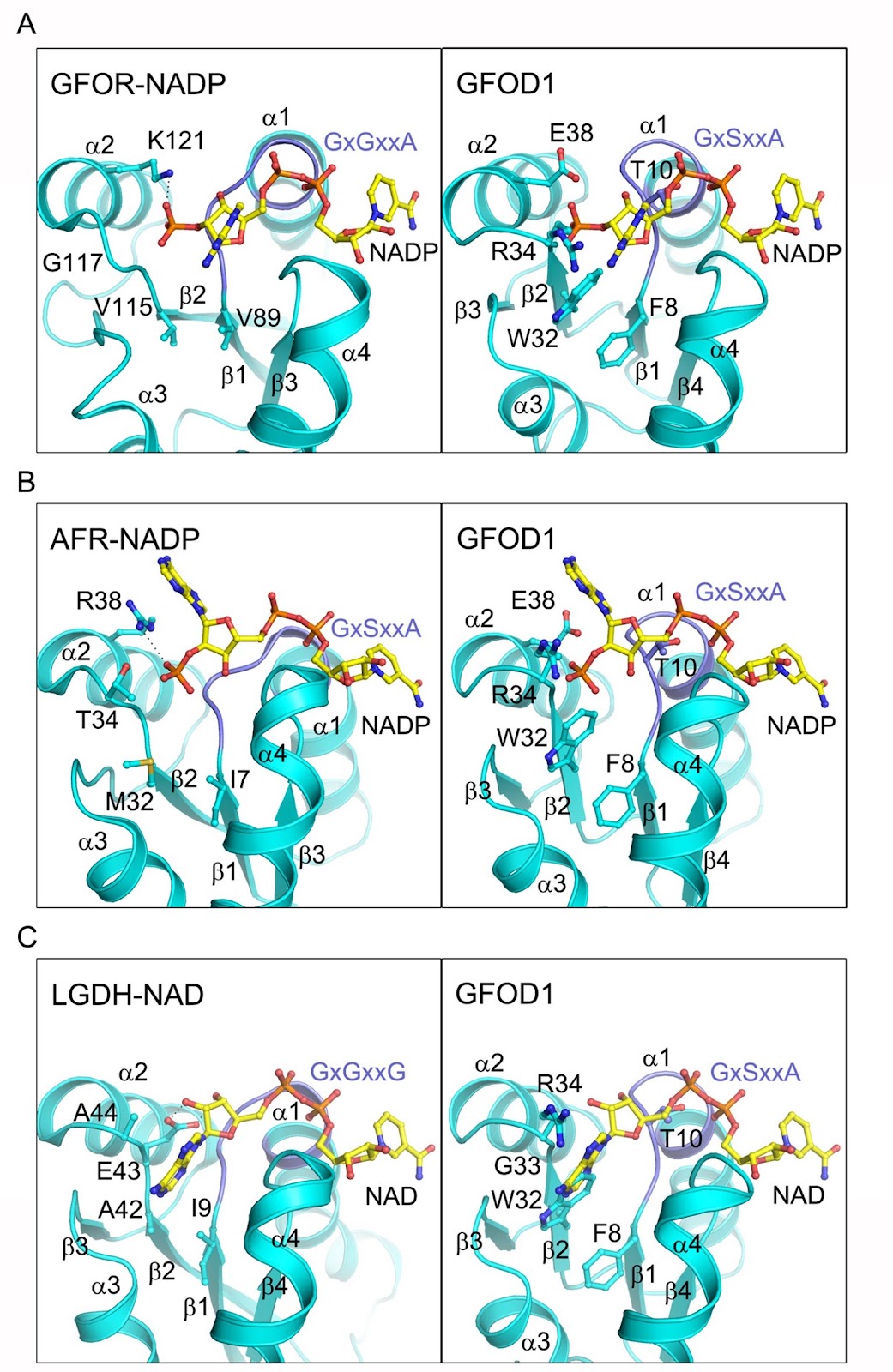
The glucose-fructose oxidoreductase/inositol dehydrogenase/rhizopine catabolism protein (Gfo/Idh/MocA) family includes a variety of oxidoreductases with a wide range of substrates that utilize NAD or NADP as redox cofactor. Human contains two members of this family, namely glucose-fructose oxidoreductase domain-containing protein 1 and 2 (GFOD1 and GFOD2). While GFOD1 exhibits low tissue specificity, it is notably expressed in the brain, potentially linked to psychiatric disorders and severe diseases. Nevertheless, the specific function, cofactor preference, and enzymatic activity of GFOD1 remain largely unknown. In this work, we find that GFOD1 does not bind to either NAD or NADP. Crystal structure analysis unveils that GFOD1 exists as a typical homodimer resembling other family members, but lacks essential residues required for cofactor binding, suggesting that it may function as a pseudoenzyme. Exploration of GFOD1-interacting partners in proteomic database identifies NF-κB inhibitor-interacting Ras-like 2 (NKIRAS2) as one potential candidate. Co-immunoprecipitation (co-IP) analysis indicates that GFOD1 interacts with both GTP- and GDP-bound forms of NKIRAS2. The predicted structural model of the GFOD1-NKIRAS2 complex is validated in cells using point mutants and shows that GFOD1 selectively recognizes the interswitch region of NKIRAS2. These findings reveal the distinct structural properties of GFOD1 and shed light on its potential functional role in cellular processes.

Molecular insight into the potential functional role of pseudoenzyme GFOD1 via interaction with NKIRAS2
Graphical Abstract
Original ResearchVol. 56, Issue 9 • pp. 1373-1386DOI: 10.3724/abbs.2024111

Lactate activates CCL18 expression via H3K18 lactylation in macrophages to promote tumorigenesis of ovarian cancer

Authors: Jinrui Sun, Qinmei Feng, Yue He, Ming Wang, Yumei Wu

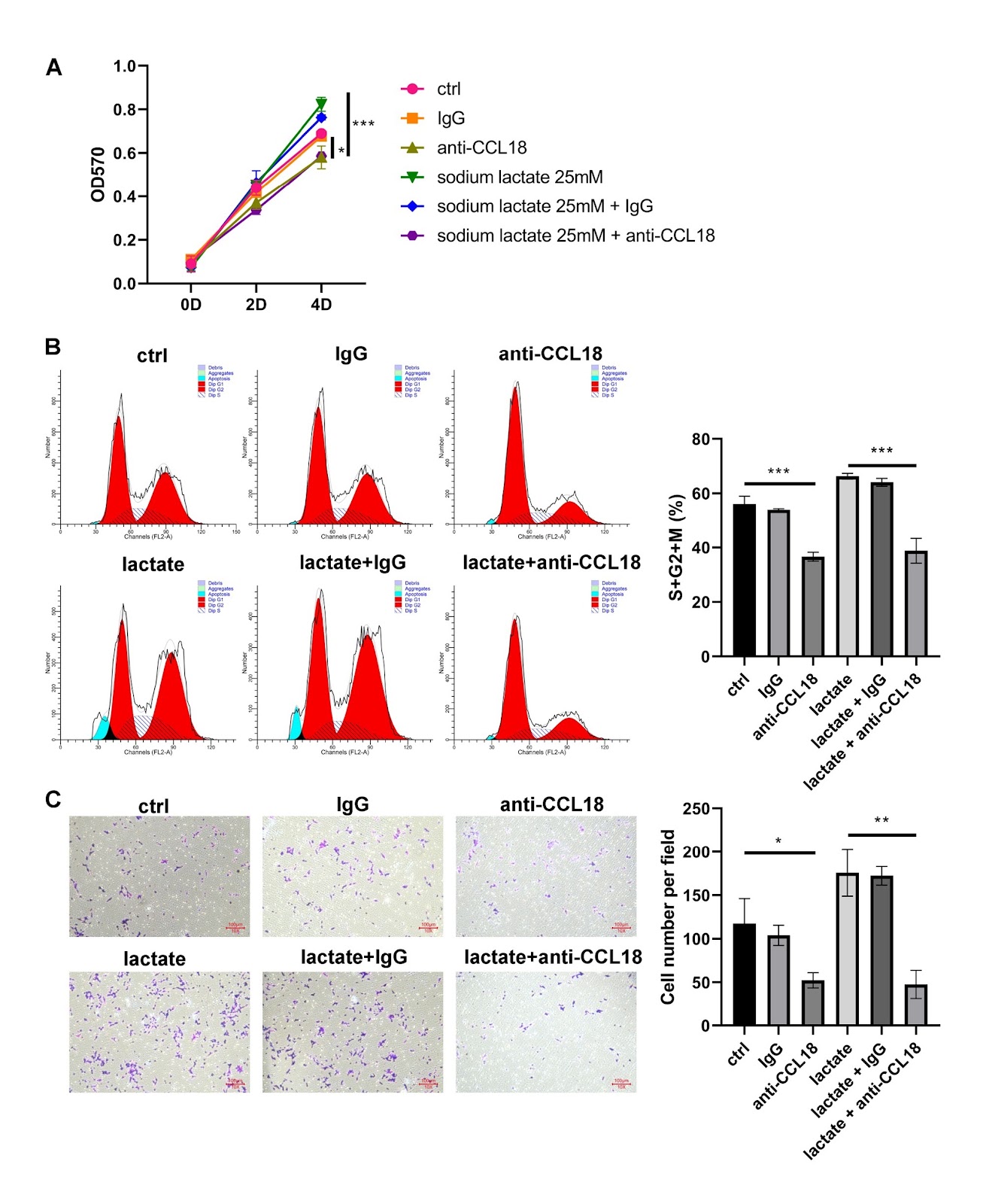
This study investigates the role of lactate in the genesis and progression of ovarian cancer (OV) and explores the underlying mechanisms. Serum lactate levels show a positive correlation with tumor grade and poor prognosis in patients with OV. Bioinformatics analysis identifies CCL18 as a lactate-related gene in OV. CCL18 is up-regulated in cancerous tissues and positively related to serum lactate levels in OV patients. THP-1 cells are exposed to phorbol-12-myristate-13-acetate for M0 macrophage induction. The results of RT-qPCR and ELISA for M1/M2 macrophage-related markers and inflammatory cytokines show that the exposure of lactate to macrophages induces M2 polarization. Based on the coculture of OV cells with macrophages, lactate-treated macrophages induces a significant increase in the proliferation and migration of OV cells. However, these effects can be reversed by silencing of Gpr132 in macrophages or treatment with anti-CCL18 antibody. Experiments using the xenograft model verify that the oncogenic role of lactate in tumor growth and metastasis relies on Gpr132 and CCL18. ChIP-qPCR and luciferase reporter assays reveal that lactate regulates CCL18 expression via H3K18 lactylation. In conclusion, lactate is a potential therapeutic target for OV. It is involved in tumorigenesis by activating CCL18 expression via H3K18 lactylation in macrophages.

Lactate activates CCL18 expression via H3K18 lactylation in macrophages to promote tumorigenesis of ovarian cancer
Graphical Abstract
Original ResearchVol. 56, Issue 9 • pp. 1289-1299DOI: 10.3724/abbs.2024084

FOXM1 mediates methotrexate resistance in osteosarcoma cells by promoting autophagy

Authors: Luoyang Wang, Dongchang Zhai, Lei Tang, Hui Zhang, Xinlong Wang, Ning Ma, Xiaoyue Zhang, Mingguo Cheng, Ruowu Shen

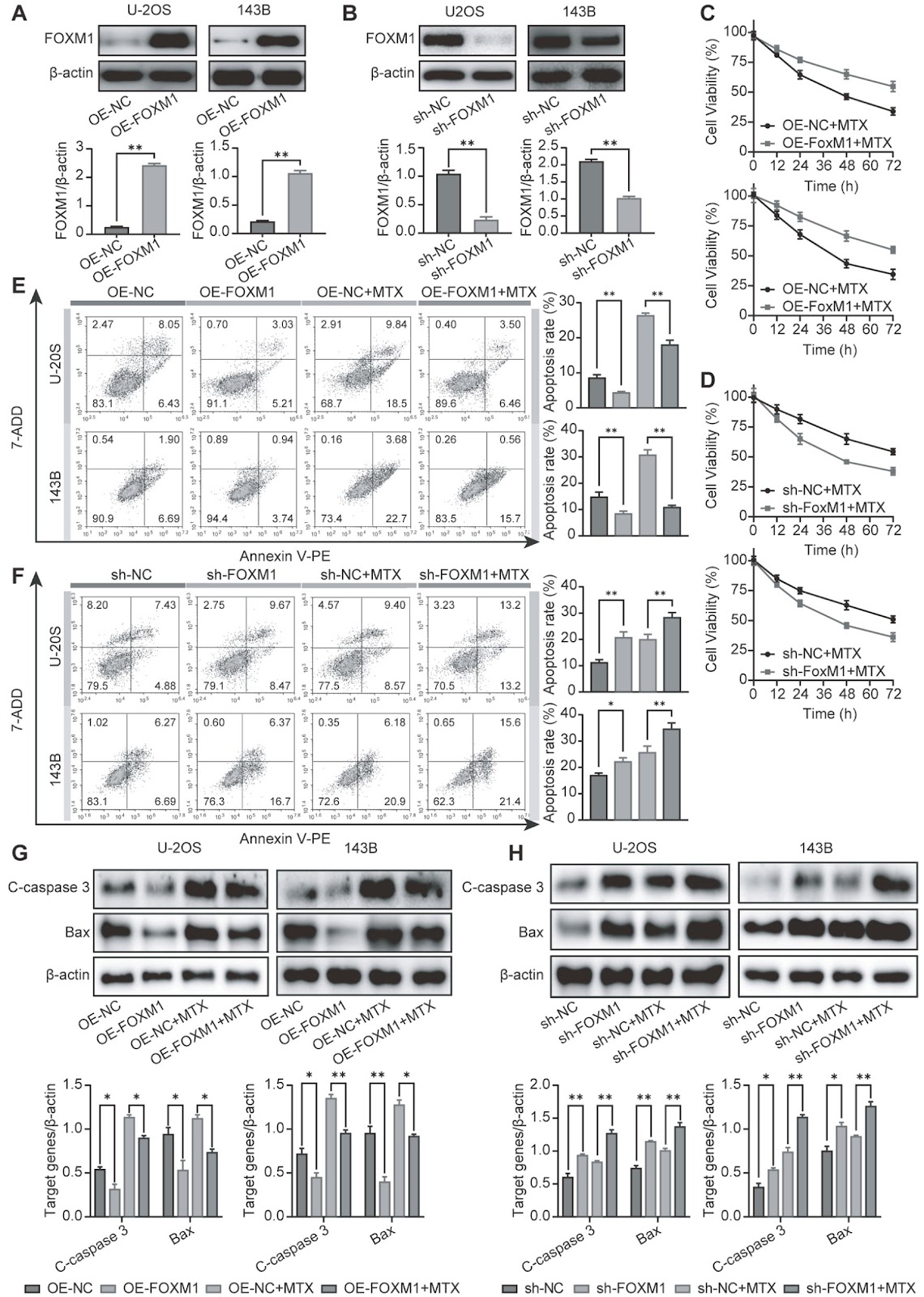
Osteosarcoma (OS) is a primary bone cancer mostly found in adolescents and elderly individuals. The treatment of OS is still largely dependent on traditional chemotherapy. However, the high incidence of drug resistance remains one of the greatest impediments to limiting improvements in OS treatment. Recent findings have indicated that the transcription factor FOXM1 plays an important role in various cancer-related events, especially drug resistance. However, the possible role of FOXM1 in the resistance of OS to methotrexate (MTX) remains to be explored. Here, we find that FOXM1, which confers resistance to MTX, is highly expressed in OS tissues and MTX-resistant cells. FOXM1 overexpression promotes MTX resistance by enhancing autophagy in an HMMR/ATG7-dependent manner. Importantly, silencing of FOXM1 or inhibiting autophagy reverses drug resistance. These findings demonstrate a new mechanism for FOXM1-induced MTX resistance and provide a promising target for improving OS chemotherapy outcomes.

FOXM1 mediates methotrexate resistance in osteosarcoma cells by promoting autophagy
Graphical Abstract
Original ResearchVol. 56, Issue 9 • pp. 1311-1322DOI: 10.3724/abbs.2024049

Aminophylline suppresses chronic renal failure progression by activating SIRT1/AMPK/mTOR-dependent autophagy

Authors: Xin Liao, Jieyi Lu, Zhifeng Huang, Jinai Lin, Miao Zhang, Huanru Chen, Xiaoqing Lin, Xia Gao, Sitang Gong

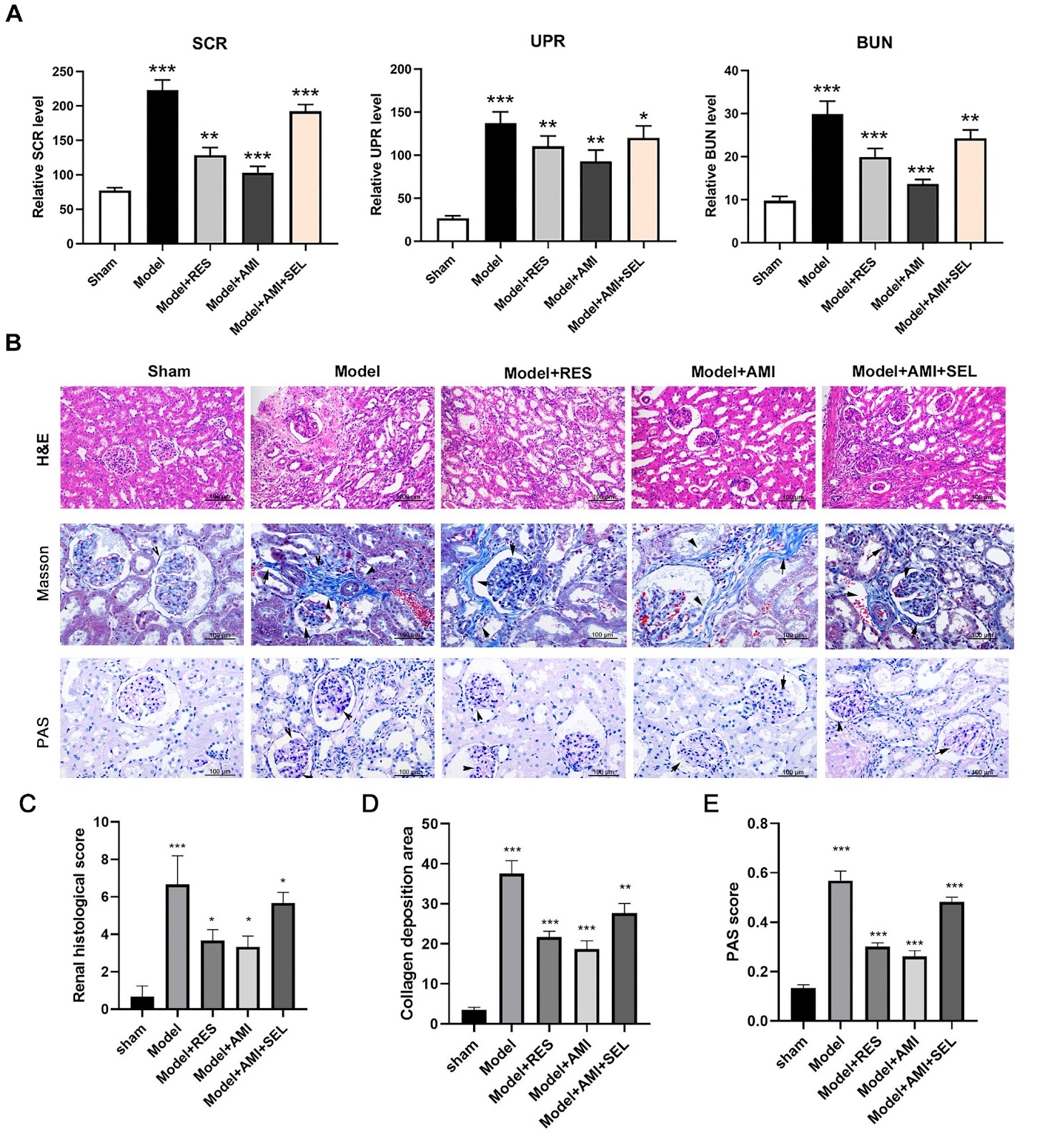
Chronic renal failure (CRF) is a severe syndrome affecting the urinary system for which there are no effective therapeutics. In this study, we investigate the effects and mechanisms of aminophylline in preventing CRF development. A rat model of chronic renal failure is established by 5/6 nephrectomy. The levels of serum creatinine (SCR), urinary protein (UPR), and blood urea nitrogen (BUN) are detected by ELISA. Histological evaluations of renal tissues are performed by H&E, Masson staining, and PAS staining. Functional protein expression is detected by western blot analysis or immunofluorescence microscopy. Glomerular cell apoptosis is determined using the TUNEL method. Results show that Aminophylline significantly reduces the levels of SCR, UPR, and BUN in the CRF model rats. Histological analyses show that aminophylline effectively alleviates renal tissue injuries in CRF rats. The protein expression levels of nephrin, podocin, SIRT1, p-AMPK, and p-ULK1 are greatly increased, while p-mTOR protein expression is markedly decreased by aminophylline treatment. Additionally, the protein level of LC3B in CRF rats is significantly increased by aminophylline. Moreover, aminophylline alleviates apoptosis in the glomerular tissues of CRF rats. Furthermore, resveratrol promotes SIRT1, p-AMPK, and p-ULK1 protein expressions and reduces p-mTOR and LC3B protein expressions in CRF rats. Selisistat (a SIRT1 inhibitor) mitigates the changes in SIRT1, p-AMPK, p-ULK1, p-mTOR, and LC3B expressions induced by aminophylline. Finally, RAPA alleviates renal injury and apoptosis in CRF rats, and 3-MA eliminates the aminophylline-induced inhibition of renal injury and apoptosis in CRF rats. Aminophylline suppresses chronic renal failure progression by modulating the SIRT1/AMPK/mTOR-mediated autophagy process.

Aminophylline suppresses chronic renal failure progression by activating SIRT1/AMPK/mTOR-dependent autophagy
Graphical Abstract
Original ResearchVol. 56, Issue 9 • pp. 1365-1372DOI: 10.3724/abbs.2024091

Hepatitis E virus infection upregulates ING5 expression in vitro and in vivo

Authors: Wanqiu Zhao, Yueping Xia, Tengyuan Li, Huichan Liu, Guo Zhong, Dongxue Chen, Wenhai Yu, Yunlong Li, Fen Huang

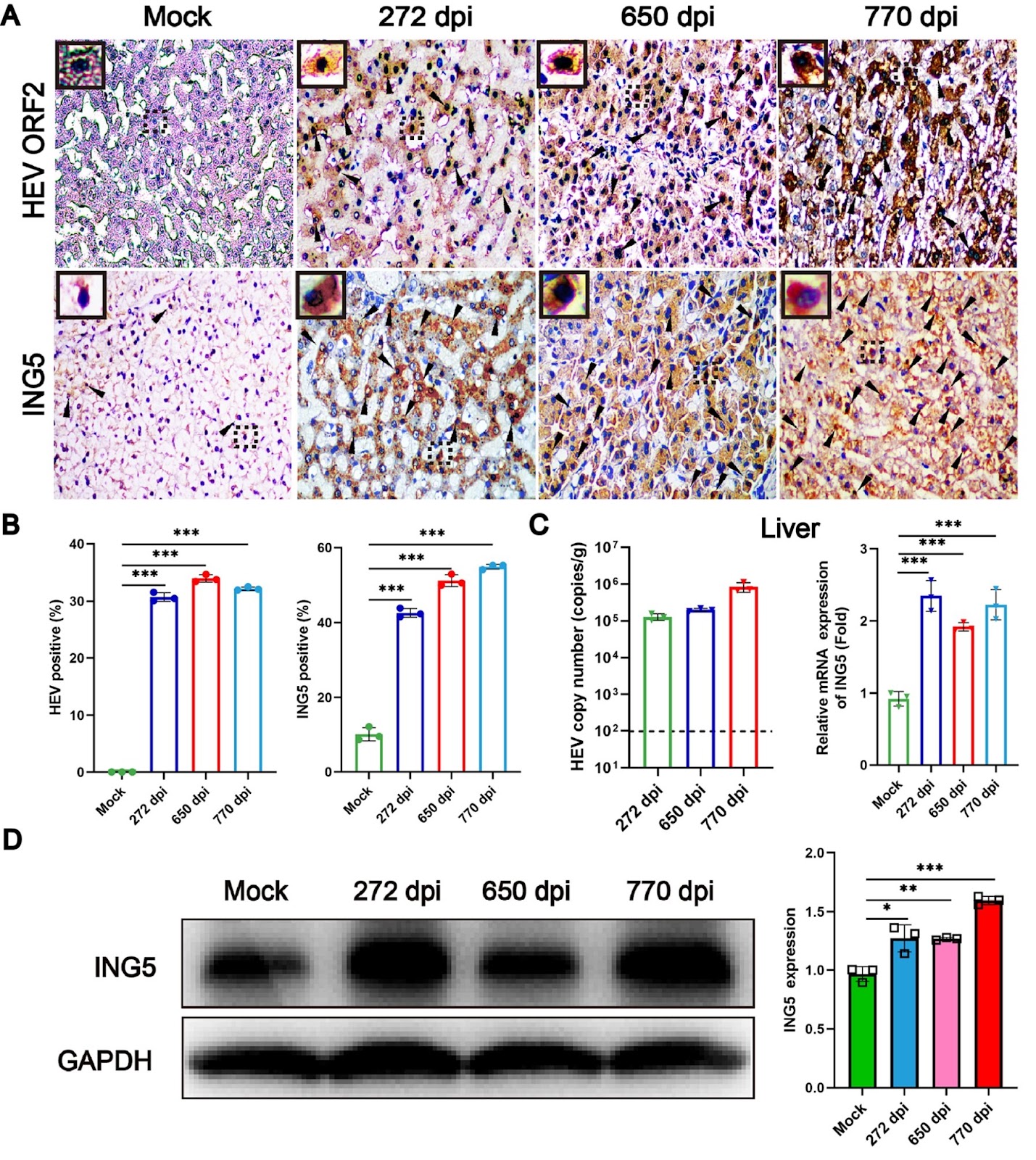
Hepatitis E virus (HEV) is the major pathogen of viral hepatitis. Immunocompromised individuals infected by HEV are prone to chronic hepatitis and increase the risk of hepato-cellular carcinoma (HCC). Inhibitor of growth family member 5 (ING5) is a tumor suppressor that is expressed at low levels in cancer tumors or cells. However, the underlying relationship between ING5 and HEV infection is unclear. In the present study, acute and chronic HEV animal models are used to explore the interaction between ING5 and HEV. Notably, the expression of ING5 is significantly increased in both the livers of acute HEV-infected BALB/c mice and chronic HEV-infected rhesus macaques. In addition, the relationship between HEV infection and ING5 expression is further identified in human hepatoma (HepG-2) cells. In conclusion, HEV infection strongly upregulates ING5 expression both in vivo and in vitro, which has significant implications for further understanding the pathogenic mechanism of HEV infection.

Hepatitis E virus infection upregulates ING5 expression in vitro and in vivo
Graphical Abstract
Original ResearchVol. 56, Issue 9 • pp. 1340-1351DOI: 10.3724/abbs.2024050

GroEL triggers NLRP3 inflammasome activation through the TLR/NF-κB p-p65 axis in human periodontal ligament stem cells

Authors: Li Zhang, Mengmeng Duan, Xiaohua Pu, Huiling Zheng, Xinjie Ning, Ying Tu, Chunming Xu, Demao Zhang, Chengcheng Liu, Jing Xie

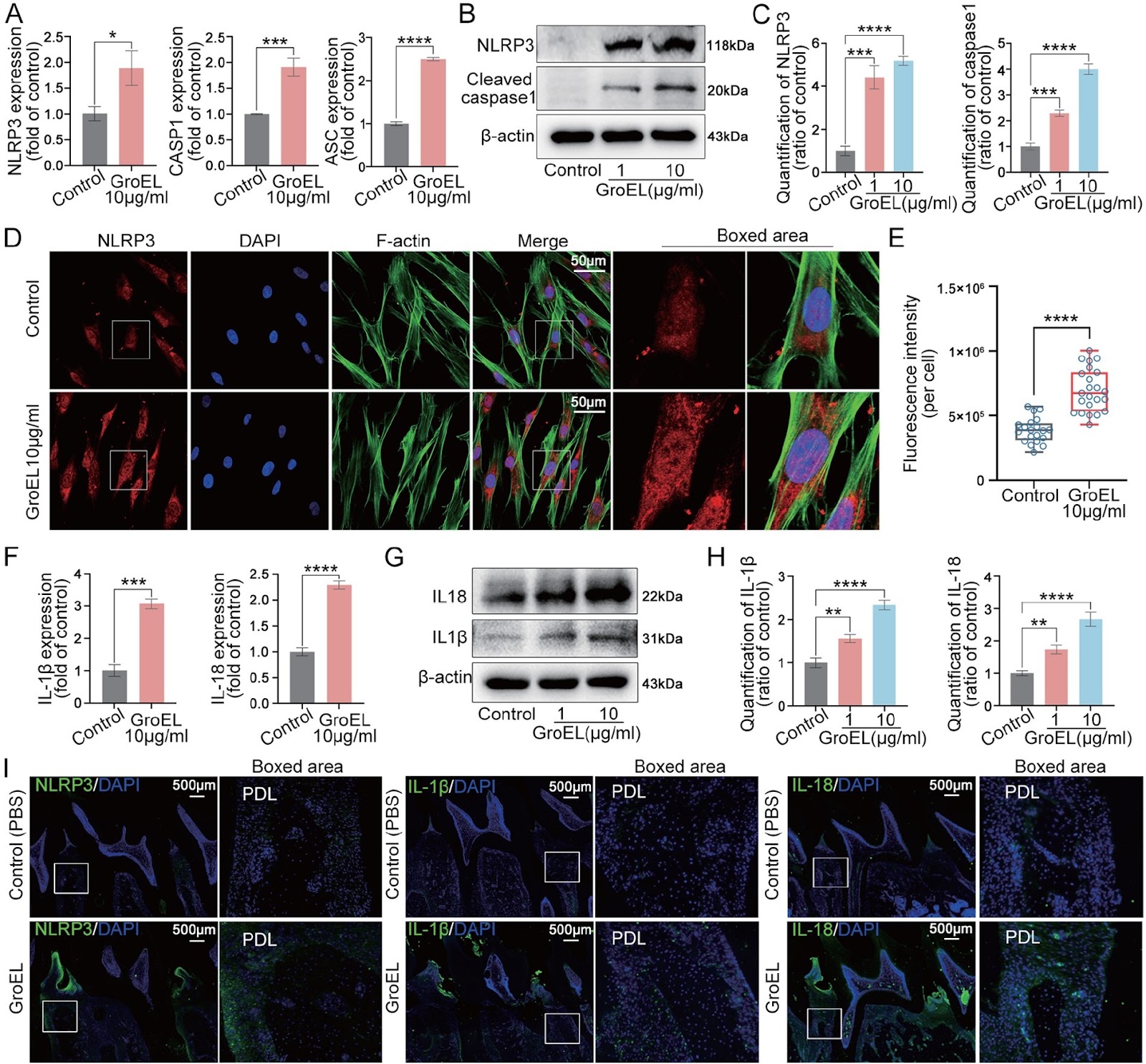
The interaction between bacteria and the host plays a vital role in the initiation and progression of systemic diseases, including gastrointestinal and oral diseases, due to the secretion of various virulence factors from these pathogens. GroEL, a potent virulence factor secreted by multiple oral pathogenic bacteria, is implicated in the damage of gingival epithelium, periodontal ligament, alveolar bone and other peripheral tissues. However, the underlying biomechanism is still largely unknown. In the present study, we verify that GroEL can trigger the activation of NLRP3 inflammasome and its downstream effector molecules, IL-1β and IL-18, in human periodontal ligament stem cells (hPDLSCs) and resultantly induce high activation of gelatinases (MMP-2 and MMP-9) to promote the degradation of extracellular matrix (ECM). GroEL-mediated activation of the NLRP3 inflammasome requires the participation of Toll-like receptors (TLR2 and TLR4). High upregulation of TLR2 and TLR4 induces the enhancement of NF-κB (p-p65) signaling and promotes its nuclear accumulation, thus activating the NLRP3 inflammasome. These results are verified in a rat model with direct injection of GroEL. Collectively, this study provides insight into the role of virulence factors in bacteria-induced host immune response and may also provide a new clue for the prevention of periodontitis.

GroEL triggers NLRP3 inflammasome activation through the TLR/NF-κB p-p65 axis in human periodontal ligament stem cells
Graphical Abstract
Original ResearchVol. 56, Issue 9 • pp. 1406-1409DOI: 10.3724/abbs.2024115

Identification and validation of disease severity-related circular RNA in acute pancreatitis

Authors: Jiarong Li, Zefang Sun, Caihong Ning, Chiayen Lin, Dingcheng Shen, Gengwen Huang, Shuai Zhu, Lu Chen

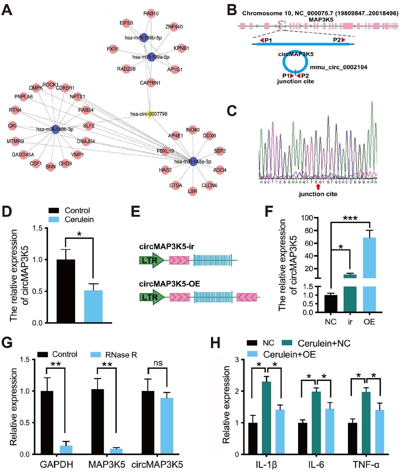
Acute pancreatitis arises from the activation of digestive enzymes in pancreatic acinar cells, leading to autodigestion of the pancreas and surrounding tissues. It is a common digestive tract emergency which requires hospitalization, and its incidence is increasing worldwide. In the past decade, several advances have been made in the treatment of acute pancreatitis. However, there is still a lack of efficacious drugs for clinical practice, and the limited value of existing biomarkers for early warning of the severity of acute pancreatitis is a major obstacle. Thus, there is an urgent need to gain a better understanding of the molecular mechanisms of acute pancreatitis. Circular RNAs (circRNAs) are a unique class of RNA molecules that are covalently closed. Ongoing investigations have provided evidence that circRNAs govern downstream target expression by acting as miRNA sponges, functioning as transcription factors, interacting with RNA-binding proteins, and regulating alternative splicing. These mechanisms support the pivotal role of circRNAs in a wide variety of physiological and pathological conditions, such as innate immunity, inflammation, neuronal function, and tumorigenesis. To explore the role of circular RNA in acute pancreatitis, we employed circRNA microarray technology (Arraystar Human circRNA Array V2) to examine the circRNA expression profile in the blood of three acute pancreatitis patients and three healthy controls. Clinical acute pancreatitis samples were obtained from Xiangya Hospital, Central South University. This study was approved by the Ethics Committee of Xiangya hospital (No. 2019010008). Normal control patients were recruited from among individuals who had visited Xiangya Hospital for a routine checkup. Written informed consent was obtained from all participants or their legal representatives for publication of data. The diagnosis and severity classification of acute pancreatitis were performed according to the American Gastroenterological Association guidelines and the Revised Atlanta Classification (RAC). circRNAs with a fold change ≥1.5 and a P value<0.05 were considered to be differentially expressed. As shown in Figure 1A, the two groups presented different expression profiles. We found that 91 circRNAs were significantly differentially expressed in the blood of acute pancreatitis patients, with 10 circRNAs exhibiting increased expression and 81 exhibiting decreased expression (Figure 1B,C). Among the differentially expressed circRNAs in acute pancreatitis, downregulated circRNAs are more prevalent than upregulated circRNAs, and the differential expression is more significant. Therefore, the present study focused on downregulated circRNAs. We selected circRNAs that are downregulated at least 2.5-fold and excluded those with fewer than 1000 bases to ensure the accuracy of qPCR. Based on these criteria, we identified nine circRNAs (circ_0006554, circ_0007798, circRNA_405815, circ_0001847, circ_0069748, circ_0001850, circ_0008417, circ_0002560, and circ_0000008) for validation by qPCR (Applied Biosystems, Foster City, USA) in blood samples from 30 acute pancreatitis patients (10 patients each with mild acute pancreatitis, moderate severe acute pancreatitis, and severe acute pancreatitis) and 15 healthy individuals. The levels of circ_0007798, circ_0001847, and circ_0069748 were significantly lower in acute pancreatitis patients than in normal controls, while the remaining circRNAs were not significantly differentially expressed (Figure 1D). In addition, the levels of circ_0007798 increased gradually with the severity of acute pancreatitis, suggesting that circ_0007798 is associated with the clinical severity of the disease (Figure 1E). Differential circRNAs have been studied for the diagnosis of pancreatic diseases. The expression level of circ_0007798 can be used to grade the severity of acute pancreatitis and provide individualized treatment. Furthermore, homology analysis (NCBI blast) revealed that circ_0007798 has a high degree of conservation between rats and humans according to the basic local alignment search tool. In conclusion, according to the

Identification and validation of disease severity-related circular RNA in acute pancreatitis
Graphical Abstract
Original ResearchVol. 56, Issue 9 • pp. 1352-1364DOI: 10.3724/abbs.2024024

The mechanism by which piR-000699 targets SLC39A14 regulates ferroptosis in aging myocardial ischemia/reperfusion injury

Authors: Hongyang Chi, Yue’e Chai, Lingju Ma, Yichen Wang, Qianqian Wu, Lexin Wang, Junjie Zhai, Fufun Ma, Yancheng Tian, Ning Qi, Jianhong Peng, Youjuan Fu, Xiaoling Yang, Hui Huang, Shengchao Ma

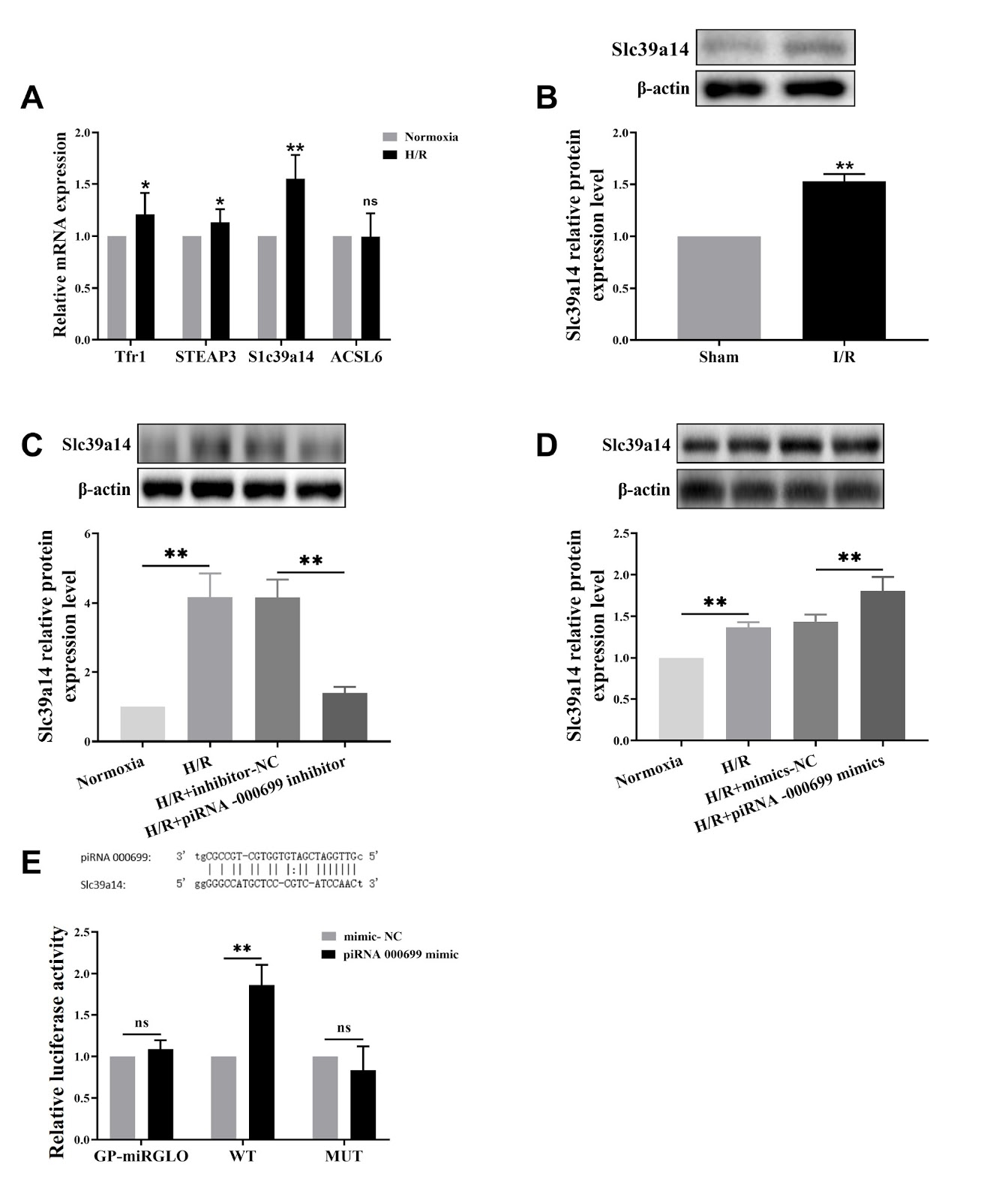
Myocardial ischemia/reperfusion (I/R) injury is a classic type of cardiovascular disease characterized by injury to cardiomyocytes leading to different types of cell death. The degree of irreversible myocardial damage is closely related to age, and ferroptosis is involved in cardiomyocyte damage. However, the mechanisms underlying ferroptosis regulation in aging myocardial I/R injury are still unclear. The present study aims to explore the underlying mechanism of piRNA regulation in ferroptosis. Using left anterior descending coronary artery ligation in an aging rat model and a D-galactose-induced rat cardiomyocyte line (H9C2) to construct an aging cardiomyocyte model, we investigate whether ferroptosis occurs after reperfusion injury in vitro and in vivo. This study focuses on the upregulation of piR-000699 after hypoxia/reoxygenation treatment in aging cardiomyocytes by observing hypoxia/reoxygenation (H/R) injury indicators and ferroptosis-related indicators and clarifying the role of piR-000699 in H/R injury caused by ferroptosis in aging cardiomyocytes. Bioinformatics analysis reveals that SLC39A14 is a gene that binds to piR-000699. Our data show that ferroptosis plays an important role in I/R injury both in vivo and in vitro. Furthermore, the results show the potential role of piR-000699 in regulating SLC39A14 in ferroptosis in aging cardiomyocytes under hypoxia/reoxygenation conditions. Together, our results reveal that the mechanism by which piR-000699 binds to SLC39A14 regulates ferroptosis in aging myocardial I/R injury.

The mechanism by which piR-000699 targets SLC39A14 regulates ferroptosis in aging myocardial ischemia/reperfusion injury
Graphical Abstract
Original ResearchVol. 56, Issue 9 • pp. 1267-1277DOI: 10.3724/abbs.2024010

TRMT13 inhibits the growth of papillary thyroid cancer by targeting ANAPC4

Authors: Lianyong Liu, Yan Wang, Mei Zou, Shiwei Chen, Fengying Wu, Xiangqi Li

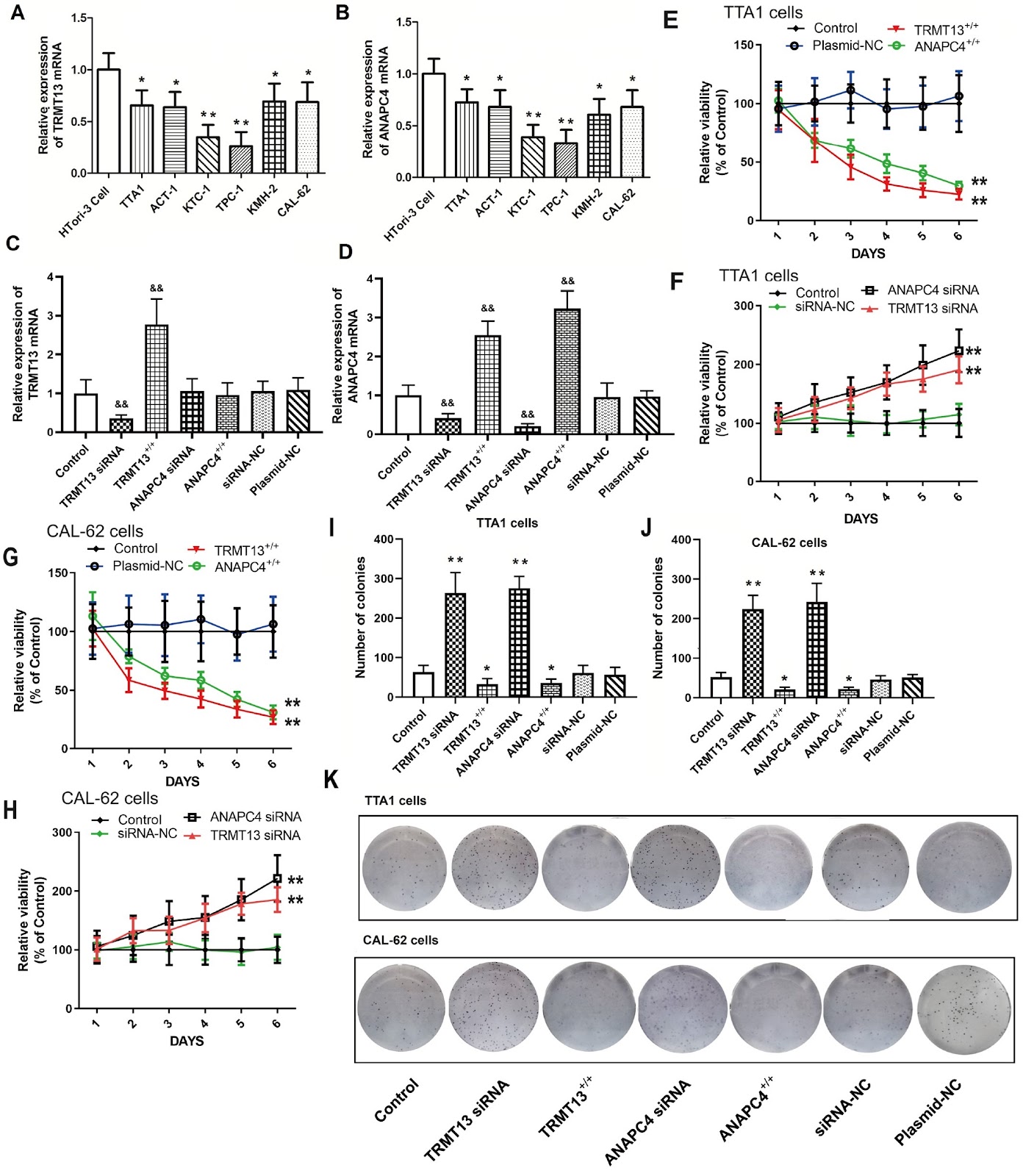
The recently discovered gene TRMT13 encodes a type of RNA methylase and is a member of the CCDC family (also called CCDC76). Here, we delineate its role in papillary thyroid cancer (PTC). Bioinformatics analysis shows significant TRMT13 and ANAPC4 downregulation in PTC and reveals that the expression levels of both genes are linearly correlated. Subsequent analyses confirm that both TRMT13 and ANAPC4 expressions are downregulated in PTC tissues and that this change in expression has a significant impact on cancer diagnosis. We conduct assays on PTC cells subjected to TRMT13 and ANAPC4 silencing or overexpression to assess the biological effects of these genes. We also perform rescue experiments to validate the regulatory effects of TRMT13 on ANAPC4. A nude mouse tumor model is used to evaluate the effects of TRMT13 and ANAPC4 on PTC tumorigenesis. TRMT13 expression is decreased in PTC tissues and cell lines and is positively correlated with that of ANAPC4. Cell assays reveal that TRMT13/ANAPC4 attenuates the malignancy of PTC cells by restraining cell proliferation, migration and invasion, while rescue experiments corroborate that ANAPC4 is a downstream target of TRMT13. In the nude mouse xenograft model, both TRMT13 and ANAPC4 inhibit tumor growth, and TRMT13 and ANAPC4 expression levels are significantly associated with survival. Taken together, these findings lead to the conclusion that TRMT13 inhibits PTC growth via ANAPC4, indicating a new role of TRMT13 and providing insights into the tRNA methyltransferase and coiled-coil domain-containing protein families.

TRMT13 inhibits the growth of papillary thyroid cancer by targeting ANAPC4
Graphical Abstract
Original ResearchVol. 56, Issue 9 • pp. 1300-1310DOI: 10.3724/abbs.2024042

Proanthocyanidins isolated from lotus seed skin mitigate glycolipid metabolism disorder through the p38/Nrf2/NF-κB signaling pathway

Authors: Yuhang Yi, Tao Li, Chenghao lv, Wenjiang He, Wenzhi Li, Xixin Zhou, Si Qin

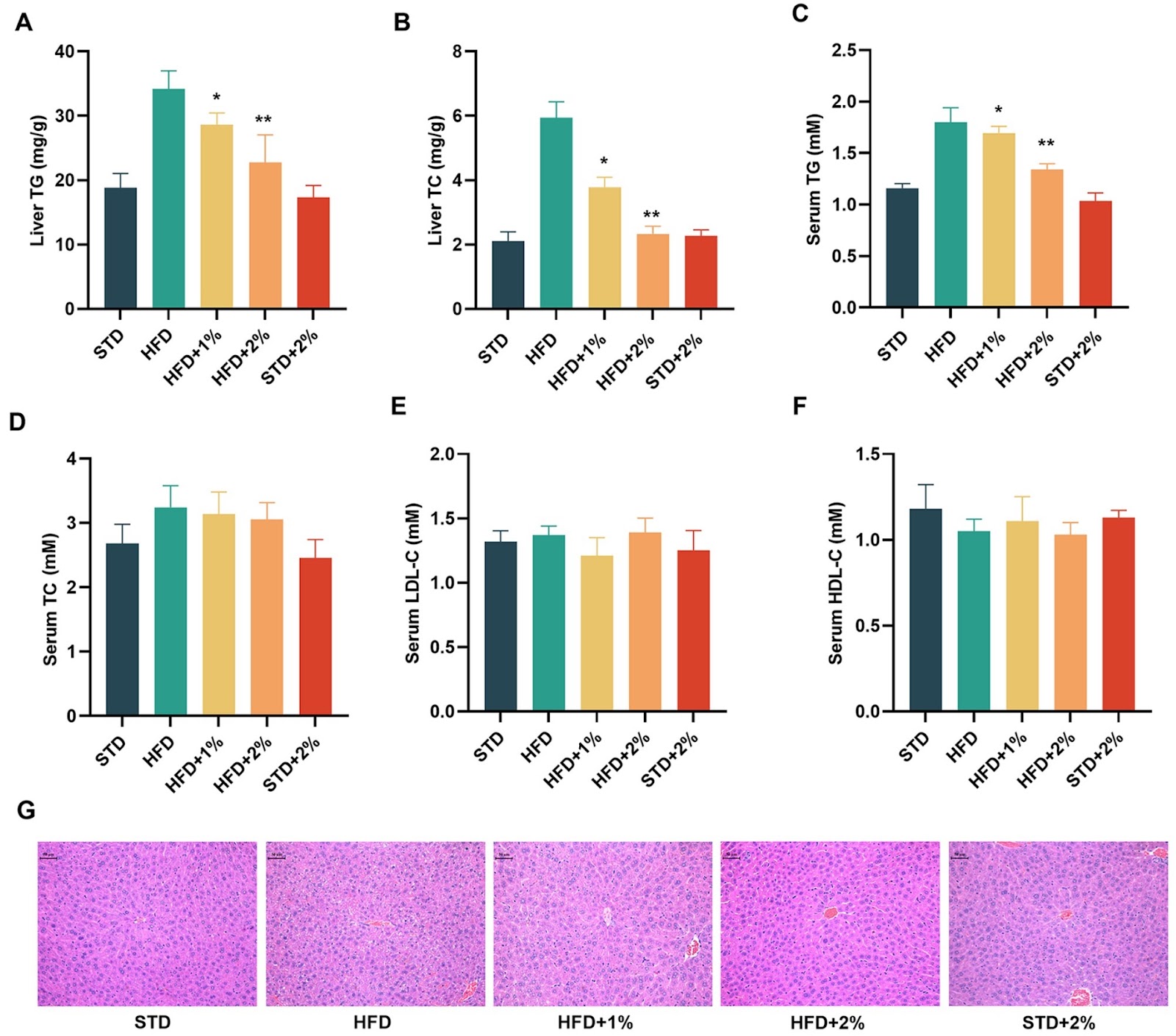
Lotus seed skin extract is rich in flavonoids, making it a promising candidate for developing health products. In a previous study, we found that proanthocyanidins from lotus seed skin, particularly proanthocyanidin B1 (PB1), can indirectly activate the Nrf2 signaling pathway, exerting an antioxidant effect. In this study, we isolate proanthocyanidins from lotus seed skin (PLS) using ethanol extraction and RP-HPLC identification, and investigate its effects on glycolipid metabolism both in vivo and in vitro. Our results demonstrate that PLS reduces body weight in high-fat diet (HFD) mice by decreasing feed efficiency. PLS also normalizes serum glucose, insulin secretion, glycosylated hemoglobin (HbA1c), and intraperitoneal glucose tolerance (IPGTT). Furthermore, PLS significantly improves blood lipid parameters and inhibits the expressions of six proinflammatory factors, including IL-1α, IL-1β, IL-3, IL-6, IFN-γ and TNF-α in HFD mice. Additionally, analysis of fresh liver tissues reveals that PLS and PB1 induce the expressions of antioxidant proteins such as HO-1 and NQO1 by activating the p38-Nrf2 signaling pathway and inhibiting the NF-κB signaling pathway. In conclusion, proanthocyanidins from lotus seed skin regulate glycolipid metabolism disorders by targeting the p38/Nrf2/NF-κB signaling pathway. Our study offers a new approach for the high-value comprehensive utilization of lotus seed skin by-products and precise dietary intervention for metabolic syndrome.

Proanthocyanidins isolated from lotus seed skin mitigate glycolipid metabolism disorder through the p38/Nrf2/NF-κB signaling pathway
Graphical Abstract
Original ResearchVol. 56, Issue 9 • pp. 1278-1288DOI: 10.3724/abbs.2024066

The rod cell, a small form of Candida albicans, possesses superior fitness to the host gut and adaptation to commensalism

Authors: Yinxing Xu, Wencheng Zhu, Baodi Dai, Hui Xiao, Jiangye Chen

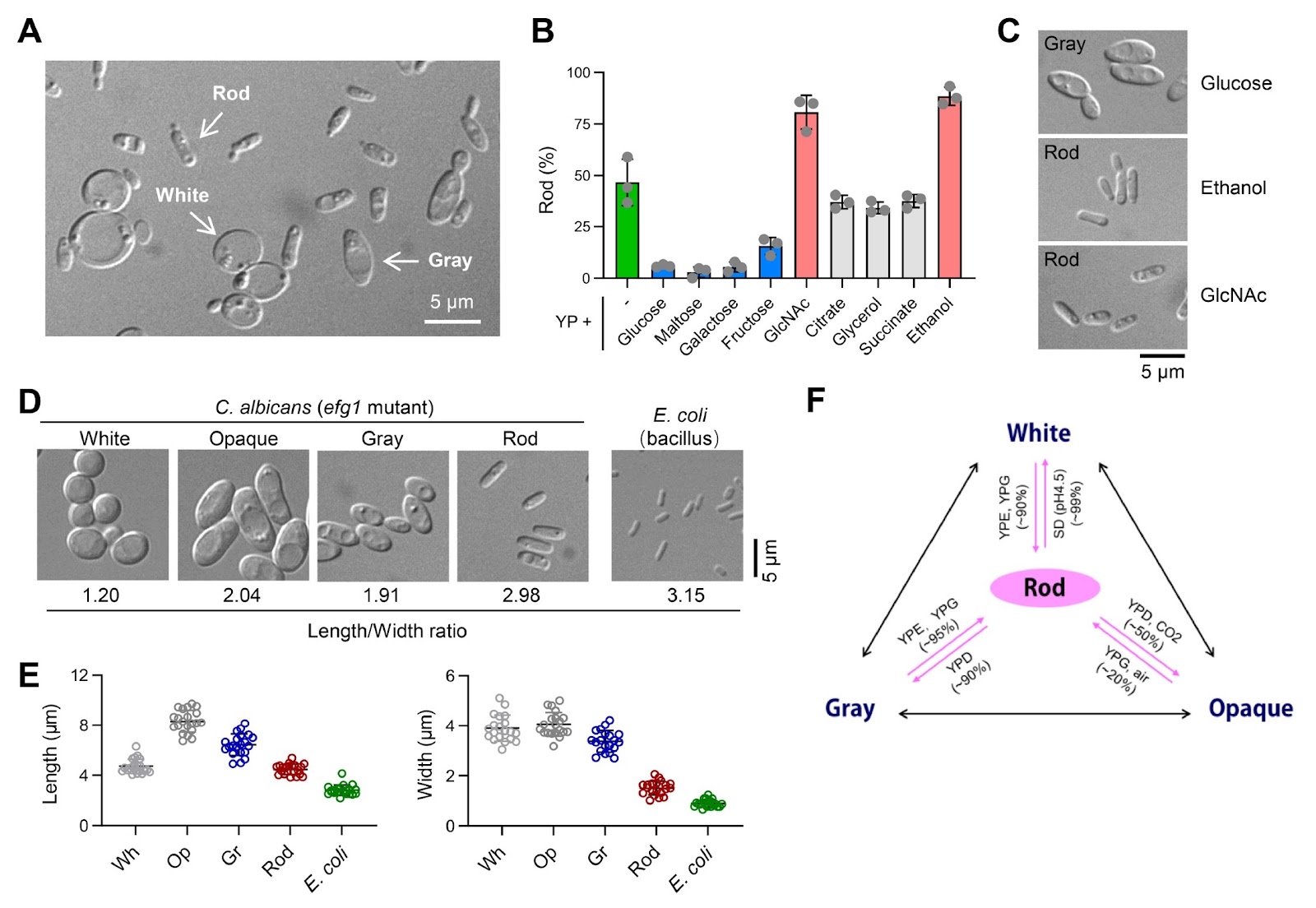
Candida albicans deploys various morphological forms through complex switching mechanisms, ensuring its survival and thriving as a commensal or pathogen in vastly different human niches. In this study, we demonstrate that a novel ''rod'' morphological form of C. albicans coexists and is interchangeable with previously reported white, gray, and opaque forms, constituting a tetra-stable phenotypic switching system. Rod cells arise from the efg1 mutant of SC5314 cells or from the clinical BJ1097 strain cultured under glucose-free conditions. They are characterized by a distinct gene expression profile and can be stably maintained through in vitro passaging or in vivo inhabitation of the gastrointestinal (GI) tract of mice. Remarkably, the majority of the efg1 mutant cells become rod cells in N-acetylglucosamine (GlcNAc)-containing medium, and the GlcNAc sensor Ngs1 is instrumental in converting the white or gray cells to the rod cells. Conversely, glucose inhibits rod cells through Cph1; consequently, the loss of Cph1 in the efg1 mutant cells permits their conversion to rod cells in glucose-replete media. Notably, rod cells of the efg1/cph1 mutant display superior adaptation and longer persistence in the murine GI environment than wild-type white cells. Taken together, these findings establish rod cells as a previously unappreciated form that is not only morphologically and transcriptionally distinguishable but also defined by specific genetic and environmental determinants, shedding light on complex fungus-host interactions.

The rod cell, a small form of Candida albicans, possesses superior fitness to the host gut and adaptation to commensalism
Graphical Abstract
Original ResearchVol. 56, Issue 8 • pp. 1118-1129DOI: 10.3724/abbs.2024127

O-glycosylation of SARS-CoV-2 spike protein by host O-glycosyltransferase strengthens its trimeric structure

Authors: Zhijue Xu, Han Zhang, Jiaqi Tian, Xin Ku, Rumeng Wei, Jingli Hou, Can Zhang, Fang Yang, Xia Zou, Yang Li, Hiroyuki Kaji, Sheng-Ce Tao, Atsushi Kuno, Wei Yan, Lin-Tai Da, Yan Zhang

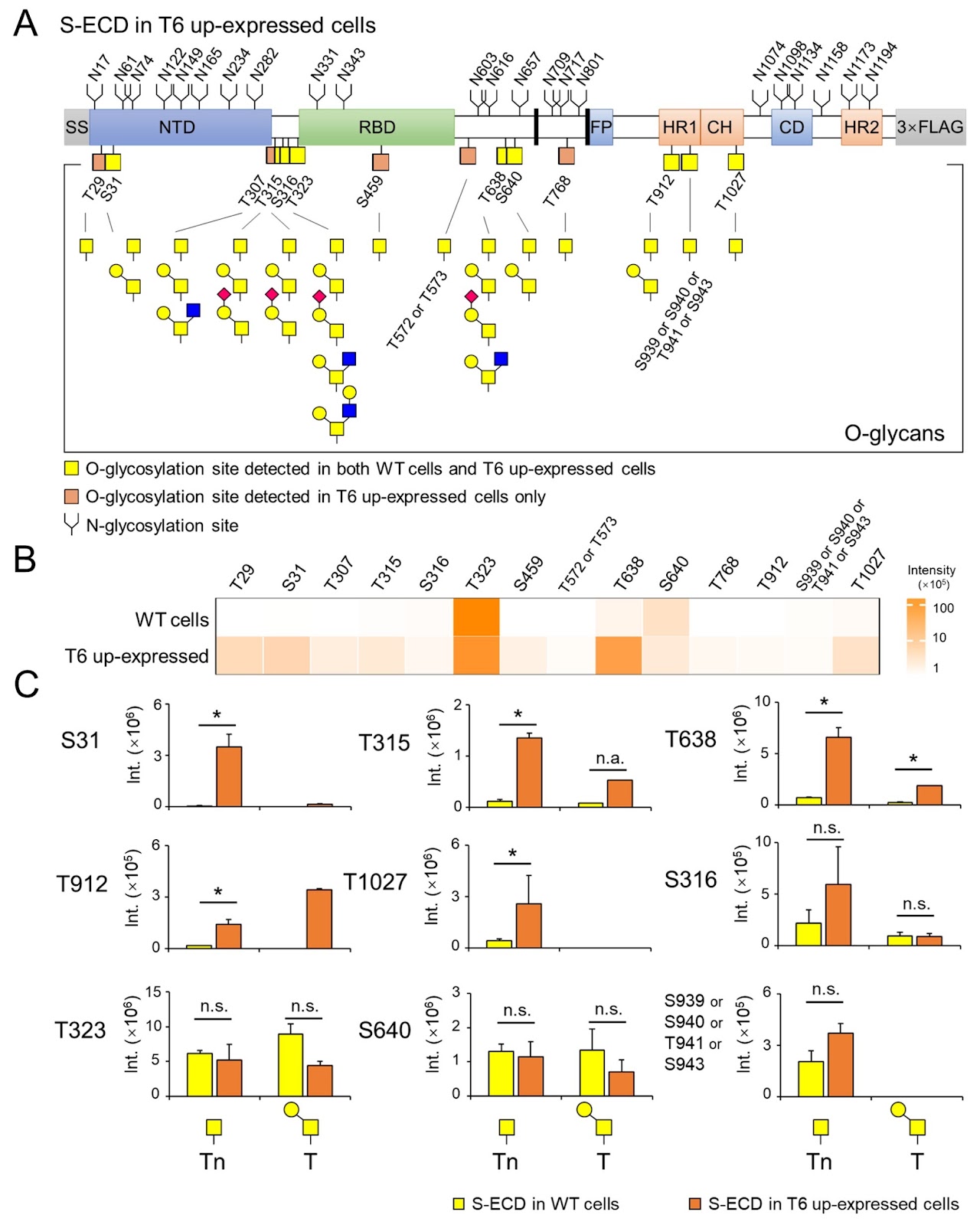
Protein O-glycosylation, also known as mucin-type O-glycosylation, is one of the most abundant glycosylation in mammalian cells. It is initially catalyzed by a family of polypeptide GalNAc transferases (ppGalNAc-Ts). The trimeric spike protein (S) of SARS-CoV-2 is highly glycosylated and facilitates the virus’s entry into host cells and membrane fusion of the virus. However, the functions and relationship between host ppGalNAc-Ts and O-glycosylation on the S protein remain unclear. Herein, we identify 15 O-glycosites and 10 distinct O-glycan structures on the S protein using an HCD-product-dependent triggered ETD mass spectrometric analysis. We observe that the isoenzyme T6 of ppGalNAc-Ts (ppGalNAc-T6) exhibits high O-glycosylation activity for the S protein, as demonstrated by an on-chip catalytic assay. Overexpression of ppGalNAc-T6 in HEK293 cells significantly enhances the O-glycosylation level of the S protein, not only by adding new O-glycosites but also by increasing O-glycan heterogeneity. Molecular dynamics simulations reveal that O-glycosylation on the protomer-interface regions, modified by ppGalNAc-T6, potentially stabilizes the trimeric S protein structure by establishing hydrogen bonds and non-polar interactions between adjacent protomers. Furthermore, mutation frequency analysis indicates that most O-glycosites of the S protein are conserved during the evolution of SARS-CoV-2 variants. Taken together, our finding demonstrate that host O-glycosyltransferases dynamically regulate the O-glycosylation of the S protein, which may influence the trimeric structural stability of the protein. This work provides structural insights into the functional role of specific host O-glycosyltransferases in regulating the O-glycosylation of viral envelope proteins.

O-glycosylation of SARS-CoV-2 spike protein by host O-glycosyltransferase strengthens its trimeric structure
Graphical Abstract
Original ResearchVol. 56, Issue 8 • pp. 1130-1144DOI: 10.3724/abbs.2024100

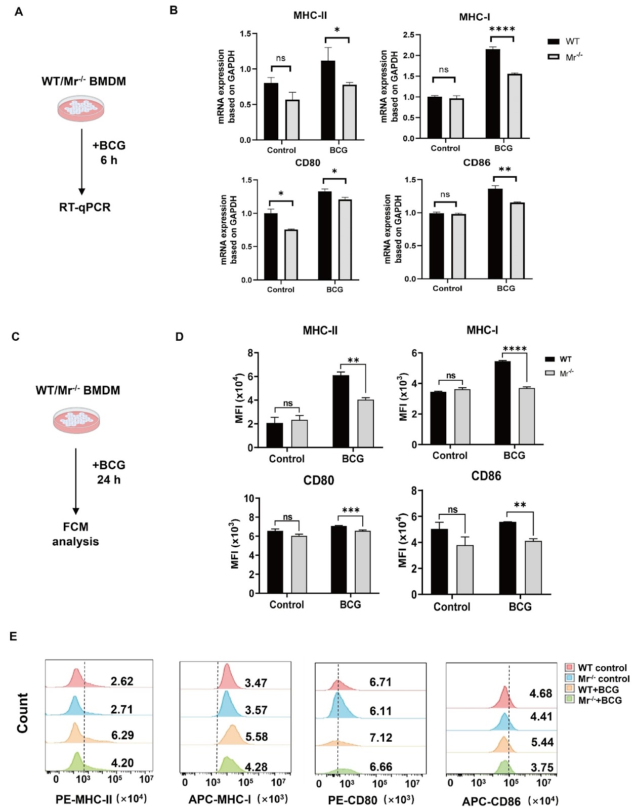
Macrophages exploit the mannose receptor and JAK-STAT1-MHC-II pathway to drive antigen presentation and the antimycobacterial immune response after BCG vaccination

Authors: Ying Zhang, Dandan Xu, Qi Nie, Jing Wang, Dan Fang, Yan Xie, Huang Xiong, Qin Pan, Xiao-Lian Zhang

Tuberculosis (TB), caused by Mycobacterium tuberculosis (M.tb), remains one of the leading causes of fatal infectious diseases worldwide. The only licensed vaccine, Mycobacterium bovis Bacillus Calmette-Guérin (BCG), has variable efficacy against TB in adults. Insufficiency of immune cell function diminishes the protective effects of the BCG vaccine. It is critical to clarify the mechanism underlying the antimycobacterial immune response during BCG vaccination. Macrophage mannose receptor (MR) is important for enhancing the uptake and processing of glycoconjugated antigens from pathogens for presentation to T cells, but the roles of macrophage MR in the BCG-induced immune response against M.tb are not yet clear. Here, we discover that macrophage MR deficiency impairs the antimycobacterial immune response in BCG-vaccinated mice. Mechanistically, macrophage MR triggers JAK-STAT1 signaling, which promotes antigen presentation via upregulated MHC-II and induces IL-12 production by macrophages, contributing to CD4+ T cell activation and IFN-γ production. MR deficiency in macrophages reduces the vaccine efficacy of BCG and increases susceptibility to M.tb H37Ra challenge in mice. Our results suggest that MR is critical for macrophage antigen presentation and the antimycobacterial immune response to BCG vaccination and offer valuable guidance for the preventive strategy of BCG immunization.

Macrophages exploit the mannose receptor and JAK-STAT1-MHC-II pathway to drive antigen presentation and the antimycobacterial immune response after BCG vaccination
Graphical Abstract
Original ResearchVol. 56, Issue 8 • pp. 1145-1155DOI: 10.3724/abbs.2024123

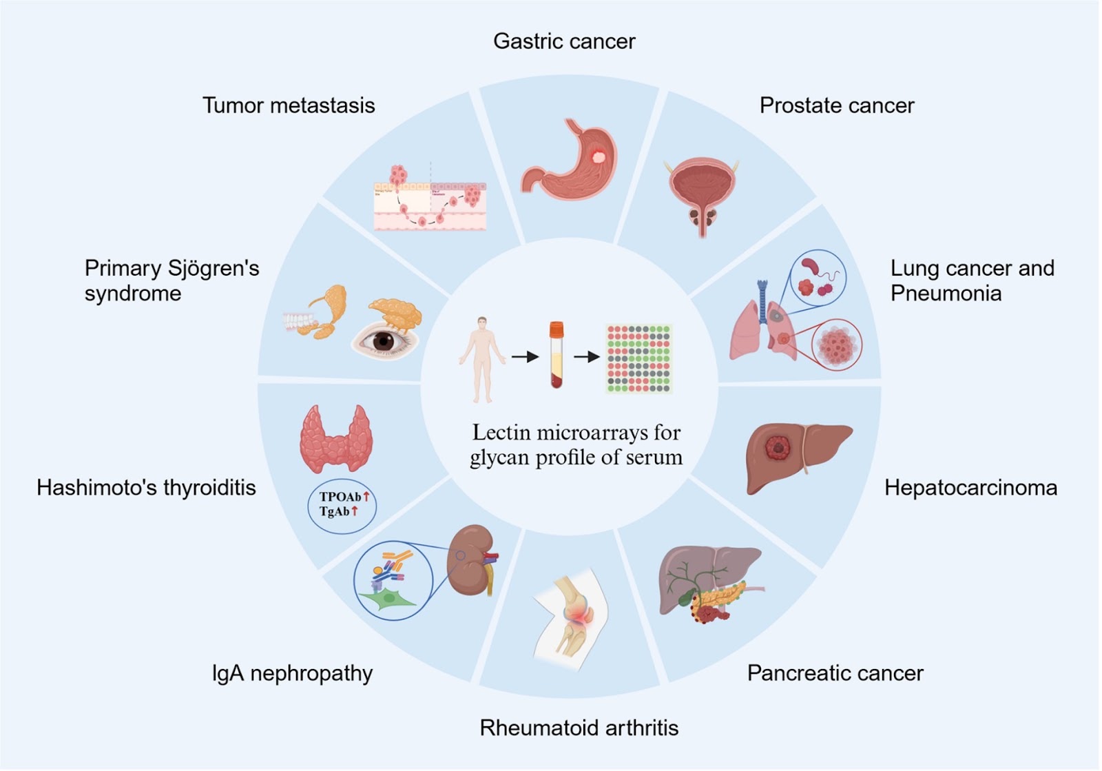
Deciphering disease through glycan codes: leveraging lectin microarrays for clinical insights

Authors: Hangzhou Yang, Zihan Lin, Bo Wu, Jun Xu, Sheng-Ce Tao, Shumin Zhou

Glycosylation, a crucial posttranslational modification, plays a significant role in numerous physiological and pathological processes. Lectin microarrays, which leverage the high specificity of lectins for sugar binding, are ideally suited for profiling the glycan spectra of diverse and complex biological samples. In this review, we explore the evolution of lectin detection technologies, as well as the applications and challenges of lectin microarrays in analyzing the glycome profiles of various clinical samples, including serum, saliva, tissues, sperm, and urine. This review not only emphasizes significant advancements in the high-throughput analysis of polysaccharides but also provides insight into the potential of lectin microarrays for diagnosing and managing diseases such as tumors, autoimmune diseases, and chronic inflammation. We aim to provide a clear, concise, and comprehensive overview of the use of lectin microarrays in clinical settings, thereby assisting researchers in conducting clinical studies in glycobiology.

Deciphering disease through glycan codes: leveraging lectin microarrays for clinical insights
Graphical Abstract