🧬 SinoBioData Academic Portal
📚 Peer-Reviewed Translated Literature

All Biomedical & Clinical Articles (Page 51)

Browse complete peer-reviewed translations from top Chinese biomedical, oncology, and genomics journals. Read verified previews and download full authentic clinical reports.

Published Research Papers

Showing 24 of 1542 peer-reviewed translated articles (Page 51 of 65)

Efficient production of recombinant mAbs mediated by a MAR-enhanced transposon vector combined with blasticidin selection in CHO cellsGraphical AbstractVerified
Acta Biochimica et Biophysica Sinica

Efficient production of recombinant mAbs mediated by a MAR-enhanced transposon vector combined with blasticidin selection in CHO cells

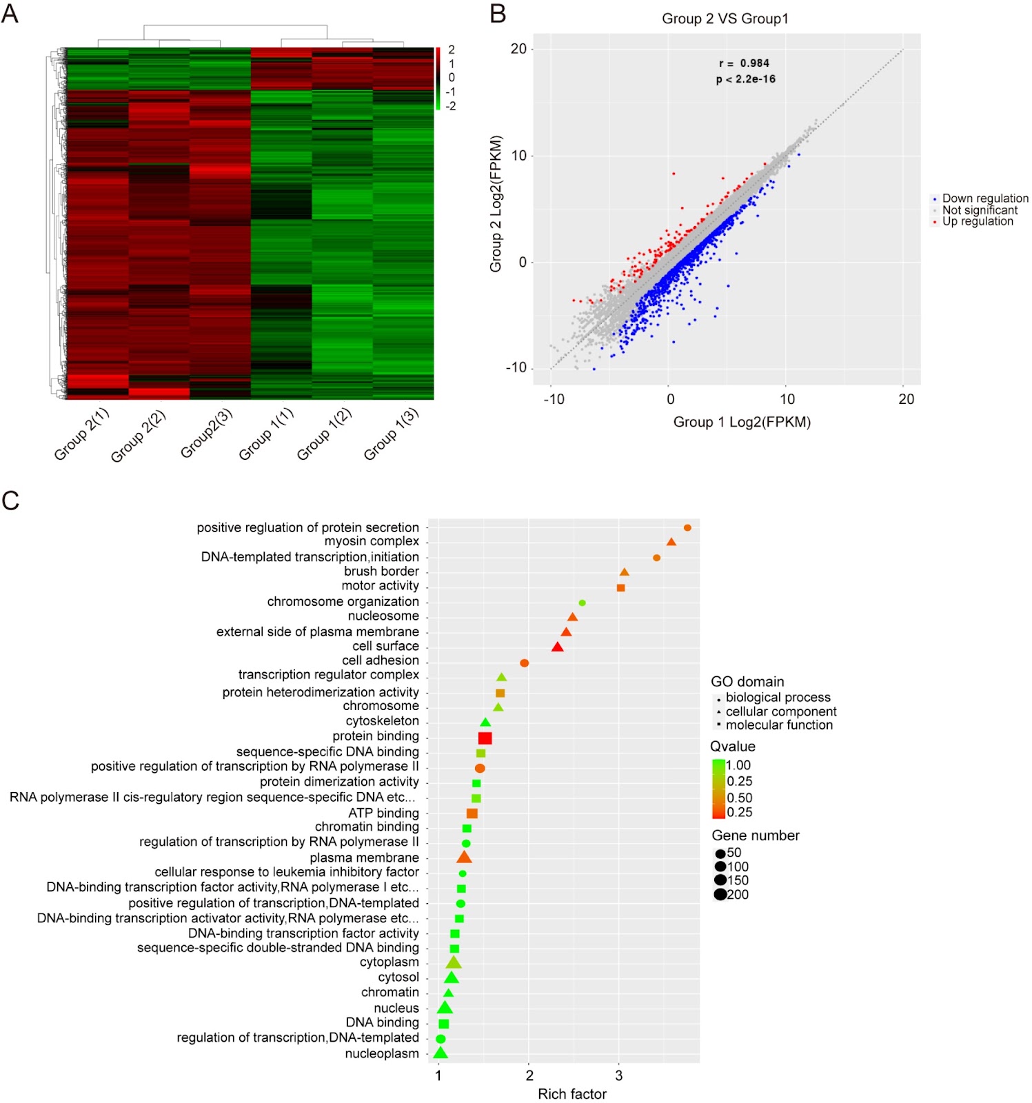
Recombinant antibodies, primarily produced in Chinese hamster ovary (CHO) cells, are widely used to treat various diseases. For industrial production, a rapid and efficient method to screen stable, high-expressing clones is essential. However, conventional screening based on random integration is often cumbersome and labor intensive. This study establishes a novel strategy for generating stable, high-yielding clones by combining a MAR-based piggyBac (PB) transposon semitargeted integration system with blasticidin (BSD) selection. Compared to the random integrated vector pMAR-mAb, the MAR-PB system increases the titers (3.95- to 5.61-fold) and specific protein productivity (Qp; 4.28- to 6.07-fold) of four monoclonal antibodies in stable cell pools. When compared to PB-only vectors, the MAR-PB transposon system enhances the titers (by up to 2.50-fold) and Qp (1.96- to 2.77-fold), respectively. The increased antibody production correlates with elevated mRNA expression. Furthermore, this approach increases the proportion of high-expressing clones by more than 10-fold and significantly improves volumetric yield. Importantly, this approach promotes the long-term stability of recombinant mAb expression for over 60 generations. Transcriptome analysis reveals that the system modulates genes involved in DNA binding, transcriptional regulation, and protein binding. In conclusion, the MAR-based PB transposon system combined with BSD selection presents a significant improvement for efficiently generating high-yielding and stable CHO cell clones, offering a valuable tool for recombinant antibody production.

Read Full Abstract10.3724/abbs.2025251
Corrigendum to: Downregulating integrin subunit alpha 7 (ITGA7) promotes proliferation, invasion, and migration of papillary thyroid carcinoma cells through regulating epithelial-to-mesenchymal transitionGraphical AbstractVerified
Acta Biochimica et Biophysica Sinica

Corrigendum to: Downregulating integrin subunit alpha 7 (ITGA7) promotes proliferation, invasion, and migration of papillary thyroid carcinoma cells through regulating epithelial-to-mesenchymal transition

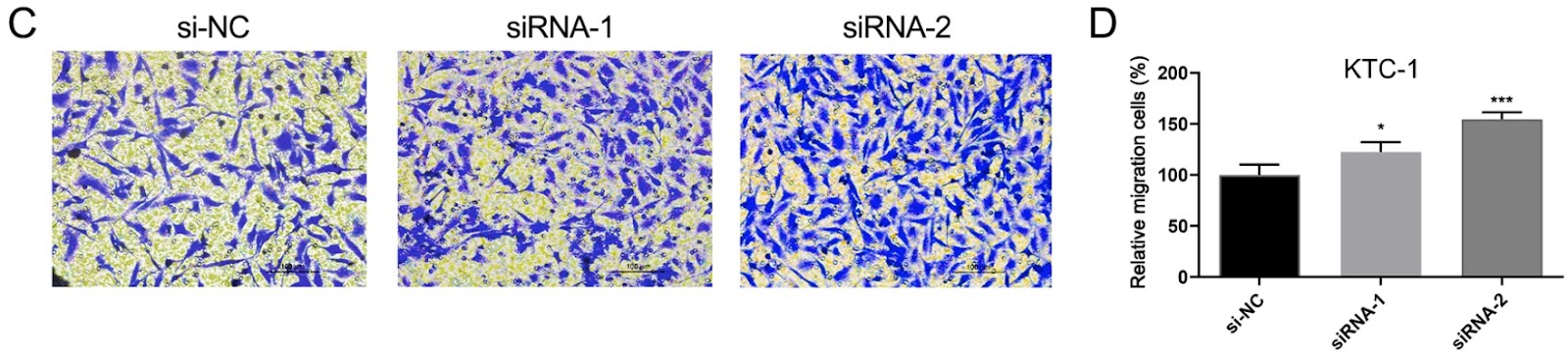
This is a corrigendum to the original article published in Acta Biochim Biophys Sin 2020, 52(2): 116–124. The authors correct an error in Figure 4 of the original manuscript. The correct figure is provided, and the authors apologize for the error.

Read Full Abstract10.3724/abbs.2024200
Scaffold Compound T4015 Attenuates Pulmonary Fibrosis via Suppressing JAK/STAT and NF-ÎșB SignalingGraphical AbstractVerified
Acta Biochimica et Biophysica Sinica2026

Scaffold Compound T4015 Attenuates Pulmonary Fibrosis via Suppressing JAK/STAT and NF-ÎșB Signaling

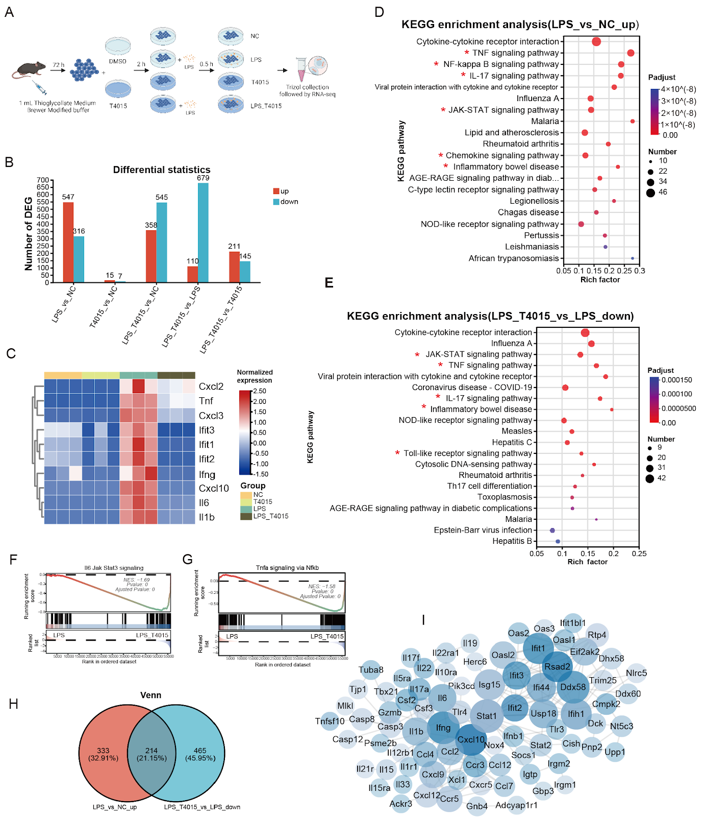
Pulmonary fibrosis (PF) is a life-threatening interstitial lung disease characterized by scarring and inflammation in lung tissues. Aberrant activation of the JAK/STAT and NF-ÎșB signaling pathways is critical in initiating and sustaining the inflammatory processes that drive fibrotic progression. In this study, we identify a novel small-molecule compound, T4015, a 4-indolyl-2-phenylaminopyrimidine derivative, as a dual-pathway inhibitor targeting both JAK/STAT and NF-ÎșB signaling. Dual-luciferase reporter assays demonstrate the potent inhibitory activity of T4015 against these pathways. T4015 effectively suppresses the phosphorylation of STAT3, JAK1, and TYK2 induced by IL-6 and IFN-ÎČ, while suppressing LPS-induced NF-ÎșB activation in macrophages. Transcriptome sequencing and pathway enrichment analyses further confirm that T4015 downregulates multiple inflammation-related signaling cascades, including the JAK/STAT, NF-ÎșB, TNF, IL-17, and Toll-like receptor pathways. In a mouse model of bleomycin-induced PF, T4015 treatment significantly improves survival, attenuates collagen deposition, and reduces the expression of pro-inflammatory and profibrotic markers such as IL-6, CCL2, and COL1. Molecular docking and target prediction analyses suggest that T4015 exhibits strong binding affinity for multiple kinases within the JAK/STAT and NF-ÎșB networks, including JAK1, TYK2, JAK2, JAK3, RIPK1, IRAK1/4, TAB1, and ZAP70. Collectively, these results highlight T4015 as a promising therapeutic candidate for PF through its simultaneous inhibition of the JAK/STAT and NF-ÎșB signaling pathways.

Read Full Abstract10.3724/abbs.2026035
Special issue: advances in immunology and its applicationsGraphical AbstractVerified
Acta Biochimica et Biophysica Sinica

Special issue: advances in immunology and its applications

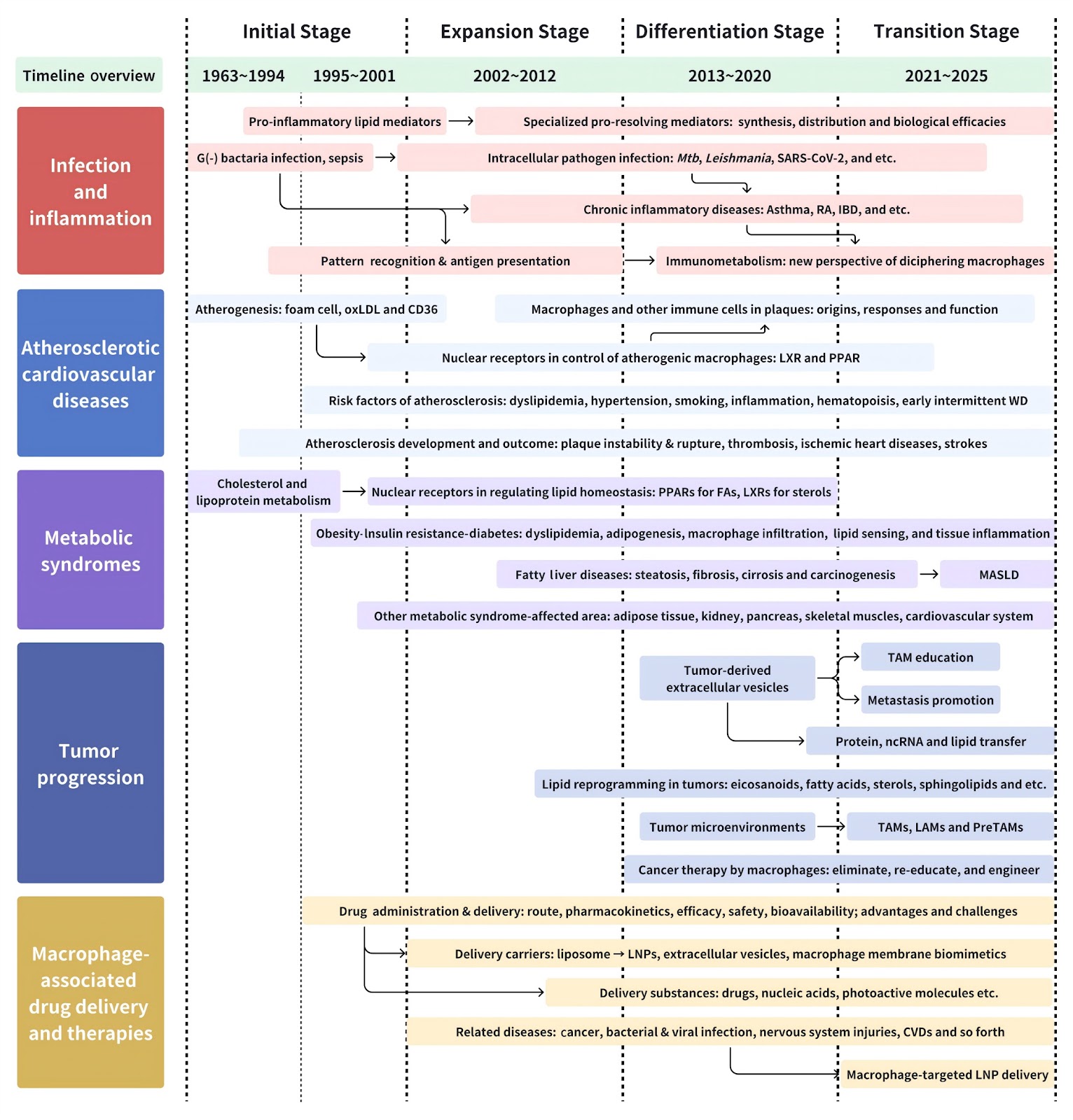
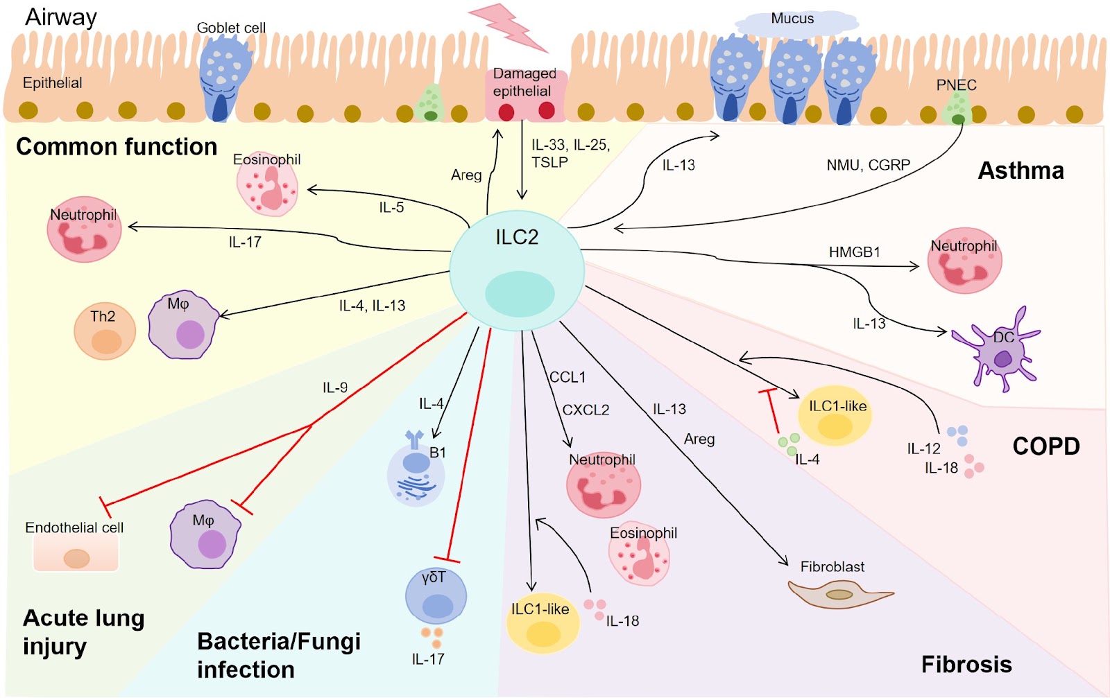
Innate immunity and adaptive immunity play crucial roles in regulating inflammation-related diseases such as tumors, infections, and autoimmune diseases. The interplay between innate immunity and adaptive immunity ensures the maintenance of tissue homeostasis and effective clearance of invading pathogens or tumor cells. However, dysregulation in each type of immune cells or their communications leads to pathological conditions, ranging from chronic inflammation to malignant progression. In recent years, the field of immunology has witnessed a paradigm shift, moving from merely describing immune cell signaling or phenotypes to actively exploring strategies that reshape immune functions for therapeutic benefit. Exploring effectors or strategies to reshape T cell function for the improved anti-tumor and anti-infection efficacy has emerged as a critical research direction. This includes the development and optimization of chimeric antigen receptor T-cell (CAR-T) therapies for clinical applications. While CAR-T therapy has revolutionized the treatment of hematologic malignancies, its success in solid tumors remains limited. Chen et al. [1] dissect the distinct signaling mechanisms of chimeric antigen receptors (CARs) compared to T cell receptors (TCRs). They elucidate how CAR-T cells, despite overcoming MHC restrictions, encounter significant hurdles such as inefficient tumor infiltration and the hostility of the immunosuppressive tumor microenvironment (TME). To address these challenges, the authors underscore several innovative strategies, such as optimizing receptor clustering to facilitate immune synapse formation and integrating novel co-stimulatory domains to augment therapeutic efficacy in solid tumors. Moving beyond engineering, fundamental insights into microenvironmental stress and T cell intrinsic signaling are pivotal. While oxidative stress is a well-established concept in the TME, Ji and Xiao [2] draw attention to the phenomenon of “reductive stress”. They explain how a surplus of intracellular reducing agents disrupts redox balance, creating a reductive environment that significantly influences immune cell differentiation and tumor survival. In a complementary study, Shi et al. [3] investigate the intrinsic role of pattern recognition receptor (PRR) signaling within T cells. Although PRRs are historically classified as innate sensors, this review highlights their critical function in adaptive immunity, discussing how T cells utilize PRRs to interpret endogenous danger signals and microbial cues to regulate cytokine release and proliferation. Additionally, Ma et al. [4] provide a comprehensive update on T cell immunoglobulin and mucin-containing molecule 3 (TIM-3), an important immune checkpoint. By detailing its expression profile across T cells, NK cells, and myeloid lineages, they propose that rational combination therapies targeting TIM-3 alongside other checkpoints offer a promising avenue to surmount current resistance mechanisms. This issue also highlights the regulation of immunity by neurotransmitters and biological rhythms, illustrating the profound integration of the immune system with physiological networks. Fan and Zhao [5] summarize the neurotransmitter-receptor landscape in T cell tumor immunology. They elaborate on how specific neurotransmitters, including glutamate, acetylcholine, GABA, and serotonin, could dictate T cell activation and differentiation within the TME. The authors advocate for targeting these neuro-immune axes, such as through ÎČ-blockers or glutamate receptor inhibitors, as a new frontier for enhancing cancer immunotherapy. From a different perspective, Sun et al. [6] assess the influence of circadian rhythms on the TME and immunotherapy outcomes. They present evidence indicating that the efficacy of treatments like immune checkpoint blockade depends on administration timing, suggesting that future clinical protocols should incorporate chronobiology to maximize patient benefit. Further exploring this dimension, Zhao et al. [7] examine the bidirectional circadian dialogue between the host and the gut microbiota. They describe how modern lifestyle factors, such as shift work and irregular dietary habits, disrupt this synchronization, resulting in compromised barrier integrity and systemic metabolic disorders. This issue also focuses on the function of various innate immune cells, including ILCs, macrophages, and dendritic cells (DCs), as well as their crosstalk with T cells in maintaining homeostasis across lung, gut, and aging contexts. Chen et al. [8] concentrate on Group 2 innate lymphoid cells (ILC2s), which functionally mirror Th2 cells. They underscore the context-specific roles of ILC2s in pulmonary diseases, explaining how these cells react to environmental alarmins such as IL-33 and thymic stromal lymphopoietin (TSLP). The review also discusses targeting ILC2 plasticity as a potential therapeutic intervention for lung inflammation. Recent findings suggest lipid metabolism as a crucial determinant of innate immune function. Huang et al. [9] offer an updated survey of lipid-regulated immunobiology in macrophages, examining how
Read Full Abstract10.3724/abbs.2025247
A bifunctional aptamer-siRNA chimera targeting ACE2 for the inhibition of SARS-CoV-2 S pseudovirus entry and replicationGraphical AbstractVerified
Acta Biochimica et Biophysica Sinica

A bifunctional aptamer-siRNA chimera targeting ACE2 for the inhibition of SARS-CoV-2 S pseudovirus entry and replication

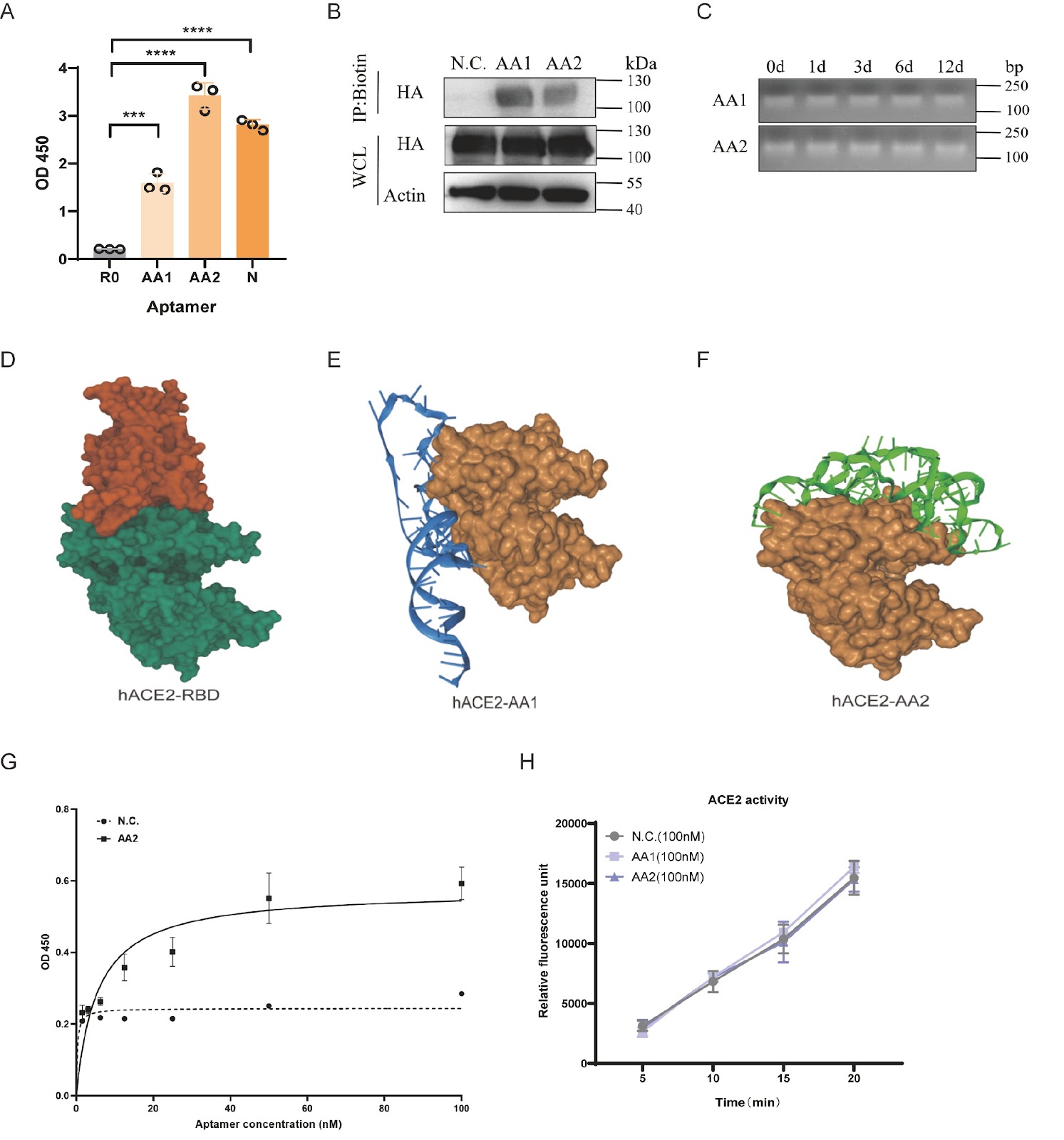
The relentless evolution of severe acute respiratory syndrome coronavirus 2 (SARS-CoV-2) and the emergence of immune-evasive variants underscore an urgent need for novel therapeutic strategies that are resilient to viral mutations. Targeting conserved host factors essential for viral entry represents a promising approach to overcome this challenge. Here, we report the development of a bifunctional therapeutic platform targeting the primary human receptor for SARS-CoV-2, angiotensin-converting enzyme 2 (ACE2). Using systematic evolution of ligands by exponential enrichment (SELEX), we isolate a high-affinity DNA aptamer, designated AA2, that binds to human ACE2 with a dissociation constant (Kd) of 5.41 ± 1.23 nM. Molecular docking and competitive binding assays confirm that AA2 sterically hinders the interaction between the viral spike receptor-binding domain (RBD) and ACE2. Consequently, AA2 demonstrates potent neutralization of SARS-CoV-2 S pseudovirus entry into host cells. To achieve a synergistic antiviral effect, we engineer an aptamer-siRNA chimera (AsiC) by conjugating AA2 to a short interfering RNA (siRNA) targeting the GFP coding region of the pseudovirus genome. This AsiC construct significantly represses viral replication compared to aptamer or siRNA treatment alone, validating a dual mechanism of action that combines receptor blockade with targeted gene silencing. This study establishes a robust proof-of-concept for an ACE2-targeted AsiC, representing a new class of dual-function antiviral therapeutics with the potential to effectively combat current and future ACE2-dependent coronaviruses.

Read Full Abstract10.3724/abbs.2026087
Immune checkpoint TIM-3 in tumor immunotherapyGraphical AbstractVerified
Acta Biochimica et Biophysica Sinica

Immune checkpoint TIM-3 in tumor immunotherapy

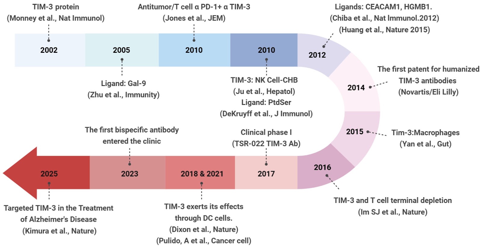
Over the past decade, immunotherapy has emerged as a pivotal therapeutic strategy in cancer treatment. Immune checkpoint inhibitors (ICIs), such as CTLA-4 and PD-1 monoclonal antibodies, have demonstrated remarkable clinical efficacy in different types of cancer. However, the overall success rate of immune checkpoint therapies remains low. Investigating alternative immune checkpoint molecules is imperative. T-cell immunoglobulin and mucin-containing molecule-3 (TIM-3), which is expressed in T cells, natural killer (NK) cells, macrophages, and dendritic cells, has gained recognition as a promising candidate for tumor immunotherapy. Targeting TIM-3 represents a promising approach for cancer immunotherapy, particularly through the rational design of novel combination therapies with other ICIs. In this review, we present a comprehensive summary of the research advancements concerning the role of TIM-3 in regulating immune responses in different cell types and explore theoretical frameworks for targeting TIM-3 to achieve more effective immunotherapeutic breakthroughs.

Read Full Abstract10.3724/abbs.2025235
Safari in the RNA world: a special issue focused on RNA biogenesis, functions, and technologiesGraphical AbstractVerified
Acta Biochimica et Biophysica Sinica

Safari in the RNA world: a special issue focused on RNA biogenesis, functions, and technologies

RNA is one of the most essential biopolymers in cells. According to the central dogma, messenger RNAs (mRNAs) transmit genetic information from DNA to proteins through a complex process, facilitated by key non-coding RNAs (ncRNAs) such as ribosomal RNAs (rRNAs) and transfer RNAs (tRNAs). In addition to these essential RNAs, a wide variety of ncRNAs have been discovered, each playing key roles in gene regulation and contributing to the complexity of the RNA landscape. All RNAs undergo intricate processing and modifications before maturation and transport to their respective cellular compartments, where they perform their functions. Regulation of these processes often results in the generation of multiple isoforms from a single gene, further diversifying the RNA landscape. Understanding the mechanisms of RNA biogenesis and the functional roles of RNAs in both physiological and pathological contexts is essential for unraveling how cells respond to developmental and environmental cues, with profound implications for biomedicine. This special issue features 12 expert reviews in RNA research, each offering a comprehensive summary of the latest advances in RNA biology from their respective perspectives.

Read Full Abstract10.3724/abbs.2024234
An updated overview of lipid-regulated immunobiology in macrophagesGraphical AbstractVerified
Acta Biochimica et Biophysica Sinica

An updated overview of lipid-regulated immunobiology in macrophages

Macrophages are well known for their widespread distribution, diverse roles, and involvement in multiple pathophysiological contexts, thereby constructing an immunological front line. Meanwhile, constant efforts over the past few decades have unveiled diverse reprogramming patterns of lipid metabolism as crucial, response- and context-specific drivers of macrophage functions and fate. Here, we take a bird’s-eye view of major fields across the research landscape of lipid-regulated macrophages; review the latest advances in understanding how alterations in several lipid subclasses, especially their fatty acyl composition and oxidative status, direct macrophage-mediated responses and pathology outcomes; and summarize representative insights that have deciphered the lipidome composition of macrophages or profiled specific lipid species under different scenarios. We hope that this review provides readers with a handy grip to learn and explore the field of lipid-regulated immunobiology, exemplified by but not limited to macrophages.

Read Full Abstract10.3724/abbs.2025239
T cell-intrinsic PRR signaling in immunity and pathologyGraphical AbstractVerified
Acta Biochimica et Biophysica Sinica

T cell-intrinsic PRR signaling in immunity and pathology

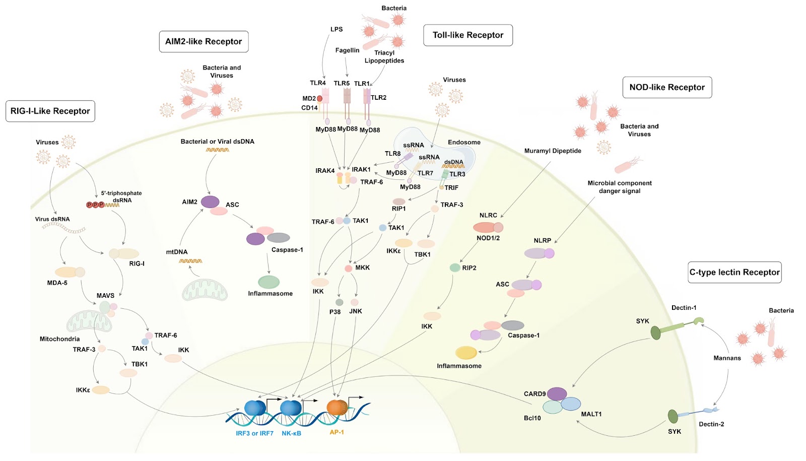
The immune system orchestrates a delicate balance between robust defense against pathogens and restraint to prevent tissue damage, with T cells serving as central mediators of adaptive immunity. The canonical pathway for T-cell activation hinges on the precise recognition of peptide antigens presented by major histocompatibility complex (MHC) molecules via the T-cell receptor (TCR), which is complemented by essential co-stimulatory signals. However, this model alone cannot fully explain the nuanced contextualization of immune responses, particularly how T cells integrate signals related to the nature of the threat. Pattern recognition receptors (PRRs), which are traditionally studied in innate immune cells, are recognized as critical regulators of T cell function, challenging the conventional dichotomy between innate and adaptive immunity. T cell-intrinsic PRR signaling integrates endogenous danger signals and microbes to modulate critical processes, including cytokine production, proliferation, and polarization, thereby shaping immune responses and disease outcomes in contexts ranging from viral infections to chronic inflammation and cancer. However, the molecular mechanisms underlying PRR-mediated T cell regulation and their contributions to immune homeostasis or pathology remain incompletely understood. This study investigates the role of T cell-intrinsic PRR signaling in shaping immune responses and its implications for disease. By elucidating key signaling pathways and their impact on T cell function, we aim to offer novel insights into the complex regulation of T cell-mediated immunity and uncover an underappreciated paradigm for immune-related disorders, providing new insights into the pathogenesis of inflammatory and neoplastic diseases.

Read Full Abstract10.3724/abbs.2025227
USP18-stabilized ELF3 drives glycolysis and malignant progression in lung adenocarcinomaGraphical AbstractVerified
Acta Biochimica et Biophysica Sinica2026

USP18-stabilized ELF3 drives glycolysis and malignant progression in lung adenocarcinoma

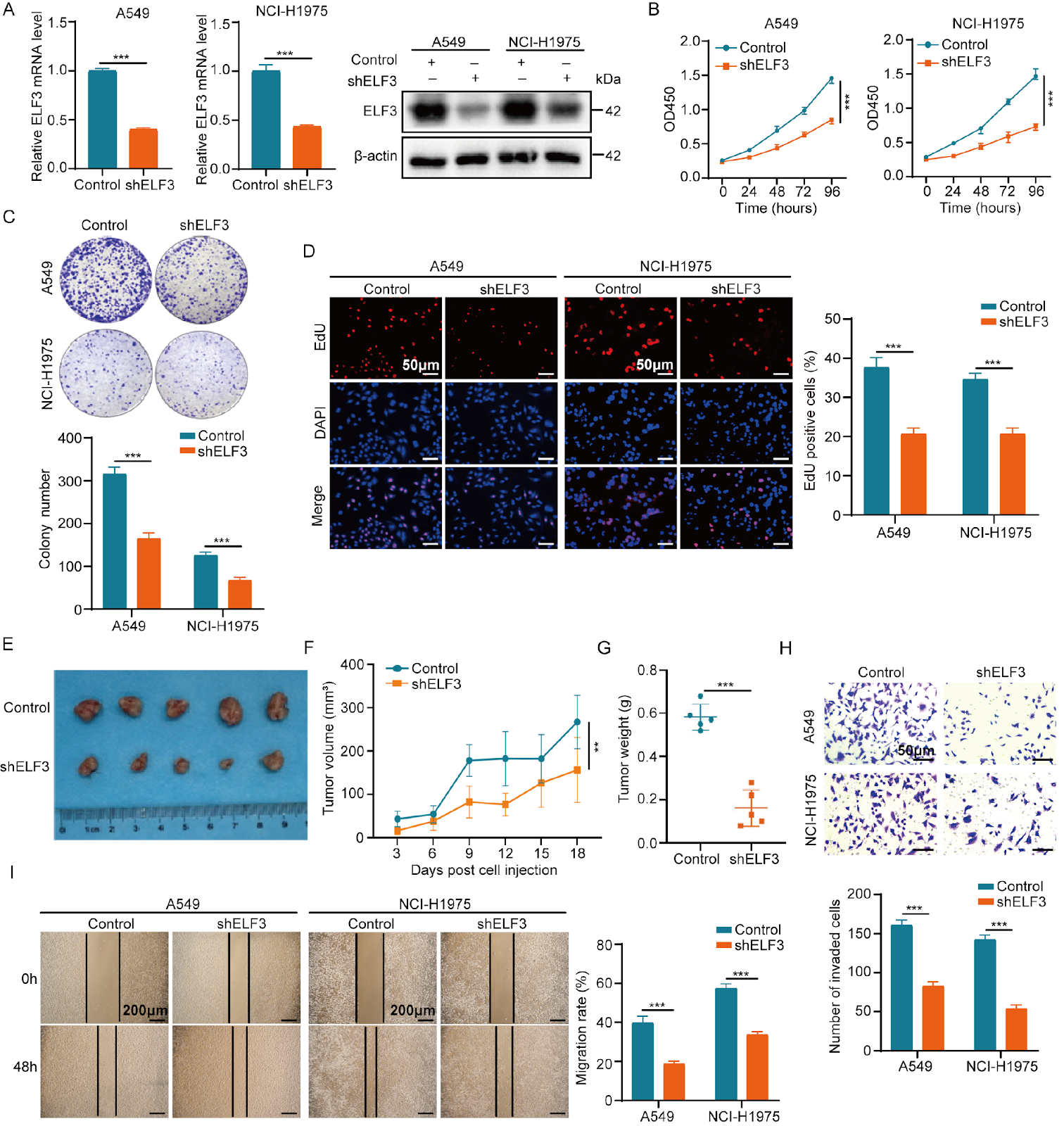
E74-like ETS transcription factor 3 (ELF3) has been implicated in various tumorigenesis and inflammatory diseases. However, its expression profile and role in lung adenocarcinoma (LUAD) remain poorly defined. In the present study, through comprehensive clinical and experimental analyses, we aim to clarify the association between ELF3 overexpression in LUAD tissues and poor prognosis. Functional assays reveal that ELF3 knockdown inhibits the proliferation, migration, and invasion of LUAD cells, while ELF3 overexpression enhances these functions. Pathway enrichment analysis indicates that ELF3 influences the metabolic processes of LUAD. Mechanistically, ELF3 exerts oncogenic effects by regulating the transcription of hexokinase 2 (HK2) and glucose transporter type 1 (GLUT1). High-throughput screening reveals that dacinostat, by targeting the active site of the ELF3 protein, attenuates the glycolytic, proliferative, and metastatic abilities of LUAD cells. Additionally, ubiquitin-specific peptidase 18 (USP18) strengthens the stability of the ELF3 protein and influences the malignant biological behavior of LUAD through ELF3. In conclusion, the USP18/ELF3/HK2 and USP18/ELF3/GLUT1 axes play critical roles in glucose metabolism, proliferation, and metastasis of LUAD cells. Dacinostat inhibits the malignant progression of LUAD by targeting ELF3, providing strong evidence for developing novel therapeutic strategies targeting ELF3.

Read Full Abstract10.3724/abbs.2026082
Circadian rhythm in immunotherapy and cellular therapy: impacts on the tumor microenvironmentGraphical AbstractVerified
Acta Biochimica et Biophysica Sinica

Circadian rhythm in immunotherapy and cellular therapy: impacts on the tumor microenvironment

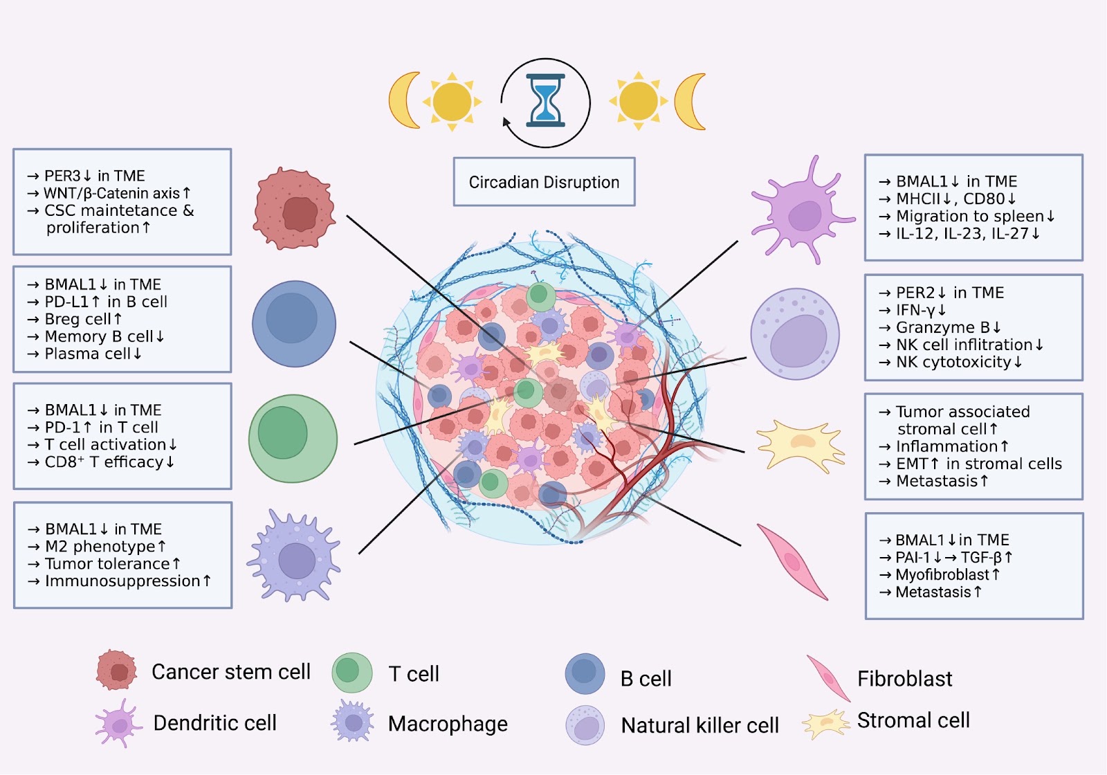
Immunotherapy, including cellular therapy, has emerged as a crucial pillar in cancer treatment, complementing established modalities such as surgery, chemotherapy and radiotherapy. The clinical observation that immunotherapy is effective in only a limited proportion of patients inspires mechanistic research on the complicated regulatory network within the tumor microenvironment (TME). Circadian regulation significantly affects immune cell behavior, including the activity of immune cells and cytokine production, and emerging evidence suggests the key role of circadian regulation in the TME, which subsequently affects the effectiveness of immunotherapy. Results from preclinical and clinical studies indicate that appropriate timing of adoptive cellular therapy and immune checkpoint blockade therapy improves their efficacy. Therefore, understanding the molecular mechanism of the circadian rhythm together with its role in immunotherapy is essential for optimizing cellular function, proliferation and persistence in the TME. Here, we review how circadian rhythms influence immunotherapy and the TME across different stages of tumor progression. Future clinical protocols may integrate concepts of circadian rhythm and immunotherapy to enhance treatment response.

Read Full Abstract10.3724/abbs.2025203
The context-dependent role of group 2 innate lymphoid cells in lung diseasesGraphical AbstractVerified
Acta Biochimica et Biophysica Sinica

The context-dependent role of group 2 innate lymphoid cells in lung diseases

Group 2 innate lymphoid cells (ILC2s), a subset of innate lymphoid cells (ILCs) lacking antigen-specific receptors and functionally mirroring T helper 2 (Th2) cells, are indispensable components of the innate immune system that lack antigen-specific receptors but phenotypically and functionally mirror T helper 2 (Th2) cells, particularly in their expression of the transcription factor GATA3 and the secretion of type 2 cytokines for mediating type 2 immune responses. ILC2s are tissue-resident cells in mucosal tissues, including the lung, where they play crucial roles in maintaining tissue homeostasis and regulating immune responses. ILC2s are poised to respond to environmental signals such as IL-25, IL-33, and TSLP, which activate and expand ILC2s. Their functions are highly context-dependent and influenced by interactions with other immune cells. In this review, we summarize recent findings on the roles of ILC2s in lung diseases, highlighting their typical characteristics and their responsiveness to environmental signals in the context of pulmonary pathology. We also discuss potential therapeutic strategies targeting ILC2s, which may offer new avenues for the treatment of inflammatory lung diseases. Understanding the mechanisms by which ILC2s contribute to lung disease progression will provide valuable insights for the development of novel diagnostic (e.g., ILC2 phenotypic markers) and therapeutic approaches (e.g., targeting ILC2 plasticity or alarmin-ILC2 signaling axes).

Read Full Abstract10.3724/abbs.2025243
Altered gut microbial dynamics and the antivascular remodeling effect of carnosine in hypobaric hypoxic pulmonary hypertension ratsGraphical AbstractVerified
Acta Biochimica et Biophysica Sinica

Altered gut microbial dynamics and the antivascular remodeling effect of carnosine in hypobaric hypoxic pulmonary hypertension rats

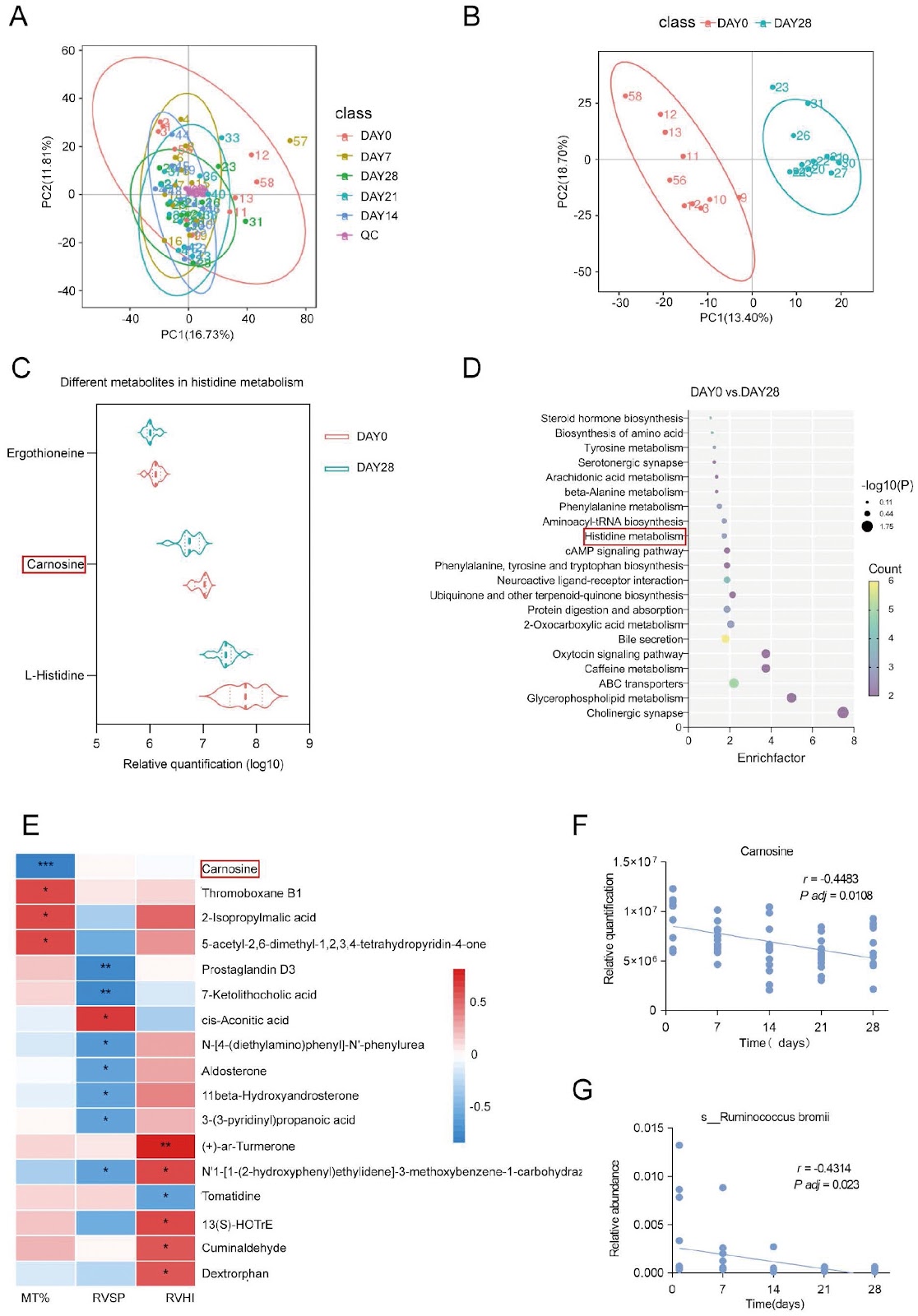
Exposure to chronic hypobaric hypoxia provokes marked alterations in the gut microbiota and its metabolome, yet the functional significance of histidine-derived metabolites in hypobaric hypoxic pulmonary hypertension (PH) remains underexplored. Here, we employ 16S rDNA, metagenomic, and untargeted metabolomic sequencing to characterize longitudinal shifts in the fecal microbiota and metabolites during hypobaric hypoxic PH development in Sprague-Dawley rats. Fecal carnosine levels and the abundance of its producer, Ruminococcus bromii, both decrease significantly over 28 days of hypobaric hypoxia (P < 0.05). Spearman correlation shows that carnosine is inversely correlated with the percentage of pulmonary arteriole media thickness (MT%; r = –0.8741, P < 0.001). Therapeutic supplementation with carnosine restores systemic and pulmonary antioxidant defenses and attenuates vascular remodeling without altering right ventricular pressures. In vitro, carnosine inhibits hypoxia-induced pulmonary artery smooth muscle cell (PASMC) proliferation and migration and suppresses nuclear factor erythroid 2-related factor 2 (Nrf2) accumulation. These findings reveal dynamic gut-lung crosstalk in hypobaric hypoxic PH and nominate carnosine as a metabolite-based intervention to mitigate hypoxia-driven pulmonary vascular remodeling.

Read Full Abstract10.3724/abbs.2025237
SPP1 facilitates sorafenib resistance in hepatocellular carcinoma by upregulating aerobic glycolysis in endothelial cellsGraphical AbstractVerified
Acta Biochimica et Biophysica Sinica

SPP1 facilitates sorafenib resistance in hepatocellular carcinoma by upregulating aerobic glycolysis in endothelial cells

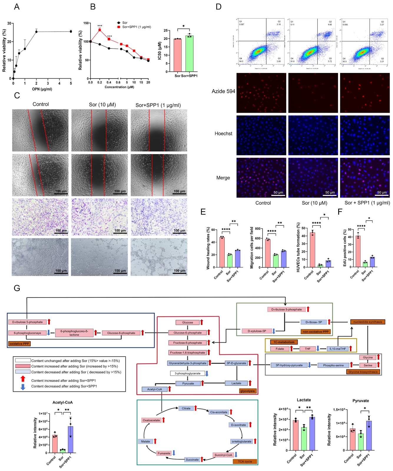
The occurrence of resistance to sorafenib, a first-line treatment for hepatocellular carcinoma (HCC), significantly limits its clinical efficacy. Therefore, investigating the potential mechanism of sorafenib resistance in HCC is highly important for developing HCC treatment strategies. In the present study, we identify that SPP1 (encoding osteopontin; OPN) is significantly elevated in sorafenib-resistant HCC. Furthermore, the upregulation of SPP1 is related to vascular invasion, advanced disease stage and poor prognosis in HCC patients. As the IC50 value of sorafenib increases in HepG2 cells, the SPP1 protein secreted by the cells is significantly upregulated, which subsequently facilitates the proliferation of human umbilical vein endothelial cells (HUVECs) and resistance to sorafenib. Further studies reveal that SPP1 induces resistance to sorafenib in HepG2 cells by upregulating glycolysis in HUVECs and further producing lactate. Mechanistically, SPP1 increases the expressions of the glucose transporter GLUT1 and the key glycolytic enzymes PFK1 and PKM2 in HUVECs, resulting in lactate accumulation, which in turn promotes the phosphorylation levels of BRAF and ERK as well as HIF-1α expression in HepG2 cells, leading to sorafenib resistance in HCC. Notably, SPP1 silencing can inhibit the proliferation and invasion of sorafenib-resistant HepG2 cells both in vitro and in vivo. Importantly, lactate derived from HUVECs plays a more dominant role in sorafenib resistance than does SPP1 in HepG2 cells. In summary, SPP1 enhances sorafenib resistance in HepG2 cells through promoting aerobic glycolysis in HUVECs, suggesting that the SPP1-aerobic glycolysis axis might be a prognostic biomarker as well as a potential therapeutic target for sorafenib-resistant HCC.

Read Full Abstract10.3724/abbs.2025245
Intestinal aging-related immune dysfunction: mechanisms and interventionsGraphical AbstractVerified
Acta Biochimica et Biophysica Sinica

Intestinal aging-related immune dysfunction: mechanisms and interventions

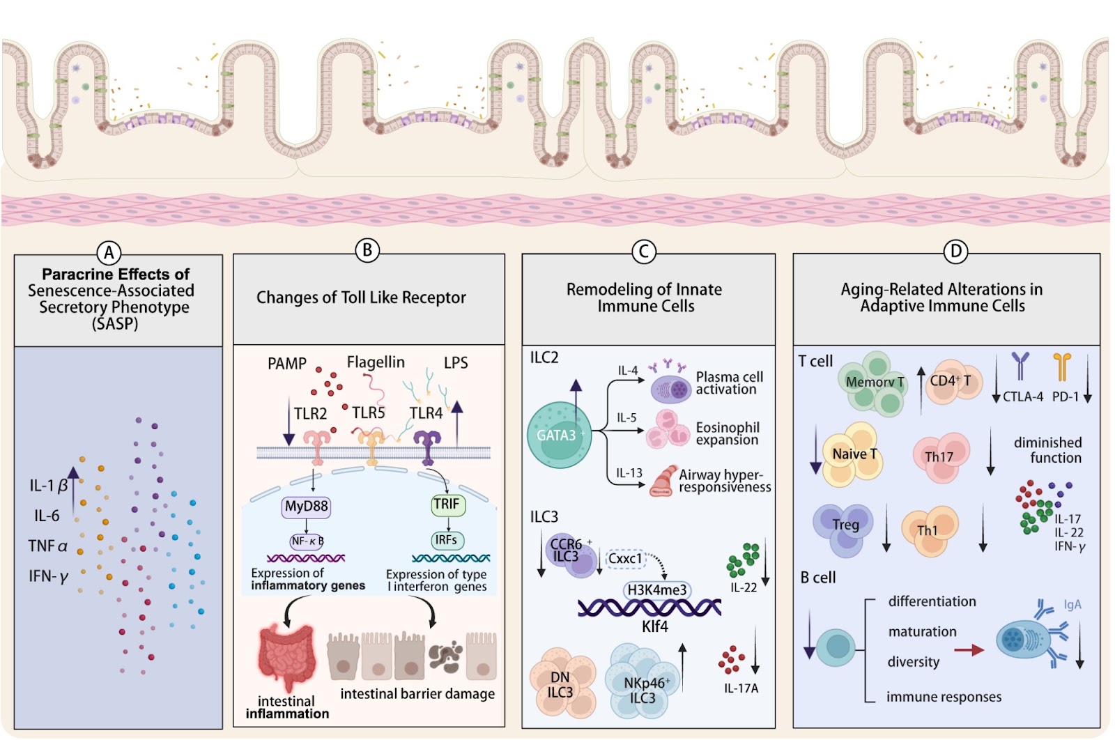
Intestinal immunosenescence, a hallmark of organismal aging, has emerged as a critical biological process impacting the health of elderly individuals. This review systematically examines the core mechanisms underlying intestinal immunosenescence, including immune cell dysfunction, imbalances in immune-microbiota interactions, and impaired barrier function. We analyze its associations with infectious diseases, chronic inflammation, and neurodegenerative disorders, summarizing recent advances in dietary interventions, microecological therapy, and other emerging strategies. By integrating cutting-edge technologies, we prospect the development of precision interventions aimed at delaying intestinal immunosenescence, thereby providing a theoretical basis for improving the healthspan of the aging population.

Read Full Abstract10.3724/abbs.2025157
Metabolic crosstalk between intestinal microbiota and dendritic cells: from homeostasis to inflammationGraphical AbstractVerified
Acta Biochimica et Biophysica Sinica

Metabolic crosstalk between intestinal microbiota and dendritic cells: from homeostasis to inflammation

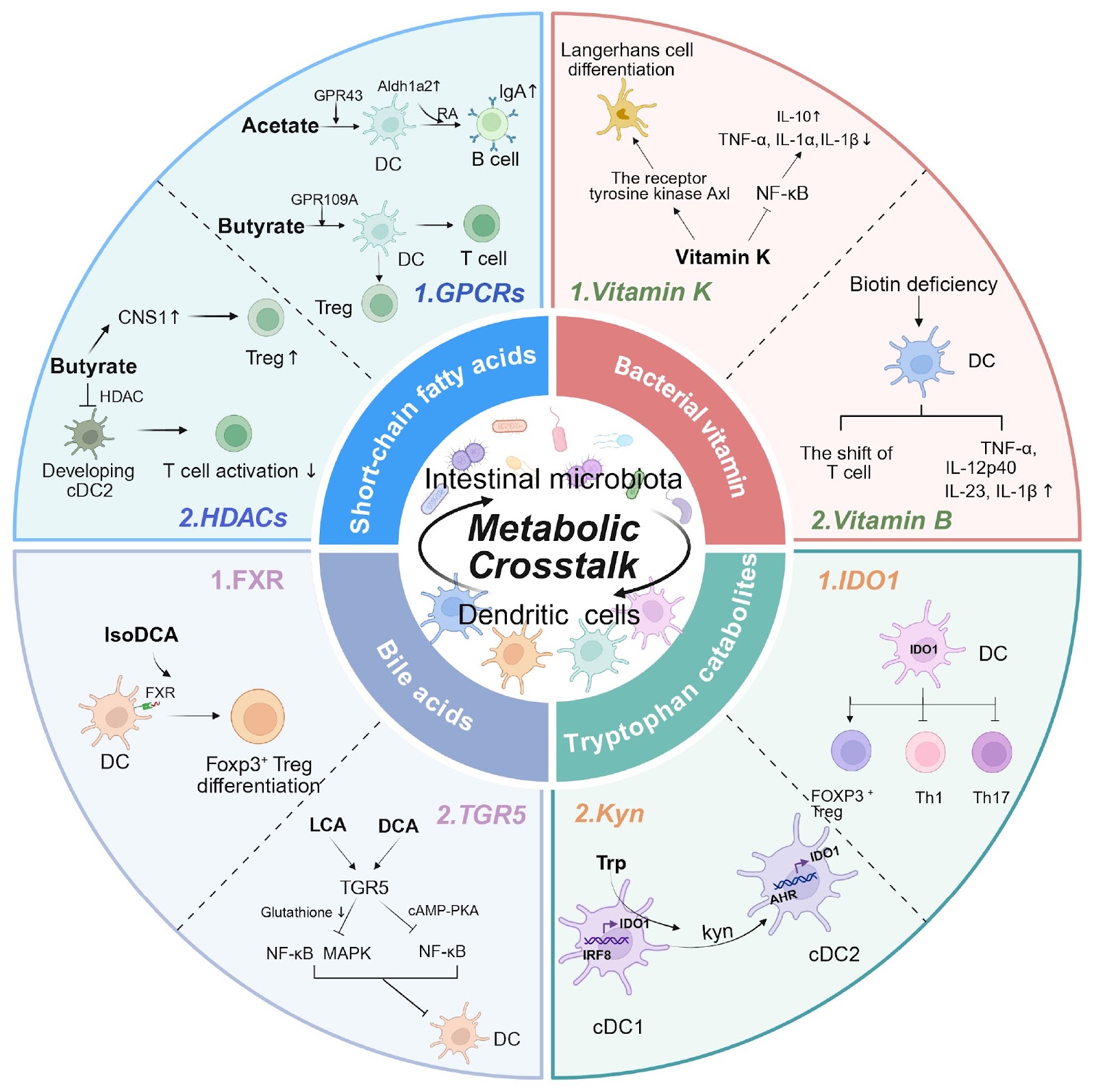
The intestinal microbiota plays critical roles in regulating immunity and inflammation through intricate interactions between microbial metabolites and diverse immune cells. Dendritic cells (DCs), the most potent professional antigen-presenting cells, are essential for sensing the complicated microbiota environment and subsequently initiating and regulating adaptive immune responses. While the commensal microbiota typically mediates DC-triggered immune tolerance and thus the maintenance of intestinal homeostasis, epithelial injury or pathogenic infection generally drives the proinflammatory function of DCs, contributing to harmful inflammation and intestinal disorders. Various microbiota metabolites (such as short-chain fatty acids, bile acids, and tryptophan derivatives) play critical roles in modulating the developmental and functional diversity of DCs through metabolic, epigenetic, or signaling reprogramming. In this review, we discuss the metabolic crosstalk between the intestinal microbiota and DCs and its pivotal function in orchestrating the balance between intestinal homeostasis and pathogenic inflammation. We also discuss future directions to better elucidate the microbiota-DC dialog in intestinal immunity and develop therapeutic approaches for manipulating the microbiota-DC axis against inflammatory disorders.

Read Full Abstract10.3724/abbs.2025231
Dysregulated immunometabolism in gut inflammationGraphical AbstractVerified
Acta Biochimica et Biophysica Sinica

Dysregulated immunometabolism in gut inflammation

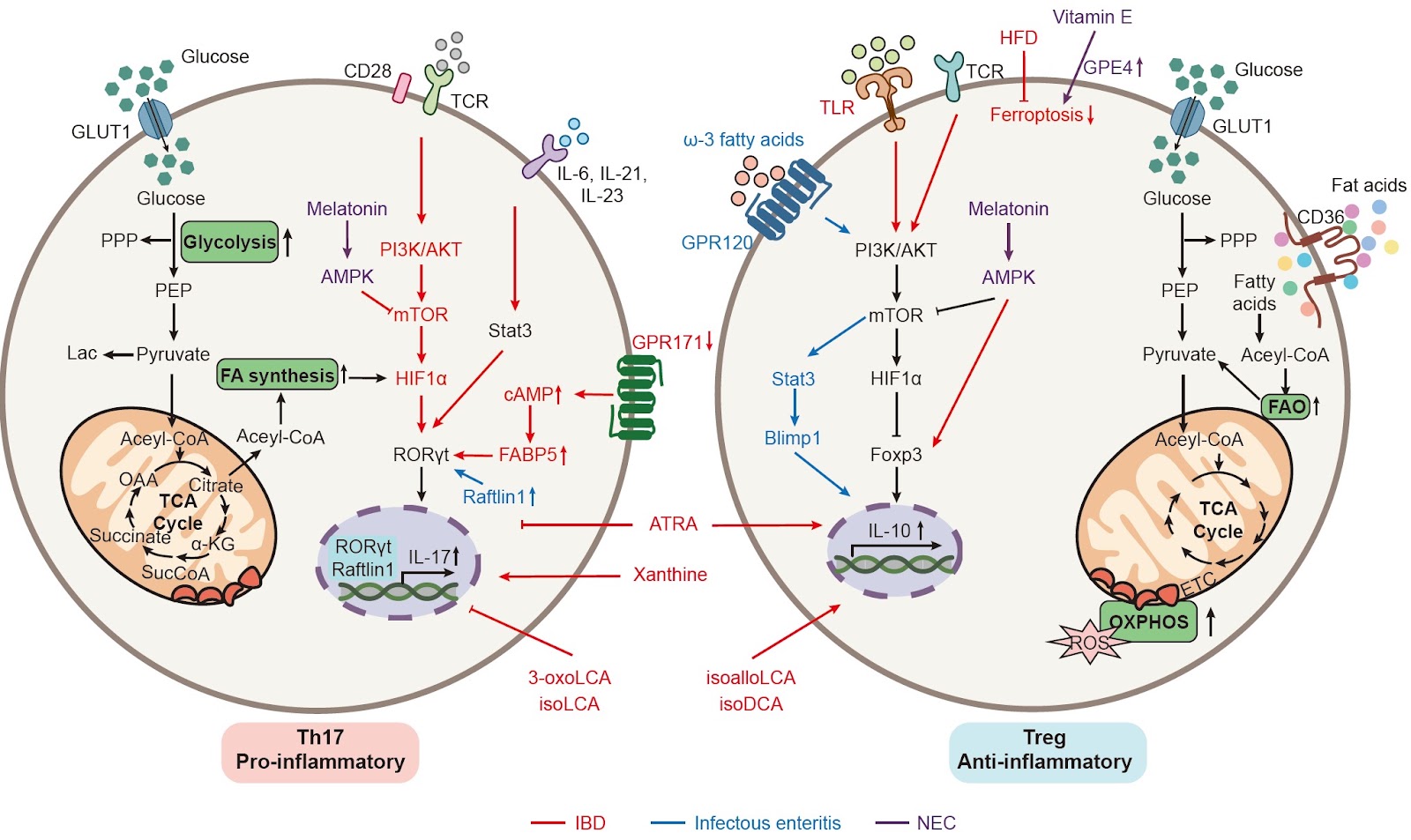
Gut inflammatory diseases, including inflammatory bowel disease (IBD), infectious enteritis, and other inflammatory conditions, are among the most common non-neoplastic intestinal disorders. Their pathogenesis is often driven by an imbalance between pro-inflammatory and anti-inflammatory signals, with immune cells playing pivotal roles in maintaining this equilibrium. Immune cells in the gut exhibit complex, multifaceted functions: they eliminate pathogens, promote tissue repair, and counteract tumors, but excessive immune activation can exacerbate tissue damage and disease progression. Notably, metabolic reprogramming in inflammatory contexts serves as a key regulator of immune cell function and phenotypic switching. This includes alterations in cellular energy metabolism (e.g., macrophage polarization via disrupted glycolysis or fatty acid oxidation) and the modulation of immune responses by microenvironmental metabolites (e.g., bile acid-mediated Th17/Treg balance). While alterations in immune cell function and composition within the inflammatory milieu are well-established, the significance of disease-associated metabolic reprogramming—specifically how metabolism regulates immune cell function—has garnered increasing attention. This review explores how cellular metabolic reprogramming, changes in the metabolic microenvironment, and gut dysbiosis collectively influence the differentiation, proliferation, and function of immune cells in various intestinal inflammatory diseases, as well as their impact on disease progression.

Read Full Abstract10.3724/abbs.2025192
Development of a colloidal gold immunochromatographic strip based on GAPDH for Pentatrichomonas hominis in dogsGraphical AbstractVerified
Acta Biochimica et Biophysica Sinica

Development of a colloidal gold immunochromatographic strip based on GAPDH for Pentatrichomonas hominis in dogs

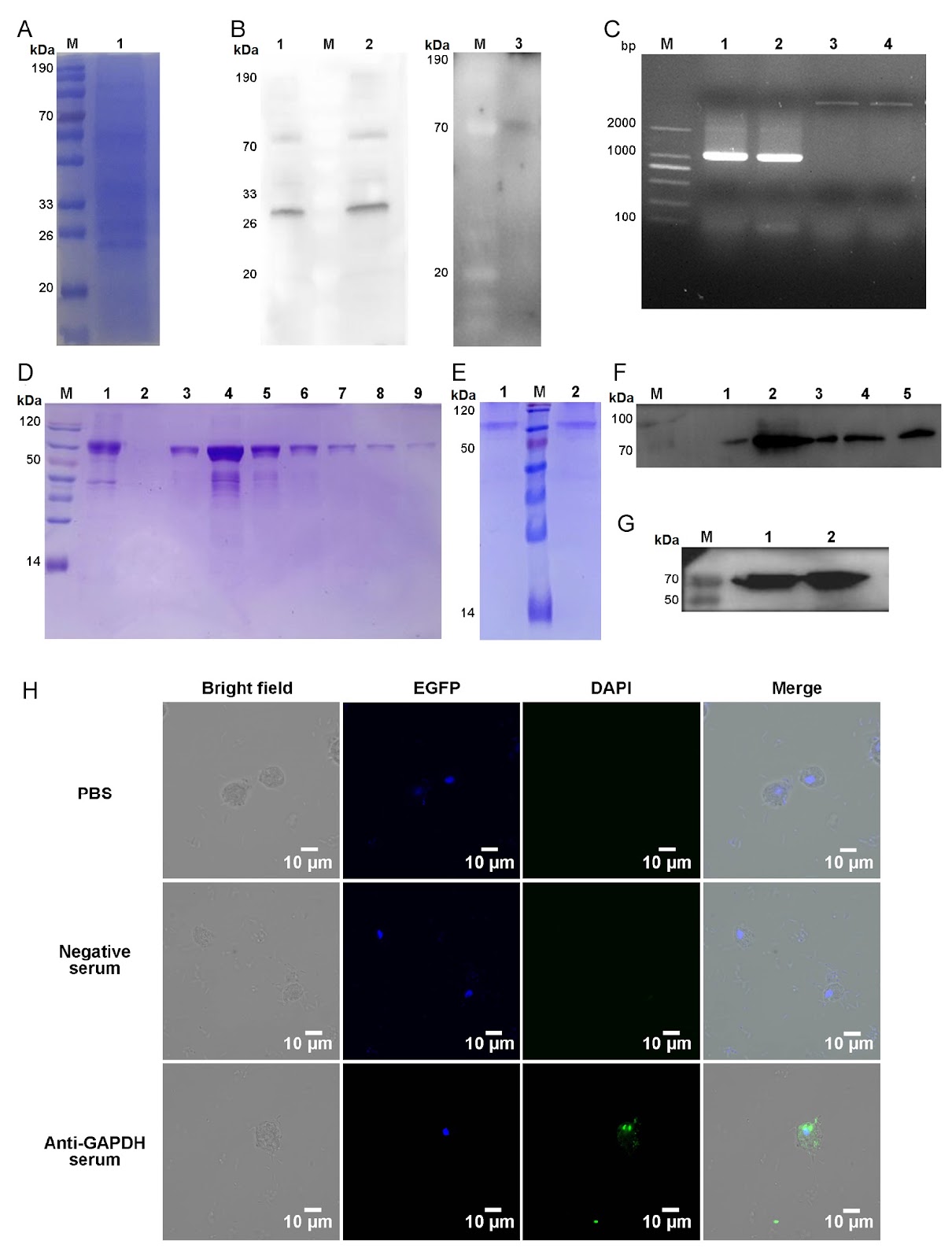
Pentatrichomonas hominis is a zoonotic protozoan belonging to the family Trichomonadidae that primarily inhabits the cecum and colon. Although traditionally regarded as an opportunistic pathogen, P. hominis is increasingly recognized for its pathogenic potential, including roles in animal diarrheal disease and the induction of intestinal epithelial damage and chronic inflammation in mice. A previous study further identified a significant correlation between P. hominis infection and colorectal cancer, underscoring its growing clinical and public health significance. This parasite infects a wide range of hosts, among which dogs, displaying infection rates as high as 47.4%, are regarded as a potential zoonotic reservoir because of their close contact with humans. Consequently, establishing reliable detection methods for P. hominis in dogs is essential for veterinary practice and public health surveillance. Current methods for detecting P. hominis infections in dogs mainly include direct smear microscopy and polymerase chain reaction (PCR)-based techniques. Although direct smear microscopy is straightforward, it frequently exhibits low sensitivity. In comparison, PCR demonstrates high sensitivity and specificity, yet it relies on specialized equipment, trained operators, and extended processing time. Recently, reported nucleic acid detection approaches, such as recombinase polymerase amplification coupled with lateral flow dipstick (RPA-LFD) and RPA-CRISPR/Cas12a assays, have enhanced the efficiency and accessibility of molecular detection for P. hominis. However, these methods still necessitate nucleic acid extraction, controlled temperature conditions, and operational complexity. In addition, some emerging detection technologies, such as microfluidic chips and nanozyme-based detection systems, offer advantages of high throughput and sensitivity but have not yet been widely applied in the field of detection of parasitic infection. Although immunoassays, including enzyme-linked immunosorbent assay (ELISA) and colloidal gold immunochromatographic strips, have been successfully used for the detection of infections of some intestinal protozoans, such as Giardia, there have been no reports on their application for detecting P. hominis infections, primarily due to the lack of specific detection antigens. To address the detection need, liquid chromatography-tandem mass spectrometry (LC-MS/MS) was used as the primary discovery tool to identify specific detected antigens. Using this targeted proteomics approach, we precisely identified immunoreactive proteins and selected glyceraldehyde 3 phosphate dehydrogenase (GAPDH), an immunogenic and species-specific antigen in related parasites, as the candidate antigen. Based on this identified antigen, we subsequently developed and evaluated the detection performance in both indirect ELISA and colloidal gold immunochromatographic strips using recombinant GAPDH. This study aimed to identify novel antigens for immunodetection of P. hominis and to establish a practical, on-site method for detecting dog infections, thereby facilitating further epidemiological and clinical research.

Read Full Abstract10.3724/abbs.2026043
Ribosomal protein L8 promotes melanoma progression by regulating the cell cycle and metastasisGraphical AbstractVerified
Acta Biochimica et Biophysica Sinica2026

Ribosomal protein L8 promotes melanoma progression by regulating the cell cycle and metastasis

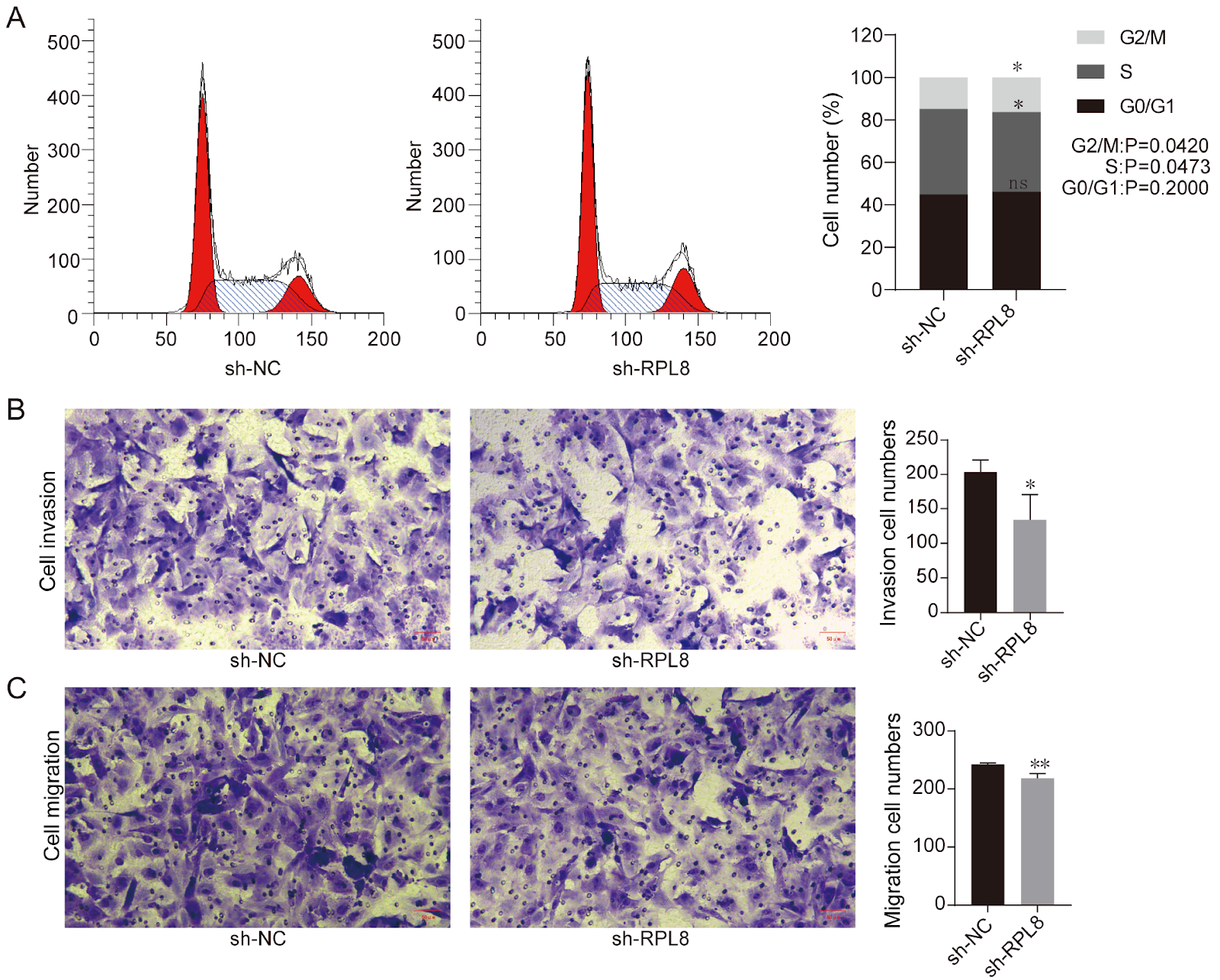
Melanoma is an aggressive skin cancer with poor prognosis in advanced stages due to its metastatic potential. Although treatment modalities such as surgical resection, targeted therapy, immunotherapy, radiotherapy, and chemotherapy have conferred long-term survival benefits to certain patients, not all individuals respond favorably, underscoring the urgent need to develop predictive biomarkers. Ribosomal proteins have been implicated in tumor progression through extraribosomal functions. Ribosomal protein L8 (RPL8) is a unique ribosomal protein that warrants further exploration. Here, we investigated the role of RPL8 in melanoma progression. Using clinical data from the GDC and GEO databases, we found that high RPL8 expression is associated with poor prognosis in melanoma patients. Immunohistochemical analysis confirmed higher RPL8 expression in melanoma tissues compared to normal tissues. To elucidate the functional impact, we established an A875 melanoma cell line with stable RPL8 knockdown (shRPL8) using lentiviral vectors. RT-qPCR and western blot analysis confirmed efficient knockdown. CCK-8 assays showed that RPL8 knockdown significantly inhibited cell proliferation. Flow cytometry analysis revealed that RPL8 knockdown led to an increase in the proportion of cells in the G2/M phase and a decrease in the S phase, indicating cell cycle arrest. Furthermore, transwell assays demonstrated that RPL8 knockdown reduced cell invasion and migration. These findings suggest that RPL8 promotes melanoma progression by regulating the cell cycle and metastasis, and may serve as a potential therapeutic target and prognostic biomarker for melanoma.

Read Full Abstract10.3724/abbs.2026099
Phillyrin protects against myocardial ischemia/reperfusion injury by promoting KNL1 K605 acetylation to inhibit the p53/p21 pathwayGraphical AbstractVerified
Acta Biochimica et Biophysica Sinica

Phillyrin protects against myocardial ischemia/reperfusion injury by promoting KNL1 K605 acetylation to inhibit the p53/p21 pathway

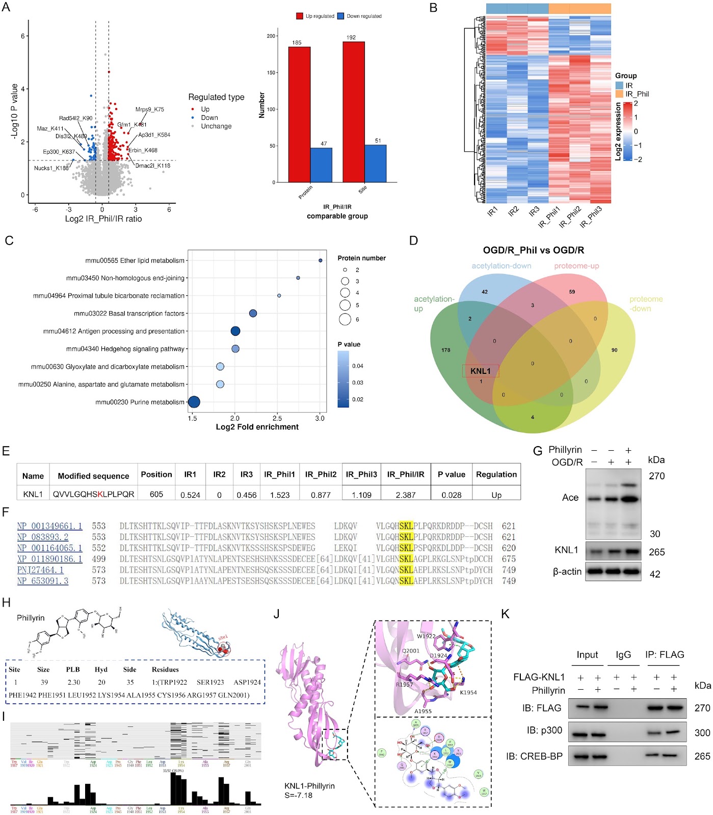
Reperfusion therapy is critical for acute myocardial infarction but is often accompanied by myocardial ischemia/reperfusion injury (MIRI). Phillyrin, a natural lignan from Forsythia suspensa, exerts anti-inflammatory and antioxidant effects; however, its role and mechanism in MIRI remain unclear. In this study, HL-1 cardiomyocytes are subjected to oxygen-glucose deprivation/reperfusion (OGD/R). Cell viability, apoptosis, oxidative stress, and inflammation are measured after phillyrin treatment. Multiomics (mRNA-seq, proteomics, and acetylproteomics) is used to identify key targets and pathways. Molecular docking, co-immunoprecipitation, site-directed mutagenesis, and western blot analysis are used to validate posttranslational regulation. A mouse MIRI model is established to confirm the in vivo cardioprotective effects of phillyrin. Phillyrin preserves cell viability and reduces apoptosis, oxidative stress, and inflammation in OGD/R-injured HL-1 cells. Multiomics integration reveals that phillyrin acts primarily through posttranslational regulation and highlights kinetochore scaffold 1 (KNL1) as the only protein that is both upregulated and hyperacetylated at lysine 605 (K605). Mechanistically, phillyrin may bind to the KNL1 C-terminus and enhance the interaction between KNL1 and acetyltransferase p300/CBP. KNL1 K605R mutation and Knl1 knockdown reduce KNL1 protein expression and reverse the inhibitory effects of phillyrin on p53 pathway-mediated apoptosis, oxidative stress, and inflammation. In mouse MIRI models, phillyrin reduces infarct size, myocardial damage, and cardiomyocyte apoptosis; these effects are abolished by knockdown of Knl1. Therefore, phillyrin promotes KNL1 acetylation at K605 to increase KNL1 protein expression, thereby inhibiting p53 signaling and alleviating apoptosis, oxidative stress, and inflammation in MIRI. This study identifies KNL1 acetylation at K605 as a novel posttranslational modification target for cardioprotection.

Read Full Abstract10.3724/abbs.2026104
Fabrication of fusogenic and magnet-responsive cells for transplantation of an intact mitochondrial networkGraphical AbstractVerified
Acta Biochimica et Biophysica Sinica2026

Fabrication of fusogenic and magnet-responsive cells for transplantation of an intact mitochondrial network

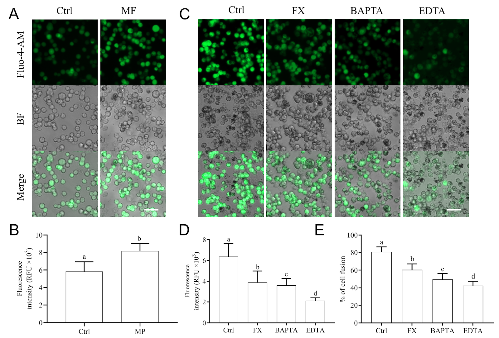
Mitochondrial transplantation is a promising treatment for many diseases associated with mitochondrial defects or aging; however, a reliable method for mitochondrial transfer remains urgently needed. In this study, we assemble fusogenic and magnet-responsive cells (FMRCs), which are enucleated stem cells loaded with Fe3O4 nanoparticles and further incorporated fusogenic vesicular stomatitis virus glycoprotein G (VSV-G). Mitochondrial transplantation from FMRCs via fusion in the presence of a magnetic force restores normal mitotic activity, mitochondrial membrane potential, ROS levels and ATP production in cells subjected to partial mtDNA depletion or in cybrids harboring mtDNA with a 4977-bp deletion. SNP tracing and qPCR analysis of the mitochondrial and nuclear genomes unequivocally demonstrate that exogenous mitochondria are able to reside stably and predominately. Mitochondrial transplantation stimulate autophagy and thus the clearance of defective endogenous counterparts, resulting in lower mtDNA heteroplasmy. These results suggest that FMRCs are excellent vehicles for mitochondrial transplantation and could be used for the treatment of aging and mitochondria-associated diseases.

Read Full Abstract10.3724/abbs.2026031
Integrating genetically encoded fluorescent sensors to elucidate the spatiotemporal choreography of necrosis by sodium overloadGraphical AbstractVerified
Acta Biochimica et Biophysica Sinica

Integrating genetically encoded fluorescent sensors to elucidate the spatiotemporal choreography of necrosis by sodium overload

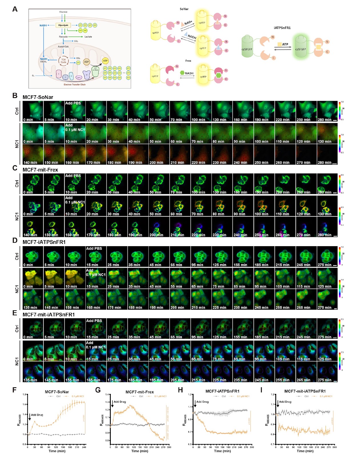
Necrosis by sodium overload (NECSO) is a distinct cell death modality induced by the chemical regulator necrocide 1 (NC1), which targets the transient receptor potential melastatin 4 (TRPM4) channel to drive excessive sodium influx and potassium efflux. This unique necrosis is characterized by the exchange of monovalent ions, a restrained ATP supply, redox disorder and a final membrane rupture as main features. Real-time monitoring of metabolic and redox causality during the whole process in living cells has been technically challenging. To bridge this gap, we integrate an advanced toolset of genetically encoded fluorescent sensors to monitor monovalent ions, energy metabolites, and redox equivalents with high spatiotemporal resolution. By directing these sensors to specific subcellular compartments, we successfully capture the real-time choreography of potassium loss specifically induced by NC1 via TRPM4. Furthermore, we establish a precise detection paradigm for evaluating energy currency by integrating sensors for NADH and ATP that are capable of subcellular imaging. We observe general and rapid NADH accumulation along with an ATP shortage in the mitochondria and cytosol. A concomitant reduction in mitochondrial oxidative stress is observed. This study not only elucidates the metabolic progression of a peculiar type of necrosis but also establishes a robust methodological framework for applying genetically encoded sensors to broader physiological and toxicological research.

Read Full Abstract10.3724/abbs.2026102
PD-1 blockade elicits a systemic immune response but not in the tumor of TNBC miceGraphical AbstractVerified
Acta Biochimica et Biophysica Sinica2026

PD-1 blockade elicits a systemic immune response but not in the tumor of TNBC mice

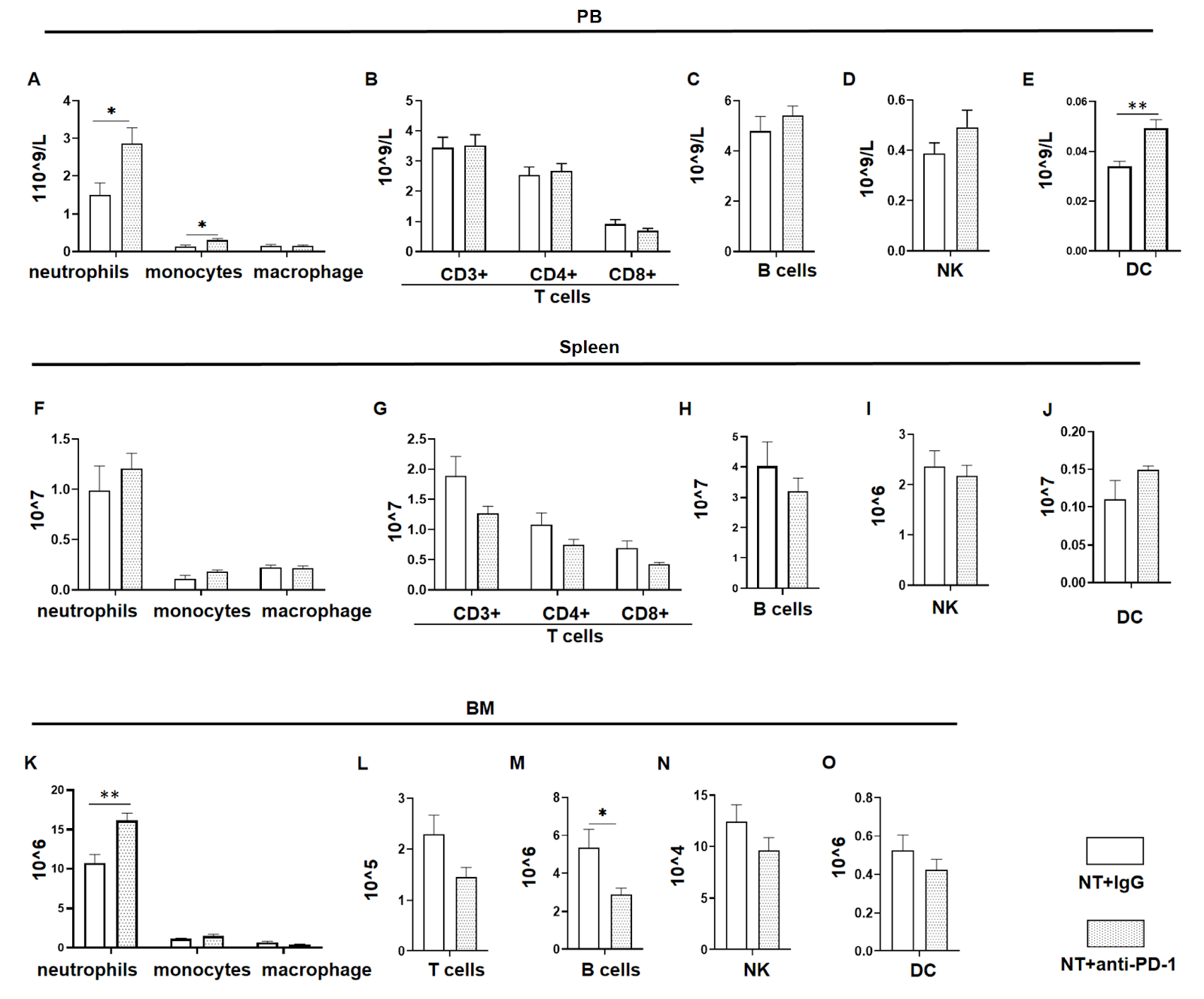
Triple-negative breast cancer (TNBC) is an aggressive subtype of breast cancer with limited treatment options due to the absence of hormone receptors and HER2 amplification. Immune checkpoint blockade, particularly targeting PD-1/PD-L1, has emerged as a promising therapeutic strategy. However, the response rate of TNBC patients to this monotherapy remains low. This study explores the systemic effect of PD-1 blockade on the immune and hematopoietic systems in 4T1 TNBC mice and demonstrates its limited efficacy in reducing the tumor burden and changing the number of tumor-infiltrating immune cells. However, PD-1 blockade increases systemic immune activity, as demonstrated by increased T cells and DCs in the peripheral blood, which may be associated with inflammatory side effects of this treatment. In addition, PD-1 blockade does not rescue the hematopoietic damage caused by TNBC, highlighting a limitation in long-term response. Furthermore, PD-1 blockade in tumor-free mice leads to an increase in hematopoietic stem/progenitor cells, suggesting that PD-1 blockade may yield better benefits post-tumor resection.

Read Full Abstract10.3724/abbs.2025240
FGF10 is essential for postnatal meibomian gland development in miceGraphical AbstractVerified
Acta Biochimica et Biophysica Sinica2026

FGF10 is essential for postnatal meibomian gland development in mice

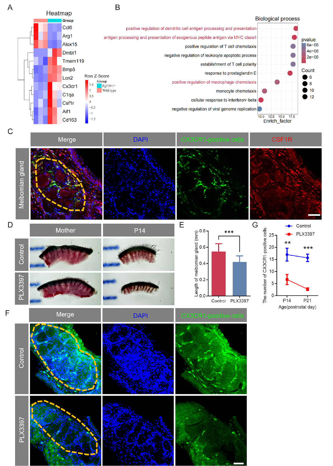
Fibroblast growth factor 10 (FGF10) plays a critical role in ocular surface homeostasis, yet its function in early meibomian gland (MG) development remains largely unknown. Here, we generated an Fgf10 mutant mouse model with deletion of exon 2, leading to loss of function. Adult Fgf10+/− mice exhibited lacrimal gland agenesis, smaller Harderian glands, increased corneal fluorescein staining, and reduced tear volume. Histological analysis revealed multilayered hyperplastic epithelium in Harderian glands and MG atrophy. Time-series Oil Red O staining showed shorter, thinner, and disordered MGs in Fgf10+/− mice at P14 and P21, with unrecoverable defects at P135. RNA sequencing of MGs at P14 and P21 revealed significant dysregulation of macrophage-related genes and immune-related pathways, including antigen processing and presentation and macrophage chemotaxis. Using Cx3cr1GFP/+ reporter mice, we observed a significant reduction in CX3CR1-positive cells in the inter-acinar stroma of Fgf10+/− MGs. Pharmacological ablation of CSF1R-expressing cells with PLX3397 in wild-type mice recapitulated the MG developmental defects, confirming that FGF10 acts through immune cells to regulate MG development. Collectively, our findings establish that FGF10 haploinsufficiency leads to severe glandular malformations, impaired tear production, and compromised corneal integrity, highlighting the essential role of FGF10 in postnatal MG development and immune cell regulation.

Read Full Abstract10.3724/abbs.2026066