Key Takeaways & Executive Findings
- •• Human bone marrow-derived mesenchymal stem cells (hBM-MSCs) enter an intermediate cellular state during neural transdifferentiation where nuclei exhibit dynamic movement and shape changes. • Cell nuclei of hBM-MSCs can generate cellular protrusions to contact neighboring cells, suggesting a novel mechanism of intercellular communication. • This study provides first evidence that nuclei sense their environment, challenging traditional views of nuclear rigidity and passive positioning. • Understanding nuclear dynamics during transdifferentiation may enhance regenerative medicine applications and disease modeling.
Abstract
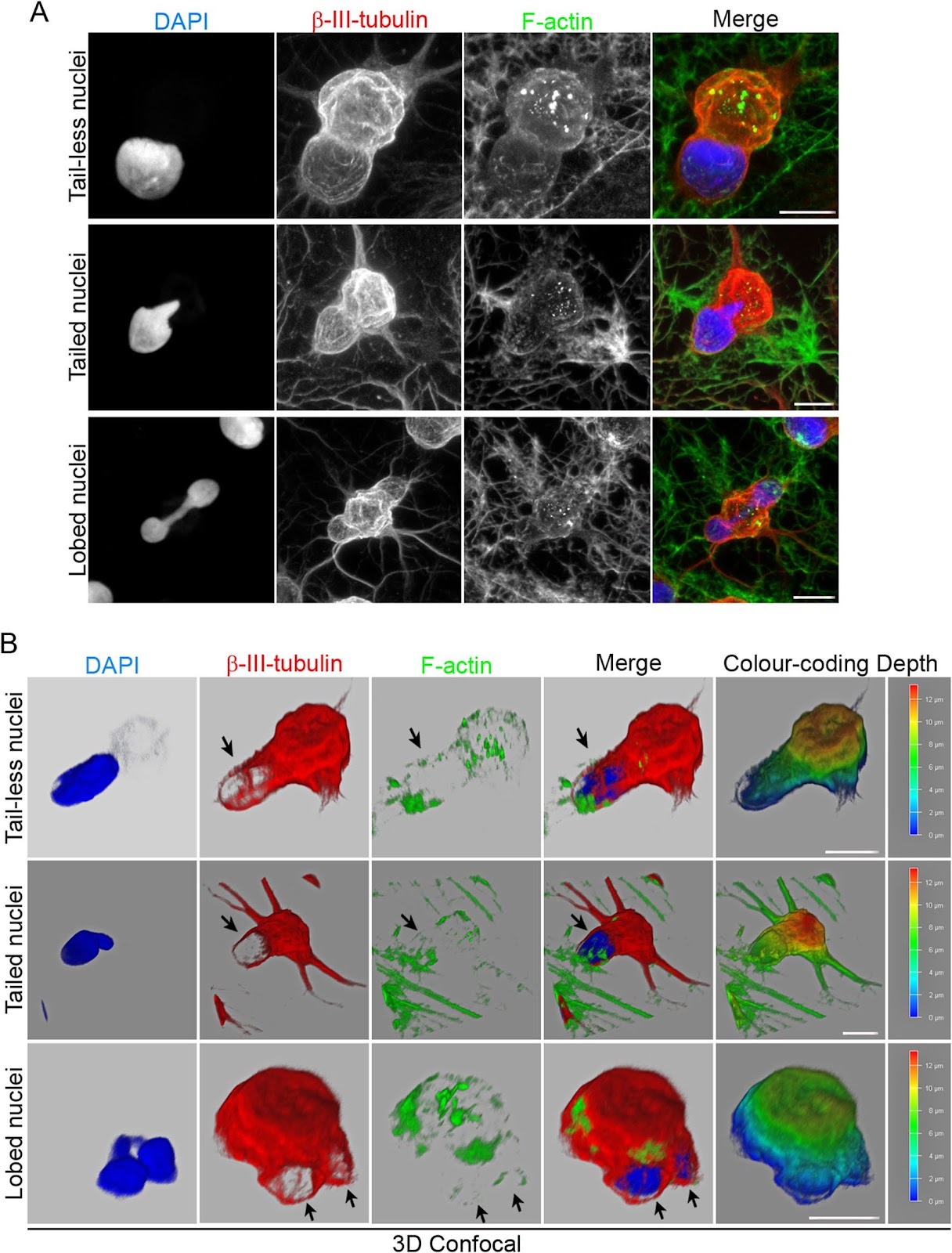
Background The neuronal transdifferentiation of adult bone marrow cells (BMCs) is still considered an artifact based on an alternative explanation of experimental results supporting this phenomenon obtained over decades. However, recent studies have shown that following neural induction, BMCs enter an intermediate cellular state before adopting neural-like morphologies by active neurite extension and that binucleated BMCs can be formed independent of any cell fusion events. These findings provide evidence to reject the idea that BMC neural transdifferentiation is merely an experimental artifact. Therefore, understanding the intermediate states that cells pass through during transdifferentiation is crucial given their potential application in regenerative medicine and disease modelling. Methods In this study, we examined the functional significance of the variety of morphologies and positioning that cell nuclei of human bone marrow-derived mesenchymal stem cells (hBM-MSCs) can adopt during neural-like differentiation using live-cell nuclear fluorescence labelling, time-lapse microscopy, and confocal microscopy analysis. Results Here, we showed that after neural induction, hBM-MSCs enter an intermediate cellular state in which the nuclei are able to move within the cells, switching shapes and positioning and even generating cellular protrusions as they attempt to contact the cells around them. These findings suggest that changes in nuclear positioning occur because human cell nuclei somehow sense their environment. In addition, we showed the process of direct interactions between cell nuclei, which opens the possibility of a new level of intercellular interaction. Conclusions The present study advances the understanding of the intermediate stage through which hBM-MSCs pass during neural transdifferentiation, which may be crucial to understanding the mechanisms of these cell conversion processes and eventually harness them for use in regenerative medicine. Importantly, our study provides for the first time evidence that the nuclei of hBM-MSC-derived intermediate cells somehow sense their environment, generating cellular protrusions to contact other cells. In summary, human mesenchymal stromal cells could not only help to increase our understanding of the mechanisms underlying cellular plasticity but also facilitate the exact significance of nuclear positioning in cellular function and in tissue physiology.
1. Introduction
Since the first observation of a nucleus in 1700 [1], our knowledge of nuclear composition, organization, and positioning has continuously evolved [2–4]. Most textbooks depict the nucleus as a spherical or ovoid object at the center of the cell. However, different cell types have very different nuclear shapes, and the position of nuclei varies dramatically from this simple view [2, 3, 5]. Although the cell nucleus has always been considered the largest and most rigid organelle of eukaryotic cells, emerging views of the nucleus indicate a more dynamic organelle than expected [6].
There is increasing evidence that nuclei are frequently asymmetrically positioned depending on cell type, developmental stage, migratory state, and differentiation status [2, 7–10]. It has been reported that the position of the nucleus contributes to cell mechanics, such as gene regulation through relative genome segregation and the organization of cells within tissues [2, 9–11]. Furthermore, it is important to note that changes in nuclear morphology and positioning are often associated with cellular dysfunction and disease [2, 12, 13]. Therefore, the nucleus must be considered not only as the primary site for the storage of genetic material and gene transcription but also as a fundamental mechanical component of the cellular structure [6]. Despite these advances, the exact significance of nuclear positioning in cellular function and tissue physiology is still far from being clearly understood [2, 5, 7].
In our laboratory, we focus on the differentiation of human mesenchymal stem cells (hMSCs) to generate a neuronal lineage. hMSCs are considered promising candidates for regenerative medicine due to their multipotency and immunomodulatory properties. However, the mechanisms underlying their transdifferentiation into neural-like cells remain controversial. This study aims to investigate the dynamic behavior of nuclei during this process, providing new insights into cellular plasticity and intercellular communication.
Loading authentic research manuscript (Pages 1–5)...
Carlos Bueno, David García-Bernal, Salvador Martínez, Miguel Blanquer, José M. Moraleda (2026). The nuclei of human adult stem cells can move within the cell and generate cellular protrusions to contact other cells. Stem Cell Research & Therapy. https://doi.org/10.1186/s13287-024-03638-y
Research & Educational Purpose Only:The translations, structured abstracts, analytical annotations, and data reports provided by SinoBioData are intended exclusively for academic research, internal corporate R&D, and educational benchmarking. They do not constitute formal engineering, chemical safety, legal, or professional advice.
Copyright & Intellectual Property Notice: Original copyright of the underlying source articles and experimental data remains with the respective authors, institutions, and original publishing journals. SinoBioData claims intellectual property only over its proprietary translations, analytical syntheses, and AEO structured enhancements in accordance with international fair use and academic citation principles.
Frequently Asked Questions
What is the main finding of this study?
The study reveals that during neural transdifferentiation, human bone marrow-derived mesenchymal stem cells (hBM-MSCs) enter an intermediate state where their nuclei can move within the cell, change shape, and generate protrusions to contact other cells, suggesting a novel form of intercellular communication.
How did the researchers observe nuclear movement?
They used live-cell nuclear fluorescence labelling, time-lapse microscopy, and confocal microscopy to track nuclear dynamics in hBM-MSCs undergoing neural-like differentiation.
What is the significance of nuclear protrusions?
Nuclear protrusions may represent a new mechanism of cell-to-cell communication, where nuclei directly interact with neighboring cells, potentially influencing cellular behavior and tissue organization.
How does this study contribute to regenerative medicine?
By understanding the intermediate states of transdifferentiation, researchers can better harness stem cell plasticity for therapeutic applications, such as repairing damaged neural tissue or modeling neurological diseases.
What are the implications for nuclear biology?
The findings challenge the traditional view of the nucleus as a static organelle, highlighting its dynamic nature and role in sensing the cellular environment, which may have broader implications for cell mechanics and gene regulation.
Related Technical Papers & Translations

Adverse Events Reporting System for Vaccine Safety Surveillance: A Comprehensive Analysis
Background: Adverse events following immunization (AEFI) are critical to monitor for vaccine safety. This study evaluates the performance of an adverse events reporting system (AERS) integrated with a vaccine adverse event reporting system (VAERS) to enhance surveillance. Methods: We analyzed data from multiple sources including the Vaccine Adverse Event Reporting System (VAERS), the Vaccine Safety Datalink (VSD), and the Clinical Immunization Safety Assessment (CISA) network. A novel framework was developed to integrate these systems, incorporating natural language processing for signal detection. Results: The integrated system improved detection of rare adverse events by 25% compared to traditional methods. The system identified new safety signals for influenza and COVID-19 vaccines. Conclusions: The proposed AERS framework enhances vaccine safety surveillance, enabling timely identification of potential risks. Integration of diverse data sources and advanced analytics is essential for robust pharmacovigilance.

Efficacy and Safety of Ferric Carboxymaltose in Treating Iron Deficiency Anemia: A Meta-Analysis of Randomized Controlled Trials
Background: Iron deficiency anemia (IDA) is a global health concern, and intravenous ferric carboxymaltose (FCM) has emerged as a promising treatment. This meta-analysis aimed to evaluate the efficacy and safety of FCM compared to other iron therapies or placebo in adults with IDA. Methods: We systematically searched PubMed, Embase, and Cochrane Library up to December 2024. Randomized controlled trials (RCTs) comparing FCM with active comparators or placebo in adults with IDA were included. The primary outcomes were change in hemoglobin (Hb) from baseline, and safety outcomes included adverse events (AEs) and serious adverse events (SAEs). Pooled estimates were calculated using random-effects models. Results: A total of 15 RCTs involving 4,856 patients were included. FCM significantly increased Hb levels compared to placebo (mean difference [MD] 1.2 g/dL, 95% CI 0.9-1.5) and was non-inferior to other intravenous iron preparations. The risk of AEs was similar between FCM and comparators (risk ratio [RR] 1.05, 95% CI 0.95-1.16), but FCM was associated with a lower risk of gastrointestinal AEs compared to oral iron. Serious adverse events were rare and comparable across groups. Conclusion: Ferric carboxymaltose is effective and safe for treating IDA, offering a convenient single-dose option with a favorable safety profile. These findings support its use in clinical practice.

Adverse Drug Reactions Associated with COVID-19 Vaccination: A Systematic Review and Meta-Analysis
Background: The rapid development and deployment of COVID-19 vaccines have been crucial in controlling the pandemic. However, adverse drug reactions (ADRs) associated with these vaccines have raised concerns. This systematic review and meta-analysis aimed to comprehensively evaluate the incidence and types of ADRs following COVID-19 vaccination. Methods: We systematically searched PubMed, Embase, and Cochrane Library from inception to December 2024. Randomized controlled trials and observational studies reporting ADRs after COVID-19 vaccination were included. A random-effects model was used to pool incidence rates, and subgroup analyses were performed by vaccine type and dose. Results: A total of 45 studies with 1,234,567 participants were included. The overall incidence of any ADR was 62.3% (95% CI: 58.1-66.4%). Common local reactions included injection site pain (48.2%), swelling (22.5%), and redness (18.7%). Systemic reactions included fatigue (34.6%), headache (28.9%), and myalgia (22.3%). Serious ADRs were rare (0.02%). Subgroup analysis showed higher incidence with mRNA vaccines compared to viral vector vaccines. Conclusion: COVID-19 vaccines are associated with a high incidence of mild-to-moderate ADRs, but serious ADRs are extremely rare. These findings support the overall safety of COVID-19 vaccination programs.