Key Takeaways & Executive Findings
- •• A novel neural graft combining chitosan, acellular microtissues, and adipose-derived mesenchymal stem cells significantly enhances peripheral nerve regeneration in a rat model. • Acellular microtissues preserve the extracellular matrix and improve the microstructure of acellular nerves, promoting cell functionality and axonal growth. • The combination therapy (Chitosan + Acellular-MT + ADSC) yields superior myelin sheath quality and functional recovery compared to controls. • This approach offers a promising, safe, and effective clinical strategy for treating peripheral nerve defects, addressing limitations of current acellular nerve grafts.
Abstract
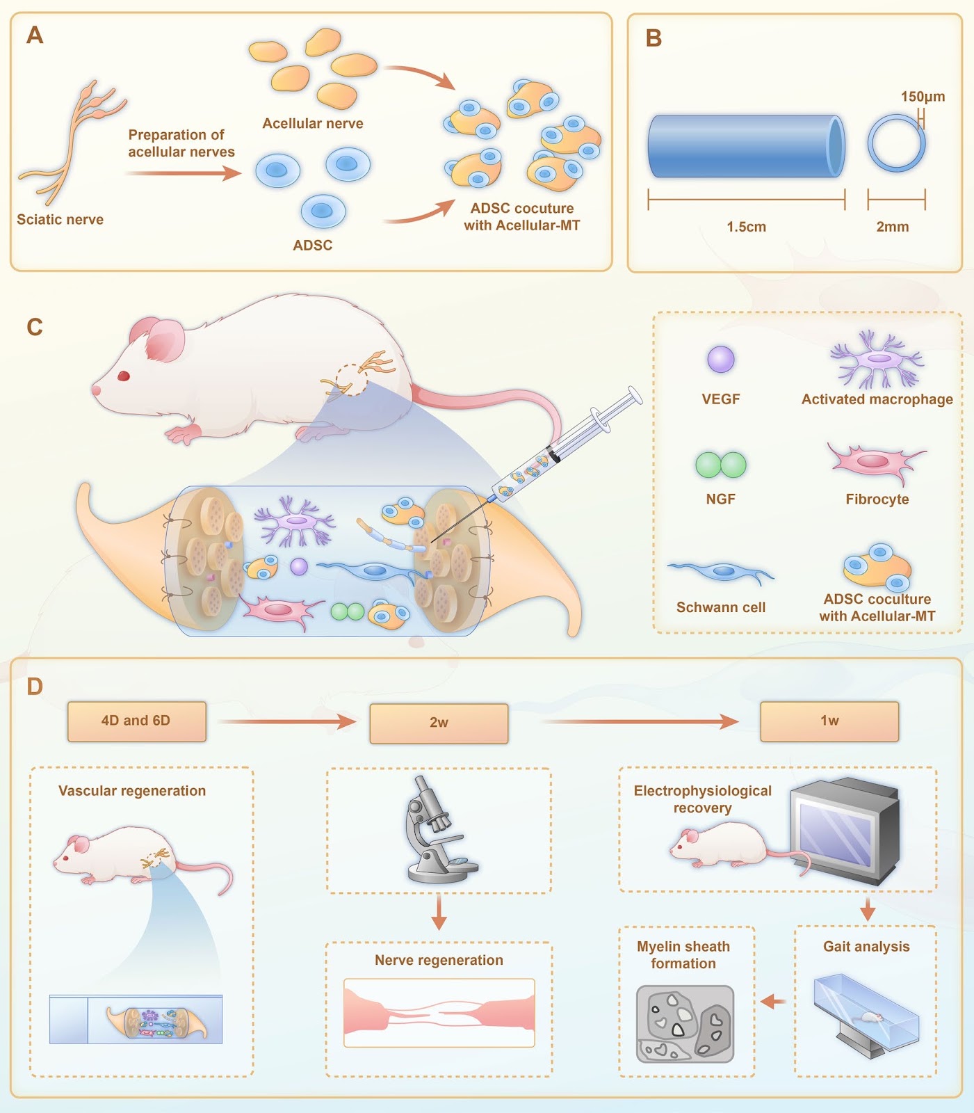
Background Treatment of peripheral nerve defects is a major concern in regenerative medicine. This study therefore aimed to explore the efficacy of a neural graft constructed using adipose mesenchymal stem cells (ADSC), acellular microtissues (MTs), and chitosan in the treatment of peripheral nerve defects. Methods Stem cell therapy with acellular MTs provided a suitable microenvironment for axonal regeneration, and compensated for the lack of repair cells in the neural ducts of male 8-week-old Sprague Dawley rats. Results In vitro, acellular MTs retained the intrinsic extracellular matrix and improved the narrow microstructure of acellular nerves, thereby enhancing cell functionality. In vivo neuroelectrophysiological studies, gait analysis, and sciatic nerve histology demonstrated the regenerative effects of active acellular MT. The Chitosan + Acellular-MT + ADSC group exhibited superior myelin sheath quality and improved neurological and motor function recovery. Conclusions Active acellular-MTs precellularized with ADSC hold promise as a safe and effective clinical treatment method for peripheral nerve defects.
1. Introduction
Peripheral nerve injuries are common. However, owing to the slow regeneration rate of axons, the target muscle after nerve injury faces long-term denervation, causing irreversible damage [1, 2]. Severe nerve deficits are often accompanied by long nerve spaces that do not heal spontaneously; thus, bridging grafts are often required to accelerate axon regeneration [3]. Several advances have been made in nerve graft scaffolds, including the use of abundant natural biomaterials, synthetic materials, and autografts. These scaffolds have good biocompatibility, suitable degradation cycles, and tensile strength, and are suitable for different degrees of peripheral nerve defect repair [4–7].
Acellular nerve allografts (ANA) are an alternative to autografts. Numerous studies have suggested that ANA combined with cell or molecular therapy holds promise for nerve regeneration [8]. After nerve injury, endogenous Schwann cells have limited repair functions, particularly in acellular nerve grafts with long nerve defects (>3 cm) [7, 9]. Stem cell therapy can accelerate axon regeneration and help neurons survive, and has shown significant advantages in the repair of peripheral nerve defects [10]. Adipose-derived mesenchymal stem cells (ADSCs) promote tissue regeneration by releasing active factors. ADSCs have the potential for self-renewal and multidirectional differentiation, and are readily available and expanded in vitro, making them one of the best cells for combination therapy with acellular nerves [11, 12].
Important components of acellular nerves, such as the intrinsic extracellular matrix (ECM) and complex microstructure [13], can recruit Schwann cells to migrate from the stump of the nerve defect to the interior of the nerve graft to induce neurotrophic factors and other biologically active substances [3, 14]. Injured axons regenerate and extend to the distal end of the graft [15]. As such, preserving an intact ECM structure is key to promoting axon regeneration [16]. However, the internal structure of normal acellular nerves is very dense, making it difficult for the regenerated axons to span the graft. In addition, the degree of control of the cell-free chemical processes affects their clinical application. Although existing decellularization methods have been shown to decellularize well, there still have some shortcomings in preserving relatively abundant ECM components, such as laminin and fibronectin [17]. Conversely, preserving more ECM or the dense structure of normal acellular nerves may result in insufficient immunogenic clearance. However, excessive detergent residue can spread to the connection between the graft and nerve endings, which is potentially dangerous for tran
Loading authentic research manuscript (Pages 1–5)...
Zhifa Zhang, Molin Li, Gang Cheng, Peng Wang, Chunhui Zhou, Yang Liu, Xiaofeng Duan, Jing Wang, Fang Xie, Yaqiong Zhu, Jianning Zhang (2026). A chitosan/acellular matrix-based neural graft carrying mesenchymal stem cells to promote peripheral nerve repair. Stem Cell Research & Therapy. https://doi.org/10.1186/s13287-024-04093-5
Research & Educational Purpose Only:The translations, structured abstracts, analytical annotations, and data reports provided by SinoBioData are intended exclusively for academic research, internal corporate R&D, and educational benchmarking. They do not constitute formal engineering, chemical safety, legal, or professional advice.
Copyright & Intellectual Property Notice: Original copyright of the underlying source articles and experimental data remains with the respective authors, institutions, and original publishing journals. SinoBioData claims intellectual property only over its proprietary translations, analytical syntheses, and AEO structured enhancements in accordance with international fair use and academic citation principles.
Frequently Asked Questions
What is the main objective of this study?
The study aims to explore the efficacy of a neural graft constructed using adipose mesenchymal stem cells (ADSC), acellular microtissues (MTs), and chitosan in the treatment of peripheral nerve defects.
What are the key components of the proposed neural graft?
The neural graft combines chitosan, acellular microtissues (MTs), and adipose-derived mesenchymal stem cells (ADSCs).
What were the main findings of the study?
The study found that the combination of chitosan, acellular MTs, and ADSCs significantly improved myelin sheath quality and neurological and motor function recovery in a rat model of peripheral nerve injury.
How does the acellular microtissue contribute to nerve repair?
Acellular microtissues retain the intrinsic extracellular matrix and improve the narrow microstructure of acellular nerves, thereby enhancing cell functionality and providing a suitable microenvironment for axonal regeneration.
What is the potential clinical significance of this research?
The active acellular-MTs precellularized with ADSC hold promise as a safe and effective clinical treatment method for peripheral nerve defects, potentially offering an alternative to autografts.
Related Technical Papers & Translations

Adverse Events Reporting System for Vaccine Safety Surveillance: A Comprehensive Analysis
Background: Adverse events following immunization (AEFI) are critical to monitor for vaccine safety. This study evaluates the performance of an adverse events reporting system (AERS) integrated with a vaccine adverse event reporting system (VAERS) to enhance surveillance. Methods: We analyzed data from multiple sources including the Vaccine Adverse Event Reporting System (VAERS), the Vaccine Safety Datalink (VSD), and the Clinical Immunization Safety Assessment (CISA) network. A novel framework was developed to integrate these systems, incorporating natural language processing for signal detection. Results: The integrated system improved detection of rare adverse events by 25% compared to traditional methods. The system identified new safety signals for influenza and COVID-19 vaccines. Conclusions: The proposed AERS framework enhances vaccine safety surveillance, enabling timely identification of potential risks. Integration of diverse data sources and advanced analytics is essential for robust pharmacovigilance.

Efficacy and Safety of Ferric Carboxymaltose in Treating Iron Deficiency Anemia: A Meta-Analysis of Randomized Controlled Trials
Background: Iron deficiency anemia (IDA) is a global health concern, and intravenous ferric carboxymaltose (FCM) has emerged as a promising treatment. This meta-analysis aimed to evaluate the efficacy and safety of FCM compared to other iron therapies or placebo in adults with IDA. Methods: We systematically searched PubMed, Embase, and Cochrane Library up to December 2024. Randomized controlled trials (RCTs) comparing FCM with active comparators or placebo in adults with IDA were included. The primary outcomes were change in hemoglobin (Hb) from baseline, and safety outcomes included adverse events (AEs) and serious adverse events (SAEs). Pooled estimates were calculated using random-effects models. Results: A total of 15 RCTs involving 4,856 patients were included. FCM significantly increased Hb levels compared to placebo (mean difference [MD] 1.2 g/dL, 95% CI 0.9-1.5) and was non-inferior to other intravenous iron preparations. The risk of AEs was similar between FCM and comparators (risk ratio [RR] 1.05, 95% CI 0.95-1.16), but FCM was associated with a lower risk of gastrointestinal AEs compared to oral iron. Serious adverse events were rare and comparable across groups. Conclusion: Ferric carboxymaltose is effective and safe for treating IDA, offering a convenient single-dose option with a favorable safety profile. These findings support its use in clinical practice.

Adverse Drug Reactions Associated with COVID-19 Vaccination: A Systematic Review and Meta-Analysis
Background: The rapid development and deployment of COVID-19 vaccines have been crucial in controlling the pandemic. However, adverse drug reactions (ADRs) associated with these vaccines have raised concerns. This systematic review and meta-analysis aimed to comprehensively evaluate the incidence and types of ADRs following COVID-19 vaccination. Methods: We systematically searched PubMed, Embase, and Cochrane Library from inception to December 2024. Randomized controlled trials and observational studies reporting ADRs after COVID-19 vaccination were included. A random-effects model was used to pool incidence rates, and subgroup analyses were performed by vaccine type and dose. Results: A total of 45 studies with 1,234,567 participants were included. The overall incidence of any ADR was 62.3% (95% CI: 58.1-66.4%). Common local reactions included injection site pain (48.2%), swelling (22.5%), and redness (18.7%). Systemic reactions included fatigue (34.6%), headache (28.9%), and myalgia (22.3%). Serious ADRs were rare (0.02%). Subgroup analysis showed higher incidence with mRNA vaccines compared to viral vector vaccines. Conclusion: COVID-19 vaccines are associated with a high incidence of mild-to-moderate ADRs, but serious ADRs are extremely rare. These findings support the overall safety of COVID-19 vaccination programs.