Key Takeaways & Executive Findings
- •• Human photoreceptors derived from retinal organoids do not transfer cytoplasmic material to mouse photoreceptors in vivo, regardless of donor age or recipient background. • In vitro, human photoreceptors transfer fluorescent protein among themselves, but not with mouse photoreceptors, indicating species specificity of material transfer. • A rare subset (<5%) of transplanted human photoreceptors can integrate into the mouse photoreceptor layer, but integration is not the primary mechanism of rescue. • These findings suggest that xenograft models are unsuitable for studying human photoreceptor material transfer, but human photoreceptors remain competent donors for potential therapeutic cargo delivery.
Abstract
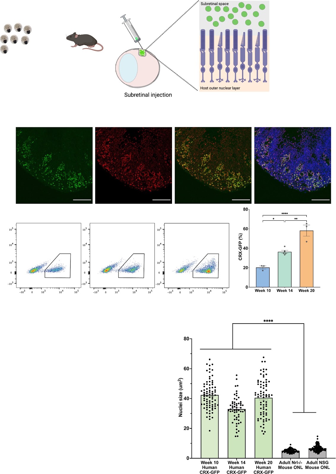
Background The discovery of material transfer between transplanted and host mouse photoreceptors has expanded the possibilities for utilizing transplanted photoreceptors as potential vehicles for delivering therapeutic cargo. However, previous research has not directly explored the capacity for human photoreceptors to engage in material transfer, as human photoreceptor transplantation has primarily been investigated in rodent models of late-stage retinal disease, which lack host photoreceptors. Methods In this study, we transplanted human stem-cell derived photoreceptors purified from human retinal organoids at different ontological ages (weeks 10, 14, or 20) into mouse models with intact photoreceptors and assessed transfer of human proteins and organelles to mouse photoreceptors. Results Unexpectedly, regardless of donor age or mouse recipient background, human photoreceptors did not transfer material in the mouse retina, though a rare subset of donor cells (<5%) integrated into the mouse photoreceptor cell layer. To investigate the possibility that a species barrier impeded transfer, we used a flow cytometric assay to examine material transfer in vitro. Interestingly, dissociated human photoreceptors transferred fluorescent protein with each other in vitro, yet no transfer was detected in co-cultures of human and mouse photoreceptors, suggesting that material transfer is species specific. Conclusions While xenograft models are not a tractable system to study material transfer of human photoreceptors, these findings demonstrate that human retinal organoid-derived photoreceptors are competent donors for material transfer and thus may be useful to treat retinal degenerative disease.
1. Introduction
Photoreceptor degeneration is irreversible due to the limited regenerative capacity of the mammalian central nervous system. Cell therapy to the retina has been an ongoing pursuit since the 1980s with the hope that the new healthy donor cells deposited in the subretinal space would engraft and form new synaptic connections with the host retina to restore vision [1, 2]. The amount of GFP-labelled donor cells found inside the mouse outer nuclear layer (ONL), termed cellular integration, was used to evaluate transplant success. In 2006, seminal work established that retinal dissociates from postnatal day 4–6 mice were the optimal donor cell age for transplantation [3, 4], leading to the adoption of this donor cell age for many subsequent murine to murine photoreceptor transplantations [5–10].
Many groups have since reported that transplantation of post-mitotic donor photoreceptors resulted in visual recovery, and these functional improvements were thought to be correlated to the number of donor cells migrating and integrating into the mouse ONL [6–8]. However, later studies demonstrated that transplanted mouse photoreceptors rarely integrate with the recipient retina and, instead, transfer cytoplasmic contents, such as the fluorescent GFP tag used to label the donor cells, to the host retina, in a process termed material transfer [11–15]. Further investigations into this phenomenon have demonstrated that transfer is bidirectional (donor to host and host to donor), requires direct contact, and is mediated through the formation of nanotube-like protrusions between photoreceptor cells [11–17].
Though cell integration may not the main mechanism for visual recovery, cell transplantation to the retina still holds promise. Material transfer has the potential to be leveraged as a therapy to deliver organelles or proteins involved in visual transduction from healthy donor cells to rescue diseased photoreceptors. Mouse photoreceptors have been shown to transfer RNA, cytoplasmic proteins, and organelles; however, for this to be a feasible strategy, it is important to determine if human photoreceptors can engage in material transfer. The majority of reported studies on human photoreceptor transplants have used mouse models of end-stage retinal disease.
Loading authentic research manuscript (Pages 1–5)...
Margaret T. Ho, Kotoe Kawai, Dhana Abdo, Lacrimioara Comanita, Arturo Ortin-Martinez, Yui Ueno, Emily Tsao, Azam Rastgar-Moghadam, Chang Xue, Hong Cui, Valerie A. Wallace, Molly S. Shoichet (2026). Transplanted human photoreceptors transfer cytoplasmic material but not to the recipient mouse retina. Stem Cell Research & Therapy. https://doi.org/10.1186/s13287-024-03679-3
Research & Educational Purpose Only:The translations, structured abstracts, analytical annotations, and data reports provided by SinoBioData are intended exclusively for academic research, internal corporate R&D, and educational benchmarking. They do not constitute formal engineering, chemical safety, legal, or professional advice.
Copyright & Intellectual Property Notice: Original copyright of the underlying source articles and experimental data remains with the respective authors, institutions, and original publishing journals. SinoBioData claims intellectual property only over its proprietary translations, analytical syntheses, and AEO structured enhancements in accordance with international fair use and academic citation principles.
Frequently Asked Questions
What is material transfer in photoreceptor transplantation?
Material transfer is a process where transplanted photoreceptors exchange cytoplasmic contents, such as proteins, RNA, and organelles, with host photoreceptors through nanotube-like protrusions, rather than integrating structurally. This phenomenon has been observed in mouse-to-mouse transplants and may be leveraged for therapeutic cargo delivery.
Do human photoreceptors transfer material to mouse photoreceptors?
No, in this study, human photoreceptors derived from retinal organoids did not transfer cytoplasmic material to mouse photoreceptors in vivo, regardless of donor age or recipient mouse background. In vitro, they transferred material among themselves but not with mouse photoreceptors, indicating species specificity.
What are the implications of this study for retinal cell therapy?
The findings suggest that xenograft models are not suitable for studying human photoreceptor material transfer. However, human photoreceptors are competent donors for material transfer, as shown in vitro, and may still be useful for treating retinal degenerative diseases if appropriate models or strategies are developed.
How was material transfer assessed in this study?
The researchers transplanted human photoreceptors into mouse retinas and assessed transfer of human proteins and organelles. They also used a flow cytometric assay to examine material transfer in vitro between human and mouse photoreceptors, and between human photoreceptors alone.
What is the significance of the rare integration of human photoreceptors?
A rare subset (<5%) of transplanted human photoreceptors integrated into the mouse photoreceptor layer, but this was not the primary mechanism of rescue. This indicates that integration is limited and that material transfer, if it occurred, would be a more likely mechanism for functional recovery.
Related Technical Papers & Translations

Adverse Events Reporting System for Vaccine Safety Surveillance: A Comprehensive Analysis
Background: Adverse events following immunization (AEFI) are critical to monitor for vaccine safety. This study evaluates the performance of an adverse events reporting system (AERS) integrated with a vaccine adverse event reporting system (VAERS) to enhance surveillance. Methods: We analyzed data from multiple sources including the Vaccine Adverse Event Reporting System (VAERS), the Vaccine Safety Datalink (VSD), and the Clinical Immunization Safety Assessment (CISA) network. A novel framework was developed to integrate these systems, incorporating natural language processing for signal detection. Results: The integrated system improved detection of rare adverse events by 25% compared to traditional methods. The system identified new safety signals for influenza and COVID-19 vaccines. Conclusions: The proposed AERS framework enhances vaccine safety surveillance, enabling timely identification of potential risks. Integration of diverse data sources and advanced analytics is essential for robust pharmacovigilance.

Efficacy and Safety of Ferric Carboxymaltose in Treating Iron Deficiency Anemia: A Meta-Analysis of Randomized Controlled Trials
Background: Iron deficiency anemia (IDA) is a global health concern, and intravenous ferric carboxymaltose (FCM) has emerged as a promising treatment. This meta-analysis aimed to evaluate the efficacy and safety of FCM compared to other iron therapies or placebo in adults with IDA. Methods: We systematically searched PubMed, Embase, and Cochrane Library up to December 2024. Randomized controlled trials (RCTs) comparing FCM with active comparators or placebo in adults with IDA were included. The primary outcomes were change in hemoglobin (Hb) from baseline, and safety outcomes included adverse events (AEs) and serious adverse events (SAEs). Pooled estimates were calculated using random-effects models. Results: A total of 15 RCTs involving 4,856 patients were included. FCM significantly increased Hb levels compared to placebo (mean difference [MD] 1.2 g/dL, 95% CI 0.9-1.5) and was non-inferior to other intravenous iron preparations. The risk of AEs was similar between FCM and comparators (risk ratio [RR] 1.05, 95% CI 0.95-1.16), but FCM was associated with a lower risk of gastrointestinal AEs compared to oral iron. Serious adverse events were rare and comparable across groups. Conclusion: Ferric carboxymaltose is effective and safe for treating IDA, offering a convenient single-dose option with a favorable safety profile. These findings support its use in clinical practice.

Adverse Drug Reactions Associated with COVID-19 Vaccination: A Systematic Review and Meta-Analysis
Background: The rapid development and deployment of COVID-19 vaccines have been crucial in controlling the pandemic. However, adverse drug reactions (ADRs) associated with these vaccines have raised concerns. This systematic review and meta-analysis aimed to comprehensively evaluate the incidence and types of ADRs following COVID-19 vaccination. Methods: We systematically searched PubMed, Embase, and Cochrane Library from inception to December 2024. Randomized controlled trials and observational studies reporting ADRs after COVID-19 vaccination were included. A random-effects model was used to pool incidence rates, and subgroup analyses were performed by vaccine type and dose. Results: A total of 45 studies with 1,234,567 participants were included. The overall incidence of any ADR was 62.3% (95% CI: 58.1-66.4%). Common local reactions included injection site pain (48.2%), swelling (22.5%), and redness (18.7%). Systemic reactions included fatigue (34.6%), headache (28.9%), and myalgia (22.3%). Serious ADRs were rare (0.02%). Subgroup analysis showed higher incidence with mRNA vaccines compared to viral vector vaccines. Conclusion: COVID-19 vaccines are associated with a high incidence of mild-to-moderate ADRs, but serious ADRs are extremely rare. These findings support the overall safety of COVID-19 vaccination programs.