🧬 SinoBioData Academic Portal
📚 Peer-Reviewed Translated Literature

All Biomedical & Clinical Articles (Page 54)

Browse complete peer-reviewed translations from top Chinese biomedical, oncology, and genomics journals. Read verified previews and download full authentic clinical reports.

Published Research Papers

Showing 24 of 1542 peer-reviewed translated articles (Page 54 of 65)

NLRP3 inflammasome activity and pyroptosis are involved in CD206+ macrophage activation by MPO anti-neutrophil cytoplasmic antibodiesGraphical AbstractVerified
Acta Biochimica et Biophysica Sinica

NLRP3 inflammasome activity and pyroptosis are involved in CD206+ macrophage activation by MPO anti-neutrophil cytoplasmic antibodies

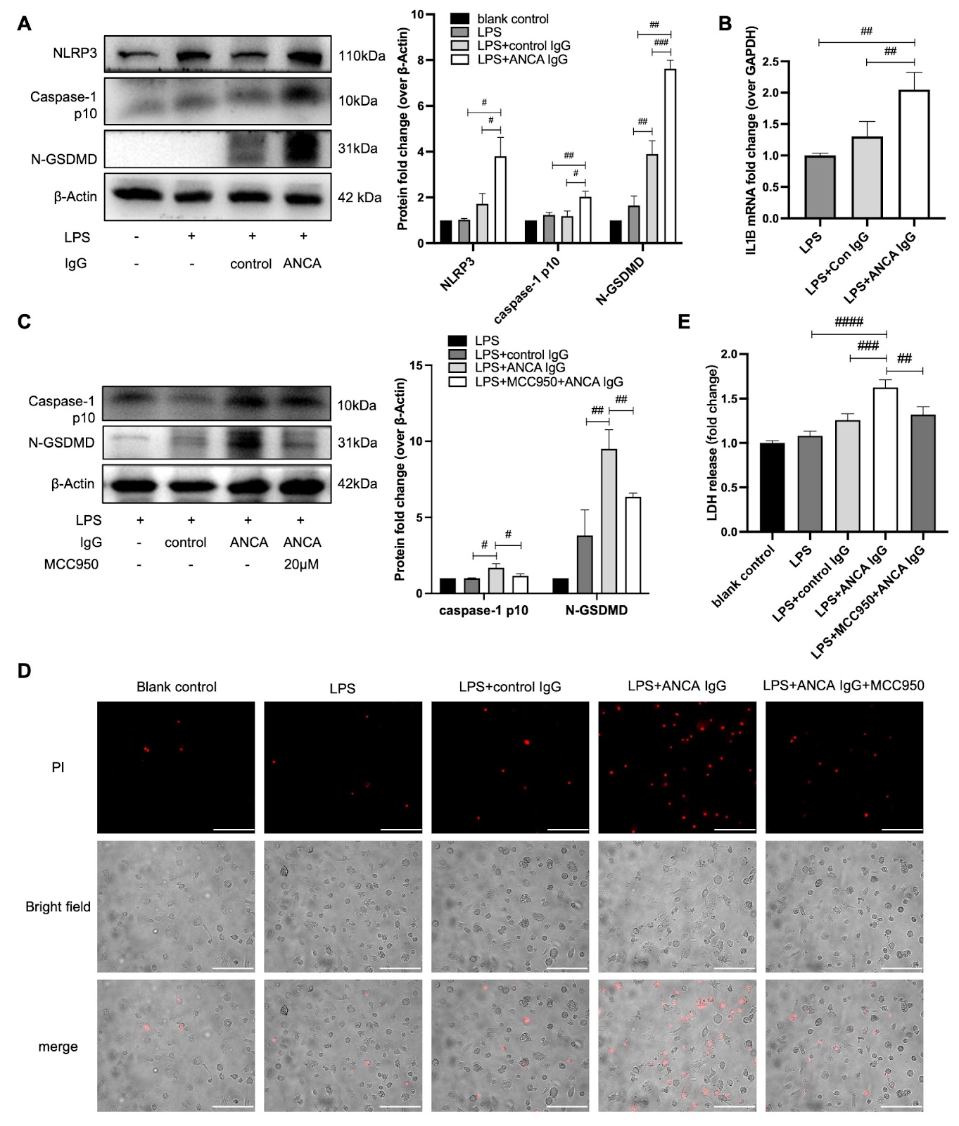
Macrophages are key players in the pathology of anti-neutrophil cytoplasmic antibody (ANCA)-associated vasculitis (AAV). Existing studies and our previous studies have documented the role of CD206-positive M2 macrophages in the inflammatory process of AAV. Inflammasome activation is a critical pathway through which macrophages release inflammatory factors. In this study, we investigate the role of the inflammasome in macrophages in AAV and explore the role of CD206 in this process. We recruit newly diagnosed AAV patients and disease controls from our department. The expression and localization of the NOD-like receptor family, pyrin domain containing 3 (NLRP3) and CD206 in the kidney are determined via immunofluorescence experiments. Myeloperoxidase (MPO)-ANCA immunoglobulin G (MPO-ANCA IgG) is purified from new-onset AAV patients with MPO-ANCA and used to treat lipopolysaccharide (LPS)-primed macrophages in vitro. Our findings reveal that NLRP3 expression is significantly elevated in the kidneys of active AAV patients, accompanied by increased cleaved caspase-1 and N-terminal gasdermin-D (GSDMD) levels in peripheral blood mononuclear cells (PBMCs). In vitro, MPO-ANCA IgG induces NLRP3 inflammasome activation and interleukin (IL)-1β production in macrophages, which is associated with increased MPO expression and JNK signaling pathway activation. Immunofluorescence analysis demonstrates partial colocalization of CD206 and NLRP3 in AAV kidneys. Furthermore, silencing of MRC1 gene, which encodes CD206, reduces inflammasome activation induced by MPO-ANCA IgG. In conclusion, our study provides evidence that MPO-ANCA IgG contributes to NLRP3 inflammasome activation and macrophage pyroptosis, with CD206 playing a critical role in this process. These findings elucidate the mechanisms underlying inflammation in AAV and suggest potential therapeutic targets.

Read Full Abstract10.3724/abbs.2025080
Exploring the antitumor effect of curcumin-piperlongumine hybrid molecule (CP) on EGFR-TKI-resistant non-small cell lung cancer using network pharmacological analysis and experimental verificationGraphical AbstractVerified
Acta Biochimica et Biophysica Sinica

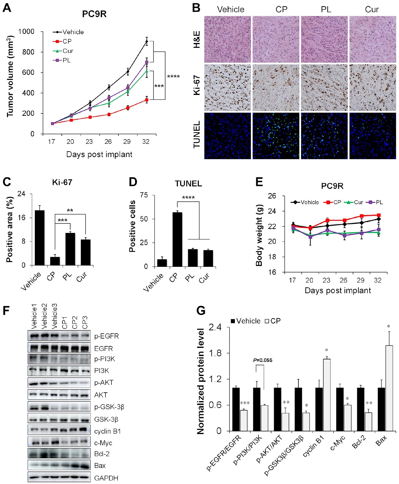
Exploring the antitumor effect of curcumin-piperlongumine hybrid molecule (CP) on EGFR-TKI-resistant non-small cell lung cancer using network pharmacological analysis and experimental verification

EGFR-tyrosine kinase inhibitor (TKI) therapy is the most effective targeted therapy for non-small cell lung cancer (NSCLC). However, drug resistance remains a significant factor in the failure of lung cancer therapy. In the present study, we utilize network pharmacology, molecular docking, in vitro and in vivo experiments to explore the targets and biological mechanisms of CP, a novel curcumin-piperlongumine hybrid molecule, in EGFR-TKI-resistant NSCLC cells. The results reveal that CP exhibits enhanced biological activity compared to its parent compounds. CP can effectively inhibit cell proliferation by arresting cell cycle in the G2/M phase and inducing apoptosis. Mechanistically, CP-induced apoptosis is partially mediated by PI3K/AKT signaling pathway. These findings highlight the potential of CP as a promising therapeutic agent for EGFR-TKI-resistant lung cancer therapy.

Read Full Abstract10.3724/abbs.2025076
Oligodendrocytes interactions with glial cells and neurons in demyelinating diseaseGraphical AbstractVerified
Acta Biochimica et Biophysica Sinica

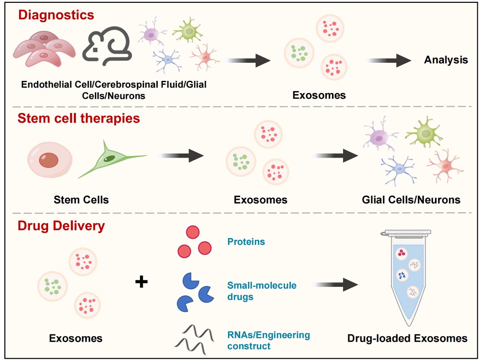
Oligodendrocytes interactions with glial cells and neurons in demyelinating disease

This review delves into the crosstalk network between oligodendrocytes and other glial cells in the context of demyelinating diseases. Oligodendrocytes, the myelin-forming cells in the central nervous system (CNS), are crucial for maintaining the function and integrity of axons and myelin sheaths. In demyelination pathologies, various factors hinder the normal differentiation of oligodendrocyte precursor cells, obstructing the myelin regeneration process, which is a primary barrier to therapeutic myelin repair. Emerging evidence highlights the critical role of glial cell interactions in CNS homeostasis and myelin regeneration, increasing interest in the treatment of demyelinating diseases. This article reviews the role of interactions between oligodendrocytes and other CNS glial cells in demyelinating and neurological diseases. Previous findings suggest that other CNS glial cells influence the survival and metabolic activity of oligodendrocytes through cell interactions, significantly affecting myelin formation and impacting demyelinating diseases characterized by myelin regeneration disorders. Targeted modulation of oligodendrocytes and their interactions with other cells at specific temporal stages may indicate a novel therapeutic direction for demyelinating diseases and offer fresh perspectives on the study of mechanisms and therapeutic approaches for related neurological conditions.

Read Full Abstract10.3724/abbs.2025105
Divergent roles of PKM2 in regulating PD-L1 and PD-L2 expression and their implications in human and mouse cancer modelsGraphical AbstractVerified
Acta Biochimica et Biophysica Sinica

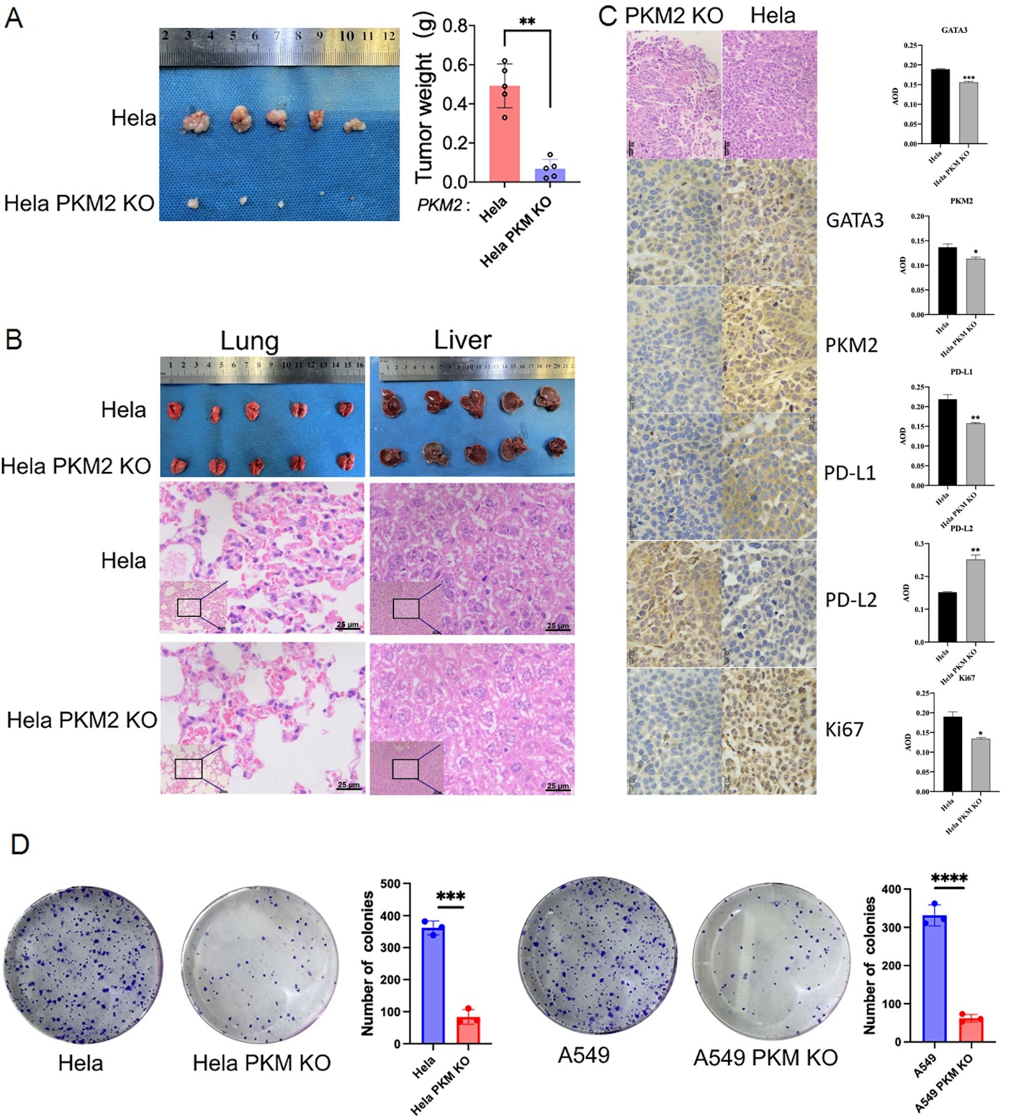
Divergent roles of PKM2 in regulating PD-L1 and PD-L2 expression and their implications in human and mouse cancer models

Cancer cells evade immune detection through checkpoint molecules like PD-L1 and PD-L2 which suppress T-cell activation. While PD-L1 is well-studied, the role of PD-L2 remains unclear. Pyruvate kinase M2 (PKM2), a metabolic enzyme, influences immune checkpoint regulation, but its role in PD-L1 and PD-L2 modulation is not well defined. Here, we investigate the role of pyruvate kinase M2 (PKM2) in modulating the immune checkpoint molecules PD-L1 and PD-L2 via GATA3 in cancer cells, with insights from both human and mouse models. We find that PKM2 enhances PD-L1 expression while inhibiting PD-L2, a dual regulatory mechanism that facilitates immune evasion. Knockdown and overexpression experiments revealed GATA3 as a key mediator. PKM2 knockout reduced GATA3 level, leading to decreased PD-L1 and increased PD-L2 expression. Chromatin immunoprecipitation (ChIP)-qPCR demonstrates that GATA3 functions as a direct transcription factor capable of binding to the promoters of PD-L1 and PD-L2. In silico analyses of 81 esophageal squamous cell carcinoma (ESCC) cases from the TCGA database demonstrate that PKM2 mRNA is unrelated to PD-L1 and PD-L2 expression but is negatively correlated with CD8+ T-cell infiltration in ESCC. To further validate these findings, we establish a xenograft model using immune-competent C57/BL6N mice, where knockdown of PKM2 results in significant downregulation of both PD-L1 and PD-L2 expression. Collectively, these findings underscore the divergent roles of PKM2 in regulating immune checkpoint expression in human and mouse cancer models and suggest that targeting the PKM2-GATA3 axis could enhance cancer immunotherapy by fine-tuning PD-L1 and PD-L2 levels.

Read Full Abstract10.3724/abbs.2025019
Modulation of Aβ42-induced toxic effects on the cultured neuronal network activity by extracellular matrix stiffnessGraphical AbstractVerified
Acta Biochimica et Biophysica Sinica

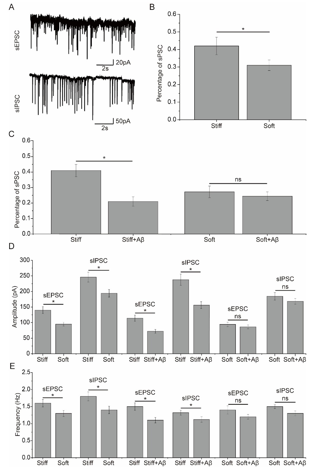
Modulation of Aβ42-induced toxic effects on the cultured neuronal network activity by extracellular matrix stiffness

Alzheimer’s disease (AD) is the most common neurodegenerative disease that usually begins with short-term memory loss, gradually progresses to cognitive dysfunction and causes loss of body function and eventual death. Mutations in the APP gene encoding the Aβ precursor protein (APP) are known to cause early-onset AD and suggest that Aβ is a major factor in AD development. Enzyme complexes, such as α-, β- and γ-secretases, catalyze various cleavage pathways to produce a variety of Aβ isoforms of different lengths. These Aβ peptides have proven toxic to the brain and accumulate in AD to form cerebral plaques. The main isoform of Aβ present in these plaques is the 42 amino acid variant known as Aβ42. A previous study revealed that changes in the stiffness of the extracellular matrix (ECM) can induce remodeling of the cytoskeleton of neurons in the brain tissues of AD patients, leading to changes in the morphology and function of neurons. ECM stiffness is unique to each specific tissue, and resident cells have developed to function optimally in microenvironments with specific ECMs. Brain tissues are reported to have a Young’s modulus of elasticity between 0.1 and 16 kPa. In patients with AD, a decrease in the elasticity of brain tissues was detected. Interestingly, the ECM is known to play an important role in cytoskeleton remodeling and neuronal function, and a stiff ECM has been reported to promote actin polymerization and stress fiber formation, whereas a soft ECM triggers actin depolymerization. However, it remains uncertain whether alterations in ECM stiffness in the AD brain contribute to Aβ-induced toxicity, particularly considering that Aβ is recognized to cause neuronal toxicity by disrupting the actin cytoskeleton, which leads to subsequent synaptic and dendritic abnormities. As such, the present study aimed to investigate the effects of substrate stiffness on Aβ-induced toxicity to the neuronal network in cultured neurons. Hippocampal neurons cultured on soft and stiff substrates were assessed for cell viability by MTT assay. When the neuronal cultures were exposed to 1 μM Aβ42 for 48 h, there was a significant decrease in the viability of the cells cultured on the stiff substrates, but there was no significant effect on the viability of the neuronal cultured on the soft substrates. In addition, the influence of Aβ42 on the number of synapses within the cultured neuronal network was analyzed using confocal immunofluorescence imaging. This analysis revealed that Aβ42 exposure induced a decrease in synaptic formation in cultured neurons, which was dependent on substrate stiffness. To evaluate the effect of substrate stiffness on Aβ42-induced toxicity to synaptic transmission in the cultured neuronal network, spontaneous Ca2+ oscillations were examined in neurons cultured on substrates with different stiffness treated with Aβ42. The percentage of neurons with spontaneous Ca2+ oscillations was significantly greater in neurons cultured on stiff substrates than in those cultured on soft substrates. After Aβ42 exposure, the percentage of neurons with spontaneous Ca2+ oscillations was significantly decreased in neurons cultured on the stiff substrates. Exposure to Aβ42 only slightly influenced the percentage of spontaneous Ca2+ oscillations in neurons cultured on the soft substrate. The amplitude and frequency of spontaneous Ca2+ oscillations were significantly greater in neurons cultured on the stiff substrates than in those cultured on the soft substrates. After exposure to Aβ42, the amplitude and frequency of spontaneous Ca2+ oscillations were significantly reduced in neurons cultured on stiff substrates. However, exposure to Aβ42 had only a weak influence on the amplitude and frequency of spontaneous Ca2+ oscillations in neurons cultured on soft substrates. To further investigate the effects of substrate stiffness on synapse function following exposure to Aβ42, spontaneous postsynaptic currents were recorded in DIV14-16 neurons cultured on stiff and soft substrates. The percentage of neurons with spontaneous postsynaptic currents was considerably greater in neurons cultured on the stiff substrates than in those cultured on soft substrates.

Read Full Abstract10.3724/abbs.2025095
Substrate topography-induced osteogenesis of bone marrow stem cells by reducing the chromatin accessibility of YBX1Graphical AbstractVerified
Acta Biochimica et Biophysica Sinica

Substrate topography-induced osteogenesis of bone marrow stem cells by reducing the chromatin accessibility of YBX1

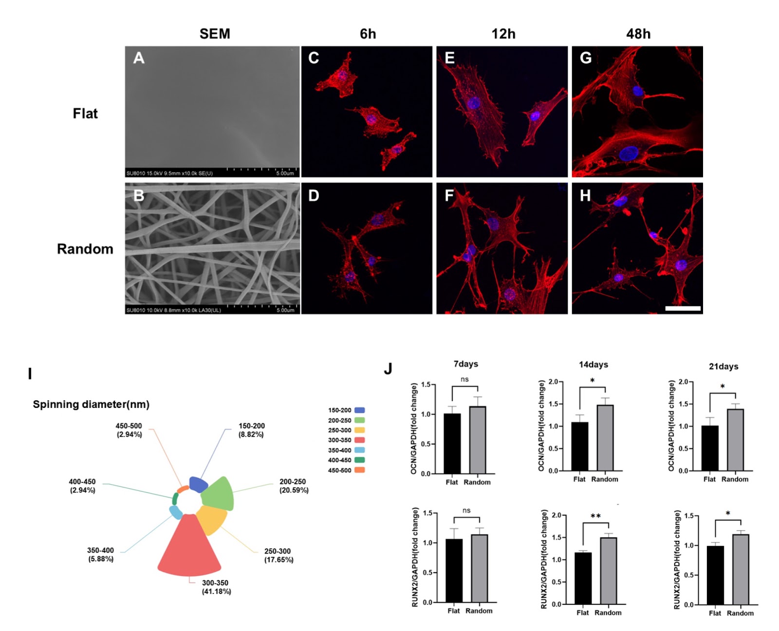
Stem cell fate is profoundly influenced by a complex interplay of biochemical and biophysical cues, with the latter increasingly recognized for its roles in cellular processes, yet the mechanisms are unclear. Since chromatin accessibility is a critical determinant in the processes of osteogenesis and bone repair, investigating the contributions of open chromatin regions (OCRs) to the intracellular signaling pathways triggered by topographical cues, which lead to osteogenic differentiation is highly valuable. This study explores the impact of the nanotopography of biomaterials on the osteogenic differentiation of human bone marrow stem cells (hBMSCs). By utilizing electrospun poly-L-lactide (PLLA) membranes with random fiber arrangements, we mimic the natural extracellular matrix (ECM) topography to study its effects on hBMSCs, contrasting them with flat PLLA controls. Through high-throughput Assay for Transposase-Accessible Chromatin with sequencing (ATAC-seq) and RNA sequencing (RNA-seq), we reveal that the nanotopography of electrospun surfaces promotes osteogenic differentiation by modulating the chromatin accessibility of the YBX1 gene promoter, leading to its upregulation. Lentiviral knockdown experiments further confirm the crucial role of YBX1, revealing a reversal of the osteogenic effects induced by nanotopography. This study emphasizes the importance of YBX1 in the osteogenic response to the surface topography of biomaterials and suggests that nanotopographical cues could be harnessed to direct stem cell fate. These findings are important for developing biomaterials that promote specific stem cell outcomes in regenerative medicine. Our results further contribute to a deeper understanding of the mechanisms underlying stem cell differentiation in response to environmental cues and pave the way for the rational design of biomaterials with enhanced osteogenic potential. By elucidating the role of chromatin accessibility and specific transcription factors such as YBX1, this study highlights the intricate interplay between cell-material interactions and the intracellular signaling pathways that govern stem cell fate.

Read Full Abstract10.3724/abbs.2025065
circ_0000389 inhibits intervertebral disc degeneration by targeting the miR-346/KLF7 axisGraphical AbstractVerified
Acta Biochimica et Biophysica Sinica

circ_0000389 inhibits intervertebral disc degeneration by targeting the miR-346/KLF7 axis

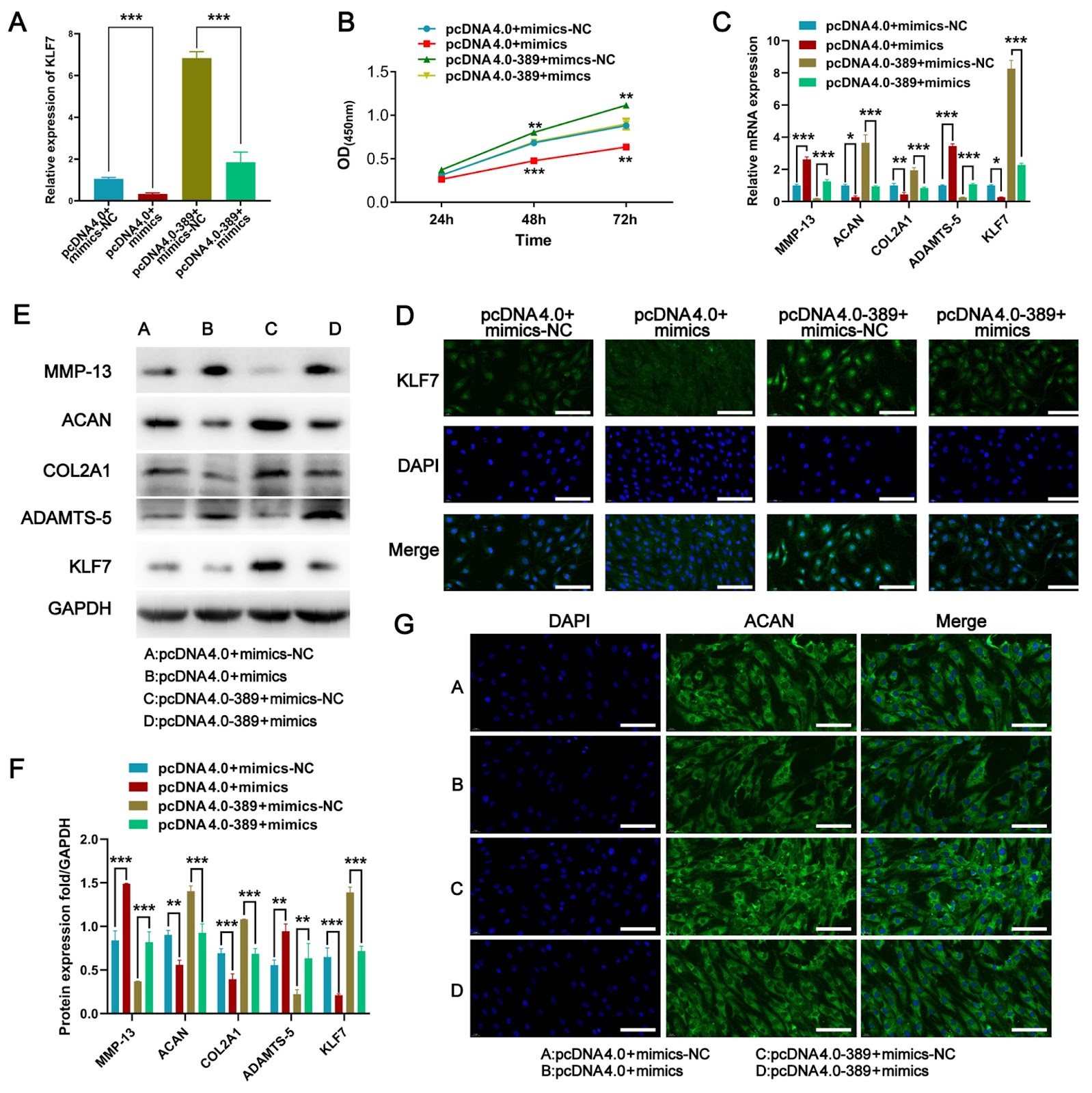
Intervertebral disc degeneration (IVDD) is a major cause of low back pain. An increasing number of studies have demonstrated that circRNAs regulate the progression of IVDD. However, the specific role of circ_0000389 in the progression of IVDD is not clear. In this study, circ_0000389 is selected by bioinformatics analysis of the GSE67566 dataset. RT-qPCR is performed to detect the expressions of circ_0000389, miR-346 and KLF7 in nucleus pulposus (NP) tissues. The proliferative capacity of nucleus pulposus cells (NPCs) is examined via CCK-8 assay. Western blot analysis of extracellular matrix (ECM) catabolism is performed in NPCs. Dual-luciferase reporter gene assays and RNA immunoprecipitation (RIP) confirm the interaction of circ_0000389 with miR-346 and KLF7. The expression of circ_0000389 is significantly downregulated in degenerating NP tissues. Functionally, circ_0000389 inhibits ECM catabolism. Mechanistically, we identify miR-346 and KLF7 as downstream target genes of circ_0000389 and miR-346, respectively. miR-346 overexpression reverses the effect of circ_0000389 on NPCs, and KLF7 overexpression reverses the effect of miR-346 on NPCs, indicating that circ_0000389 alleviates IVDD progression by regulating the miR-346/KLF7 axis. This study may provide a new therapeutic target for the treatment of IVDD.

Read Full Abstract10.3724/abbs.2025029
TRIM25 ubiquitinates and degrades p62/SQSTM1 to suppress autophagyGraphical AbstractVerified
Acta Biochimica et Biophysica Sinica

TRIM25 ubiquitinates and degrades p62/SQSTM1 to suppress autophagy

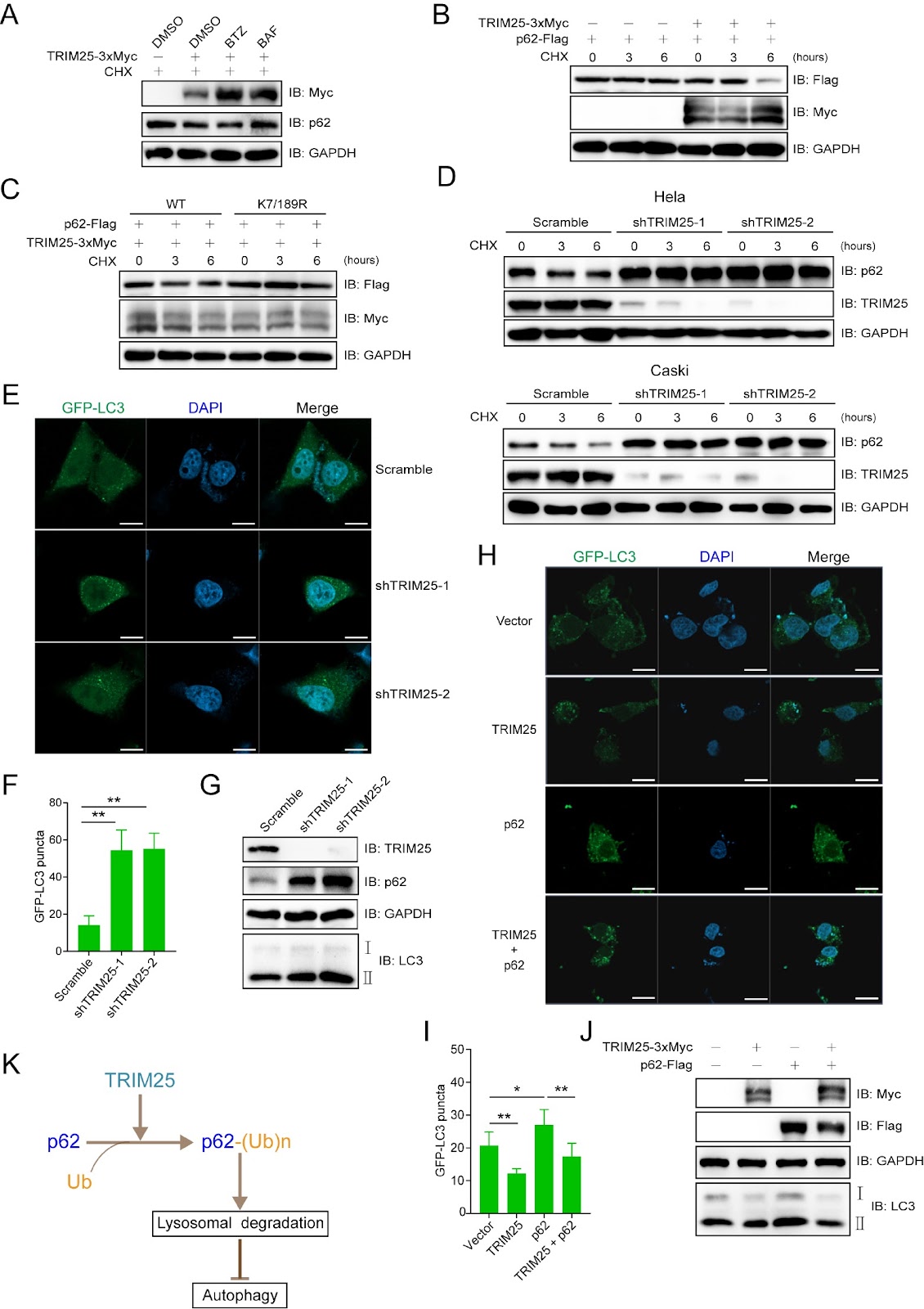
Autophagy is a conserved catabolic process in which organelles, macromolecules and pathogens are degraded via lysosomes. Sequestosome 1 (SQSTM1), also known as p62, the first autophagy receptor identified, binds to ubiquitin on targets and LC3 on phagophores, mediating the selective autophagy of ubiquitinated substrates. To identify the potential interacting partners for p62, Flag-tagged p62 was transfected into HEK293T cells and used as bait to Co-immunoprecipitate (Co-IP) with proteins that form a complex with p62. More materials and methods are provided in the Supplementary Materials and Methods. The proteins were then identified via mass spectrometry analysis. Two E3 ubiquitin ligases, TRIM25 and ITCH, were identified as the highest confidence hits in a list of identified proteins. Gene Ontology (GO) analysis revealed that p62-interacting proteins were enriched in the ubiquitin-dependent protein degradation, protein folding, oxidative phosphorylation, autophagy, etc., signalling pathways. Validation assays were then performed to test the E3 ubiquitin ligases for p62 identified in this study. Both endogenously and ectopically expressed p62 formed a complex with the E3 ubiquitin ligases TRIM25 and ITCH. GST pull-down assays revealed that recombinant TRIM25 and ITCH directly interact with p62. As detected by fluorescence microscopy analysis, mCherry-tagged TRIM25 and GFP-tagged p62 were colocalized mainly in the cytoplasm of HeLa cells. Ubiquitination assays were performed to determine whether TRIM25 and ITCH are merely interacting partners or true E3 ubiquitin ligases for p62. One potential explanation for this phenomenon is that ITCH requires assistance from a specific protein or undergoes a particular modification to activate its ability to ubiquitinate p62. Alternatively, it is conceivable that p62 needs to be modified to be ubiquitinated by ITCH. Exogenously expressed p62 was efficiently ubiquitinated by TRIM25 but not by ITCH. These results suggest that TRIM25 is an E3 ubiquitin ligase for p62, whereas ITCH is only an interacting partner. A reconstituted E. coli ubiquitination system, which has been used in several of our studies, was included in this study. The p62 proteins recovered from the E. coli ubiquitination system were subjected to mass spectrometry analysis, and fifteen Lys (K) residues of p62 were identified. As shown in Supplementary Figure S1D, K7 and K189 were validated as the two major sites for the TRIM25-mediated ubiquitination of p62 in mammalian cells. The mutant bearing simultaneous K-to-R substitutions (K7/189R) at the two Lys residues almost completely abolished the TRIM25-mediated ubiquitination of p62. Four shRNAs targeting TRIM25 were designed and tested in HeLa and Caski cells, and shTRIM25-1 and shTRIM25-2 were selected for further study. TRIM25 knockdown reduced p62 ubiquitination, which was effectively reversed by exogenously expressed TRIM25 in both HeLa and Caski cells. In HEK293T cells, the TRIM25-mediated reduction in p62 protein expression was blocked by treatment with the autophagy inhibitor bafilomycin (BAF) but not by treatment with the proteasome inhibitor bortezomib (BTZ). Upon undergoing ubiquitination, p62 enhances its interaction with LC3 through its LC3-interacting region (LIR) domain. This interaction enables p62 to be

Read Full Abstract10.3724/abbs.2025062
CDR1as modulates arrhythmia post-myocardial infarction via regulating Cav1.2Graphical AbstractVerified
Acta Biochimica et Biophysica Sinica

CDR1as modulates arrhythmia post-myocardial infarction via regulating Cav1.2

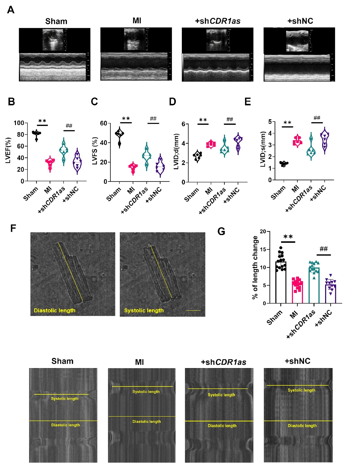
Arrhythmias, especially ventricular arrhythmias (VAs), are the primary cause of mortality following myocardial infarction (MI) and are typically attributable to electrophysiological disorders of the heart. Our previous work demonstrated that CDR1as knockdown ameliorates arrhythmias by modulating Nav1.5 and Kir6.2 channels post-MI. This study aims to explore the role of CDR1as in calcium channel remodeling subsequent to ischemic arrhythmia. We employ MI in mice by ligating the left anterior descending coronary artery (LAD) and use patch-clamp techniques to measure the Ca current (ICaL) in isolated ventricular cardiomyocytes. The results show that the expression of Cav1.2 is significantly decreased in the infarct border zone at 12 h post-MI. CDR1as knockdown via AAV9-CDR1as-shRNA administration leads to an enhancement of cardiac function and a restoration of both ICaL density and Cav1.2 expression in MI model mice. These findings indicate that targeting the CDR1as pathway to modulate calcium channels can be a viable strategy for antiarrhythmic therapy following MI.

Read Full Abstract10.3724/abbs.2025126
ATF4 promotes glutaminolysis and glycolysis in colorectal cancer by transcriptionally inducing SLC1A5Graphical AbstractVerified
Acta Biochimica et Biophysica Sinica

ATF4 promotes glutaminolysis and glycolysis in colorectal cancer by transcriptionally inducing SLC1A5

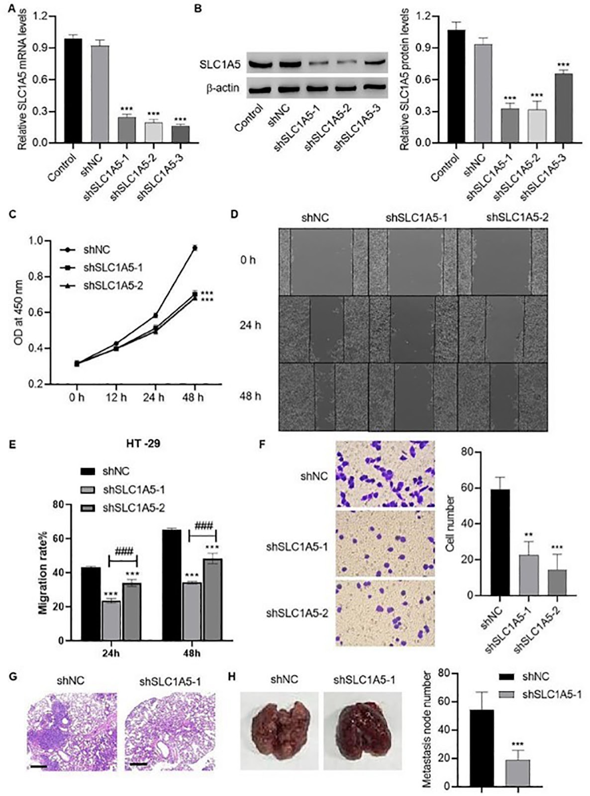
Glutaminolysis and glycolysis promote the malignant progression of colorectal cancer. The role of activating transcription factor 4 (ATF4) in solute carrier family 1 member 5 (SLC1A5)-mediated glutaminolysis and glycolysis remains to be elucidated. SLC1A5 and ATF4 expression levels are detected in colorectal cancer tissues. ATF4 is knocked down or overexpressed to assess its role in cell viability, migration and invasion. SLC1A5 is knocked down to evaluate its role in cell viability, migration, invasion, and metastasis and the metabolism of glutamine and glucose. The regulatory effect of the transcription factor ATF4 on SLC1A5 transcription and expression is determined using a luciferase reporter assay and chromatin immunoprecipitation (ChIP) techniques. Upregulated ATF4 and SLC1A5 expressions are observed in tumor tissue, which is positively correlated with the tumor, node, and metastasis (TNM) stages. ATF4-overexpressing SW480 cells show the increased cell viability, migration and invasion. Conversely, ATF4 knockdown decreases the viability, migration and invasion of HCT-116 cells. SLC1A5 knockdown inhibits viability, migration, invasion, and metastasis and the metabolism of glutamine and glucose in HT-29 cells, as well as the expressions of two key glycolytic enzymes, hexokinase 2 (HK2) and pyruvate kinase M2 (PKM2). The luciferase activity of the SLC1A5 promoter is increased by ATF4 overexpression. SLC1A5 promoter enrichment is increased by anti-ATF4 antibody immunoprecipitation in ATF4-overexpressing colorectal cells, indicating that ATF4 targets SLC1A5 to promote glutamine and glucose metabolism in these cells. In summary, the ATF4/SLC1A5 axis plays a significant role in the progression of colorectal cancer by regulating glutamine metabolism and glycolysis.

Read Full Abstract10.3724/abbs.2024226
A simple, rapid, and transgene-free strategy for the generation of transgenic pigs via precise editing of monoclonal porcine fetal fibroblastsGraphical AbstractVerified
Acta Biochimica et Biophysica Sinica

A simple, rapid, and transgene-free strategy for the generation of transgenic pigs via precise editing of monoclonal porcine fetal fibroblasts

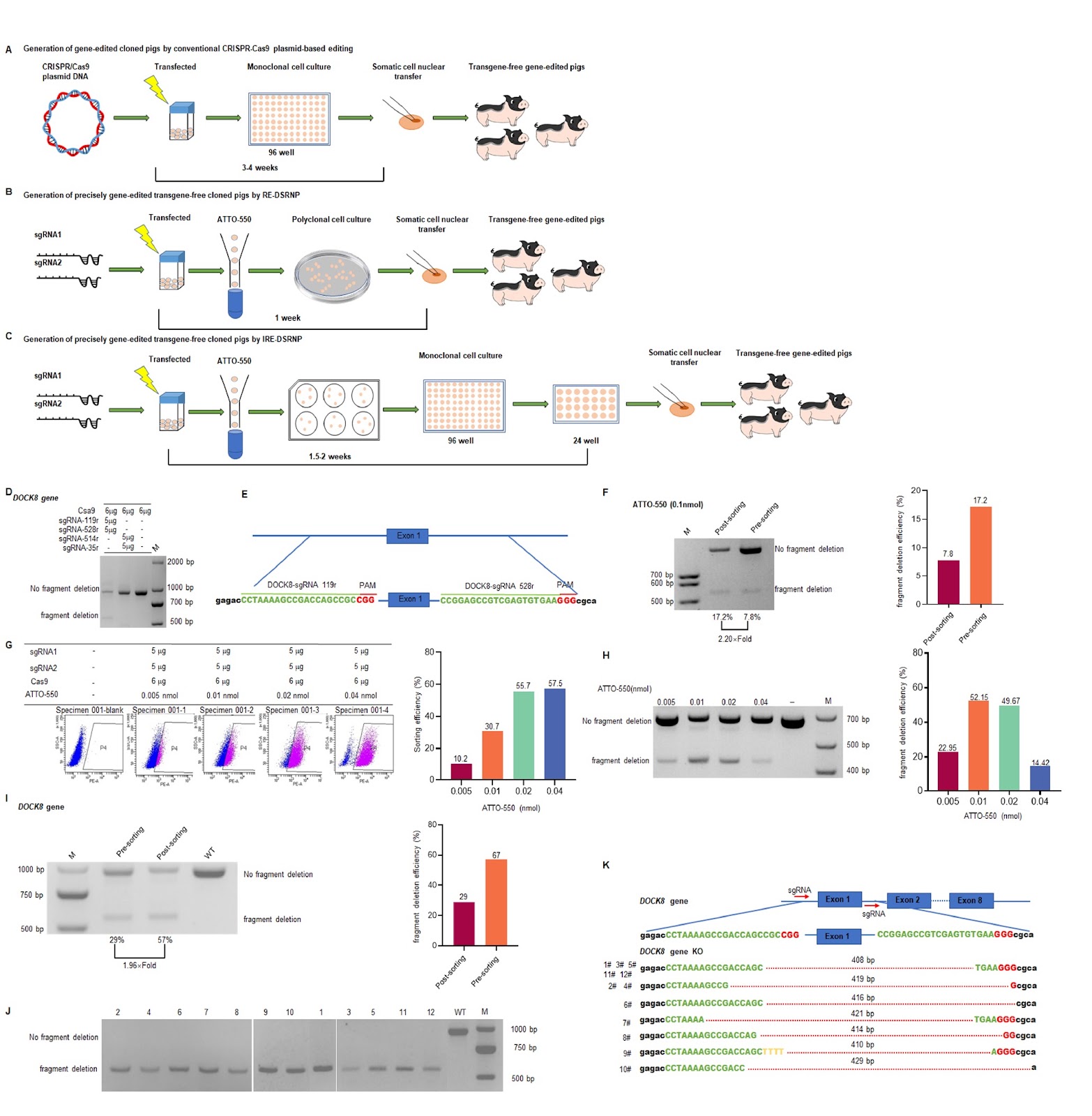
Pigs, as crucial economic livestock species, possess remarkable reproductive traits and thus play a highly significant role in promoting the progress of the livestock industry. With the advent and application of CRISPR/Cas9 technology, researchers have explored genetic editing techniques to increase swine reproductive performance, flavour profiles, and nutritional attributes. Additionally, with respect to anatomy, physiology, immunology, and genomics as well as other traits, pigs exhibit remarkable similarities to humans. Genetically edited pigs play crucial roles in human disease models, xenotransplantation, breed improvement, vaccine development, and drug assessment. Common methods deployed in the preparation of genetically edited pigs include somatic cell nuclear transfer (SCNT), microinjection and sperm-mediated approaches. For example, Shen et al. [1] successfully generated P53-knockout Diannan miniature pigs using transcription activator-like effector nucleases combined with SCNT, offering a valuable resource for preclinical oncology research. In 2019, Chen et al. [2] employed microinjection to deliver Cas9 messenger ribonucleic acid (mRNA) and single guide ribonucleic acid (sgRNA) into the cytoplasm of fertilized eggs. These authors successfully obtained both the albinism phenotype and the combined phenotype of albinism and immunodeficiency in Tibetan miniature pigs. More recently, Tenihara et al. [3] introduced the CRISPR/Cas9 protein into fertilized porcine eggs via electroporation, enabling a simple, micromanipulation-free approach for generating gene-edited pigs. Among these methods, SCNT has gained extensive interest among researchers because of its reliability. An essential aspect of SCNT is the preparation of embryonic fibroblasts to serve as donor cells. Previously, the CRISPR/Cas9 plasmid editing system served as the predominant technique to generate genetically edited embryonic fibroblasts (Figure 1A) [4]. This approach, which is distinguished by its relative simplicity, high stability, and low cost, was formerly widely utilized in the production of gene-edited pigs. However, plasmid editing is associated with several notable limitations. First, it introduces resistance genes, posing risks of inaccurate gene editing, drug resistance and biosafety concerns. Second, during the CRISPR/Cas9 editing process, there is a possibility of ongoing editing due to deoxyribonucleic acid (DNA) integration. This continuous editing can increase the likelihood of off-target effects, random mutations, and interference with DNA repair mechanisms. Third, the acquisition of positive cell lines via the plasmid editing system typically demands an extended period of in vitro cultivation (lasting 3–4 weeks), which increases the risk of apoptosis and chromosomal aberrations. Consequently, plasmid-based transfection is now largely supplanted by ribonucleoprotein (RNP) systems for gene editing. RNP systems bypass plasmids, delivering the Cas9 protein and sgRNA directly into cells, reducing off-target effects and cytotoxicity [5]. In 2022, Xu et al. [6] developed the reporter RNA-enriched dual-sgRNA CRISPR/Cas9 ribonucleoprotein (RE-DSRNP) method, a transgene-free approach using CRISPR/Cas9 RNPs enriched with ATTO550-tracrRNA (IDT, Iowa, USA) as a fluorescent RNA probe (Figure 1B). This method reduced the time needed to generate donor cells from 3-4 weeks to one week, resulting in high-efficiency WIP1 gene knockouts and the production of pigs with male reproductive disorders. However, owing to genetic diversity, not all target genes achieve 95% editing efficiency, as demonstrated by the RE-DSRNP method, with some falling below 90%. For example, DOCK8, which belongs to the DOCK family, is an atypical guanine nucleotide exchange factor that plays a crucial role in immune responses. DOCK8 deficiency syndrome, a rare hereditary disorder, often leads to combined immunodeficiency and is characterized by elevated serum immunoglobulin E levels, increased eosinophil
Read Full Abstract10.3724/abbs.2025044
HECTD3 is overexpressed in breast cancer and associated with a good prognosisGraphical AbstractVerified
Acta Biochimica et Biophysica Sinica

HECTD3 is overexpressed in breast cancer and associated with a good prognosis

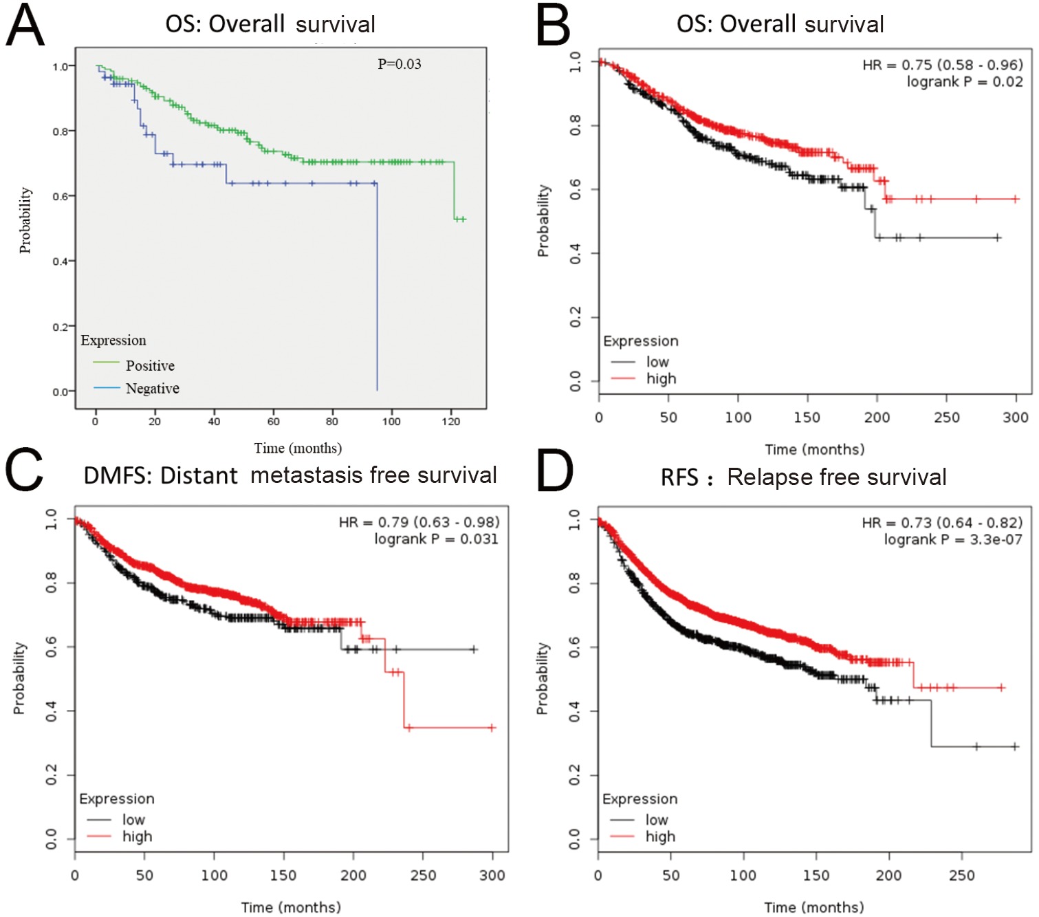
HECTD3 is an E3 ubiquitin ligase that has been implicated in cancer progression. This study investigates HECTD3 expression in breast cancer and its association with prognosis. Immunohistochemical staining was performed on 320 breast cancer samples (cohort 1) and a tissue microarray of 227 samples (cohort 2), along with 39 normal adjacent tissues. HECTD3 was overexpressed in 73.75% of cohort 1 and 75.33% of cohort 2, compared to 41.03% in normal tissues (P < 0.0001). Logistic regression analysis revealed that positive HECTD3 expression was significantly associated with lower risk of lymph node metastasis (OR = 0.37, P = 0.003), reduced risk of poor tumor differentiation (grade 3 vs 1-2, OR = 0.09, P < 0.0001), and smaller tumor size (≤2 cm vs >2 cm, OR = 0.16, P < 0.0001). These associations persisted after age adjustment. The findings suggest that HECTD3 overexpression is a favorable prognostic marker in breast cancer.

Read Full Abstract10.3724/abbs.2025073
Melatonin mitigates ovarian aging through regulation of the YTHDF2/m6A/UBE3C axisGraphical AbstractVerified
Acta Biochimica et Biophysica Sinica

Melatonin mitigates ovarian aging through regulation of the YTHDF2/m6A/UBE3C axis

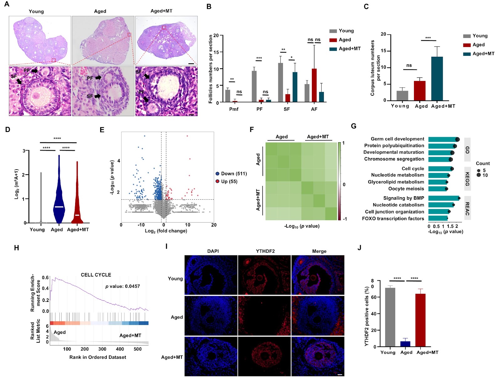
Ovarian aging is a natural process characterized by a decline in both the quantity and quality of oocytes, which subsequently leads to diminished fertility, particularly in women over the age of 35. Given the societal trend toward postponing childbirth, it is imperative to understand the molecular mechanisms that underpin ovarian aging to address infertility issues. Melatonin (MT) is recognized for its therapeutic potential in mitigating ovarian aging; however, the specific epigenetic mechanisms involved, particularly concerning m6A methylation, remain inadequately defined. Our investigation demonstrates that MT mitigates ovarian aging in murine models, significantly decreasing m6A methylation levels. In vitro analyses of ovarian granulosa (KGN) cells reveals a marked increase in YTHDF2 expression, with differentially methylated genes being notably enriched in the polyubiquitination pathway. Further examination shows that YTHDF2 enhances the expression of the E3 ligase UBE3C by modulating the m6A methylation of UBE3C mRNA, thereby reducing the expression of the P53 senescence factor and alleviating the effects of ovarian aging.

Read Full Abstract10.3724/abbs.2025090
Integrated multi-omics and experimental approaches identify fascin actin-bundling protein 1 as an unfavorable prognostic biomarker in adrenocortical carcinomaGraphical AbstractVerified
Acta Biochimica et Biophysica Sinica

Integrated multi-omics and experimental approaches identify fascin actin-bundling protein 1 as an unfavorable prognostic biomarker in adrenocortical carcinoma

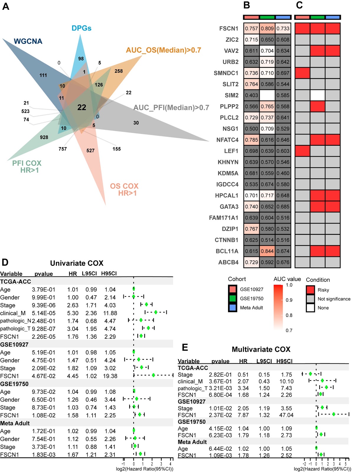
Adrenocortical carcinoma (ACC) is a rare epithelial tumor originating from adrenal cortical cells, notable for its high degree of malignancy and poor prognosis. Owing to heterogeneity, patient outcomes vary significantly. Current biomarkers for ACC risk stratification have notable limitations. However, with the advancement of multi-omics sequencing technology, we can utilize multi-omics data to explore the heterogeneity of ACC, thereby identifying novel biomarkers. In this study, we establish multicenter transcriptomics and ATAC-seq data from the TCGA and GEO databases to perform weighted gene coexpression network analysis (WGCNA) clustering and conduct comprehensive analyses of various ACC samples. These findings are integrated with univariate Cox regression, receiver operating characteristic (ROC) curve analysis, and survival analysis to identify potential biomarkers. We establish FSCN1 as an independent risk factor associated with poor ACC prognosis. ATAC-seq data demonstrate higher chromatin accessibility of FSCN1 in ACC patients with progressive disease. Immunohistochemical analysis confirms the expression of FSCN1 at the protein level, while functional cell assays reveal its role in promoting tumor invasion and proliferation. Functional enrichment analyses highlight the biological characteristics of FSCN1, and estimation of TME-infiltrating cells suggests that FSCN1 expression contributes to poor prognosis by inhibiting CD8+ T-cell infiltration within the ACC microenvironment. Finally, multi-omics analyses elucidate the role of FSCN1 at the mutation level. Taken together, our findings highlight FSCN1 as a promising novel biomarker and potential therapeutic target, underscoring its value in guiding the strategic management of ACC.

Read Full Abstract10.3724/abbs.2025067
Long noncoding RNA UCA1 knockdown inhibits cisplatin-resistant cervical cancer tumorigenesis via the miR-195-5p/IKBKB axisGraphical AbstractVerified
Acta Biochimica et Biophysica Sinica

Long noncoding RNA UCA1 knockdown inhibits cisplatin-resistant cervical cancer tumorigenesis via the miR-195-5p/IKBKB axis

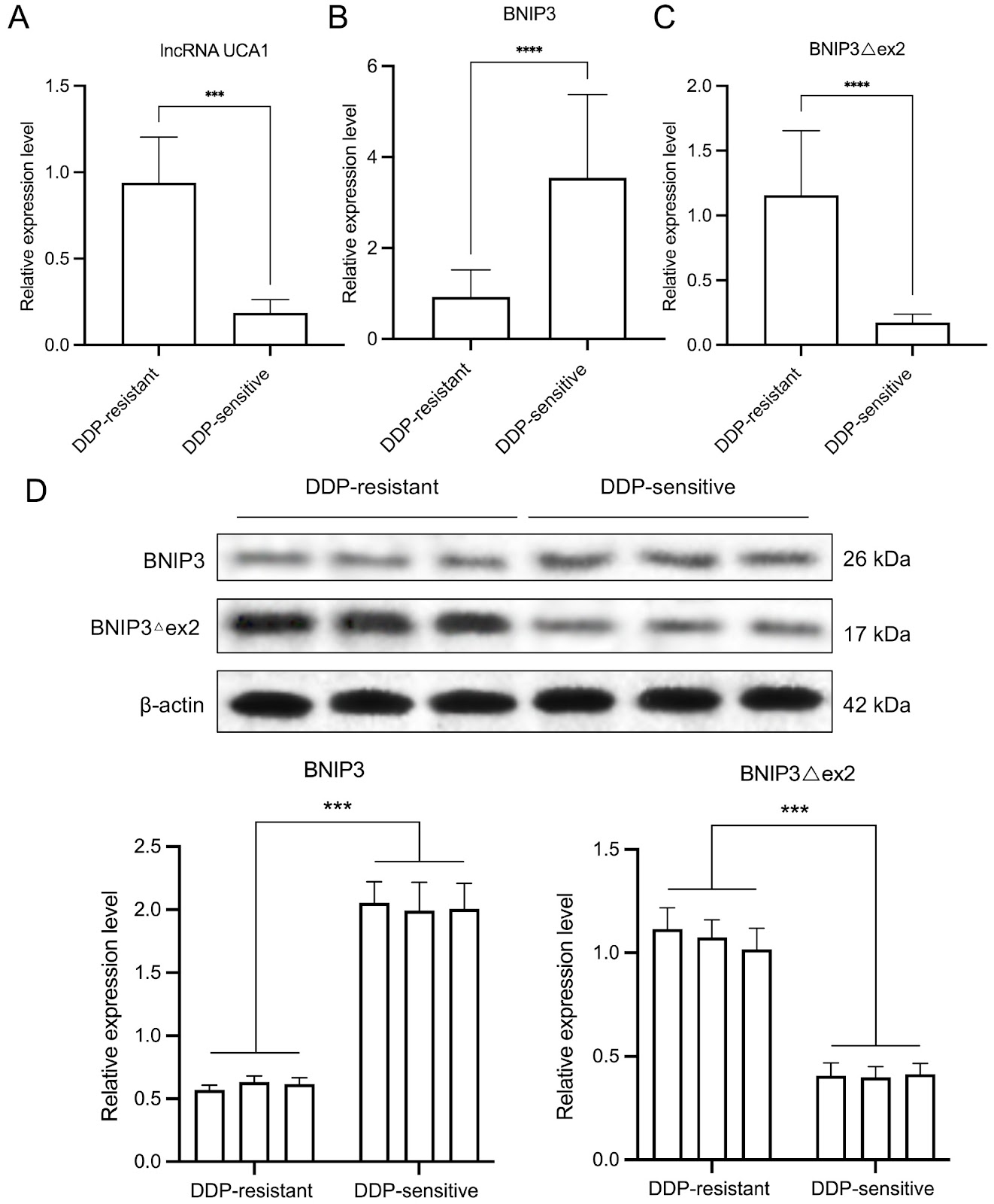
Cisplatin resistance is a major cause of poor prognosis in patients with cervical cancer. Dysregulation of long noncoding RNAs (lncRNAs) plays a key role in chemoresistance. Our results reveal that the lncRNA UCA1 is upregulated in cisplatin (DDP)-resistant cervical cancer tissues and HeLa cells. Mechanistically, the lncRNA UCA1 acts as a sponge for miR-195-5p, targeting IKBKB. UCA1 enhances proliferation, migration, and invasion while reducing apoptosis in DDP-resistant HeLa cells via the miR-195-5p/IKBKB axis. Additionally, UCA1 upregulates BNIP3Δex2 and p-p65 expressions and downregulates BNIP3 expression in DDP-resistant HeLa cells. Abnormal expressions of BNIP3Δex2 and BNIP3 significantly alter the malignant progression of HeLa/DPP cells. In vivo, UCA1 silencing inhibits growth, enhances apoptosis, and upregulates IKBKB, BNIP3Δex2, and p-p65 expressions while downregulating BNIP3 expression in subcutaneous xenografts in nude mice by targeting miR-195-5p. Overall, this study highlights a novel promising target for the treatment of DDP-resistant cervical cancer.

Read Full Abstract10.3724/abbs.2025032
Causal effects of immune cells on the efficacy and adverse drug reactions of platinum drugsGraphical AbstractVerified
Acta Biochimica et Biophysica Sinica

Causal effects of immune cells on the efficacy and adverse drug reactions of platinum drugs

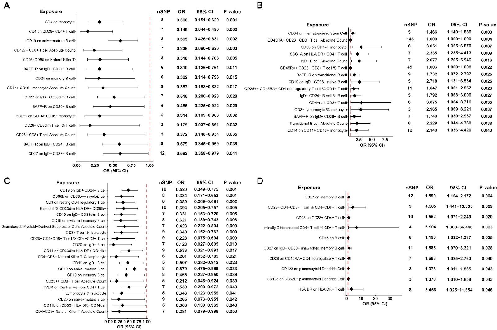
Platinum drugs are widely used in lung cancer chemotherapy, but the immune characteristics of different individuals have different effects on the sensitivity and side effects of platinum drugs. In this study, we use 731 kinds of immune cell traits of 3757 healthy individuals and 429 patients with non-small cell lung cancer (NSCLC) in Xiangya Hospital of Central South University to conduct a Mendel randomized analysis in order to find out the causal relationship between some immune cell traits and the efficacy and adverse reactions of platinum drugs. We find that CD19 on CD24+CD27+ B cell (OR = 0.598, P = 0.004) is the most significant immune cell trait as the protective factor of efficacy. HLA-DR+CD8+ T cell % lymphocyte (OR = 0.427, P = 7.55 × 10⁻⁴) and HLA-DR+CD8+ T cell % T cell (OR = 0.471, P = 0.003) are the protective factors of liver injury. CD39 on CD39+ secreting CD4+ regulatory T cell (OR = 28.729, P = 0.009) and CD3 on CD39+ resting CD4 regulatory T cell (OR = 3.024, P = 0.009) are the risk factors of renal injury. Meanwhile, B cell-related traits mainly affect gastrointestinal upset and cutaneous toxicity, while T cell-related traits mainly affect other outcome variables. These findings may promote our understanding of the relationship between the efficacy and adverse reactions of platinum drugs and the immune system, and promote future development of biomarkers for predicting the efficacy and adverse reactions of platinum drugs.

Read Full Abstract10.3724/abbs.2025052
Modulation of ferroptosis via YY1-SLC7A11 axis in hepatic ischemia-reperfusion injury pathogenesisGraphical AbstractVerified
Acta Biochimica et Biophysica Sinica

Modulation of ferroptosis via YY1-SLC7A11 axis in hepatic ischemia-reperfusion injury pathogenesis

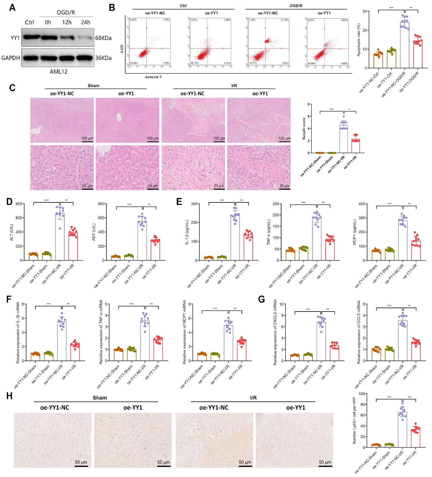
YY1 is a crucial transcription factor and plays significant roles in biological processes. However, the mechanisms of YY1 action in ischemia-reperfusion injury and its regulatory role in ferroptosis have not been extensively studied. This study aims to elucidate the molecular mechanism by which NEDD4L-mediated degradation of YY1 through ubiquitination suppresses SLC7A11 transcription, leading to the promotion of cellular ferroptosis and exacerbation of hepatic ischemia-reperfusion injury (IRI), via the integration of multiple omics sequencing datasets. An IRI-I/R mouse model is established, followed by proteomic sequencing to identify proteins that are differentially expressed during IRI. The altered expression of YY1 is validated, and in vivo and in vitro experiments are used to assess its impact on IRI damage. The E3 ligase NEDD4L, which regulates YY1 ubiquitination, is identified and validated via the UbiBrowser 2.0 database. The ubiquitination types of YY1 and its sites are screened and confirmed through in vitro experiments. Transcriptional sequencing of YY1-overexpressing cell lines is conducted to analyze the involvement of the downstream transcription factor SLC7A11 in IRI, followed by validation of its regulatory role. The results show that YY1 is downregulated in liver tissues during IRI and is expressed primarily in liver cells. YY1 overexpression alleviates liver tissue and liver cell IRI both in vitro and in vivo. Upregulation of E3 ligase expression during IRI promotes the K63-linked ubiquitination of YY1 at the K339 site, leading to proteasomal degradation of YY1. RNA-seq analysis and experimental validation demonstrate that YY1 suppresses IRI-induced ferroptosis via the transcriptional regulation of downstream target genes. YY1 positively regulates SLC7A11 transcription, inhibits IRI-induced ferroptosis and ameliorates liver injury. In summary, the E3 ubiquitin ligase NEDD4L facilitates YY1 protein degradation through ubiquitination, suppressing the transcription of the ferroptosis inhibitor SLC7A11, thus promoting IRI-related ferroptosis and exacerbating liver injury.

Read Full Abstract10.3724/abbs.2025093
Citronellal improves endothelial dysfunction by affecting the stability of the GCH1 proteinGraphical AbstractVerified
Acta Biochimica et Biophysica Sinica

Citronellal improves endothelial dysfunction by affecting the stability of the GCH1 protein

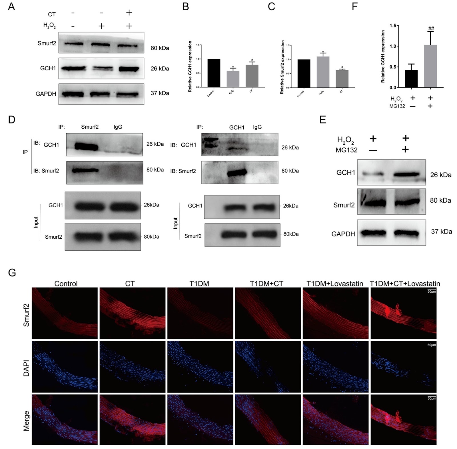
Endothelial dysfunction (ED) serves as the pathological basis for various cardiovascular diseases. Guanosine triphosphate cyclopyrrolone 1 (GCH1) emerges as a pivotal protein in sustaining nitric oxide (NO) production within endothelial cells, yet it undergoes degradation under oxidative stress, contributing to endothelial cell dysfunction. Citronellal (CT), a monoterpenoid, has been shown to ameliorate endothelial dysfunction induced by in atherosclerosis rats. However, whether CT can inhibit the degradation of GCH1 protein is not clear. It has been reported that ubiquitination may play a crucial role in regulating GCH1 protein levels and activities. However, the specific E3 ligase for GCH1 and the molecular mechanism of GCH1 ubiquitination remain unclear. Using data-base exploration analysis, we find that the levels of the E3 ligase Smad-ubiquitination regulatory factor 2 (Smurf2) negatively correlate with those of GCH1 in vascular tissues and HUVECs. We observe that Smurf2 interacts with GCH1 and promotes its degradation via the proteasome pathway. Interestingly, ectopic Smurf2 expression not only decreases GCH1 levels but also reduces cell proliferation and reactive oxygen species (ROS) levels, mostly because of increased GCH1 accumulation. Furthermore, we identify BH4/eNOS as downstream of GCH1. Taken together, our results indicate that CT can obviously improve vascular endothelial injury in Type 1 diabetes mellitus (T1DM) rats and reverse the expressions of GCH1 and Smurf2 proteins in aorta of T1DM rats. Smurf2 promotes ubiquitination and degradation of GCH1 through proteasome pathway in HUVECs. We conclude that the Smurf2-GCH1 interaction might represent a potential target for improving endothelial injury.

Read Full Abstract10.3724/abbs.2024086
Lutonarin attenuates LPS-induced intestinal epithelial barrier dysfunction: a functional and transcriptomic analysisGraphical AbstractVerified
Acta Biochimica et Biophysica Sinica

Lutonarin attenuates LPS-induced intestinal epithelial barrier dysfunction: a functional and transcriptomic analysis

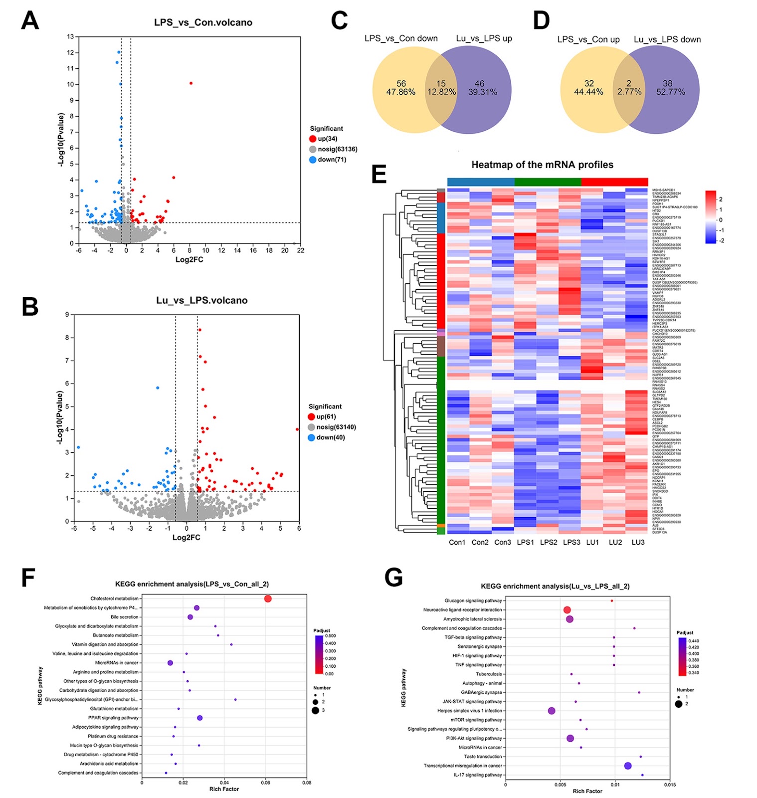
Inflammation, while essential for host defense, can contribute to diseases like inflammatory bowel disease (IBD) when dysregulated. IBD involves chronic intestinal inflammation linked to bacterial infection and disruption of the intestinal epithelial barrier (IEB). The IEB maintains intestinal homeostasis, preventing the entry of harmful substances like lipopolysaccharide (LPS) while allowing nutrient absorption. Preserving IEB function is crucial in managing various diseases. Current IBD treatments often have adverse effects, including facial swelling and acne from corticosteroids, and allergic reactions, nausea, elevated liver tests, and pancreatitis from immunomodulators, increasing interest in natural products. Flavonoids, a diverse class of plant metabolites, have shown promising anti-inflammatory properties. Lutonarin (LU), a flavonoid abundant in barley seedlings, has exhibited various biological activities, including anti-inflammatory effects in LPS-stimulated macrophages. However, its specific impacts on IEB function remain unclear. This study aims to investigate the protective effects of LU against LPS-induced IEB dysfunction using an in vitro Caco-2 cell monolayer model. To assess the possible cytotoxicity of LU and optimize the suitable concentration, the viability of Caco-2 cells was assessed by CCK-8 assay. LU concentrations up to 96 μM did not significantly affect cell viability after 24 and 48 h of exposure. To evaluate the protective effect of LU against LPS-induced cytotoxicity, Caco-2 cells were pretreated with LU (3‒96 μM) for 48 h prior to exposure to 15 μg/mL LPS for 24 h. LPS significantly reduced cell viability, while LU pretreatment attenuated this reduction in a concentration-dependent manner. Concentrations of 12 μM LU were selected for subsequent experiments to minimize potential off-target effects. The Caco-2 cell monolayer model, a well-established in vitro system for investigating IEB function, was employed to study the effects of LU on IEB integrity. Barrier integrity was evaluated using transepithelial electrical resistance (TEER) measurements. LPS treatment significantly reduced TEER, indicating impaired barrier function. Pretreatment with 12 μM LU preserved TEER values, suggesting a protective effect against LPS-induced barrier disruption. Furthermore, paracellular permeability was evaluated using fluorescein isothiocyanate-dextran 4 (FITC-dextran, 4 kDa). LPS significantly increased FITC-dextran (FD4) flux, indicating increased permeability. LU pretreatment markedly attenuated this effect, confirming its ability to prevent LPS-induced permeability changes. To detect if LPS and LU pretreatment changes the expressions of tight junction (TJ) proteins Zonula occludens-1 (ZO-1) and Occludin, real-time qPCR and immunofluorescence staining assays were applied. LPS treatment significantly reduced mRNA expression levels of TJ proteins ZO-1 and Occludin. LU pretreatment effectively mitigated this downregulation, restoring their expression to levels comparable to the CON group.

Read Full Abstract10.3724/abbs.2025069
PGC7 maintains the pluripotency of F9 embryonic carcinoma cells by promoting Nanog translationGraphical AbstractVerified
Acta Biochimica et Biophysica Sinica

PGC7 maintains the pluripotency of F9 embryonic carcinoma cells by promoting Nanog translation

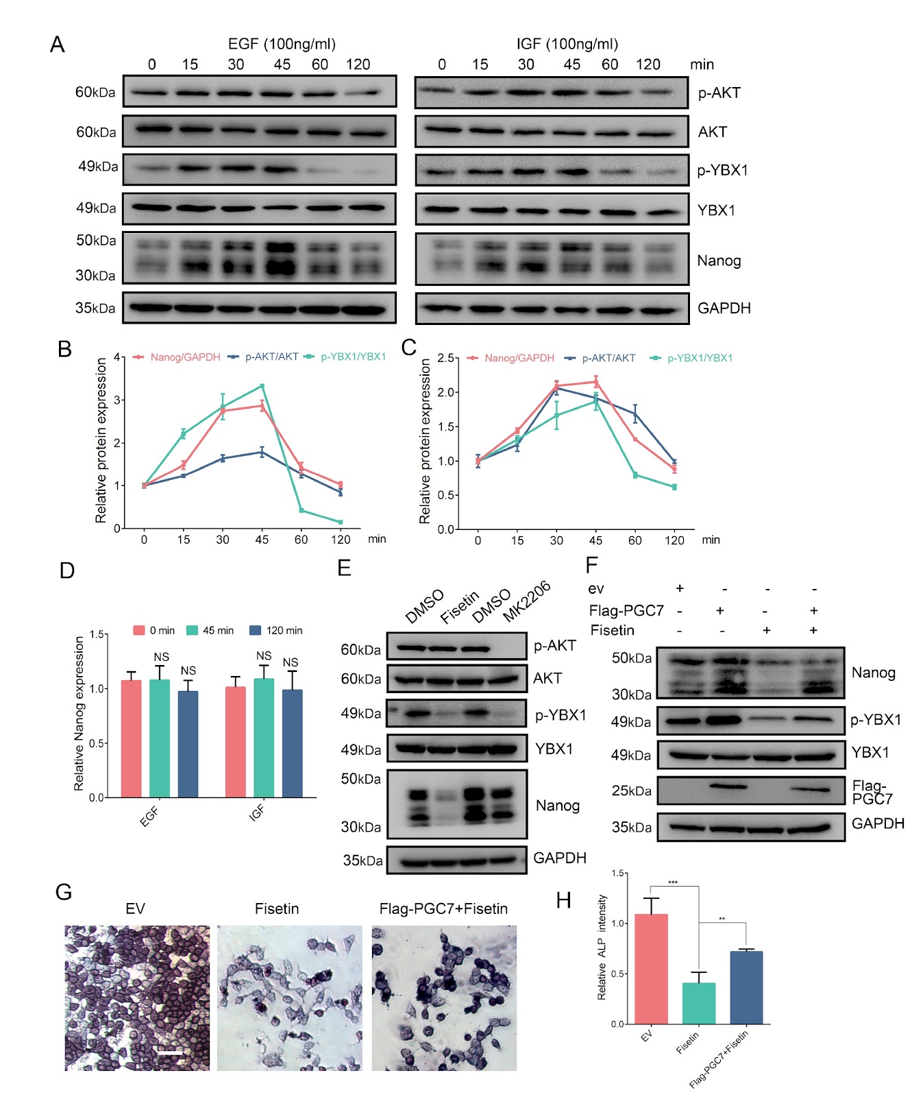
Primordial germ cell 7 (PGC7) is prominently expressed in primordial germ cells (PGCs) and embryonic stem cells (ESCs), serving as a pivotal marker for discerning stem cell pluripotency. However, the role of PGC7 in regulating core pluripotency factors remains unclear. In this study, the expression dynamics of PGC7 and pluripotency-associated proteins are systematically evaluated by quantitative reverse transcription PCR (RT-qPCR) and western blot analysis. Complementary experimental approaches including confocal immunofluorescence and Co-immunoprecipitation (Co-IP) assays are subsequently employed to establish subcellular colocalization patterns and elucidate the molecular mechanisms associated with PGC7 function. The results show that PGC7 is closely associated with the pluripotency status of F9 embryonal carcinoma (EC) cells. Notably, PGC7 can counteract the decrease in pluripotency induced by retinoic acid (RA). Ectopic expression of PGC7 in F9 EC cells enhances the translation of Nanog. Mechanistic analysis reveal that PGC7 activates Y-box binding protein 1 (YBX1) phosphorylation by enhancing the interaction between YBX1 and AKT1. The subsequent phosphorylation of YBX1 reduces its binding to Nanog mRNA and promotes the translation of Nanog. These results shed light on a previously unknown role of PGC7 in supporting the translation of Nanog, offering valuable insights into the functions of PGC7 in F9 EC cells.

Read Full Abstract10.3724/abbs.2025035
Morroniside promotes skin wound re-epithelialization by facilitating epidermal stem cell proliferation through GLP-1R-mediated upregulation of β-catenin expressionGraphical AbstractVerified
Acta Biochimica et Biophysica Sinica

Morroniside promotes skin wound re-epithelialization by facilitating epidermal stem cell proliferation through GLP-1R-mediated upregulation of β-catenin expression

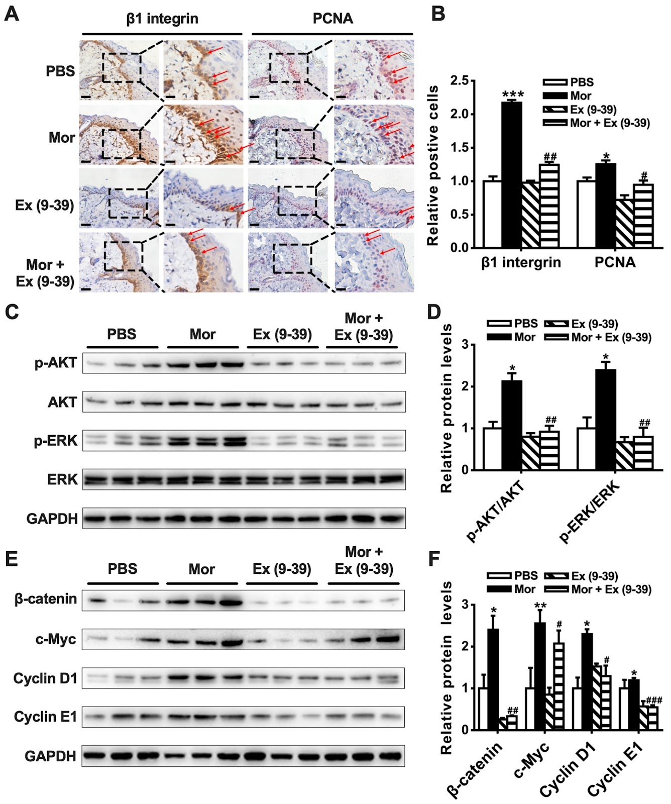
Epidermal stem cells (EpSCs) play a vital role in skin wound healing through re-epithelialization. Identifying chemicals that can promote EpSC proliferation is helpful for treating skin wounds. This study investigates the effect of morroniside on cutaneous wound healing in mice and explores the underlying mechanisms. Application of 10‒50 μg/mL of morroniside to the skin wound promotes wound healing in mice. In vitro studies demonstrate that morroniside stimulates the proliferation of mouse and human EpSCs in a time- and dose-dependent manner. Mechanistic studies reveal that morroniside promotes the proliferation of EpSCs by facilitating the cell cycle transition from the G1 to S phase. Morroniside increases the expression of β-catenin via the glucagon-like peptide-1 receptor (GLP-1R)-mediated PKA, PKA/PI3K/AKT and PKA/ERK signaling pathways, resulting in an increase in cyclin D1 and cyclin E1 expression, either directly or by upregulating c-Myc expression. This process ultimately leads to EpSC proliferation. Administration of morroniside to mouse skin wounds increases the phosphorylation of AKT and ERK, the expressions of β-catenin, c-Myc, cyclin D1, and cyclin E1, as well as the proliferation of EpSCs, in periwound skin tissue, and accelerates wound re-epithelialization. These effects of morroniside are mediated by the GLP-1R. Overall, these results indicate that morroniside promotes skin wound healing by stimulating the proliferation of EpSCs via increasing β-catenin expression and subsequently upregulating c-Myc, cyclin D1, and cyclin E1 expressions through GLP-1R signaling pathways. Morroniside has clinical potential for treating skin wounds.

Read Full Abstract10.3724/abbs.2024070
(Pro)renin receptor promotes cardiomyocyte senescence via tripartite motif-containing 24-mediated stabilization of p53 in diabetic cardiomyopathyGraphical AbstractVerified
Acta Biochimica et Biophysica Sinica

(Pro)renin receptor promotes cardiomyocyte senescence via tripartite motif-containing 24-mediated stabilization of p53 in diabetic cardiomyopathy

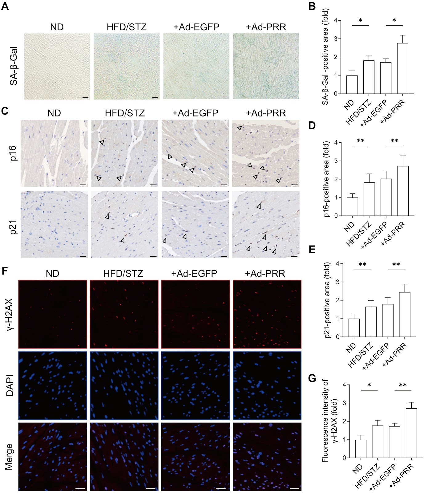
Diabetic cardiomyopathy (DCM) is a major complication of diabetes and a leading contributor to heart failure, in which cardiomyocyte senescence plays an increasingly recognized role. However, the underlying mechanisms driving this process remain poorly defined. Here, we identify the (pro)renin receptor (PRR) as a critical mediator of cardiomyocyte senescence in DCM. Using a high-fat diet and streptozotocin (STZ)-induced DCM mouse model, as well as primary cardiomyocytes exposed to high glucose and palmitic acid, we demonstrate that PRR expression is significantly upregulated in diabetic hearts and closely associated with key senescence markers, including SA-β-gal, γ-H2AX, p16, and p21. PRR overexpression exacerbates these senescence phenotypes and promotes the secretion of profibrotic senescence-associated secretory phenotype factors, contributing to increased myocardial fibrosis and cardiac dysfunction. Mechanistically, PRR stabilizes the p53 protein by inhibiting tripartite motif-containing 24 (TRIM24)-mediated ubiquitination and proteasomal degradation, thereby activating the p53-p21 axis. These findings reveal a novel role of the PRR in diabetic myocardial senescence and provide potential therapeutic targets for attenuating DCM progression.

Read Full Abstract10.3724/abbs.2025185
Zinc fingers are responsible for the efficient control of KLF7 on the transcription of genes in the NF-κB signaling pathway and fatty acid β-oxidationGraphical AbstractVerified
Acta Biochimica et Biophysica Sinica

Zinc fingers are responsible for the efficient control of KLF7 on the transcription of genes in the NF-κB signaling pathway and fatty acid β-oxidation

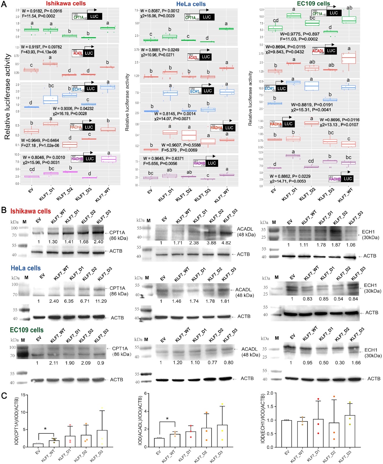
Krüppel-like factors (KLFs) are a family of 18 transcriptional regulators characterized by three highly conserved C2H2 zinc fingers at their C-terminal regions. KLF7, a member of this family, plays a crucial role in cell proliferation, differentiation, and the development of the nervous system, adipogenesis, diabetes, and various cancers. Studies have shown that KLF7 aggravates metabolic disorders by impeding insulin secretion and sensitivity. The nuclear factor kappa-B (NF-κB) signaling cascade is essential for inflammatory responses, while fatty acid β-oxidation is vital for metabolism. Both are linked to insulin resistance, obesity, and cardiovascular diseases. A functional link between KLF7 and the NF-κB signaling pathway has been demonstrated. In rheumatoid arthritis, KLF7 activates NF-κB signaling pathway, leading to increased cell proliferation and the production of proinflammatory cytokines, including interleukin 6 (IL-6), IL-1β, and IL-17A. In adipose tissue, KLF7 may initiate NF-κB signaling pathway by upregulating protein kinase Cζ, causing significant IL-6 secretion. KLF7 also reduces oleate-induced lipid droplets in chicken preadipocytes, indicating its role in fatty acid metabolism. Studies in mice showed that KLF7 regulates the transcription of genes of the rate-limiting glycolytic enzyme phosphofructokinase liver type (PFKL) and the fatty acid β-oxidation enzyme acyl-CoA dehydrogenase long-chain (ACADL) in cardiomyocytes independently of peroxisome proliferator-activated receptor (PPAR) γ, thereby altering heart metabolism. Additionally, KLF7 may promote cervical cancer progression by enhancing fatty acid utilization efficiency at least via ACADL upregulation. Overall, KLF7 is crucial for the regulation of fatty acid β-oxidation. KLF7 is a ring-shaped protein with zinc fingers forming the protruding part of ring surface. The lack of the third zinc finger domain in KLF7 affected its function in chicken preadipocytes. However, the importance of zinc finger domains for the regulatory function of human KLF7 remains unclear. In this study, we engineered an overexpression vector for wild-type KLF7 (pCMV-myc-KLF7_WT) and three vectors for KLF7 mutants with different zinc finger deletions (pCMV-myc-KLF7_D1, pCMV-myc-KLF7_D2, and pCMV-myc-KLF7_D3). These vectors were constructed using primers shown in Supplementary Table S1 and cDNA from HEK293T cells. Western blot analysis in the HEK293T, Ishikawa, HeLa, and EC109 cells (Pricella, Wuhan, China) showed that, unlike cells transfected with the empty vector (EV) of pCMV-myc (Clontech, Mountain View, USA), Myc-tagged proteins appeared at expected sizes in cells transfected with either the wild-type KLF7 or any of the three mutant KLF7 overexpression plasmids after 48 h (Figure 1A and Supplementary Figure S1). The impacts of overexpressing various KLF7 isoforms on gene transcription related to the NF-κB signaling pathway and fatty acid β-oxidation were evaluated using luciferase reporter assays, real-time PCR, and western blot analysis in Ishikawa, HeLa, and EC109 cells 48 h post-transfection. Details of the luciferase reporter assay transfection protocol are provided in Supplementary Table S2, the oligonucleotide sequences for real-time PCR are shown in Supplementary Table S3, and the antibodies for western blot analysis are listed in Supplementary Table S4. Compared to EV group, wild-type KLF7 overexpression significantly boosted NF-κB pathway activity in HeLa and EC109 cells (P < 0.05, Figure 1B). Furthermore, wild-type KLF7 overexpression significantly elevated IL-6 and TNF-α expressions in Ishikawa and HeLa cells (P < 0.05, Figure 1C), confirming previous findings that KLF7 enhances inflammation via the NF-κB pathway [4,5]. Cells transfected with KLF7 overexpression plasmids lacking zinc fingers showed significant differences in NF-κB pathway activities compared to those transfected with the wild-type KLF7 overexpression plasmid (P < 0.05, Figure 1B). In HeLa and EC109 cells, the absence of zinc fingers reduced NF-κB signaling activity, with the reduction proportional to the number of zinc fingers lost (P < 0.05, Figure 1B). In Ishikawa cells, losing one or two zinc fingers increased NF-κB activity compared to the wild-type KLF7 (P < 0.05, Figure 1B). Additionally, the ability of KLF7 overexpression to increase IL-6 and TNF-α expression decreased with the loss of zinc fingers.

Read Full Abstract10.3724/abbs.2025053
FOXP3 targets KIF5A to increase lactate production and promote docetaxel resistance in lung adenocarcinomaGraphical AbstractVerified
Acta Biochimica et Biophysica Sinica

FOXP3 targets KIF5A to increase lactate production and promote docetaxel resistance in lung adenocarcinoma

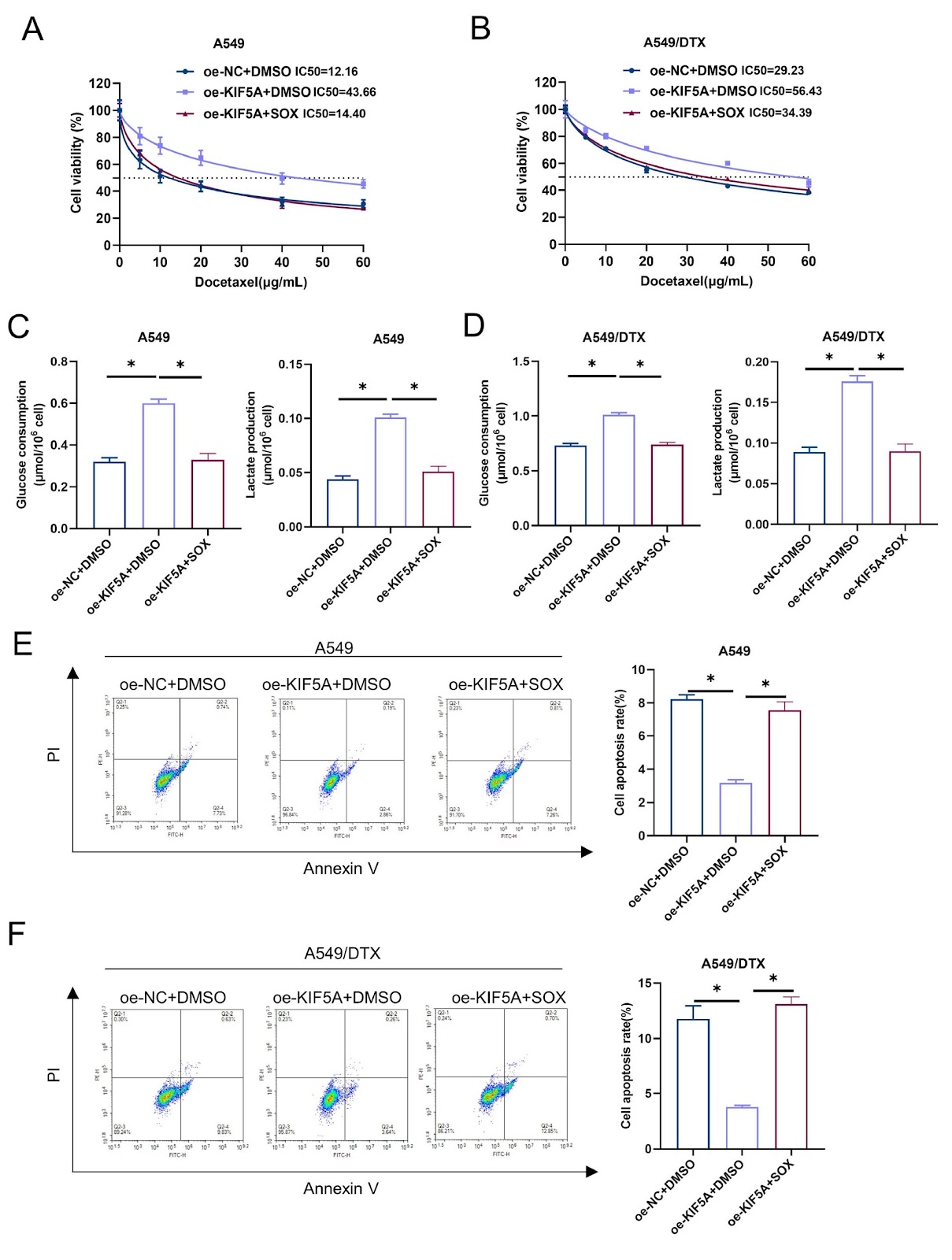
A prominent cause of cancer-related fatalities with a poor prognosis is lung adenocarcinoma (LUAD). KIF5A, a crucial member of the kinesin superfamily, is linked to drug resistance in malignancies. This work aims to investigate the mechanism of KIF5A in docetaxel (DTX) resistance in LUAD cells. The results of bioinformatics analysis, qRT-PCR and western blot analysis show that KIF5A, which is involved in the glycolysis pathway, is highly expressed in LUAD and is positively correlated with glycolysis-related genes. We further verify that silencing of KIF5A inhibits DTX resistance, glycolysis, and lactate production in LUAD cells via cell counting kit-8 (CCK-8), flow cytometry, Seahorse XFe 96, lactate, and glucose assays. Mechanistically, KIF5A promotes DTX resistance in LUAD, and this effect is attenuated upon the addition of an LDHA inhibitor. Chromatin immunoprecipitation and dual-luciferase reporter assays reveal that FOXP3 transcriptionally activates KIF5A. Knockdown of FOXP3 reduces lactate production and enhances DTX sensitivity in LUAD, which is restored upon simultaneous overexpression of KIF5A. Our findings reveal that FOXP3 increases DTX resistance in LUAD cells by enhancing lactate production through the upregulation of KIF5A level. In conclusion, our study provides a novel treatment target for improving chemosensitivity in LUAD.

Read Full Abstract10.3724/abbs.2024082