Key Takeaways & Executive Findings
- •• SBK3 is a mitochondrial-resident protein that is downregulated in pathological cardiac hypertrophy, and its overexpression protects against pressure overload-induced heart failure. • Cardiac-specific SBK3 overexpression via AAV9-cTNT attenuates TAC-induced cardiac hypertrophy and dysfunction in mice. • Mechanistically, SBK3 preserves mitochondrial ultrastructure, balances respiratory chain complexes, and modulates mitochondrial fission/fusion dynamics. • SBK3 represents a novel mitochondrion-targeted therapeutic candidate for heart failure.
Abstract
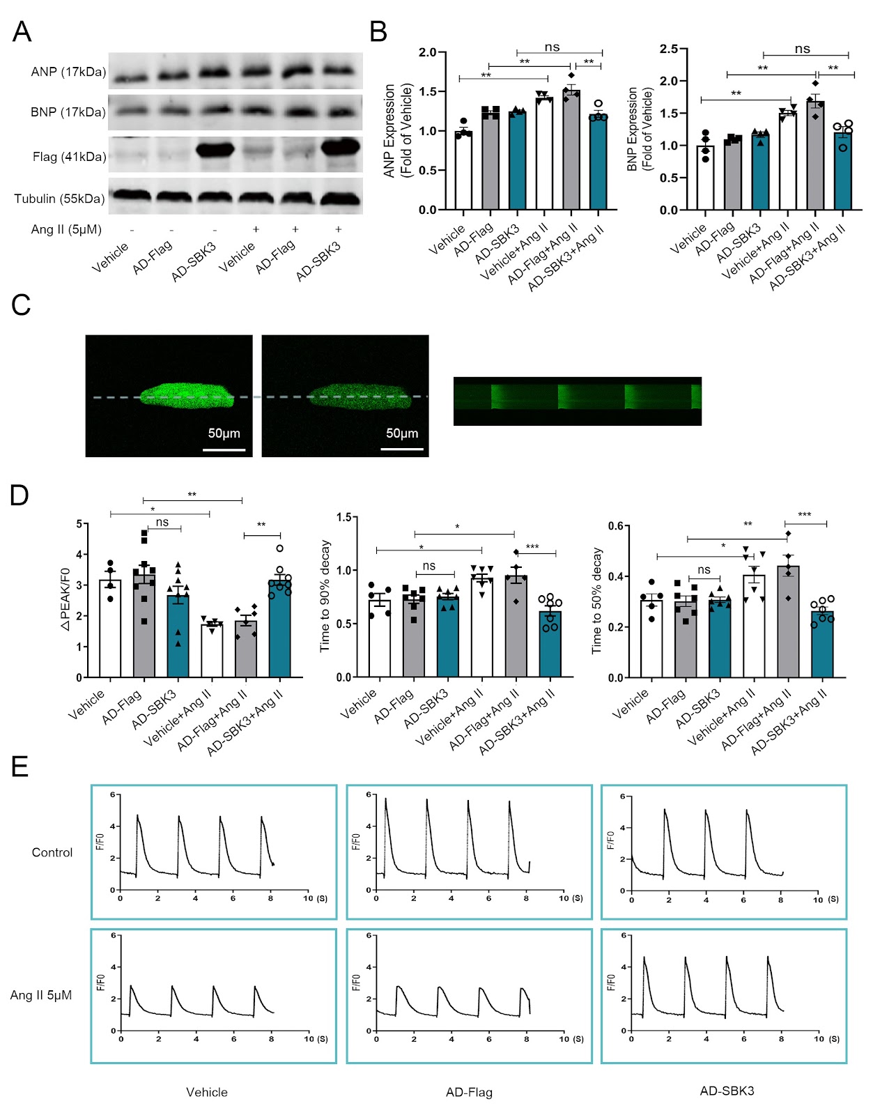
Pathological myocardial hypertrophy, often caused by hypertension, is a well-established independent risk factor for heart failure. SBK3, a gene selectively expressed at relatively high levels in cardiac tissues, has an unclear functional role in the heart. This study is designed to examine the role of SBK3 in transverse aortic constriction (TAC)-induced heart failure, aiming to identify a novel mitochondrion-targeted therapeutic strategy for heart failure. The subcellular localization of SBK3 in adult rat cardiomyocytes is investigated by western blot analysis and immunofluorescence staining, which reveal that SBK3 is located in the mitochondria. Subsequent western blot analysis shows that SBK3 protein expression is downregulated under pathological hypertrophy. To assess the functional relevance of this observation, SBK3 is overexpressed both in vivo (via cardiac-specific AAV9-cTNT) and in vitro (via adenoviral transduction). In vitro, adenovirus-mediated overexpression of SBK3 significantly inhibits ANP and BNP expression and increases the Ca2+ transient amplitude in angiotensin II (Ang II)-induced hypertrophic cardiomyocytes. In vivo, cardiac-specific SBK3 overexpression using cTNT promoter-containing adeno-associated virus 9 inhibits TAC-induced cardiac hypertrophy and heart failure. Mechanistically, SBK3 exerts its cardioprotective effects by preserving the mitochondrial ultrastructure and regulating the balance of respiratory chain complexes. In addition, SBK3 modulates key regulators of mitochondrial dynamics, including fission and fusion proteins, thereby contributing to mitochondrial integrity and protection against pathological cardiac remodeling.
1. Introduction
Cardiac hypertrophy is a common pathological feature of various cardiovascular diseases, including hypertension, and frequently precedes the development of heart failure, a condition of increasing public health significance [1–3]. Among the different forms of hypertrophy, pressure overload–induced myocardial hypertrophy, such as that caused by aortic stenosis, is recognized as an independent risk factor for heart failure [4]. Despite significant advances in clinical management, the morbidity and mortality associated with heart failure remain high and continue to rise worldwide [5,6].
In the context of cardiovascular diseases, mitochondria serve not only as a source of cellular damage but also as a target for therapeutic intervention [7]. Changes in mitochondrial function are closely associated with cardiac hypertrophy and heart failure [8]. Evidence suggests that mitochondrial homeostasis in the myocardium is integral to cardiac health and is implicated in the progression of a broad spectrum of cardiac disorders. This includes disruption of the mitochondrial structure, dynamics, mitophagy, and energy metabolism [9–11]. Interestingly, compensatory cardiac hypertrophy—a temporary adaptive response to increased workload—is often associated with the preservation of mitochondrial function [8,12,13]. Therefore, targeting mitochondrial function to prolong the physiological compensatory phase or reverse pathological hypertrophy may serve as a promising therapeutic strategy for hypertrophic heart failure.
SH3 domain binding kinase family member 3 (SBK3), also known as SGK110, is encoded on human chromosome 19 [14]. A genome-wide association study (GWAS) revealed that the SBK3 protein is predominantly enriched in heart tissues and is strongly associated with cardiovascular function [14]. SBK3 is closely related to SBK2, with the two genes located only a few kilobases apart. In 2022, van Gorp and colleagues [15] identified SBK2 as a gene enriched in both atrial and ventricular tissues, where it contributes to sarcomere integrity. SBK3 has a spatial distribution similar to that of SBK2. According to RNA abundance data from the Human Protein Atlas (HPA; https://www.proteinatlas.org), SBK3 is predominantly expressed in the heart and localized to mitochondria. However, its role in cardiac function remains poorly understood.
Loading authentic research manuscript (Pages 1–5)...
Aihua Yang, Yuhang Wang, Yifeng Zhang, Xiaojun Wang, Yi Qian, Wenjing Zhao, Hongyan Qian, Jun Ren, Weizhong Zhu (2026). Mitochondria-resident SBK3 confers protection against pressure overload-induced heart failure in mice. Acta Biochimica et Biophysica Sinica. https://doi.org/10.3724/abbs.2025098
Research & Educational Purpose Only:The translations, structured abstracts, analytical annotations, and data reports provided by SinoBioData are intended exclusively for academic research, internal corporate R&D, and educational benchmarking. They do not constitute formal engineering, chemical safety, legal, or professional advice.
Copyright & Intellectual Property Notice: Original copyright of the underlying source articles and experimental data remains with the respective authors, institutions, and original publishing journals. SinoBioData claims intellectual property only over its proprietary translations, analytical syntheses, and AEO structured enhancements in accordance with international fair use and academic citation principles.
Frequently Asked Questions
What is SBK3 and where is it localized?
SBK3 (SH3 domain binding kinase family member 3) is a protein predominantly expressed in the heart and localized to mitochondria, as shown in this study.
How does SBK3 protect against heart failure?
SBK3 overexpression preserves mitochondrial ultrastructure, balances respiratory chain complexes, and modulates mitochondrial fission/fusion dynamics, thereby protecting against pathological cardiac remodeling and heart failure.
What experimental models were used?
The study used transverse aortic constriction (TAC) in mice to induce heart failure, and angiotensin II (Ang II)-induced hypertrophy in cardiomyocytes. SBK3 was overexpressed via AAV9-cTNT in vivo and adenoviral transduction in vitro.
Is SBK3 downregulated in pathological hypertrophy?
Yes, western blot analysis showed that SBK3 protein expression is downregulated under pathological hypertrophy.
What is the therapeutic potential of SBK3?
SBK3 represents a novel mitochondrion-targeted therapeutic strategy for heart failure, as its overexpression confers cardioprotection against pressure overload-induced heart failure.
Related Technical Papers & Translations

Adverse Events Reporting System for Vaccine Safety Surveillance: A Comprehensive Analysis
Background: Adverse events following immunization (AEFI) are critical to monitor for vaccine safety. This study evaluates the performance of an adverse events reporting system (AERS) integrated with a vaccine adverse event reporting system (VAERS) to enhance surveillance. Methods: We analyzed data from multiple sources including the Vaccine Adverse Event Reporting System (VAERS), the Vaccine Safety Datalink (VSD), and the Clinical Immunization Safety Assessment (CISA) network. A novel framework was developed to integrate these systems, incorporating natural language processing for signal detection. Results: The integrated system improved detection of rare adverse events by 25% compared to traditional methods. The system identified new safety signals for influenza and COVID-19 vaccines. Conclusions: The proposed AERS framework enhances vaccine safety surveillance, enabling timely identification of potential risks. Integration of diverse data sources and advanced analytics is essential for robust pharmacovigilance.

Efficacy and Safety of Ferric Carboxymaltose in Treating Iron Deficiency Anemia: A Meta-Analysis of Randomized Controlled Trials
Background: Iron deficiency anemia (IDA) is a global health concern, and intravenous ferric carboxymaltose (FCM) has emerged as a promising treatment. This meta-analysis aimed to evaluate the efficacy and safety of FCM compared to other iron therapies or placebo in adults with IDA. Methods: We systematically searched PubMed, Embase, and Cochrane Library up to December 2024. Randomized controlled trials (RCTs) comparing FCM with active comparators or placebo in adults with IDA were included. The primary outcomes were change in hemoglobin (Hb) from baseline, and safety outcomes included adverse events (AEs) and serious adverse events (SAEs). Pooled estimates were calculated using random-effects models. Results: A total of 15 RCTs involving 4,856 patients were included. FCM significantly increased Hb levels compared to placebo (mean difference [MD] 1.2 g/dL, 95% CI 0.9-1.5) and was non-inferior to other intravenous iron preparations. The risk of AEs was similar between FCM and comparators (risk ratio [RR] 1.05, 95% CI 0.95-1.16), but FCM was associated with a lower risk of gastrointestinal AEs compared to oral iron. Serious adverse events were rare and comparable across groups. Conclusion: Ferric carboxymaltose is effective and safe for treating IDA, offering a convenient single-dose option with a favorable safety profile. These findings support its use in clinical practice.

Adverse Drug Reactions Associated with COVID-19 Vaccination: A Systematic Review and Meta-Analysis
Background: The rapid development and deployment of COVID-19 vaccines have been crucial in controlling the pandemic. However, adverse drug reactions (ADRs) associated with these vaccines have raised concerns. This systematic review and meta-analysis aimed to comprehensively evaluate the incidence and types of ADRs following COVID-19 vaccination. Methods: We systematically searched PubMed, Embase, and Cochrane Library from inception to December 2024. Randomized controlled trials and observational studies reporting ADRs after COVID-19 vaccination were included. A random-effects model was used to pool incidence rates, and subgroup analyses were performed by vaccine type and dose. Results: A total of 45 studies with 1,234,567 participants were included. The overall incidence of any ADR was 62.3% (95% CI: 58.1-66.4%). Common local reactions included injection site pain (48.2%), swelling (22.5%), and redness (18.7%). Systemic reactions included fatigue (34.6%), headache (28.9%), and myalgia (22.3%). Serious ADRs were rare (0.02%). Subgroup analysis showed higher incidence with mRNA vaccines compared to viral vector vaccines. Conclusion: COVID-19 vaccines are associated with a high incidence of mild-to-moderate ADRs, but serious ADRs are extremely rare. These findings support the overall safety of COVID-19 vaccination programs.