🧬 SinoBioData Academic Portal
🏛️ Indexed Academic JournalOriginal: 中国组织工程研究

Chinese Journal of Tissue Engineering Research

Premier Chinese Biomedical Journal indexed in SinoBioData: Chinese Journal of Tissue Engineering Research (中国组织工程研究).

Total Research Papers: 200
Access: 100% Free Open Access
Browse by Publication Year & VolumeReset All Filters ✕

Published Research PapersFiltered: Year 2026 • Vol 1901

Showing 32 of 200 peer-reviewed papers with full Graphical Abstracts.

Original ResearchVol 1901, Issue 29 • pp. 100-112DOI: 10.12307/2026.21365Jan 15, 2026

Exosomes and neuropathic pain: visualization analysis on literature

Authors: MA Jing, HAN Jing, SHI Linyu, WU Yuwei, YUAN Haiguang, LI Yongfeng

BACKGROUND: Neuropathic pain has a complex pathogenesis and limited clinical intervention outcomes. In recent years, exosomes have gradually emerged as a focal point in the study of neuropathic pain due to their unique intercellular communication functions and molecular delivery capabilities. OBJECTIVE: To systematically review research progress on exosomes in the field of neuropathic pain using bibliometric methods, summarize the knowledge framework and research hotspots, and provide theoretical foundations and translational strategies for advancing this field. METHODS: Based on the Web of Science Core Collection (WOSCC) database, literature on exosomes and neuropathic pain was retrieved from 2012-01-01 to 2025-03-31. VOSviewer and CiteSpace software were used for keyword co-occurrence, cluster analysis, burst detection, and collaboration network visualization. RESULTS AND CONCLUSION: A total of 313 articles were included. Among 42 countries or regions, the United States and China made significant contributions. Shanghai Jiao Tong University and Nantong University were the most prolific institutions. The most productive and co-cited journals were Neural Regeneration Research and International Journal of Molecular Sciences, respectively. A total of 382 authors were identified; Zhang Zhenggang had the most articles, and Zhang Yi had the most citations. Key high-frequency keywords included 'nerve regeneration', 'neuroinflammation', 'schwann cell', 'regenerative medicine', and 'spinal cord injury', which are key research areas for future development. Through bibliometric analysis, this study mapped the research trends of exosomes in neuropathic pain. Exosomes hold broad prospects in elucidating basic mechanisms and developing novel precision therapies for neuropathic pain. Future efforts should integrate multi-omics and engineered exosome platforms to advance this field.
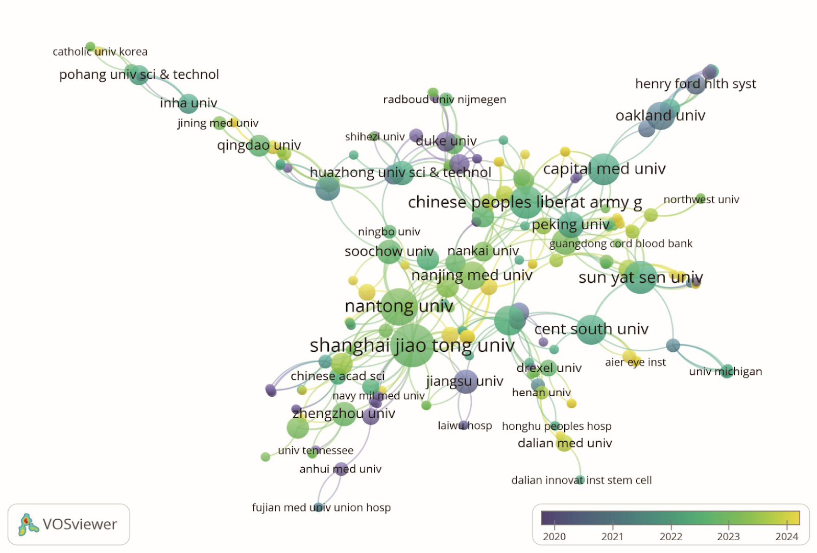

Exosomes and neuropathic pain: visualization analysis on literature
Graphical Abstract
Original ResearchVol 1901, Issue 29 • pp. 100-112DOI: 10.12307/2026.21364Jan 15, 2026

Exosomes promote diabetic wound healing: a visual analysis of research hotspots and evolutionary trends

Authors: Jian Xichao, Shao Jingjie, Tang Shihan, Qi Fang, Deng Chengliang

BACKGROUND: Diabetes wound is one of the serious complications of diabetes patients, and its complex pathological mechanism and clinical treatment dilemma is still a major challenge. In recent years, exosomes have become a new focus in the field of diabetes wound research because they play a key role in intercellular communication, immune regulation, and tissue repair. OBJECTIVE: To investigate the research hotspots and evolutionary trends of exosomes in diabetic wound healing. METHODS: A systematic search was conducted in the Web of Science core collection to identify English literature focusing on exosomes in diabetic wound healing and published between the inception of the database and December 31, 2024. The annual publication volume was analyzed to track changes over time. Visual analyses using VOSviewer and CiteSpace software were performed on the retrieved literature to examine key aspects such as authors, countries, institutions, journals, and keywords, providing insights into the current research landscape and evolving hot topics in exosomes for diabetic wound healing. RESULTS AND CONCLUSION: From 2014 to 2024, a total of 424 publications on exosome-promoted diabetic wound healing were produced, contributed by 2,883 authors from 46 countries and featured in 199 journals. In the realm of exosome-promoted diabetic wound healing, China had the highest number of publications, followed by the United States. The journals 'Journal of Nanobiotechnology' and 'Advanced Healthcare Materials' published the most papers and had high influence. Author Chen Zhenbing and Huazhong University of Science and Technology were the most productive author and institution, respectively, but the researcher clusters have not yet reached a certain scale, and future collaboration needs to be strengthened. Global research focus mainly concentrated on 10 thematic clusters including adipose stem cells, diabetic wounds, diabetic wound healing, wound healing, endoplasmic reticulum stress, microvesicles, collagen, proteomics, and diabetic foot infection. The research hotspots in this field are undergoing a transition from molecular mechanisms to systematic interventions. Future research hotspots will focus on angiogenesis, macrophages, antibacterial, and hydrogels. On this basis, integrating multidisciplinary technologies to achieve more effective precision treatment and optimize management strategies for diabetic wounds.

Exosomes promote diabetic wound healing: a visual analysis of research hotspots and evolutionary trends
Graphical Abstract
Original ResearchVol 1901, Issue 29 • pp. 100-112DOI: 10.12307/2026.21363Jan 15, 2026

Effects of human umbilical cord blood mesenchymal stem cells on pain and function in patients with knee osteoarthritis: a meta-analysis

Authors: Liu Yanzhe, Liu Hua, Yang Tubao, Liu Yupeng

OBJECTIVE: To conduct a meta-analysis concerning the effects of human umbilical cord blood mesenchymal stem cells on pain and function in patients with knee osteoarthritis. METHODS: Using the Chinese search terms “human umbilical cord blood, mesenchymal stem cells, knee joint-related diseases” and the English search terms “human cord blood, mesenchymal stem cell, MSC, knee osteoarthritis, knee joint disease, knee joint disorders, knee OA,” we conducted searches in the CNKI, WanFang, VIP, PubMed, Elsevier, and Web of Science databases. The search timeframe spanned from the establishment of each database until June 13, 2024. The quality of the included literature was assessed using the Cochrane Risk of Bias tool and the ROBINS-I tool. For meta-analysis, the Revman software was utilized, calculating mean differences for continuous variables and relative risks for dichotomous variables, along with 95% confidence intervals. RESULTS: Three randomized controlled trials and three case-control studies were included, totaling 248 subjects, with moderate quality. Meta-analysis showed: (1) The visual analog scale score in the experimental group was lower than that in the control group, with a significant difference (χ²=44.98, P < 0.001, I²=91%); (2) The Western Ontario and McMaster Universities Osteoarthritis Index in the experimental group was lower than that in the control group, with a significant difference (χ²=16.84, P < 0.001, I²=88%); (3) The Lysholm knee function score in the experimental group was higher than that in the control group, with a significant difference (χ²=0.12, P=0.73, I²=0%); (4) The incidence of adverse reactions in the experimental group was higher than that in the control group, with a significant difference (χ²=4.99, P < 0.001, I²=20%), with a combined risk difference of 0.21, translating to a number needed to treat of 5. CONCLUSION: Human umbilical cord blood mesenchymal stem cells can reduce pain and improve knee function in patients with knee osteoarthritis, achieving a good balance between safety and efficacy.

Effects of human umbilical cord blood mesenchymal stem cells on pain and function in patients with knee osteoarthritis: a meta-analysis
Graphical Abstract
Original ResearchVol 1901, Issue 29 • pp. 100-112DOI: 10.12307/2026.21362Jan 15, 2026

In vitro simulation of cellular exercise environments: advancements in methodology and signal simulation

Authors: CHEN Bingao, CHEN Hongbao, XIE Hao, DING Xinglei, YUAN Yu, ZHANG Jiahao, BAN Weikang, XU Shenghao, YUAN Yang

BACKGROUND: With an increasing understanding of the health benefits of exercise, research on the mechanisms of exercise intervention has become a focal point. Traditional studies rely on in vivo animal models or multi-omics techniques to indirectly infer exercise intervention mechanisms, but the research is not in-depth enough, and many disease models cannot achieve the prescribed exercise intensity. Therefore, in vitro cell-based exercise environment simulation techniques are of particular significance. Existing technologies primarily focus on the replication of single signals, failing to comprehensively simulate the interaction of multi-dimensional signals during exercise, which limits the understanding of exercise adaptation mechanisms. OBJECTIVE: To explore the technological advancements in in vitro cell-based exercise environment simulation, analyze the advantages of existing signal simulation techniques, and propose a new framework integrating multi-dimensional signals to promote the precise replication of exercise mechanisms and application research in related fields. METHODS: This study conducted a search in the PubMed and Web of Science databases using keywords such as Exercise, Physiology, Molecular Signals, Myokines, Exerkines, etc. After initial screening and removal of duplicates, 5,046 relevant articles were identified, and 99 were finally included after further screening. RESULTS AND CONCLUSION: Existing in vitro cell exercise simulation techniques have made some progress in simulating specific attributes of exercise (e.g., mechanical stretching, electrical signals), but they still fail to fully replicate the multi-dimensional signal interactions during exercise. By integrating multiple signals such as mechanical forces, electrophysiological stimuli, and biological factors, future simulation technologies are expected to more realistically reproduce the effects of exercise on cellular metabolism, gene expression, and phenotypic remodeling, providing a more precise experimental platform for studying exercise mechanisms. Furthermore, innovations and optimizations in in vitro exercise simulation technologies will provide important support for sports medicine, drug development, and regenerative medicine.

In vitro simulation of cellular exercise environments: advancements in methodology and signal simulation
Graphical Abstract
Original ResearchVol 1901, Issue 29 • pp. 100-112DOI: 10.12307/2026.21336Jan 15, 2026

Human umbilical cord mesenchymal stem cell transplantation protects against reproductive damage induced by high-altitude hypoxia exposure in male mice

Authors: Cui Shuo, Li Xiujuan, Wang Wenting, Yang Lihong, He Sheng, Lei Lijian, Xie Jun

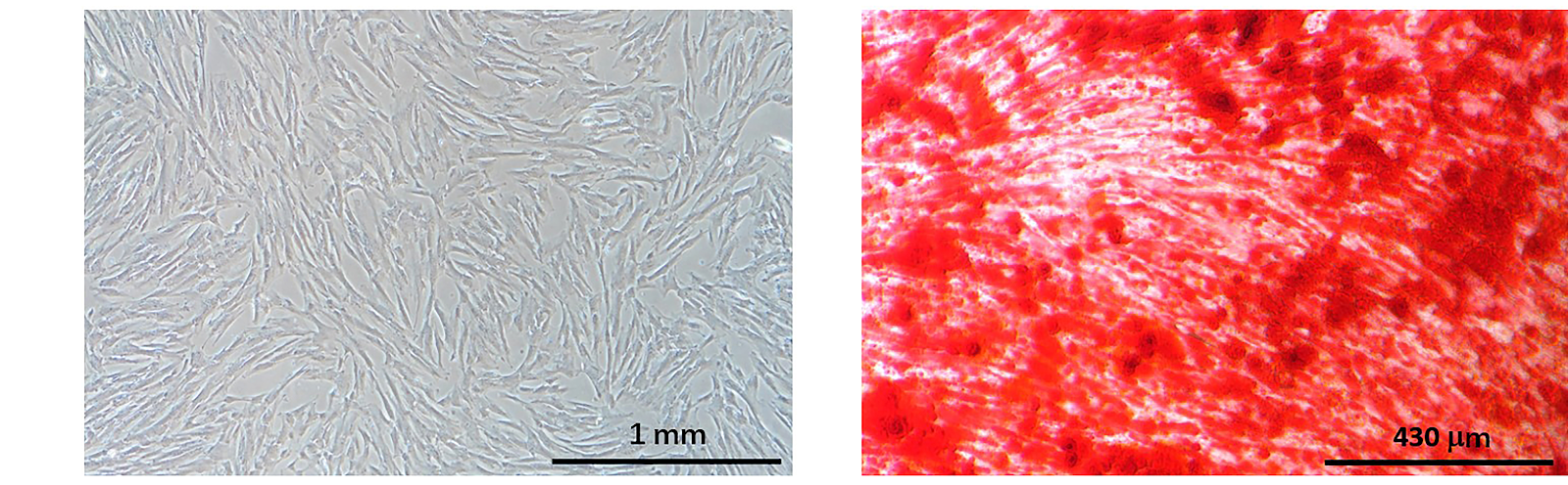
BACKGROUND: High-altitude hypoxia has been reported to damage the male reproductive system, but whether stem cells can protect against male reproductive damage caused by high-altitude hypoxia has not been reported. OBJECTIVE: To investigate the preventive effect of human umbilical cord mesenchymal stem cell transplantation on reproductive damage in hypoxia-exposed male mice. METHODS: Human umbilical cord mesenchymal stem cells were isolated and cultured, and three-lineage differentiation and flow cytometry identification were performed. Twenty-one C57BL/6 male mice were randomly divided into control, hypoxia, and stem cell groups (n=7). The hypoxia and stem cell groups were exposed to a chronic intermittent hypoxia model simulating an altitude of 5,000 m (11.1% oxygen). In the stem cell group, 1×10^6 human umbilical cord mesenchymal stem cells were injected via the tail vein once a week for 6 weeks, while the other groups received PBS. Body mass, food intake, and water intake were monitored. After hypoxia exposure, testicular tissue was analyzed for morphology, ultrastructure, reactive oxygen species levels, and mitochondrial membrane potential; epididymal tissue was analyzed by hematoxylin-eosin staining and sperm motility; and the homing ability of stem cells was observed by DiL fluorescence tracing. RESULTS AND CONCLUSION: Human umbilical cord mesenchymal stem cell transplantation significantly improved water and food intake in hypoxic mice but had no significant effect on body mass. Morphological analysis showed that hypoxia caused edema of the testis and epididymis and shedding of spermatogenic cells, while stem cell transplantation alleviated these structural damages and reversed mitochondrial swelling and atrophy in germ cells. Additionally, stem cell transplantation significantly inhibited hypoxia-induced increase in reactive oxygen species, restored mitochondrial membrane potential, and improved sperm motility. Tracing experiments showed that after entering the mice, stem cells mainly accumulated in lung tissue, with low homing to the testis. In conclusion, human umbilical cord mesenchymal stem cell transplantation can protect the structure and function of germ cell mitochondria, reduce hypoxia-induced testicular and epididymal edema, and thereby restore spermatogenesis and sperm motility.

Human umbilical cord mesenchymal stem cell transplantation protects against reproductive damage induced by high-altitude hypoxia exposure in male mice
Graphical Abstract
Original ResearchVol 1901, Issue 29 • pp. 100-112DOI: 10.12307/2026.21358Jan 15, 2026

Mechanisms and clinical strategies of mesenchymal stem cell-derived extracellular vesicles intervening in cell regulatory networks to treat pulmonary fibrosis

Authors: Ding Yan, Nie Hongguang, Sun Yu

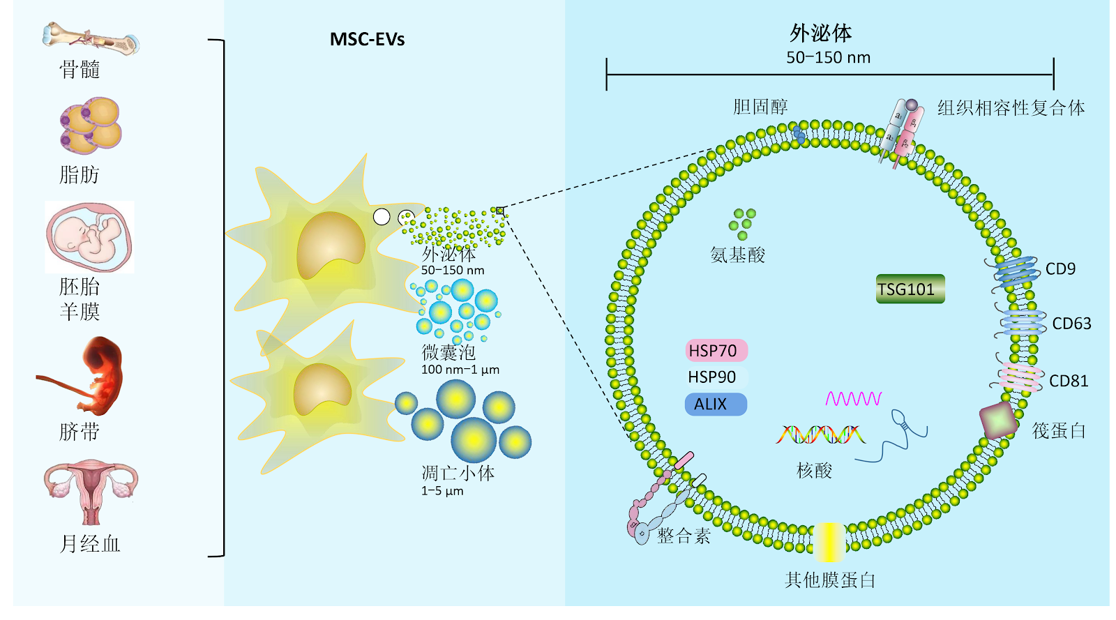
BACKGROUND: Pulmonary fibrosis is a chronic progressive lung disease characterized by abnormal deposition of extracellular matrix, with current therapeutic options remaining limited. Extracellular vesicles derived from mesenchymal stem cells, with their lipid membrane structure, can cross the internal barriers in the body and directly deliver various anti-fibrotic, immunomodulatory factors (such as growth factors, immunomodulatory cytokines, and chemokines), lipids and nucleic acids (mRNAs and miRNAs) and other bioactive substances to target cells in the lungs. OBJECTIVE: To systematically review the core mechanisms of mesenchymal stem cell extracellular vesicles in the treatment of pulmonary fibrosis, summarize and elaborate on how extracellular vesicles directly deliver the bioactive substances they carry to different target cells in the lungs, demonstrating their unique advantages in regulating the pulmonary fibrosis microenvironment, and provide a theoretical basis for future use of mesenchymal stem cell-derived extracellular vesicles in the treatment of pulmonary fibrosis. METHODS: A computer-based search was conducted in CNKI, PubMed, clinicaltrials.gov, and the Chinese Clinical Trial Registry. English search terms included "Mesenchymal stem cells, Extracellular vesicles, Pulmonary fibrosis, Alveolar epithelium, Microvascular endothelium, Macrophages, Neutrophils"; Chinese search terms included "间充质干细胞,细胞外囊泡,肺纤维化,上皮细胞,血管内皮细胞,巨噬细胞,中性粒细胞". A total of 56 articles were included for summary. RESULTS AND CONCLUSION: In alveolar epithelial cells, epithelial-mesenchymal transition is inhibited by regulating signaling pathways such as protein kinase B/glycogen synthase kinase 3β and transforming growth factor β/Smad, and specific miRNAs (e.g., miR-466f-3p, let-7) block pro-fibrotic pathway networks. In fibroblasts and endothelial cells, miR-21-5p and miR-218/miR-214-3p respectively interfere with fibroblast activation and endothelial-mesenchymal transition. Meanwhile, they reprogram monocytes, regulate macrophage polarization, inhibit dendritic cell maturation, and balance Th17/Treg responses, reshaping the immune microenvironment. Furthermore, engineered modifications (targeting peptide modification, drug co-loading) of mesenchymal stem cell-derived extracellular vesicles can enhance precise targeting of multiple cell subtypes in diseased areas, while vesicle heterogeneity, standardized production, and in vivo dynamic tracing remain key bottlenecks for clinical translation. Future research should combine single-cell sequencing and spatial multi-omics to deeply analyze the mechanisms of mesenchymal stem cell-derived extracellular vesicles in intervening in cell regulatory networks and develop novel cell-targeted clinical strategies for pulmonary fibrosis.

Mechanisms and clinical strategies of mesenchymal stem cell-derived extracellular vesicles intervening in cell regulatory networks to treat pulmonary fibrosis
Graphical Abstract
Original ResearchVol 1901, Issue 29 • pp. 100-112DOI: 10.12307/2026.21359Jan 15, 2026

Application and prospects of precision-medicine-driven breast cancer organoids in therapeutic drug discovery

Authors: Mou Jiancheng, Luo Jie, Liu Haotian, Yang Zhuotao, Mu Yuxiao, Qian Da, Meng Xuli

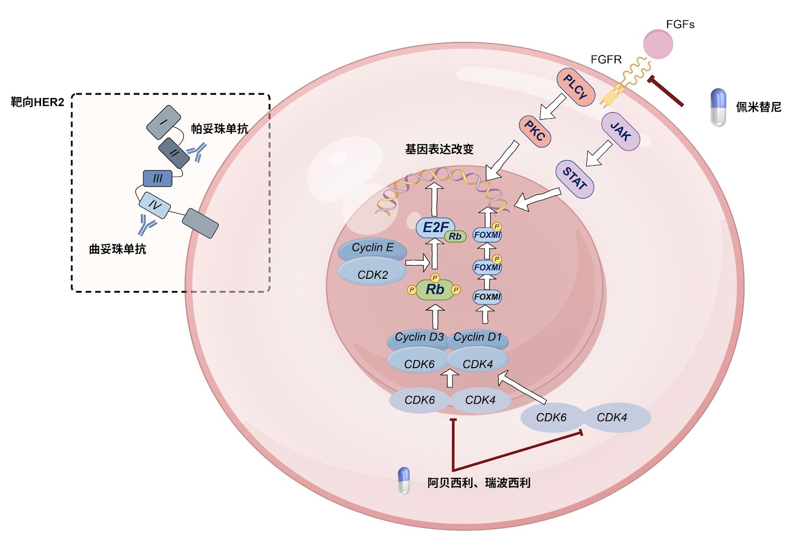
BACKGROUND: Breast cancer organoids, as a novel in vitro model, can not only simulate the biological characteristics of breast cancer but also to some extent reproduce the impact of the tumor microenvironment on the tumor, facilitating research on breast cancer and further promoting precision medicine. OBJECTIVE: To review the application status of breast cancer organoids in the field of therapeutic drugs, including chemotherapy, targeted therapy, and immunotherapy, over the past few years, and to discuss the existing limitations in order to further promote their application in breast cancer treatment. METHODS: The first author conducted a search in the China National Knowledge Infrastructure (CNKI) and PubMed databases in June 2025 for relevant literature published from January 2010 to June 2025. Chinese search terms included '类器官,乳腺类器官,乳腺癌类器官,乳腺癌模型实验验证,精准治疗,靶向治疗,化疗,免疫治疗,药物敏感性'; English search terms included 'organoid, breast organoid, breast cancer organoid, breast cancer experimental model, precision medicine, targeted therapy, chemotherapy, immunotherapy, drug sensitivity'. A total of 58 articles were included for review. RESULTS AND CONCLUSION: (1) Compared with traditional breast cancer cell experiments, which lack verification of tissue structure and cell-cell interactions as well as in vivo microenvironment, breast cancer organoids have diverse sources of primary tumor cells and continuously innovating culture systems. They can simulate cell-cell interactions and reproduce the biological characteristics of breast cancer and its tumor microenvironment, making breast cancer organoids one of the most promising tools in breast cancer research. This also provides greater potential for improving treatment resistance in clinical breast cancer patients through drug sensitivity screening. (2) The application of drug sensitivity test results from breast cancer organoids in clinical practice has yielded promising outcomes. By testing drug sensitivity in breast cancer organoids to common chemotherapeutic agents, targeted drugs, and immunotherapeutic drugs, the antitumor mechanisms and synergistic effects of multiple drugs can be verified, avoiding the use of drugs with primary resistance and high toxicity in patients, thereby enabling personalized treatment plans and evidence-based precision medicine strategies. (3) Breast cancer organoids still have limitations in practical applications such as drug screening and treatment, including low model construction success rates, difficulty in model growth, and lack of angiogenesis processes. To overcome these limitations, it is necessary to increase the source tissue volume, improve the culture system, and innovate culture techniques, which will facilitate comprehensive therapeutic drug selection through breast cancer organoids and aid in personalized precision medicine.

Application and prospects of precision-medicine-driven breast cancer organoids in therapeutic drug discovery
Graphical Abstract
Original ResearchVol 1901, Issue 29 • pp. 100-112DOI: 10.12307/2026.21361Jan 15, 2026

Mechanism of acupuncture regulating proliferation and differentiation of stem cells

Authors: LI Wenfang, DONG Mengwei, JIN Haizhu, YANG Wanpeng, BA Te, NAN Nan, LIU Yang, HAO Huiqin

BACKGROUND: Stem cells have the potential for self-renewal and multi-directional differentiation, which can enhance tissue repair through direct differentiation or paracrine and immunomodulatory microenvironments. Acupuncture can promote the proliferation and differentiation of stem cells through multi-pathway synergy, which expands the application range of acupuncture. OBJECTIVE: To review the types of stem cells and their differentiation potential, and to explore the role and mechanism of acupuncture in promoting stem cell proliferation and differentiation. METHODS: Articles published before March 2025 were searched in PubMed and CNKI databases using English search terms 'Electroacupuncture, Acupuncture, Neural stem cells, Bone marrow mesenchymal stem cells, Adipose mesenchymal stem cells' and Chinese search terms '电针, 针刺, 神经干细胞, 骨髓间充质干细胞, 脂肪间充质干细胞'. Literature related to acupuncture promoting stem cell proliferation and differentiation was included, while irrelevant content was excluded. Finally, 76 articles were selected for analysis. RESULTS AND CONCLUSION: (1) Stem cells belong to the category of kidney essence, and stem cells and kidney essence play a synergistic role, which can be used to guide the treatment of diseases with kidney essence deficiency, explaining modern medicine with traditional Chinese medicine theory. (2) Acupuncture can activate multiple signaling pathways and regulate growth factor expression to promote the proliferation and differentiation of endogenous or exogenous stem cells, providing new ideas for clinical treatment. (3) Although acupuncture plays an important role in stem cell proliferation and differentiation, it also faces many challenges in application; the establishment of stem cell engineering standards and the innovative development of acupuncture therapy are the primary prerequisites for in-depth exploration of the combined mechanism of acupuncture and stem cell therapy, ensuring the stability and efficiency of treatment effects.

Mechanism of acupuncture regulating proliferation and differentiation of stem cells
Graphical Abstract
Original ResearchVol 1901, Issue 29 • pp. 100-112DOI: 10.12307/2026.21360Jan 15, 2026

Innovative application of kidney organoids in acute kidney injury

Authors: Huang Zhengbo, Ou Min, Li Guoshun, Duan Fuhui, Liu Jianqi, Lou Juxiang, Zhao Yanxiu, Su Xiaoyan

BACKGROUND: In recent years, the application of kidney organoid technology in acute kidney injury has gradually become a research hotspot. Traditional animal models have species differences from humans, and physiological and pathological processes of their kidneys cannot fully represent the human situation. Kidney organoid technology forms 3D kidney models through stem cell culture, which can simulate the complex structure and function of human kidneys. It has shown great potential in disease modeling and mechanism exploration of acute kidney injury, prediction of drug nephrotoxicity, and exploration of regeneration and repair mechanisms. OBJECTIVE: To summarize the application progress of kidney organoids in acute kidney injury research, providing new technical means and research strategies for the prevention and treatment of acute kidney injury. METHODS: Literature related to organoids and acute kidney injury was searched in CNKI and PubMed databases. Chinese search terms included "acute kidney injury, organoid, pluripotent stem cells, 3D bioprinting, kidney-on-a-chip, regenerative medicine, kidney transplantation"; English search terms included "acute kidney injury, organoid, pluripotent stem cells, 3D bioprinting, kidney-on-a-chip, regenerative medicine, kidney transplantation". All retrieved literature were original research articles and relevant reviews, with the search time limit from database inception to April 2025. Finally, 99 articles were screened for analysis and summary. RESULTS AND CONCLUSION: (1) The cell sources for inducing kidney organoid formation reported in the literature mainly include pluripotent stem cells, embryonic stem cells, and urine-derived stem cells. These induced kidney organoids play important roles in in vitro drug screening, kidney development, and disease modeling. (2) 3D bioprinting and kidney-on-a-chip technology are emerging techniques for constructing kidney organoids. 3D bioprinting can precisely and specifically construct complex multicellular structures, while kidney-on-a-chip technology has characteristics such as high gas permeability, sensitivity, and low cost, which can extend organoid lifespan, increase biocompatibility, and are suitable for preclinical drug development and toxicity screening. (3) The combination of gene editing technology with kidney organoid models brings new perspectives and tools for kidney disease research, drug development, and regenerative medicine. It can construct kidney organoids with specific reporter genes or sensitive indicators, and amplify and classify specific kidney cell types in kidney organoids. (4) Kidney organoids show unique advantages in disease simulation, drug evaluation, and exploration of regenerative therapeutic strategies for acute kidney injury. They can serve as in vitro models to study the toxicity mechanisms of drugs such as cisplatin, doxorubicin, and red yeast rice supplements that cause acute kidney injury, screen high-throughput drugs and therapeutic targets, and also play an important role in the field of renal transplantation regenerative medicine.

Innovative application of kidney organoids in acute kidney injury
Graphical Abstract
Original ResearchVol 1901, Issue 29 • pp. 100-112DOI: 10.12307/2026.21354Jan 15, 2026

Stem cell-derived exosomes modulate the inflammatory microenvironment and enhance regenerative capacity of oligodendrocytes

Authors: ZHANG Xixian

BACKGROUND: The dynamic interplay between the inflammatory microenvironment and oligodendrocytes following neural injury constitutes a central pathological feature in neurodegenerative and demyelinating diseases. Stem cell-derived exosomes, leveraging their inherent low immunogenicity, efficient barrier-penetrating capacity, and targeted delivery of diverse pro-repair factors, play a pivotal role in modulating oligodendrocyte differentiation and the inflammatory microenvironment, thereby facilitating neural repair and regeneration. OBJECTIVE: To investigate the mechanisms by which stem cell-derived exosomes regulate the inflammatory microenvironment to enhance oligodendrocyte survival, differentiation, and myelin repair. It seeks to establish a novel "cell-free therapy" paradigm, utilizing exosome-mediated multi-component synergy (miRNAs, proteins, and metabolites) and microenvironmental adaptation for treating neurological disorders. METHODS: Literature searches were conducted in the China National Knowledge Infrastructure, PubMed, and WanFang databases, covering publications from 2010 to 2025. Chinese search terms included "exosomes, stem cells, engineered, diagnosis, inflammatory microenvironment, oligodendrocytes, signaling pathways," while English terms comprised "stem cell-derived exosomes, oligodendrocytes, inflammatory microenvironment, signaling pathway, regulatory mechanisms." Irrelevant studies were excluded, and 65 articles meeting inclusion criteria were systematically reviewed according to the inclusion and exclusion criteria. RESULTS AND CONCLUSION: (1) The biological characteristics of exosomes and their roles in the central nervous system were summarized, analyzing the impact of the inflammatory microenvironment on oligodendrocytes and the regulatory mechanisms of exosomes, including miRNA-mediated signaling pathway regulation, anti-inflammatory factor secretion, and immune cell function modulation. (2) The regulatory mechanisms of the inflammatory microenvironment on oligodendrocyte biological behavior and their roles in disease pathogenesis were elaborated. (3) An engineered exosome delivery system based on targeting peptide modification and functional molecule loading, combined with traditional Chinese medicine active ingredient regulation strategies, was proposed to construct a novel cell-free therapy paradigm. (4) The deep interaction between the exosome functional network and myelin homeostasis was explained at the molecular level, providing new therapeutic directions for the development of exosome-targeted delivery systems to intervene in central nervous system demyelinating diseases.

Stem cell-derived exosomes modulate the inflammatory microenvironment and enhance regenerative capacity of oligodendrocytes
Graphical Abstract
Original ResearchVol 1901, Issue 29 • pp. 100-112DOI: 10.12307/2026.21353Jan 15, 2026

Exosomal miRNA as an early diagnostic biomarker and potential therapeutic target for cerebral small vessel disease

Authors: LIU Yuxuan, GUAN Dongsheng, WANG Jing, REN Yihan

BACKGROUND: In recent years, microRNA (miRNA) has received extensive attention in the pathogenesis and diagnosis and treatment of cerebral small vessel disease, and is involved in the regulation of various pathological processes of cerebral small vessel disease. OBJECTIVE: To review the role of miRNA in the pathogenesis, diagnosis, and treatment of cerebral small vessel disease, and to provide effective therapeutic targets and new potential biomarkers for the early diagnosis of cerebral small vessel disease. METHODS: “microRNA, cerebral small vessel disease, blood-brain barrier, chronic cerebral hypoperfusion, inflammation, apoptosis, diagnosis, biomarkers” were used as English search terms for PubMed search. “Exosomal miRNA, cerebral small vessel disease” were used as Chinese search terms for CNKI search. The search time limit was from inception to January 2025. Through the preliminary screening of reading titles and abstracts, the literature with poor relevance and duplicate content was excluded, and finally 72 articles were included for inductive discussion. RESULTS AND CONCLUSION: (1) Through the excavation and discussion of the biological functions and characteristics of exosomal miRNAs, it was confirmed that exosomal miRNAs are important related components in the occurrence and progression of cerebral small vessel disease diseases. (2) Exosomal miRNA participates in the regulation of various pathological processes of cerebral small vessel disease, playing an important role in the pathological mechanism of cerebral small vessel disease by protecting the blood-brain barrier, improving chronic cerebral hypoperfusion, reducing inflammatory responses, and inhibiting apoptosis. (3) Exosomal miRNA can intervene at different stages of pathological development by targeting multiple signaling pathways, effectively targeting different pathological links of cerebral small vessel disease. (4) The combined use of multiple exosomal miRNAs can effectively improve the progression of cerebral small vessel disease. Different miRNAs play different roles at various stages of the pathological mechanism, and constructing a miRNA interaction network is of great significance for regulating the development of cerebral small vessel disease. (5) Exosomal miRNAs are widely and stably present in various body fluids, and their specific and significant expression in urine, serum, blood and other body fluids of patients with cerebral small vessel disease can serve as an effective basis for diagnosis. (6) Currently, the main clinical treatment is to inject miRNA mimics or antagonists to regulate downstream target gene expression. How to achieve the most effective therapeutic effect of miRNA still requires further research. (7) miRNA as exosomal content has great application prospects in the treatment of cerebral small vessel disease. Future research should further explore its mechanism of action to provide effective ideas and further optimize clinical treatment plans for cerebral small vessel disease.

Exosomal miRNA as an early diagnostic biomarker and potential therapeutic target for cerebral small vessel disease
Graphical Abstract
Original ResearchVol 1901, Issue 29 • pp. 100-112DOI: 10.12307/2026.21356Jan 15, 2026

Advantages and potential of cell-derived exosomes in oral tissue regeneration

Authors: Li Jiapeng, Zuleina·Abula, Jia Qianqian, Nigare·Yunusijiang, Sun Jiaqi, Zhao Jin, Wu Zeyu

BACKGROUND: Stem cells show a great potential in oral tissue regeneration but face challenges such as immune rejection and tumor formation. Exosomes are nanoscale extracellular vesicles secreted by cells, reducing immunogenicity and tumor risks while maintaining stem cell functions, such as promoting angiogenesis and tissue repair. OBJECTIVE: To summarize the mechanisms and roles of exosomes in oral tissue regeneration, explore exosome engineering strategies and the challenges and future directions in the application of exosomes in oral regenerative medicine. METHODS: The relevant literature published from the WanFang and PubMed databases from their inception to 2025 was searched using Chinese search terms “stem cells, exosomes, dental pulp regeneration, periodontal regeneration” and English search terms “exosomes, stem cells, dentistry, regenerate.” Finally, 94 articles were included for review and analysis. RESULTS AND CONCLUSION: (1) Exosomes have lower immunogenicity and no tumorigenic risk compared with stem cells. They are more stable and easier to store and transport. Additionally, exosomes can penetrate dense tissues for targeted delivery, avoiding ethical and immune rejection issues associated with stem cell therapy, making them a safer and more effective treatment option. (2) Exosomes have shown significant efficacy in regenerating dental pulp, periodontal tissues, craniofacial bone, salivary glands, nerves, and skin, promoting tissue repair and regeneration through multiple mechanisms, demonstrating broad application prospects. (3) Engineering strategies such as preconditioning, isolation and purification, and targeted modification can enhance exosome function, improving therapeutic potential and clinical feasibility. However, current technologies still have limitations, and further optimization is needed to promote widespread application of exosomes.

Advantages and potential of cell-derived exosomes in oral tissue regeneration
Graphical Abstract
Original ResearchVol 1901, Issue 29 • pp. 100-112DOI: 10.12307/2026.21345Jan 15, 2026

Single-cell sequencing data identifies differentially expressed genes and immune cell subtypes in periodontitis patients

Authors: Qiu Xuedi, Guo Chao, He Jiayue, Zhou Zheng

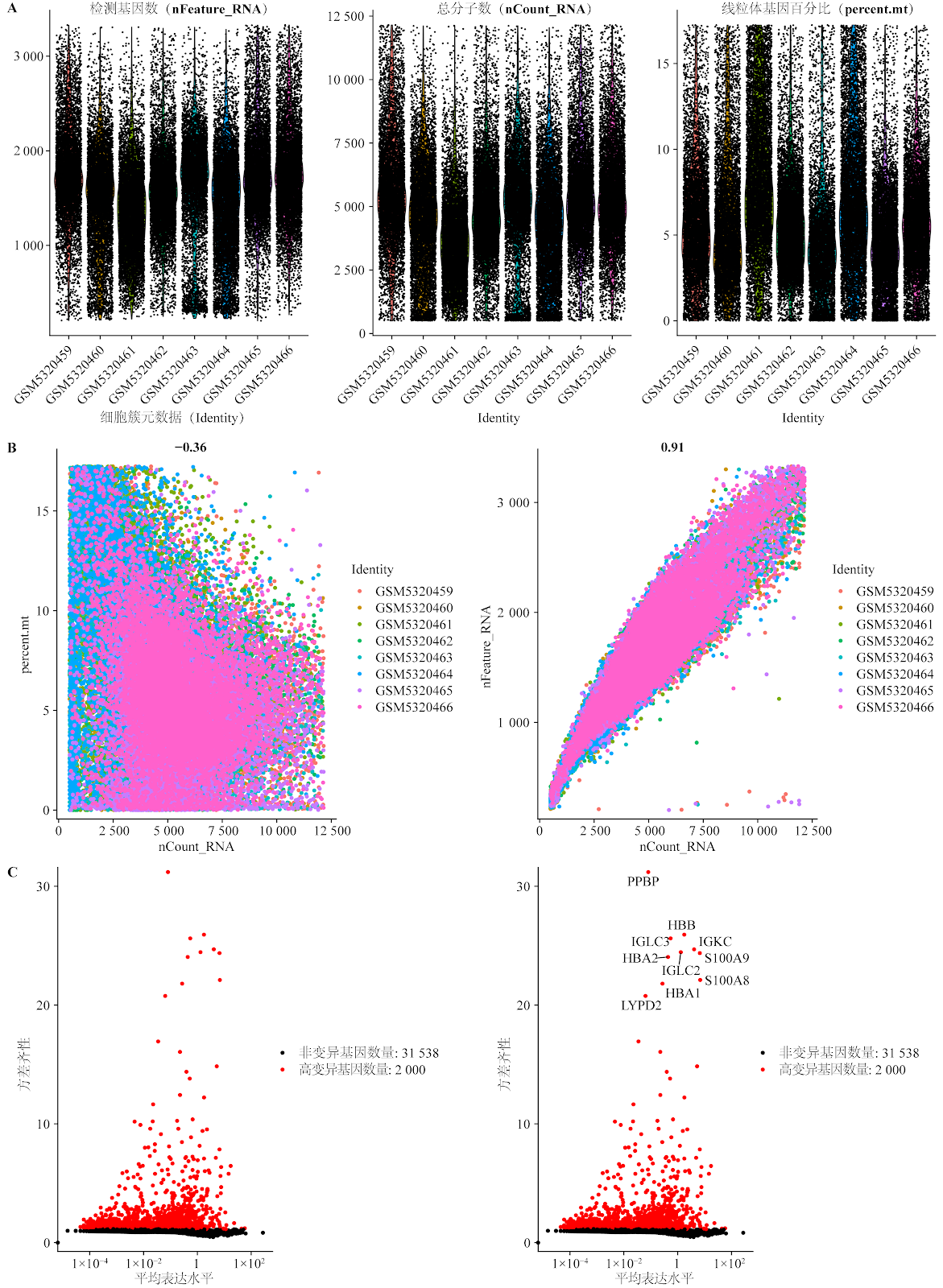
BACKGROUND: Periodontitis is a chronic inflammatory disease. Previous research has predominantly focused on specific immune cells or cytokines. Therefore, systematically elucidating its immune mechanisms and discovering novel therapeutic targets hold significant implications. OBJECTIVE: To analyze the expression profiles of periodontitis-associated immune cell subpopulations and identify key differentially expressed genes with a causal relationship to the disease, thereby exploring potential molecular mechanisms and key genes involved in periodontitis and immune cell dynamics. METHODS: Single-cell RNA sequencing data from the GEO database were used to analyze immune cell subset heterogeneity and identify differentially expressed genes. Mendelian randomization analysis was performed using expression quantitative trait loci data to infer causal relationships between immune cell gene expression and periodontitis risk. Pathway enrichment and immune infiltration analyses were performed on the identified causal genes to reveal the associations between differentially expressed genes and immune cells with the development and progression of periodontitis. CellChat trajectory analysis was used to explore intercellular communication. To validate key findings, gingival tissue samples were collected from 20 patients with periodontitis diagnosed by the Department of Stomatology at The First Affiliated Hospital of Shihezi University (periodontitis group) and 20 healthy gingival tissue samples from patients undergoing orthodontic or impacted tooth extraction (control group). RT-qPCR and immunohistochemistry were used to detect the expression of key genes. RESULTS AND CONCLUSION: Comprehensive analysis identified 23 immune cell clusters in periodontitis and three key genes with significant causal relationships to periodontitis risk: annexin A1 (ANXA1), solute carrier family 11 member 1 (SLC11A1), and vimentin (VIM). Pathway enrichment analysis revealed their involvement in key immune regulatory mechanisms. Further analyses of immune subtype receptor-ligand interactions and key cell subtype trajectories characterized the distinct roles of ANXA1, SLC11A1, and VIM in disease progression. Compared with healthy controls, the mRNA expression levels of ANXA1, SLC11A1, and VIM were upregulated in periodontitis tissues (P < 0.05). This study reveals the key roles of immune cell subpopulations in periodontitis and validates causal genes (ANXA1, SLC11A1, VIM) associated with the disease.

Single-cell sequencing data identifies differentially expressed genes and immune cell subtypes in periodontitis patients
Graphical Abstract
Original ResearchVol 1901, Issue 29 • pp. 100-112DOI: 10.12307/2026.21351Jan 15, 2026

Action mechanism of mesenchymal stem cells and their derivatives in the treatment of liver fibrosis

Authors: Fan Longyu, Yuan Xiao, Xie Yanan, Yin Xiaoxuan

BACKGROUND: Multiple chronic liver diseases that fail to heal will progress to the stage of liver fibrosis. If not treated in a timely manner, they will eventually develop into liver cancer, severely threatening the safety of patients' lives. However, there is currently no specific drug for the treatment of liver fibrosis. Recent studies have demonstrated that mesenchymal stem cell therapy has significant advantages over traditional treatment protocols, providing a new direction for the treatment of liver fibrosis. OBJECTIVE: To review the mechanisms of action of mesenchymal stem cells and their derivatives in the treatment of liver fibrosis. METHODS: The Chinese and English keywords "mesenchymal stem cells, mesenchymal stromal cells, MSCs, liver fibrosis, hepatic fibrosis, hepatocyte death, liver cell death, hepatocyte-like cells, immunomodulation, macrophage, hepatic stellate cells, clinical trials, clinical studies" were used and searched in CNKI and PubMed databases, a total of 81 eligible articles were selected for this review. RESULTS AND CONCLUSION: Through summarizing existing studies, the mechanisms by which mesenchymal stem cells and their derivatives exert anti-fibrotic effects and delay disease progression have been identified. These specific mechanisms include reducing hepatocyte death, differentiating into hepatocyte-like cells, regulating immune responses, and inhibiting hepatic stellate cell activation, confirming that mesenchymal stem cells and their derivatives can serve as a new direction for the treatment of liver fibrosis-related diseases.

Action mechanism of mesenchymal stem cells and their derivatives in the treatment of liver fibrosis
Graphical Abstract
Original ResearchVol 1901, Issue 29 • pp. 100-112DOI: 10.12307/2026.21341Jan 15, 2026

Overexpression of collagen triple helix repeat-containing protein 1 promotes proliferation and osteogenic differentiation of human periodontal ligament stem cells

Authors: ZHOU Rui, ZHANG Xuesong, YANG Donghong, JIN Yinan, YANG Yuqi, YE Zhihui

BACKGROUND: Collagen triple helix repeat-containing protein 1 (CTHRC1) is a positive regulator of bone formation. However, its role and underlying mechanisms in periodontal ligament stem cells (PDLSCs) remain unclear. OBJECTIVE: To investigate the effects of CTHRC1 on the proliferation and osteogenic differentiation of PDLSCs and its mechanism of action. METHODS: Human PDLSCs were isolated and cultured in vitro, and cells were transfected with a lentiviral vector overexpressing CTHRC1. The effect of CTHRC1 overexpression on the proliferation activity of PDLSCs was determined by CCK-8 assay and flow cytometry. The effect of CTHRC1 overexpression on the osteogenic differentiation of PDLSCs was determined by alkaline phosphatase (ALP) activity and Alizarin Red staining. Western blot was used to detect the expression of extracellular signal-regulated kinase 1/2 (ERK1/2) and phosphorylated ERK1/2 (p-ERK1/2) after CTHRC1 overexpression. After blocking the ERK1/2 signaling pathway, the expression of osteogenic differentiation-related factors Runt-related transcription factor 2 (Runx2), osteocalcin (OCN), and Osterix was detected by Western blot and qRT-PCR. RESULTS AND CONCLUSION: CCK-8 and flow cytometry results showed that CTHRC1 overexpression promoted the proliferation of PDLSCs. ALP activity and Alizarin Red staining showed that CTHRC1 overexpression promoted the osteogenic differentiation of PDLSCs. Western blot results showed that CTHRC1 overexpression activated the ERK1/2 signaling pathway. Western blot and qRT-PCR results showed that CTHRC1 overexpression promoted the expression of Runx2, OCN, and Osterix at both protein and mRNA levels. When the ERK signaling pathway was inhibited by the specific inhibitor PD98059, the upregulation of osteogenic-related factors was partially suppressed. These results suggest that overexpression of CTHRC1 can promote the proliferation and osteogenic differentiation of PDLSCs, and its osteogenic differentiation effect may be related to the activation of the ERK1/2 signaling pathway.

Overexpression of collagen triple helix repeat-containing protein 1 promotes proliferation and osteogenic differentiation of human periodontal ligament stem cells
Graphical Abstract
Original ResearchVol 1901, Issue 29 • pp. 100-112DOI: 10.12307/2026.21347Jan 15, 2026

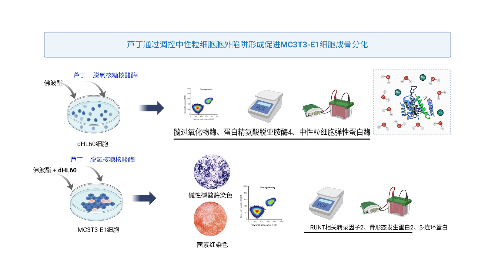
Rutin promotes osteogenic differentiation of MC3T3-E1 cells: regulating the formation of neutrophil extracellular traps

Authors: LI Jie, LIU Yang, WANG Dayu, WAN Qiang, ZHU Jiayi, FENG Wenjun, CHEN Jinlun, JIE Ke, HUANG Yiwei, XIN Pengfei, ZENG Jianchun, ZENG Yirong, ZHANG Haitao

BACKGROUND: Rutin can effectively prevent osteoporosis, but its mechanism of action remains unclear. OBJECTIVE: To investigate the effect of rutin on osteogenesis of MC3T3-E1 cells under the action of neutrophil extracellular traps. METHODS: (1) Human myeloid leukemia dHL60 cells were stimulated with phorbol 12-myristate 13-acetate to induce neutrophil extracellular trap formation. dHL60 cells were divided into 4 groups: control group received Hank's balanced salt solution; the other three groups received 50 nmol/L phorbol 12-myristate 13-acetate; the latter two groups additionally received 250 μmol/L rutin or 250 μmol/L rutin plus 5 U/mL DNase I. Apoptosis of dHL60 cells was detected by flow cytometry; mRNA and protein expression of neutrophil extracellular trap marker genes were detected by RT-qPCR and western blot. (2) MC3T3-E1 cells were divided into 6 groups: control group received Hank's balanced salt solution; the other five groups received 50 nmol/L phorbol 12-myristate 13-acetate; dHL60 cells and 50 nmol/L phorbol 12-myristate 13-acetate; 100 μmol/L rutin; dHL60 cells, 50 nmol/L phorbol 12-myristate 13-acetate, and 250 μmol/L rutin; dHL60 cells, 50 nmol/L phorbol 12-myristate 13-acetate, 250 μmol/L rutin, and 5 U/mL DNase I. Apoptosis of MC3T3-E1 cells under neutrophil extracellular traps was detected by flow cytometry; alkaline phosphatase staining and alizarin red staining were used to determine osteogenic and mineralization abilities; RT-qPCR and western blot were used to detect osteogenic-related gene and protein expression. RESULTS AND CONCLUSION: (1) Compared with the blank control group, rutin significantly inhibited the mRNA and protein expression of protein arginine deiminase 4, myeloperoxidase, and neutrophil elastase in dHL60 cells (P < 0.000 1); compared with the rutin group, the combination of rutin and DNase I had a more significant downregulation effect (P < 0.05), indicating that rutin can significantly inhibit neutrophil extracellular trap formation. (2) After inducing neutrophil extracellular traps from dHL60 and co-culturing with MC3T3-E1, the mRNA and protein expression of Runt-related transcription factor 2, β-catenin, and bone morphogenetic protein 2 in MC3T3-E1 cells were significantly downregulated (P < 0.000 1), and the apoptosis rate significantly increased (P < 0.000 1), indicating that neutrophil extracellular traps can significantly inhibit osteogenic ability and promote apoptosis of MC3T3-E1 cells in vitro. After intervention with rutin alone or rutin combined with DNase I, the apoptosis and osteogenic ability of MC3T3-E1 cells under neutrophil extracellular traps were significantly improved, and the effect of rutin combined with DNase I was more significant than rutin alone, indicating that rutin may inhibit neutrophil extracellular trap formation, thereby improving the osteogenic ability of MC3T3-E1 cells. (3) Molecular docking and molecular dynamics simulations showed that rutin binds well to protein arginine deiminase 4, myeloperoxidase, and neutrophil elastase target proteins, indicating that rutin can target and inhibit neutrophil extracellular trap formation.

Rutin promotes osteogenic differentiation of MC3T3-E1 cells: regulating the formation of neutrophil extracellular traps
Graphical Abstract
Original ResearchVol 1901, Issue 29 • pp. 100-112DOI: 10.12307/2026.21346Jan 15, 2026

Cerebrospinal fluid-contacting neurons differentiating into motor neurons promote functional recovery in spinal cord-injured mice

Authors: Tang Min, Shangguan Zeyu, Li Qizhe, Tan Wei, Li Qing

BACKGROUND: Cell transplantation is one of the effective approaches for repairing spinal cord injury. Our research team previously found that transplanted cerebrospinal fluid-contacting neurons can survive and promote motor function recovery in mice with spinal cord injury. However, whether these transplanted cerebrospinal fluid-contacting neurons differentiate into functional neurons and thereby facilitate motor function recovery remains unclear. OBJECTIVE: To investigate whether transplanted cerebrospinal fluid-contacting neurons differentiate into functional neurons in vivo and contribute to motor function recovery after spinal cord injury. METHODS: Primary cells containing cerebrospinal fluid-contacting neurons were isolated from the cervical spinal cord of C57BL/6 neonatal mice within 24 hours of birth and cultured adherently. Cells were transduced with a lentivirus carrying a multimodal imaging gene, and cerebrospinal fluid-contacting neurons were selected and purified using puromycin. Differentiation was induced with serum-containing differentiation medium, and expression of neuronal marker NeuN and motor neuron marker ChAT was detected by immunofluorescence. Thirty C57BL/6 mice were randomly divided into three groups: transplantation group and PBS group underwent T10 spinal cord injury by clip compression, while sham group only had laminectomy. One week after injury, cerebrospinal fluid-contacting neurons were transplanted in situ in the transplantation group, and an equal volume of PBS was injected in the PBS group. At 1, 4, and 8 weeks after transplantation, immunofluorescence was used to detect expression of motor neuron marker ChAT in spinal cord tissue. At 8 weeks, immunofluorescence was used to detect synaptic marker SYN, inhibitory transmitter marker GAD65/67, and excitatory transmitter marker vGLUT1; hematoxylin-eosin staining was used to observe spinal cord morphology; BMS motor function score and footprint analysis were used to assess motor function recovery. RESULTS AND CONCLUSION: (1) Cerebrospinal fluid-contacting neurons expressed neural stem cell characteristics in vitro and could differentiate into motor neurons. (2) Transplanted cerebrospinal fluid-contacting neurons could survive long-term in vivo and differentiate into motor neurons. (3) The proportion of cerebrospinal fluid-contacting neurons differentiating into motor neurons was highest at 8 weeks (P < 0.0001). (4) At 8 weeks after transplantation, cerebrospinal fluid-contacting neurons co-expressed SYN, GAD65/67, and vGLUT1, indicating synaptic connections with host neurons. (5) BMS scores of PBS group were consistently lower than those of transplantation group (P < 0.001); footprint analysis showed more coordinated gait in transplantation group with only toe dragging, while PBS group showed obvious hindlimb dragging. (6) Hematoxylin-eosin staining showed large cavities in the injured area of PBS group, while cavities were reduced in transplantation group. These results indicate that transplanted cerebrospinal fluid-contacting neurons can differentiate into motor neurons both in vitro and in vivo, form synaptic connections, and thereby improve motor function in spinal cord-injured mice.

Cerebrospinal fluid-contacting neurons differentiating into motor neurons promote functional recovery in spinal cord-injured mice
Graphical Abstract
Original ResearchVol 1901, Issue 29 • pp. 100-112DOI: 10.12307/2026.21348Jan 15, 2026

Protective effect of optimization of the whole blood separation process to prepare therapeutic-grade platelet lysate on cardiomyocytes from hypoxic injury

Authors: Jiang Lihong, Lin Fuwen, Chen Ying, Huang Yuchen, Peng Shaojing, Chen Jierun, Su Changshan, Zhong Zhoulin

BACKGROUND: Platelets are important blood resources, yet in routine blood bank processes they are often filtered out along with white blood cells as medical waste. Optimizing whole blood separation processes to prepare platelet lysate products and exploring their applications in tissue engineering and regenerative medicine is of great value. OBJECTIVE: To optimize whole blood separation to prepare therapeutic-grade platelet lysate and to investigate the protective effect of platelet lysate on hypoxic injury of cardiomyocytes. METHODS: Platelets were isolated from 21 qualified whole blood units under closed blood bag and tubing conditions, and 21 platelet lysates were prepared by freeze-thawing. The mass concentration ranges of platelet-derived growth factor AA, platelet-derived growth factor BB, platelet-derived growth factor AB, vascular endothelial growth factor, epidermal growth factor, insulin-like growth factor 1, fibroblast growth factor, and transforming growth factor beta 1 in platelet lysates were measured using enzyme-linked immunosorbent assay kits. Bacterial contamination was assessed by colony culture method and mycoplasma contamination by PCR detection kit. A cardiomyocyte hypoxia model was established to evaluate the protective effect of platelet lysate on hypoxic injury. RESULTS AND CONCLUSION: (1) The mass concentration ranges of major growth factors and cytokines in platelet lysates were: platelet-derived growth factor AA 12.86-24.17 μg/L, platelet-derived growth factor BB 0.25-0.32 μg/L, platelet-derived growth factor AB 85.09-114.91 μg/L, vascular endothelial growth factor 10.57-58.37 μg/L, epidermal growth factor 0.43-0.69 μg/L, insulin-like growth factor 1 106-204.9 μg/L, fibroblast growth factor 0.03-0.06 μg/L, and transforming growth factor beta 1 124.17-192.38 μg/L. (2) Colony culture and mycoplasma detection results were negative. (3) Low volume fraction (1%) platelet lysate yielded the highest proliferation efficiency of cardiomyocytes; low volume fraction (1%) platelet lysate stimulated cardiomyocytes to produce high levels of superoxide dismutase and glutathione peroxidase to protect cardiomyocytes. This study established a method for preparing therapeutic-grade platelet lysate by optimizing the whole blood separation process, which can improve the utilization rate of blood resources. Platelet lysate has high levels of major growth factors and can significantly promote the repair of hypoxic injured cardiomyocytes.

Protective effect of optimization of the whole blood separation process to prepare therapeutic-grade platelet lysate on cardiomyocytes from hypoxic injury
Graphical Abstract
Original ResearchVol 1901, Issue 29 • pp. 100-112DOI: 10.12307/2026.21344Jan 15, 2026

miR-9 regulates the differentiation of neural stem cells in mouse cerebral cortex

Authors: LIU Yingzhao, MA Yanxia, LIN Yaofa, ZHANG Guoqiao, MIAO Weiliang, JIA Yanli, CHI Chenshen, SONG Wangsheng, LI Di, LIU Chenglong, ZHANG Haonan

BACKGROUND: Neural stem cells located in the ventricular zone and subventricular zone are crucial for cortical neurodevelopment and the treatment of neurodegenerative diseases. However, their precise regulatory mechanisms remain incompletely understood. miRNA-9 is one of the most abundantly expressed miRNAs in the vertebrate embryonic and adult brain, playing diverse roles during development. Nevertheless, the role of miR-9 in neural stem cell differentiation remains unclear. OBJECTIVE: To investigate the role of miR-9 in regulating the differentiation of neural stem cells in the ventricular zone and subventricular zone. METHODS: Neural stem cells were isolated from the ventricular zone and subventricular zone of embryonic day 14.5 ICR mice and cultured in proliferation medium for 3-4 days to form neurospheres. Stemness was identified by Pax6/Nestin immunofluorescence double staining. The expression profile of miR-9 was detected by qRT-PCR in telencephalon tissues at embryonic days 12.5, 14.5, 16.5, 18.5 and postnatal days 0, 7, as well as in embryonic day 14.5 neural stem cells cultured in vitro. Neural stem cells were transfected with miR-9 inhibitor or mimic using transfection reagents. After 24 hours, cells were differentiated for 3-4 days (neurons) and 6-8 days (glial cells). The differentiation of each lineage was quantified by immunofluorescence staining for Tuj1 (neuronal marker), myelin basic protein (oligodendrocyte marker), and glial fibrillary acidic protein (astrocyte marker). RESULTS AND CONCLUSION: qRT-PCR results showed that miR-9 was highly expressed in early embryonic telencephalon (E12.5-E14.5) and gradually decreased with development (E16.5 to P7). In E14.5 neural stem cells, miR-9 expression level was close to 90% of the internal reference RNU6B. Functional experiments showed that compared with the control group, the miR-9 inhibition group had decreased proportions of Tuj1-positive neurons and myelin basic protein-positive oligodendrocytes, while the proportion of glial fibrillary acidic protein-positive astrocytes increased. Conversely, the miR-9 overexpression group had increased proportions of Tuj1-positive neurons and myelin basic protein-positive oligodendrocytes, and decreased proportion of glial fibrillary acidic protein-positive astrocytes, with significant differences (P < 0.001). These results indicate that miR-9 plays a bidirectional regulatory role in neural stem cell differentiation: (1) It participates in the temporal regulation of neurogenesis through developmental stage-specific expression patterns (high early, downregulated later); (2) It maintains the balance of trilineage differentiation by promoting neuronal and oligodendrocyte differentiation while inhibiting astrocyte generation.

miR-9 regulates the differentiation of neural stem cells in mouse cerebral cortex
Graphical Abstract
Original ResearchVol 1901, Issue 29 • pp. 100-112DOI: 10.12307/2026.21343Jan 15, 2026

Role and mechanism of emodin in slowing down the senescence of HT-22 cells induced by high glucose

Authors: Rao Binchan, Xu Yongjie, Xu Mengling, Chen Di, Zhu Liying, Yang Siyuan, Li Xing, Wang Zhengrong, Pan Wei

BACKGROUND: The occurrence of diabetic encephalopathy may be closely related to neuronal aging, but its underlying molecular mechanism is not fully understood. Therefore, exploring the role of neuronal senescence in diabetic encephalopathy is of great significance for further revealing the pathogenesis of diabetic encephalopathy. OBJECTIVE: To investigate the effect and mechanism of emodin on senescence of HT-22 cells under high glucose conditions. METHODS: HT-22 cells were divided into control group (glucose concentration 25 mmol/L), high glucose group (glucose concentration 55 mmol/L), and high glucose + emodin group (glucose concentration 55 mmol/L, emodin concentration 100 µmol/L) and cultured for 48 h. The growth state of cells in each group was observed under microscope; CCK-8 assay was used to detect cell viability; ELISA was used to detect telomerase reverse transcriptase activity; RT-qPCR and western blot were used to detect the expression of senescence-related proteins P53, P21, and P16; immunofluorescence, RT-qPCR and western blot were used to detect the expression of lamin A/C. RESULTS AND CONCLUSION: Compared with the control group, the high glucose group showed obvious growth inhibition under microscope, characterized by decreased cell number, increased cell volume, and flattened morphology; compared with the high glucose group, the high glucose + emodin group showed significantly increased cell number and more regular morphology. Compared with the control group, cell viability was significantly decreased in the high glucose group (P < 0.0001); compared with the high glucose group, cell viability was significantly increased in the high glucose + emodin group (P < 0.0001). Compared with the control group, telomerase reverse transcriptase activity was significantly decreased in the high glucose group (P < 0.001). Compared with the control group, the expression levels of P53, P21, and P16 were significantly increased in the high glucose group (P < 0.05); compared with the high glucose group, the expression levels of P53, P21, and P16 were significantly decreased in the high glucose + emodin group (P < 0.05). Compared with the control group, the expression level of lamin A/C was significantly decreased in the high glucose group (P < 0.0001); compared with the high glucose group, the expression level of lamin A/C was significantly increased in the high glucose + emodin group (P < 0.05). The results indicate that emodin may slow down the senescence of HT-22 cells induced by high glucose by upregulating the expression of lamin A/C.

Role and mechanism of emodin in slowing down the senescence of HT-22 cells induced by high glucose
Graphical Abstract
Original ResearchVol 1901, Issue 29 • pp. 100-112DOI: 10.12307/2026.21342Jan 15, 2026

Non-coding RNA-activated by DNA damage promotes proliferation and inhibits apoptosis of induced pluripotent stem cell-derived cardiomyocytes

Authors: Huan Kanghui, Jiang Yujian, Bian Weihua

BACKGROUND: Although cell transplantation offers a promising approach for the treatment of myocardial infarction, the low transplantation rate limits its application. Therefore, promoting the proliferation of transplanted cells and reducing apoptosis are the key issues to be solved urgently to improve the therapeutic effect. OBJECTIVE: To investigate the effects of non-coding RNA-activated by DNA damage (NORAD) on the proliferation of human induced pluripotent stem cell-derived cardiomyocytes and their apoptosis induced by oxygen-glucose deprivation/reoxygenation, as well as the effects of transplanting NORAD-overexpressing human induced pluripotent stem cell-derived cardiomyocytes (hiPSC-NORADOECMs) on cardiac function in a murine model of myocardial infarction. METHODS: The expression of NORAD in the hearts of mice at different ages (3 days old and 8 weeks old) was measured by real-time fluorescence quantitative polymerase chain reaction (RT-qPCR). A cellular model of hiPSC-NORADOECMs was established by infecting human induced pluripotent stem cells with a lentiviral vector designed to specifically upregulate NORAD, followed by directed differentiation into cardiomyocytes. The overexpression efficiency was confirmed by RT-qPCR. Proliferation was assessed by immunofluorescence staining for Ki67. Apoptosis was induced by oxygen-glucose deprivation/reoxygenation (OGD/R). Intracellular reactive oxygen species (ROS) levels were measured by flow cytometry, and the expression of apoptosis-related proteins (Bax, Bcl-2, Cleaved Caspase-3) was detected by western blot. In vivo, hiPSC-NORADOECMs were transplanted into the infarcted myocardium of mice, and cardiac function was evaluated by echocardiography after 4 weeks. RESULTS AND CONCLUSION: NORAD expression was significantly higher in 3-day-old neonatal mouse hearts compared with 8-week-old adult hearts. hiPSC-NORADOECMs were successfully generated, showing increased Ki67 expression compared with control cells. Overexpression of NORAD inhibited OGD/R-induced ROS production, decreased Bax and Cleaved Caspase-3 protein levels, and increased Bcl-2 levels. Transplantation of hiPSC-NORADOECMs significantly improved cardiac function in myocardial infarction mice. These findings indicate that NORAD overexpression promotes hiPSC-CM proliferation and inhibits apoptosis by reducing ROS production, thereby enhancing the reparative capacity of hiPSC-CMs in myocardial infarction.

Non-coding RNA-activated by DNA damage promotes proliferation and inhibits apoptosis of induced pluripotent stem cell-derived cardiomyocytes
Graphical Abstract
Original ResearchVol 1901, Issue 29 • pp. 100-112DOI: 10.12307/2026.21340Jan 15, 2026

Retrospective analysis of central nervous system diseases related to non-primary infiltration after allogeneic hematopoietic stem cell transplantation

Authors: Chen Shiyu, Zhang Xiaohan, Li Xiaoqing, Du Xin

BACKGROUND: Allogeneic hematopoietic stem cell transplantation may be complicated by central nervous system diseases not related to primary disease infiltration. There is no clear conclusion on the clinical symptoms, possible causes, and prognosis. OBJECTIVE: To explore the clinical characteristics, risk factors, and prognosis of non-primary infiltration-related central nervous system diseases after allogeneic hematopoietic stem cell transplantation, in order to provide evidence-based basis for early clinical diagnosis, etiological intervention, and prognosis improvement. METHODS: A retrospective analysis was conducted on clinical data, laboratory characteristics, and treatment processes of 298 patients with hematological diseases who developed non-primary infiltration-related central nervous system diseases after allogeneic hematopoietic stem cell transplantation from January 2015 to June 2024. They were divided into a non-primary infiltration-related central nervous system disease group (n=19) and a control group (without such diseases, n=279). Risk factors were analyzed statistically, and clinical symptoms, possible causes, and prognosis were evaluated. RESULTS AND CONCLUSION: (1) Among 298 patients, 19 developed non-primary infiltration-related central nervous system diseases, with an incidence of 6.4%. (2) The median onset time was 16 days (2-45 days) after transplantation. The main initial symptom was convulsions, accompanied by elevated blood pressure, headache, visual decline, consciousness disorders, and psychiatric behavioral abnormalities. (3) Univariate analysis showed significant associations between the occurrence of these diseases and granulocyte engraftment time, platelet engraftment time, history of central nervous system leukemia before transplantation, and grade III-IV graft-versus-host disease. Etiological analysis revealed: calcineurin inhibitor-related encephalopathy (2 cases), central nervous system injury (4 cases), central nervous system infection (4 cases), transplantation-associated thrombotic microangiopathy (4 cases), central nervous system graft-versus-host disease (1 case), intracranial hemorrhage (2 cases), endocrine metabolic encephalopathy (1 case), and unknown cause (1 case). (4) As of the follow-up date, the cumulative mortality in the disease group was 47% (9/19), significantly higher than 28.6% (80/279) in the control group. Further analysis showed that the estimated overall survival rates at 1 and 2 years after transplantation were significantly lower in the disease group than in the control group. In conclusion, non-primary infiltration-related central nervous system diseases after allogeneic hematopoietic stem cell transplantation are caused by multiple transplantation-related factors. Timely identification of pathogenic factors and precise diagnosis and treatment are crucial for improving the prognosis of patients with these complications.

Retrospective analysis of central nervous system diseases related to non-primary infiltration after allogeneic hematopoietic stem cell transplantation
Graphical Abstract
Original ResearchVol 1901, Issue 29 • pp. 100-112DOI: 10.12307/2026.21333Jan 15, 2026

Culture and identification of adipose-derived stem cells from periprostatic adipose tissue

Authors: Zhou Shukui, Liu Jinpeng, Gao Wenlong, Yang Shengke, Liao Hong, Wu Yi, Li Zeng

BACKGROUND: Periprostatic adipose tissue is the white visceral adipose tissue that is closest to the prostate, which is part of the prostate cancer tumor microenvironment and plays a key role in the occurrence and progression of prostate cancer. OBJECTIVE: To investigate the ability of adipose-derived stem cells derived from periprostatic adipose tissue to form three-dimensional cell sheets. METHODS: Periprostatic adipose tissue was harvested from patients undergoing radical prostatectomy. Adipose-derived stem cell suspensions were prepared using a combination of enzymatic digestion and mechanical dissection. Adipose-derived stem cell proliferation was assessed using a CCK-8 assay. Expression of stem cell-associated antigens CD34/CD44/CD45/CD90/CD105 was determined by flow cytometry. Multidirectional differentiation potential of the stem cells was assessed using osteogenic/adipogenic/chondrogenic differentiation assays. Adipose-derived stem cells were cultured for three weeks in low-glucose DMEM containing 100 μg/mL vitamin C and 10% fetal bovine serum to construct cell sheets, followed by histological analysis and scanning electron microscopy. RESULTS AND CONCLUSION: Adipose-derived stem cells from periprostatic adipose tissue exhibited a long spindle or fusiform shape, aligned growth, and consistent morphology. Primary culture reached 95% confluence at 9-10 days with good cell viability, and no obvious senescence was observed up to passage 15. Flow cytometry showed expression rates of CD44, CD90, and CD105 at 98.24%, 84.99%, and 89.14%, respectively, while CD34 and CD45 were expressed at 0.64% and 1.02%. After 3 weeks of osteogenic, adipogenic, and chondrogenic induction, the cells could differentiate into osteoblasts, adipocytes, and chondrocytes. After continuous culture for 3 weeks, the cells formed a three-dimensional cell sheet with a smooth surface and uniform texture, rich in extracellular matrix components such as fibronectin and type I collagen. Scanning electron microscopy revealed a flat surface with aligned long spindle-shaped cells and abundant extracellular matrix deposition between cells. This study successfully isolated adipose-derived stem cells from periprostatic adipose tissue of prostate cancer patients and constructed a three-dimensional cell sheet by stimulating extracellular matrix secretion with vitamin C over 3 weeks of continuous culture.

Culture and identification of adipose-derived stem cells from periprostatic adipose tissue
Graphical Abstract
Original ResearchVol 1901, Issue 29 • pp. 100-112DOI: 10.12307/2026.21338Jan 15, 2026

Exosomes derived from human umbilical cord mesenchymal stem cells in treatment of diabetic foot ulcers

Authors: LI Tianbo, YU Zeyang, QIN Xinyuan, WANG Jiangning, GAO Lei

BACKGROUND: Exosomes derived from mesenchymal stem cells play an important role in regulating apoptosis, promoting cell regeneration, and improving the wound microenvironment, making them a hot research topic in the treatment of diabetic foot ulcers. OBJECTIVE: To explore the clinical application value of exosomes derived from human umbilical cord mesenchymal stem cells in the repair of diabetic foot ulcers. METHODS: A retrospective analysis was conducted on the data from 72 patients with diabetic foot ulcers treated between May 2022 and April 2025. Thirty-six patients received treatment with exosomes derived from human umbilical cord mesenchymal stem cells (observation group), and 36 patients were managed with vacuum-assisted closure therapy (control group). Ulcer healing rate, incidence of adverse events, serum inflammatory markers, growth factor levels, total wound healing time, and ulcer recurrence rate during follow-up were compared between the two groups after 2 weeks of treatment. RESULTS AND CONCLUSION: (1) After 2 weeks of treatment, the ulcer healing rate in the observation group (48.03±6.12)% was significantly higher than that in the control group (30.13±6.38)%, with a significant difference (P < 0.05). (2) Ulcers healed in both groups, with healing time in the observation group (30.42±2.30) d significantly shorter than that in the control group (43.94±3.46) d (P < 0.05). (3) The incidence of adverse events during treatment was 13.89% in the control group and 19.44% in the observation group, with no significant difference (P > 0.05). (4) Serum levels of interleukin-6, C-reactive protein, and procalcitonin decreased significantly in both groups, with a greater decrease in the observation group (P < 0.01); serum levels of vascular endothelial growth factor, basic fibroblast growth factor, and platelet-derived growth factor increased significantly in both groups, with a greater increase in the observation group (P < 0.01). (5) After ulcer healing, the observation group was followed for an average of 9.4 months, with 5 cases of ulcer recurrence; the control group was followed for an average of 9.8 months, with 10 cases of recurrence. The recurrence rate in the observation group was significantly lower than that in the control group (13.9% vs. 27.8%, P < 0.05). These results indicate that compared with vacuum-assisted closure therapy, application of exosomes derived from human umbilical cord mesenchymal stem cells for repairing diabetic foot ulcers can effectively inhibit inflammation, promote ulcer healing, and reduce recurrence rate without increasing the risk of related adverse events, demonstrating both efficacy and safety.

Exosomes derived from human umbilical cord mesenchymal stem cells in treatment of diabetic foot ulcers
Graphical Abstract
Original ResearchVol 1901, Issue 29 • pp. 100-112DOI: 10.12307/2026.21331Jan 15, 2026

Quercetin promotes osteogenic differentiation of senescent jaw bone marrow mesenchymal stem cells

Authors: WANG Hengxin, LI Hongkun, XU Nuo, LI Anping, WANG Xinjing, ZHANG Tong

BACKGROUND: Age-related degeneration is closely associated with bone metabolic imbalance. In the jaw, this manifests as alveolar bone resorption, tooth loosening, and even loss. Impaired osteogenic differentiation potential of senescent jaw bone marrow mesenchymal stem cells is a critical factor hindering jaw bone regeneration. Quercetin, a natural flavonoid compound, exhibits antioxidant, anti-inflammatory, and cell differentiation-regulating properties, yet effect and mechanism of quercetin in osteogenic differentiation of senescent jaw bone marrow mesenchymal stem cells remain unclear. OBJECTIVE: To investigate the effects of quercetin on the proliferation, migration, osteogenic differentiation, and senescence of aged jaw bone marrow mesenchymal stem cells. METHODS: Jaw bone marrow mesenchymal stem cells were isolated from the mandibles of 10 8-week-old SD rats and cultured using a combination of bone marrow flushing and bone slice digestion. Jaw bone marrow mesenchymal stem cells were subcultured to the third and seventh passages, serving as the young and senescent groups, respectively. The quercetin group was treated with quercetin based on the senescent group. CCK-8 assay was used to detect the effects of 0.01, 0.1, 1, 10, 100 μmol/L quercetin on proliferation of senescent jaw bone marrow mesenchymal stem cells, and the optimal concentration was selected. Cell scratch assay was used to observe cell migration ability. RT-qPCR and western blot were used to detect the expression of senescence markers. β-galactosidase staining was used to observe the proportion of positive cells. After 7 days of osteogenic induction, RT-qPCR and western blot were used to detect the expression of osteogenic markers. After 14 days of osteogenic induction, alkaline phosphatase staining was performed. After 21 days of osteogenic induction, alizarin red staining was performed. Western blot was used to detect the expression of phosphorylated protein kinase B, protein kinase B, phosphorylated mammalian target of rapamycin, and mammalian target of rapamycin. RESULTS AND CONCLUSION: Compared with the young group, the proliferation ability of the senescent group decreased. Compared with the senescent group, 1 μmol/L quercetin significantly promoted the proliferation of senescent jaw bone marrow mesenchymal stem cells (P < 0.01). Compared with the senescent group, the migration ability of senescent jaw bone marrow mesenchymal stem cells in the quercetin group was improved, the proportion of β-galactosidase positive cells was significantly reduced, and the mRNA and protein expression of senescence-related P16, P53, and P21 were decreased (P < 0.05). After osteogenic induction, compared with the senescent group, the quercetin group showed increased calcium nodule formation, alkaline phosphatase staining area, and mRNA and protein expression of alkaline phosphatase, osteopontin, and Runt-related transcription factor 2 (P < 0.05). Compared with the senescent group, the phosphorylation levels of protein kinase B and mammalian target of rapamycin in the quercetin group were significantly reduced (P < 0.05). These results indicate that quercetin can inhibit senescence of jaw bone marrow mesenchymal stem cells caused by multiple passages and promote osteogenic differentiation by regulating the protein kinase B/mammalian target of rapamycin signaling pathway.

Quercetin promotes osteogenic differentiation of senescent jaw bone marrow mesenchymal stem cells
Graphical Abstract
Original ResearchVol 1901, Issue 29 • pp. 100-112DOI: 10.12307/2026.21339Jan 15, 2026

Effect of lactylated mixed lineage kinase domain-like protein on stemness expression of breast tumor stem cells

Authors: WU Fang, TAO Xiang, HE Wenying, XIE Jixin, LIAO Hailei, WANG Libin, WANG Lijuan

BACKGROUND: Mixed lineage kinase domain-like protein is one of the key executor proteins in the necroptosis pathway and plays an important role in various diseases. However, the mechanism by which its lactylation affects the formation and differentiation of breast tumor stem cells remains unclear. OBJECTIVE: To investigate the effect of mixed lineage kinase domain-like protein K230 site lactylation on the stemness expression of breast tumor stem cells. METHODS: The differences in protein lactylation between breast tumor MCF-7 adherent cells and spheroidal stem cells were analyzed by mass spectrometry. The mixed lineage kinase domain-like protein and its lactylation sites related to tumor stem cells were screened. A mixed lineage kinase domain-like protein K230R mutant plasmid vector was constructed and transfected into MCF-7 breast tumor cells. The proliferation and migration abilities of the cells were detected by CCK-8 and scratch assays. The effect of mixed lineage kinase domain-like protein K230R mutation on the formation of breast tumor stem cells was verified by suspension spheroid formation assay. Western blot was used to detect the expression of stemness and epithelial-mesenchymal transition-related proteins. RESULTS AND CONCLUSION: The lactylation level of mixed lineage kinase domain-like protein at K230 was higher in breast tumor stem cells. Compared with the wild-type group, the K230R mutant group showed significantly reduced scratch healing rate and spheroid formation rate, significantly increased expression of epithelial-related proteins, and significantly decreased expression of mesenchymal-related and stemness-related proteins. The study indicates that lactylation of mixed lineage kinase domain-like protein at K230 promotes epithelial-mesenchymal transition and enhances stemness expression of breast tumor stem cells, thereby promoting the occurrence and development of breast tumors.

Effect of lactylated mixed lineage kinase domain-like protein on stemness expression of breast tumor stem cells
Graphical Abstract
Original ResearchVol 1901, Issue 29 • pp. 100-112DOI: 10.12307/2026.21337Jan 15, 2026

Transplantation of human umbilical cord mesenchymal stem cells to repair myelination disorders in neonatal rats with white matter injury

Authors: ZHANG Shujuan, XU Qianqian, WANG Chao, LI Yunhui, ZHU Yanping

BACKGROUND: Myelination deficits are a core feature of white matter injury in preterm infants. In recent years, human umbilical cord mesenchymal stem cells have been applied in various animal models of brain injury, demonstrating the capacity to promote myelin repair. Elucidating the regulatory mechanisms by which human umbilical cord mesenchymal stem cells enhance neural myelination will contribute to optimizing therapeutic strategies and facilitating clinical translation. OBJECTIVE: To clarify the reparative effect of human umbilical cord mesenchymal stem cells on myelination disorders caused by maturation arrest of the oligodendrocyte lineage in neonatal rats with white matter injury. METHODS: Seventy-two 2-day-old Sprague-Dawley rats were randomly divided into sham operation group, white matter injury group, and human umbilical cord mesenchymal stem cell transplantation group (n=24 per group). A neonatal rat model of white matter injury was established by combining low-dose lipopolysaccharide with hypoxia-ischemia. On day 14 after modeling, pathological changes in white matter were observed by hematoxylin-eosin staining; the positive expression, protein and mRNA levels of oligodendrocyte lineage transcription factor 2, neural/glial antigen 2, and myelin basic protein were detected by immunohistochemistry, western blot, and real-time quantitative PCR. On day 28 after modeling, myelin formation was observed by Luxol fast blue staining, and spatial learning and memory ability were tested by Morris water maze. RESULTS AND CONCLUSION: On day 14 after modeling, hematoxylin-eosin staining showed that in the white matter injury group, a large number of cells degenerated and necrosed, and nerve fibers were arranged disorderly; in the human umbilical cord mesenchymal stem cell transplantation group, cell morphology was close to normal and nerve fibers were arranged relatively neatly. On day 14 after modeling, there was no statistically significant difference in the positive expression, protein and mRNA levels of oligodendrocyte lineage transcription factor 2 among groups (P > 0.05). Compared with the sham operation group, the positive expression, protein and mRNA levels of neural/glial antigen 2 were upregulated (P < 0.05), while those of myelin basic protein were downregulated (P < 0.05) in the white matter injury group. Compared with the white matter injury group, the positive expression, protein and mRNA levels of neural/glial antigen 2 were downregulated (P < 0.05), while those of myelin basic protein were upregulated (P < 0.05) in the human umbilical cord mesenchymal stem cell transplantation group. On day 28 after modeling, Luxol fast blue staining showed that compared with the sham operation group, myelin expression was decreased in the white matter injury group (P < 0.05); compared with the white matter injury group, myelin expression was increased in the human umbilical cord mesenchymal stem cell transplantation group (P < 0.05). On day 28 after modeling, Morris water maze results showed that compared with the sham operation group, the escape latency was prolonged and the number of platform crossings was decreased in the white matter injury group (P < 0.05); compared with the white matter injury group, the escape latency was shortened and the number of platform crossings was increased in the human umbilical cord mesenchymal stem cell transplantation group (P < 0.05); there was no statistically significant difference in average swimming distance among groups (P > 0.05). These findings indicate that human umbilical cord mesenchymal stem cells can promote the maturation of the oligodendrocyte lineage, repair myelination disorders, and improve cognitive function in neonatal rats with white matter injury.

Transplantation of human umbilical cord mesenchymal stem cells to repair myelination disorders in neonatal rats with white matter injury
Graphical Abstract
Original ResearchVol 1901, Issue 29 • pp. 100-112DOI: 10.12307/2026.21332Jan 15, 2026

Human adipose multilineage-differentiating stress-enduring cells on treatment of ischemic stroke in rats

Authors: GAO Hongmei, ZHANG Kun, XIAO Dongjie, LIU Hua

BACKGROUND: Mesenchymal stem cells have shown good therapeutic effects in ischemic stroke, while the role of multilineage-differentiating stress-enduring (Muse) cells isolated from adipose-derived mesenchymal stem cells in ischemic stroke needs further study. OBJECTIVE: To explore the neurorestorative effect of intravenous administration of adipose Muse cells on ischemic stroke in rats. METHODS: The Muse cells expressing stage-specific embryonic antigen 3 were sorted by magnetic beads after long-term (4 hours) trypsin incubation of human adipose-derived mesenchymal stem cells. The middle cerebral artery occlusion model was established in rats. After successful modeling, the adipose-derived mesenchymal stem cell group and adipose-derived Muse group were injected with 200 μL adipose-derived mesenchymal stem cell suspension or adipose Muse cell suspension (containing 2×10^5 cells) via the tail vein, while the saline group received 200 μL saline. Behavioral scores were assessed at days 3 and 7 after transplantation. Hematoxylin-eosin staining was used to observe brain tissue damage at day 3. Immunofluorescence was used to detect microtubule-associated protein 2 and Ki67 expression in the damaged area. TUNEL staining was used to observe apoptosis. Western blot was used to detect growth-associated protein 43 and Bcl-2/Bax protein expression. RESULTS AND CONCLUSION: After magnetic bead sorting, flow cytometry showed that the expression rate of stage-specific embryonic antigen 3 in positively sorted cells was as high as 80%. At day 7 after transplantation, compared with the saline group, the neurological deficit score was reduced in both the adipose-derived mesenchymal stem cell group and the adipose-derived Muse group (P < 0.05). At day 3, compared with the adipose-derived mesenchymal stem cell group, the adipose-derived Muse group showed a lower neurological deficit score (P < 0.05). Hematoxylin-eosin staining showed reduced inflammatory response and vacuolation in the cerebral cortex of rats in both cell-treated groups, and immunofluorescence for microtubule-associated protein 2 showed that the adipose-derived Muse group had more obvious inhibition of neuronal loss. TUNEL and Ki67 staining showed reduced apoptosis and increased proliferation in the damaged area in both cell-treated groups. Western blot results showed that the Bcl-2/Bax ratio and growth-associated protein 43 expression were increased in both cell-treated groups, and the adipose-derived Muse group was superior to the adipose-derived mesenchymal stem cell group in inhibiting apoptosis. These results indicate that both adipose-derived mesenchymal stem cells and adipose-derived Muse cells promote neural repair in rats, with adipose-derived Muse cells playing a better role in inhibiting apoptosis.

Human adipose multilineage-differentiating stress-enduring cells on treatment of ischemic stroke in rats
Graphical Abstract
Original ResearchVol 1901, Issue 29 • pp. 100-112DOI: 10.12307/2026.21335Jan 15, 2026

Overexpression of programmed death ligand 1 enhances immunosuppressive capacity of human umbilical cord mesenchymal stromal cells against T cells

Authors: Liang Zihan, Wang Rui, Sun Lei, Jin Ranran, Lyu Pengju, Li Yalong, Cheng Chaofei, Yue Han, Shen Sining

BACKGROUND: The T cell immunosuppressive activity of mesenchymal stromal cells offers new hope for the treatment of autoimmune diseases. Amimatoside injection, a drug of human umbilical cord mesenchymal stromal cells, has been approved for the treatment of acute graft-versus-host disease (GVHD) primarily affecting the digestive tract, after steroid therapy failure, in patients aged 14 years and older. Therefore, further exploration of the T cell immunosuppressive potential of mesenchymal stromal cells can lay a foundation for the treatment of autoimmune diseases. OBJECTIVE: To investigate the effect of programmed death ligand 1 (PD-L1) gene overexpression on the inhibition of CD4+ T cell proliferation by human umbilical cord mesenchymal stromal cells (hUC-MSCs). METHODS: (1) hUC-MSCs were cultured in vitro to passages 0, 1, 2, and 3, and the percentage of PD-L1-positive cells was detected by flow cytometry. (2) hUC-MSCs were divided into experimental group and negative control group. The experimental group was transduced with lentivirus-mediated PD-L1 gene, while the negative control group was transduced with lentivirus carrying empty plasmid vector. Transfection efficiency was assessed by flow cytometry, real-time quantitative PCR, and western blot. (3) CD4+ T cells were enriched from healthy human peripheral blood using magnetic beads, labeled with carboxyfluorescein diacetate succinimidyl ester (CFSE), and co-cultured with hUC-MSCs from the experimental and negative control groups at a ratio of 5:1. The proportion of CFSE-dim CD4+ T cells was detected by flow cytometry. (4) RNA sequencing was performed on hUC-MSCs from both groups, and single-cell RNA sequencing was performed on the experimental group. Subgroups were identified based on function, and bioinformatics analysis was used to depict heatmaps of marker genes, enriched signaling pathways, and gene regulatory networks for each subgroup. RESULTS AND CONCLUSION: (1) The percentage of PD-L1-positive cells in hUC-MSCs gradually decreased with increasing passage number. (2) hUC-MSCs stably overexpressing PD-L1 were successfully constructed, with significantly increased PD-L1 expression in the experimental group. (3) Transcriptome sequencing data suggested that PD-L1-overexpressing hUC-MSCs promoted the upregulation of genes related to immune effector regulatory pathways. (4) Co-culture of CD4+ T cells with hUC-MSCs showed a significant decrease in the proportion of CD4+ T cells in the experimental group. (5) Based on single-cell RNA sequencing results, PD-L1-overexpressing hUC-MSCs could be divided into three functional subgroups with heterogeneity; PD-L1 gene expression was higher in subgroup 1, and the significantly high expression of histone methyltransferase SETDB1 might be closely related to enhanced T cell immunosuppressive function. These findings indicate that PD-L1 gene overexpression can significantly enhance the T cell immunosuppressive capacity of hUC-MSCs. Single-cell RNA sequencing can effectively classify PD-L1 gene-modified hUC-MSCs based on their functional characteristics, providing theoretical support for improving the clinical efficacy of hUC-MSCs.

Overexpression of programmed death ligand 1 enhances immunosuppressive capacity of human umbilical cord mesenchymal stromal cells against T cells
Graphical Abstract
Original ResearchVol 1901, Issue 29 • pp. 100-112DOI: 10.12307/2026.21334Jan 15, 2026

Inhibitory effect of complement 1q/tumor necrosis factor-related protein 4 on 3T3-L1 preadipocyte differentiation

Authors: Maireyanmu·Rozi, Wang Hongping, Zhang Cuiping, Xia Juan, Shen Tian, Lei Tao, Lu Jun, Gao Jie

BACKGROUND: Adipocyte differentiation is a complex biological process involving the transformation of preadipocytes into mature adipocytes. This process plays a key role in the development and progression of obesity and related metabolic diseases. In recent years, the complement C1q/tumor necrosis factor-related protein family, as a new member of the adipokine family, has become a research hotspot in the field of metabolic regulation. OBJECTIVE: To investigate the effect of complement 1q/tumor necrosis factor-related protein 4 on the differentiation of 3T3-L1 preadipocytes and its underlying mechanism. METHODS: The CCK-8 assay was used to determine the effects of different concentrations of complement 1q/tumor necrosis factor-related protein 4 on the viability of 3T3-L1 preadipocytes, and a safe concentration was selected for intervention. 3T3-L1 preadipocytes were divided into a control group, an inducer group, and a complement 1q/tumor necrosis factor-related protein 4 group. 3T3-L1 preadipocytes were first contact-inhibited for 2 days, followed by adipogenic inducer and complement 1q/tumor necrosis factor-related protein 4 treatment. On day 10 of differentiation, oil red O staining was used to observe lipid droplet formation; RT-qPCR and western blot were used to detect the mRNA and protein expression levels of CCAAT/enhancer binding protein alpha, peroxisome proliferator-activated receptor gamma, and fatty acid binding protein 4; and triglyceride and total cholesterol assay kits were used to measure intracellular triglyceride and total cholesterol levels. RESULTS AND CONCLUSION: The highest safe concentration of complement 1q/tumor necrosis factor-related protein 4 with no toxic effect on 3T3-L1 preadipocytes was 1,000 ng/mL. Compared with the control group, the inducer group showed increased lipid droplet formation, elevated triglyceride and total cholesterol levels (P < 0.05), and significantly upregulated mRNA and protein expression of CCAAT/enhancer binding protein alpha, peroxisome proliferator-activated receptor gamma, and fatty acid binding protein 4 (P < 0.05). Compared with the inducer group, the complement 1q/tumor necrosis factor-related protein 4 group showed reduced lipid droplet formation, decreased triglyceride and total cholesterol levels (P < 0.05), and significantly downregulated mRNA and protein expression of CCAAT/enhancer binding protein alpha, peroxisome proliferator-activated receptor gamma, and fatty acid binding protein 4 (P < 0.05). These results indicate that complement 1q/tumor necrosis factor-related protein 4 can inhibit the differentiation of preadipocytes into mature adipocytes, and the mechanism may be related to the downregulation of CCAAT/enhancer binding protein alpha, peroxisome proliferator-activated receptor gamma, and fatty acid binding protein 4 expression.

Inhibitory effect of complement 1q/tumor necrosis factor-related protein 4 on 3T3-L1 preadipocyte differentiation
Graphical Abstract
Original ResearchVol 1901, Issue 29 • pp. 100-112DOI: 10.12307/2026.21329Jan 15, 2026

Acanthopanax exosome-like nanovesicles promote osteogenic differentiation of human bone marrow mesenchymal stem cells

Authors: Zhong Zhuolan, Peng Zhina, Tian Xiaohong, Han Cuifei, Zhang Zihan, Chu Jiaqi

BACKGROUND: Acanthopanax and its extracts exhibit osteogenic effects, but the osteogenic potential and mechanisms of acanthopanax exosome-like nanovesicles remain unclear. OBJECTIVE: To investigate the molecular mechanism by which acanthopanax exosome-like nanovesicles promote osteogenic differentiation of human bone marrow mesenchymal stem cells and their preventive role in osteoporosis. METHODS: (1) Human bone marrow mesenchymal stem cells were extracted by gradient density centrifugation. Exosome-like nanovesicles derived from acanthopanax were isolated by differential centrifugation and sucrose gradient density centrifugation. (2) Human bone marrow mesenchymal stem cells were treated with 0, 2.5, and 5 μg/mL acanthopanax exosome-like nanovesicles. Osteogenic differentiation was assessed by alkaline phosphatase staining, Alizarin red staining, qRT-PCR, and western blot assay. (3) Key pathways were identified by transcriptome sequencing and verified using a transforming growth factor β1 receptor inhibitor. (4) An ovariectomized rat model of osteoporosis was established, and acanthopanax exosome-like nanovesicles were administered intraperitoneally for 12 weeks. Bone microarchitecture was analyzed by Micro-CT, and osteogenic protein expression was detected by histological staining. RESULTS AND CONCLUSION: (1) Acanthopanax exosome-like nanovesicles exhibited a typical cup-shaped or disc-shaped morphology. (2) In vitro experiments confirmed that acanthopanax exosome-like nanovesicles dose-dependently promoted osteogenic differentiation of bone marrow mesenchymal stem cells, as evidenced by increased alkaline phosphatase activity, enhanced mineralization nodule formation, and upregulated osteogenic gene expression. (3) Transcriptome analysis revealed that acanthopanax exosome-like nanovesicles activated the transforming growth factor β1/Smad2/3 signaling pathway, upregulating transforming growth factor β1 and phosphorylated Smad2/3 protein expression, and the transforming growth factor β1 receptor inhibitor partially suppressed their osteogenic effect. (4) Animal experiments showed that after intervention with 5 mg/kg acanthopanax exosome-like nanovesicles, bone mineral density, bone volume fraction, and trabecular thickness were significantly increased in ovariectomized rats (P < 0.05), collagen fiber formation was evident, and the expression of Runt-related transcription factor 2, osteocalcin, and transforming growth factor β1 proteins in bone tissue was upregulated. No obvious toxicity was observed in major organs by histological examination. These results indicate that acanthopanax exosome-like nanovesicles promote osteogenic differentiation of human bone marrow mesenchymal stem cells by activating the transforming growth factor β1/Smad2/3 pathway and effectively improve osteoporosis.

Acanthopanax exosome-like nanovesicles promote osteogenic differentiation of human bone marrow mesenchymal stem cells
Graphical Abstract
Original ResearchVol 1901, Issue 29 • pp. 100-112DOI: 10.12307/2026.21330Jan 15, 2026

Senescent bone marrow mesenchymal stem cells promote multiple myeloma cell proliferation through galectin-3

Authors: FAN Tingting, XIANG Miaomiao, YUAN Xiaoshuang, YANG Xu, YANG Bo, TIAN Ting, CHEN Xiaoxu, TANG Dongxin, WANG Feiqing, LIU Yang, LI Yanju

BACKGROUND: Studies showed that multiple myeloma microenvironment has the function of inducing mesenchymal stem cells to become senescent phenotype, while the effect of senescent bone marrow mesenchymal stem cells on multiple myeloma cells is rarely reported. OBJECTIVE: To investigate the effect of senescent bone marrow mesenchymal stem cells on the proliferation of multiple myeloma cells through paracrine galectin-3. METHODS: Bone marrow blood was collected from healthy donors, and bone marrow mesenchymal stem cells were extracted by Ficoll density gradient centrifugation and adherent purification. The third-generation bone marrow mesenchymal stem cells were taken and induced with 200 µmol/L hydrogen peroxide solution for 2 h, then cultured with L-DMEM complete medium for 24 h to construct a senescent bone marrow mesenchymal stem cell model. The model was identified by β-galactosidase staining and senescence gene P21 expression. RT-qPCR was used to detect the expression of galectin-3 in senescent bone marrow mesenchymal stem cells. The supernatant of senescent bone marrow mesenchymal stem cells was collected and concentrated by centrifugation to prepare conditioned medium, which was used to culture multiple myeloma cell line U266 for 24 h. CCK-8 was used to detect U266 cell proliferation, flow cytometry was used to detect U266 cell apoptosis, and RT-qPCR and western blot were used to detect BCL-2 protein and mRNA expression in U266 cells. Bone marrow from multiple myeloma patients and healthy individuals was collected, and galectin-3 levels were detected by ELISA. RESULTS AND CONCLUSION: After hydrogen peroxide induction, the number of β-galactosidase positive cells significantly increased, and the mRNA expression of P21 and galectin-3 was upregulated (P < 0.01). Compared with the control group, after culturing U266 cells with senescent bone marrow mesenchymal stem cell conditioned medium for 24 h, cell proliferation increased (P < 0.05), apoptosis rate decreased (P < 0.05), and BCL-2 protein and mRNA expression levels increased (P < 0.05). The level of galectin-3 in bone marrow of multiple myeloma patients was significantly higher than that of healthy individuals (P < 0.05). The results indicate that senescent bone marrow mesenchymal stem cells may promote the proliferation of multiple myeloma cells by upregulating BCL-2 expression through paracrine galectin-3.

Senescent bone marrow mesenchymal stem cells promote multiple myeloma cell proliferation through galectin-3
Graphical Abstract