Key Takeaways & Executive Findings
- •• Utilized ATUM-SEM volume electron microscopy to achieve comprehensive 3D ultrastructural reconstruction of rat sperm during spermiogenesis. • Nucleus volume dramatically reduces to 10% of original size, while acrosomal vesicles from Golgi converge and elongate to form a cap-like structure defining the sperm head. • Mitochondria migrate from beneath the cell membrane to the sperm tail, forming a helical mitochondrial sheath essential for sperm motility. • Reconstructed the chromatoid body, a cloud-like mRNA-storing structure, providing new insights into the molecular organization of spermatids.
Abstract
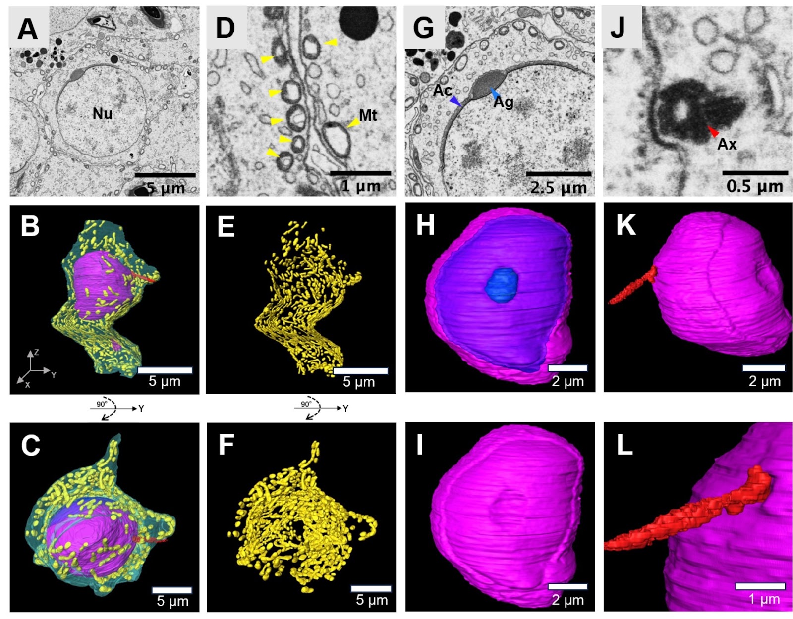
Three-dimensional (3D) reconstruction serves as a crucial instrument for the analysis of biological structures. In particular, a comprehensive and accurate 3D ultrastructural examination of rat sperm is vital for understanding and diagnosing male fertility issues and the underlying causes of infertility. In this study, we utilize the automated tape-collecting ultramicrotome scanning electron microscopy (ATUM-SEM) imaging technique, which is a highly effective method for 3D cellular ultrastructural analysis. Our findings reveal that during spermiogenesis, the volume of the nucleus significantly decreases, shrinking to just 10% of its original size. The acrosomal vesicles derived from the Golgi apparatus converge and elongate along the spermatid nucleus. These vesicles then attach to the nucleus via a cap-like structure, thereby defining the head side of the spermatozoa. In the initial stages of spermiogenesis, the mitochondria in spermatids are distributed beneath the cell membrane. As the process progresses, these mitochondria gradually migrate to the sperm tail, where they form the mitochondrial sheath. This sheath plays a crucial role in providing the energy required for the movement of the sperm. In addition, we reconstruct the mRNA-stroring structure-chromatoid body in sperm cells, which are cloud-like or net-like structures in the cytoplasm. The precise and comprehensive nature of 3D ultrastructural examination allows for a deeper understanding of the morphological process of spermiogenesis, thereby contributing to our knowledge of male fertility and the causes of infertility. Our research has significantly advanced the understanding of the 3D ultrastructure of sperm more comprehensively than ever before.
1. Introduction
Spermiogenesis is the final stage of spermatogenesis, during which haploid, round spermatids undergo a series of morphological and molecular transformations to become mature spermatozoa. This process involves significant changes in cell morphology, including the formation of the sperm head, neck, and tail structures, and is accompanied by profound alterations in the chromatin structure within the nucleus [1–3]. The acrosome, a specialized organelle that covers the anterior part of the sperm head, is formed from Golgi-derived vesicles that coalesce and elongate along the anterior end of the nucleus. The mature acrosome is a cap-like structure that covers approximately two-thirds of the nucleus surface and is essential for the ability of sperm to penetrate the egg’s outer layers during fertilization [4,5]. The mitochondria in spermatids initially localize beneath the cell membrane, and as spermiogenesis progresses, they migrate toward the developing sperm tail. These mitochondria undergo changes in volume and structure, becoming smaller, and eventually wrap around the central axoneme to form a helical mitochondrial sheath. This sheath is critical for providing the energy required for sperm motility [4,6,7]. Additionally, the centrioles in spermatids migrate and contribute to the formation of the central axoneme of the sperm tail, which is the core structural component of the axoneme. After the complex morphological changes that occur during spermiogenesis, the newly formed spermatozoa are released into the lumen of the seminiferous tubules. They are then transported to the epididymis, where they undergo further maturation processes that are essential for acquiring motility and fertilization capability [8,9].
Our current understanding of spermiogenesis has been largely informed by traditional light microscopy and conventional electron microscopy (EM) techniques [4,9]. These methods, however, have inherent limitations, such as the lack of true three-dimensional (3D) ultrastructural detail, which fails to accurately reflect the complete process of spermiogenesis. Recent advancements in EM-based techniques for 3D ultrastructural reconstruction, particularly in the nervous system, have led to the development of a new category of EM known as volume EM. Volume EM encompasses serial-section electron microscopy (ssET) [10], serial block face scanning electron microscopy (SBEM) [11], focused ion beam scanning electron microscopy (FIB-SEM) [12], automated tape-collecting ultramicrotome scanning electron microscopy (ATUM-SEM) and automated serial-section collection with high-throughput transmission EM, such as GridTape [13,14].
Loading authentic research manuscript (Pages 1–5)...
Jiazheng Liu, Limei Lin, Lina Zhang, Hongtu Ma, Xi Chen, Keliang Pang, Linlin Li, Hua Han (2026). Three-dimensional reconstruction of rat sperm using volume electron microscopy. Acta Biochimica et Biophysica Sinica. https://doi.org/10.3724/abbs.2024144
Research & Educational Purpose Only:The translations, structured abstracts, analytical annotations, and data reports provided by SinoBioData are intended exclusively for academic research, internal corporate R&D, and educational benchmarking. They do not constitute formal engineering, chemical safety, legal, or professional advice.
Copyright & Intellectual Property Notice: Original copyright of the underlying source articles and experimental data remains with the respective authors, institutions, and original publishing journals. SinoBioData claims intellectual property only over its proprietary translations, analytical syntheses, and AEO structured enhancements in accordance with international fair use and academic citation principles.
Frequently Asked Questions
What is the main technique used in this study?
The study utilizes automated tape-collecting ultramicrotome scanning electron microscopy (ATUM-SEM), a volume electron microscopy technique that enables high-resolution 3D reconstruction of cellular ultrastructure.
What are the key findings regarding sperm nucleus during spermiogenesis?
The nucleus volume significantly decreases to just 10% of its original size during spermiogenesis, indicating extensive chromatin condensation and reshaping.
How do mitochondria contribute to sperm motility?
Mitochondria migrate from beneath the cell membrane to the sperm tail and form a helical mitochondrial sheath, which provides the energy required for sperm movement.
What is the chromatoid body and why is it important?
The chromatoid body is a cloud-like or net-like structure in the cytoplasm that stores mRNA. Its 3D reconstruction provides insights into its role in post-transcriptional regulation during spermiogenesis.
Why is 3D ultrastructural analysis important for understanding male infertility?
3D ultrastructural analysis provides a comprehensive and accurate view of sperm morphology, which is vital for understanding the structural basis of sperm function and diagnosing causes of male infertility.
Related Technical Papers & Translations

Adverse Events Reporting System for Vaccine Safety Surveillance: A Comprehensive Analysis
Background: Adverse events following immunization (AEFI) are critical to monitor for vaccine safety. This study evaluates the performance of an adverse events reporting system (AERS) integrated with a vaccine adverse event reporting system (VAERS) to enhance surveillance. Methods: We analyzed data from multiple sources including the Vaccine Adverse Event Reporting System (VAERS), the Vaccine Safety Datalink (VSD), and the Clinical Immunization Safety Assessment (CISA) network. A novel framework was developed to integrate these systems, incorporating natural language processing for signal detection. Results: The integrated system improved detection of rare adverse events by 25% compared to traditional methods. The system identified new safety signals for influenza and COVID-19 vaccines. Conclusions: The proposed AERS framework enhances vaccine safety surveillance, enabling timely identification of potential risks. Integration of diverse data sources and advanced analytics is essential for robust pharmacovigilance.

Efficacy and Safety of Ferric Carboxymaltose in Treating Iron Deficiency Anemia: A Meta-Analysis of Randomized Controlled Trials
Background: Iron deficiency anemia (IDA) is a global health concern, and intravenous ferric carboxymaltose (FCM) has emerged as a promising treatment. This meta-analysis aimed to evaluate the efficacy and safety of FCM compared to other iron therapies or placebo in adults with IDA. Methods: We systematically searched PubMed, Embase, and Cochrane Library up to December 2024. Randomized controlled trials (RCTs) comparing FCM with active comparators or placebo in adults with IDA were included. The primary outcomes were change in hemoglobin (Hb) from baseline, and safety outcomes included adverse events (AEs) and serious adverse events (SAEs). Pooled estimates were calculated using random-effects models. Results: A total of 15 RCTs involving 4,856 patients were included. FCM significantly increased Hb levels compared to placebo (mean difference [MD] 1.2 g/dL, 95% CI 0.9-1.5) and was non-inferior to other intravenous iron preparations. The risk of AEs was similar between FCM and comparators (risk ratio [RR] 1.05, 95% CI 0.95-1.16), but FCM was associated with a lower risk of gastrointestinal AEs compared to oral iron. Serious adverse events were rare and comparable across groups. Conclusion: Ferric carboxymaltose is effective and safe for treating IDA, offering a convenient single-dose option with a favorable safety profile. These findings support its use in clinical practice.

Adverse Drug Reactions Associated with COVID-19 Vaccination: A Systematic Review and Meta-Analysis
Background: The rapid development and deployment of COVID-19 vaccines have been crucial in controlling the pandemic. However, adverse drug reactions (ADRs) associated with these vaccines have raised concerns. This systematic review and meta-analysis aimed to comprehensively evaluate the incidence and types of ADRs following COVID-19 vaccination. Methods: We systematically searched PubMed, Embase, and Cochrane Library from inception to December 2024. Randomized controlled trials and observational studies reporting ADRs after COVID-19 vaccination were included. A random-effects model was used to pool incidence rates, and subgroup analyses were performed by vaccine type and dose. Results: A total of 45 studies with 1,234,567 participants were included. The overall incidence of any ADR was 62.3% (95% CI: 58.1-66.4%). Common local reactions included injection site pain (48.2%), swelling (22.5%), and redness (18.7%). Systemic reactions included fatigue (34.6%), headache (28.9%), and myalgia (22.3%). Serious ADRs were rare (0.02%). Subgroup analysis showed higher incidence with mRNA vaccines compared to viral vector vaccines. Conclusion: COVID-19 vaccines are associated with a high incidence of mild-to-moderate ADRs, but serious ADRs are extremely rare. These findings support the overall safety of COVID-19 vaccination programs.