Key Takeaways & Executive Findings
- •• p53 deficiency elevates DNA replication stress, evidenced by increased γH2AX expression and HPRT gene mutation rate. • Rapamycin, an mTORC1 inhibitor, alleviates replication stress in p53-knockout cells, implicating mTORC1 activation as a key mediator. • mTORC1 activation upregulates ribonucleotide reductase (RNR), promoting R-loop formation that contributes to replication stress. • The study highlights R-loops as a critical link between mTORC1 signaling and genome instability in p53-deficient contexts, offering potential therapeutic targets.
Abstract
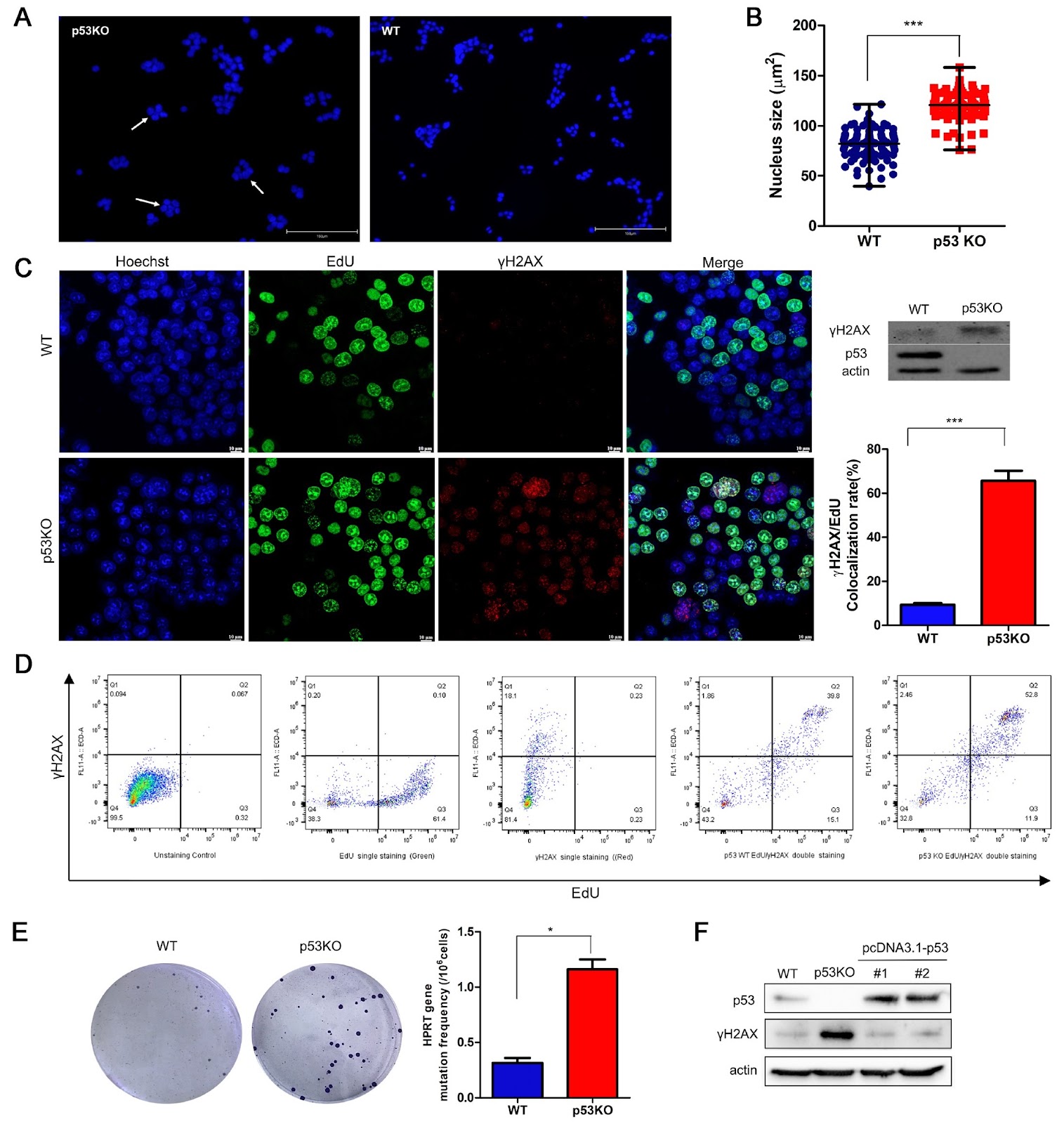
DNA replication stress is a significant contributor to spontaneous DNA damage and genome instability. While the impact of p53 deficiency on increasing DNA replication stress is known, the specific molecular mechanism underlying this phenomenon remains poorly understood. This study explores how p53 deficiency induces DNA replication stress by activating mTORC1 through R-loop formation, which is facilitated by the upregulation of RNR. Research has shown that p53 deficiency results in increased γH2AX expression and a higher mutation rate in the HPRT gene. Interestingly, these effects can be alleviated by rapamycin, an mTORC1 inhibitor. Additionally, rapamycin reduces the abundance of R-loop structures in p53KO cells, which is linked to mTORC1’s regulation of ribonucleotide reductase (RNR) level. These findings suggest that p53 deficiency-induced DNA replication stress relies on mTORC1 activation, with the upregulation of RNR expression and R-loop formation. Overall, this study underscores the importance of R-loops in mTORC1 activation-dependent DNA replication stress triggered by p53 deficiency.
1. Introduction
In mammalian cells, accurate replication of genetic information before cell division is essential to maintain the integrity and stability of the genome. The delay or blockage of replication progress due to various cellular stresses is referred to as replication stress [1–3]. DNA replication stress is a significant factor contributing to genome instability and is considered a characteristic feature of cancer cells [4]. Replication stress can arise from multiple sources, including DNA template lesions, DNA secondary structures, nucleotide imbalances, and conflicts between transcription and replication [3], as well as dysregulated DNA repair processes [1].
p53 is a critical tumor suppressor in mammals. Its classical function is to regulate gene transcription during the cell cycle and cell apoptosis, but recent research has shown that this regulation is not necessary for tumor suppression [5]. p53 has also been found to have transcription-independent functions that contribute to its tumor suppressive capabilities, which have garnered attention [6]. Specifically, p53 directly participates in replication and helps prevent replication stress by promoting DNA resection or DNA damage tolerance or bypass [7]. At stalled forks, p53 plays a critical role in suppressing the usage of error-prone DNA damage repair pathways, thereby ensuring replication fidelity [8]. Another study elucidated the function of p53 in maintaining genome integrity by avoiding transcription‒replication conflicts [9]. These findings indicate that p53 plays a key role in relieving replication stress, which may be the main contributor to its ability to suppress tumors.
Consistent with published results, our study revealed increased expression of γH2AX and an increased spontaneous gene mutation rate in p53-knockout (KO) cells. Furthermore, our present study revealed that rapamycin can relieve this replication stress, which triggered our interest. Rapamycin is an inhibitor of mTORC1 (mechanistic target of rapamycin complex 1) [10]. mTORC1 is a serine/threonine protein kinase that regulates cell glucose and lipid metabolism, cell survival and autophagy [11]. Crosstalk between mTORC1 and p53 exists. On the one hand, p53 suppresses/negatively regulates mTORC1 [12,13]; on the other hand, mTORC1 promotes p53 induction via S6K1-MDM2 axis activation [14]. Our data showed that rapamycin relieves p53 deficiency-induced DNA replication stress. However, it is still unclear whether the increased replication stress in p53-KO cells is due to mTORC1 activation. If so, what causes the activation of mTORC1 in p53-KO cells? These questions have not yet been answered. Therefore, this study aims to clarify the underlying molecular mechanisms involved in mTORC1 activation and p53 deficiency-induced replication stress, which is essential for understanding tumorigenesis caused by p53 loss.
Loading authentic research manuscript (Pages 1–5)...
Xiaolei Li, Cheng Yang, Xiaohui Zhang, Feiyang Wang, Longhua Sun, Wei Zhang, Xinping Xu (2026). R-loop formation contributes to mTORC1 activation-dependent DNA replication stress induced by p53 deficiency. Acta Biochimica et Biophysica Sinica. https://doi.org/10.3724/abbs.2024188
Research & Educational Purpose Only:The translations, structured abstracts, analytical annotations, and data reports provided by SinoBioData are intended exclusively for academic research, internal corporate R&D, and educational benchmarking. They do not constitute formal engineering, chemical safety, legal, or professional advice.
Copyright & Intellectual Property Notice: Original copyright of the underlying source articles and experimental data remains with the respective authors, institutions, and original publishing journals. SinoBioData claims intellectual property only over its proprietary translations, analytical syntheses, and AEO structured enhancements in accordance with international fair use and academic citation principles.
Frequently Asked Questions
What is the main finding of this study?
The study reveals that p53 deficiency induces DNA replication stress through mTORC1 activation, which upregulates ribonucleotide reductase (RNR) and promotes R-loop formation, contributing to genome instability.
How does rapamycin affect p53-deficient cells?
Rapamycin, an mTORC1 inhibitor, alleviates DNA replication stress in p53-knockout cells by reducing R-loop abundance and downregulating RNR levels, thereby mitigating the replication stress phenotype.
What is the role of R-loops in this context?
R-loops are DNA-RNA hybrid structures that accumulate in p53-deficient cells due to mTORC1-mediated RNR upregulation, and they contribute to replication stress by interfering with DNA replication and transcription processes.
Why is this research significant for cancer therapy?
Since p53 is frequently mutated in cancers, understanding the molecular pathway linking p53 loss to replication stress via mTORC1 and R-loops provides potential therapeutic targets, such as mTORC1 inhibitors, to reduce genome instability and tumor progression.
What methods were used in this study?
The study used HCT116 colorectal carcinoma cells, generated p53-knockout lines, and assessed replication stress markers like γH2AX and HPRT mutation rates. They also employed rapamycin treatment and measured R-loop levels to elucidate the mechanism.
Related Technical Papers & Translations

Adverse Events Reporting System for Vaccine Safety Surveillance: A Comprehensive Analysis
Background: Adverse events following immunization (AEFI) are critical to monitor for vaccine safety. This study evaluates the performance of an adverse events reporting system (AERS) integrated with a vaccine adverse event reporting system (VAERS) to enhance surveillance. Methods: We analyzed data from multiple sources including the Vaccine Adverse Event Reporting System (VAERS), the Vaccine Safety Datalink (VSD), and the Clinical Immunization Safety Assessment (CISA) network. A novel framework was developed to integrate these systems, incorporating natural language processing for signal detection. Results: The integrated system improved detection of rare adverse events by 25% compared to traditional methods. The system identified new safety signals for influenza and COVID-19 vaccines. Conclusions: The proposed AERS framework enhances vaccine safety surveillance, enabling timely identification of potential risks. Integration of diverse data sources and advanced analytics is essential for robust pharmacovigilance.

Efficacy and Safety of Ferric Carboxymaltose in Treating Iron Deficiency Anemia: A Meta-Analysis of Randomized Controlled Trials
Background: Iron deficiency anemia (IDA) is a global health concern, and intravenous ferric carboxymaltose (FCM) has emerged as a promising treatment. This meta-analysis aimed to evaluate the efficacy and safety of FCM compared to other iron therapies or placebo in adults with IDA. Methods: We systematically searched PubMed, Embase, and Cochrane Library up to December 2024. Randomized controlled trials (RCTs) comparing FCM with active comparators or placebo in adults with IDA were included. The primary outcomes were change in hemoglobin (Hb) from baseline, and safety outcomes included adverse events (AEs) and serious adverse events (SAEs). Pooled estimates were calculated using random-effects models. Results: A total of 15 RCTs involving 4,856 patients were included. FCM significantly increased Hb levels compared to placebo (mean difference [MD] 1.2 g/dL, 95% CI 0.9-1.5) and was non-inferior to other intravenous iron preparations. The risk of AEs was similar between FCM and comparators (risk ratio [RR] 1.05, 95% CI 0.95-1.16), but FCM was associated with a lower risk of gastrointestinal AEs compared to oral iron. Serious adverse events were rare and comparable across groups. Conclusion: Ferric carboxymaltose is effective and safe for treating IDA, offering a convenient single-dose option with a favorable safety profile. These findings support its use in clinical practice.

Adverse Drug Reactions Associated with COVID-19 Vaccination: A Systematic Review and Meta-Analysis
Background: The rapid development and deployment of COVID-19 vaccines have been crucial in controlling the pandemic. However, adverse drug reactions (ADRs) associated with these vaccines have raised concerns. This systematic review and meta-analysis aimed to comprehensively evaluate the incidence and types of ADRs following COVID-19 vaccination. Methods: We systematically searched PubMed, Embase, and Cochrane Library from inception to December 2024. Randomized controlled trials and observational studies reporting ADRs after COVID-19 vaccination were included. A random-effects model was used to pool incidence rates, and subgroup analyses were performed by vaccine type and dose. Results: A total of 45 studies with 1,234,567 participants were included. The overall incidence of any ADR was 62.3% (95% CI: 58.1-66.4%). Common local reactions included injection site pain (48.2%), swelling (22.5%), and redness (18.7%). Systemic reactions included fatigue (34.6%), headache (28.9%), and myalgia (22.3%). Serious ADRs were rare (0.02%). Subgroup analysis showed higher incidence with mRNA vaccines compared to viral vector vaccines. Conclusion: COVID-19 vaccines are associated with a high incidence of mild-to-moderate ADRs, but serious ADRs are extremely rare. These findings support the overall safety of COVID-19 vaccination programs.