🧬 SinoBioData Academic Portal
📚 Peer-Reviewed Translated Literature

All Biomedical & Clinical Articles (Page 53)

Browse complete peer-reviewed translations from top Chinese biomedical, oncology, and genomics journals. Read verified previews and download full authentic clinical reports.

Published Research Papers

Showing 24 of 1542 peer-reviewed translated articles (Page 53 of 65)

FOXM1 mediates methotrexate resistance in osteosarcoma cells by promoting autophagyGraphical AbstractVerified
Acta Biochimica et Biophysica Sinica

FOXM1 mediates methotrexate resistance in osteosarcoma cells by promoting autophagy

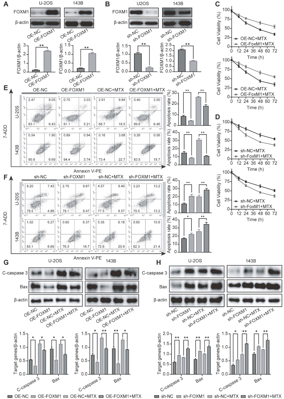
Osteosarcoma (OS) is a primary bone cancer mostly found in adolescents and elderly individuals. The treatment of OS is still largely dependent on traditional chemotherapy. However, the high incidence of drug resistance remains one of the greatest impediments to limiting improvements in OS treatment. Recent findings have indicated that the transcription factor FOXM1 plays an important role in various cancer-related events, especially drug resistance. However, the possible role of FOXM1 in the resistance of OS to methotrexate (MTX) remains to be explored. Here, we find that FOXM1, which confers resistance to MTX, is highly expressed in OS tissues and MTX-resistant cells. FOXM1 overexpression promotes MTX resistance by enhancing autophagy in an HMMR/ATG7-dependent manner. Importantly, silencing of FOXM1 or inhibiting autophagy reverses drug resistance. These findings demonstrate a new mechanism for FOXM1-induced MTX resistance and provide a promising target for improving OS chemotherapy outcomes.

Read Full Abstract10.3724/abbs.2024084
Aminophylline suppresses chronic renal failure progression by activating SIRT1/AMPK/mTOR-dependent autophagyGraphical AbstractVerified
Acta Biochimica et Biophysica Sinica

Aminophylline suppresses chronic renal failure progression by activating SIRT1/AMPK/mTOR-dependent autophagy

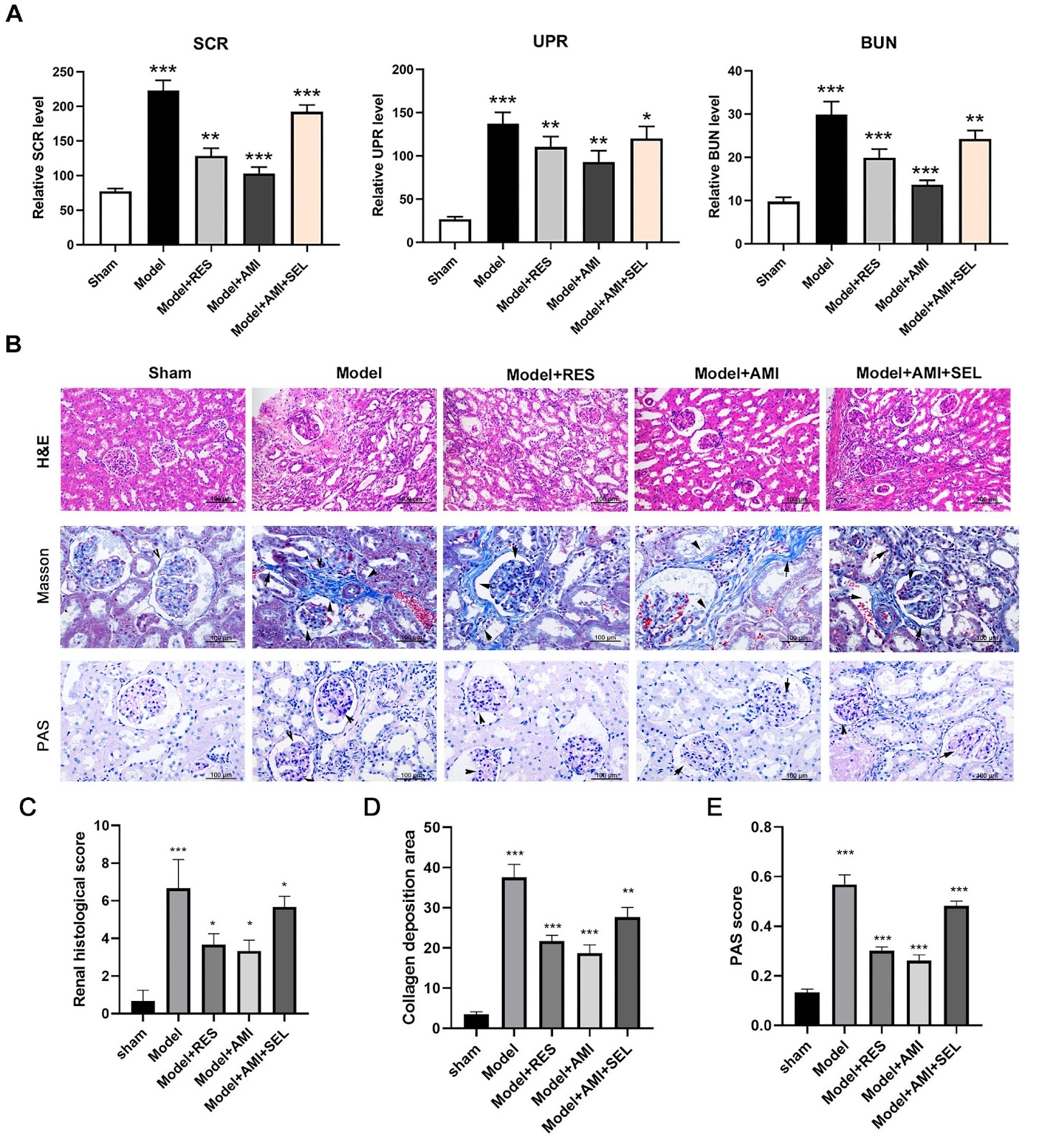
Chronic renal failure (CRF) is a severe syndrome affecting the urinary system for which there are no effective therapeutics. In this study, we investigate the effects and mechanisms of aminophylline in preventing CRF development. A rat model of chronic renal failure is established by 5/6 nephrectomy. The levels of serum creatinine (SCR), urinary protein (UPR), and blood urea nitrogen (BUN) are detected by ELISA. Histological evaluations of renal tissues are performed by H&E, Masson staining, and PAS staining. Functional protein expression is detected by western blot analysis or immunofluorescence microscopy. Glomerular cell apoptosis is determined using the TUNEL method. Results show that Aminophylline significantly reduces the levels of SCR, UPR, and BUN in the CRF model rats. Histological analyses show that aminophylline effectively alleviates renal tissue injuries in CRF rats. The protein expression levels of nephrin, podocin, SIRT1, p-AMPK, and p-ULK1 are greatly increased, while p-mTOR protein expression is markedly decreased by aminophylline treatment. Additionally, the protein level of LC3B in CRF rats is significantly increased by aminophylline. Moreover, aminophylline alleviates apoptosis in the glomerular tissues of CRF rats. Furthermore, resveratrol promotes SIRT1, p-AMPK, and p-ULK1 protein expressions and reduces p-mTOR and LC3B protein expressions in CRF rats. Selisistat (a SIRT1 inhibitor) mitigates the changes in SIRT1, p-AMPK, p-ULK1, p-mTOR, and LC3B expressions induced by aminophylline. Finally, RAPA alleviates renal injury and apoptosis in CRF rats, and 3-MA eliminates the aminophylline-induced inhibition of renal injury and apoptosis in CRF rats. Aminophylline suppresses chronic renal failure progression by modulating the SIRT1/AMPK/mTOR-mediated autophagy process.

Read Full Abstract10.3724/abbs.2024049
Hepatitis E virus infection upregulates ING5 expression in vitro and in vivoGraphical AbstractVerified
Acta Biochimica et Biophysica Sinica

Hepatitis E virus infection upregulates ING5 expression in vitro and in vivo

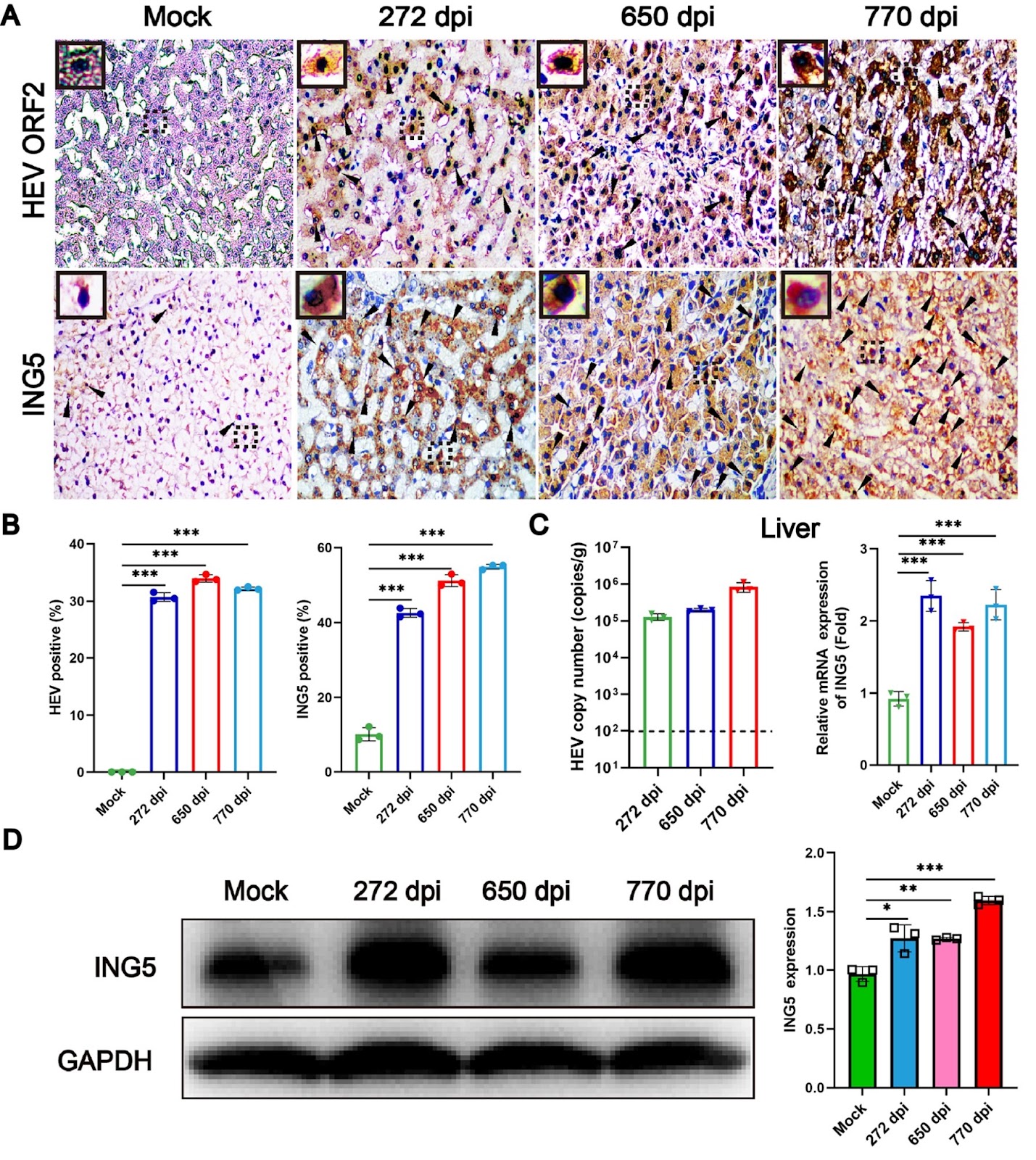
Hepatitis E virus (HEV) is the major pathogen of viral hepatitis. Immunocompromised individuals infected by HEV are prone to chronic hepatitis and increase the risk of hepato-cellular carcinoma (HCC). Inhibitor of growth family member 5 (ING5) is a tumor suppressor that is expressed at low levels in cancer tumors or cells. However, the underlying relationship between ING5 and HEV infection is unclear. In the present study, acute and chronic HEV animal models are used to explore the interaction between ING5 and HEV. Notably, the expression of ING5 is significantly increased in both the livers of acute HEV-infected BALB/c mice and chronic HEV-infected rhesus macaques. In addition, the relationship between HEV infection and ING5 expression is further identified in human hepatoma (HepG-2) cells. In conclusion, HEV infection strongly upregulates ING5 expression both in vivo and in vitro, which has significant implications for further understanding the pathogenic mechanism of HEV infection.

Read Full Abstract10.3724/abbs.2024091
GroEL triggers NLRP3 inflammasome activation through the TLR/NF-κB p-p65 axis in human periodontal ligament stem cellsGraphical AbstractVerified
Acta Biochimica et Biophysica Sinica

GroEL triggers NLRP3 inflammasome activation through the TLR/NF-κB p-p65 axis in human periodontal ligament stem cells

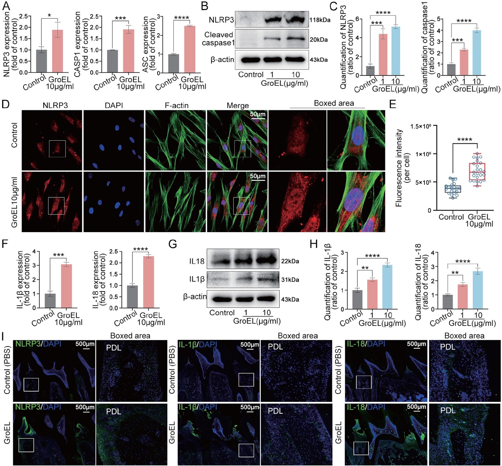
The interaction between bacteria and the host plays a vital role in the initiation and progression of systemic diseases, including gastrointestinal and oral diseases, due to the secretion of various virulence factors from these pathogens. GroEL, a potent virulence factor secreted by multiple oral pathogenic bacteria, is implicated in the damage of gingival epithelium, periodontal ligament, alveolar bone and other peripheral tissues. However, the underlying biomechanism is still largely unknown. In the present study, we verify that GroEL can trigger the activation of NLRP3 inflammasome and its downstream effector molecules, IL-1β and IL-18, in human periodontal ligament stem cells (hPDLSCs) and resultantly induce high activation of gelatinases (MMP-2 and MMP-9) to promote the degradation of extracellular matrix (ECM). GroEL-mediated activation of the NLRP3 inflammasome requires the participation of Toll-like receptors (TLR2 and TLR4). High upregulation of TLR2 and TLR4 induces the enhancement of NF-κB (p-p65) signaling and promotes its nuclear accumulation, thus activating the NLRP3 inflammasome. These results are verified in a rat model with direct injection of GroEL. Collectively, this study provides insight into the role of virulence factors in bacteria-induced host immune response and may also provide a new clue for the prevention of periodontitis.

Read Full Abstract10.3724/abbs.2024050
Identification and validation of disease severity-related circular RNA in acute pancreatitisGraphical AbstractVerified
Acta Biochimica et Biophysica Sinica

Identification and validation of disease severity-related circular RNA in acute pancreatitis

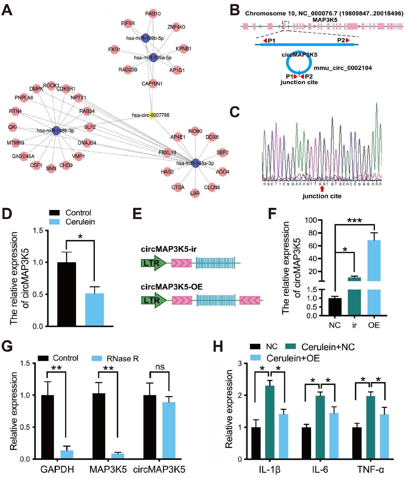
Acute pancreatitis arises from the activation of digestive enzymes in pancreatic acinar cells, leading to autodigestion of the pancreas and surrounding tissues. It is a common digestive tract emergency which requires hospitalization, and its incidence is increasing worldwide. In the past decade, several advances have been made in the treatment of acute pancreatitis. However, there is still a lack of efficacious drugs for clinical practice, and the limited value of existing biomarkers for early warning of the severity of acute pancreatitis is a major obstacle. Thus, there is an urgent need to gain a better understanding of the molecular mechanisms of acute pancreatitis. Circular RNAs (circRNAs) are a unique class of RNA molecules that are covalently closed. Ongoing investigations have provided evidence that circRNAs govern downstream target expression by acting as miRNA sponges, functioning as transcription factors, interacting with RNA-binding proteins, and regulating alternative splicing. These mechanisms support the pivotal role of circRNAs in a wide variety of physiological and pathological conditions, such as innate immunity, inflammation, neuronal function, and tumorigenesis. To explore the role of circular RNA in acute pancreatitis, we employed circRNA microarray technology (Arraystar Human circRNA Array V2) to examine the circRNA expression profile in the blood of three acute pancreatitis patients and three healthy controls. Clinical acute pancreatitis samples were obtained from Xiangya Hospital, Central South University. This study was approved by the Ethics Committee of Xiangya hospital (No. 2019010008). Normal control patients were recruited from among individuals who had visited Xiangya Hospital for a routine checkup. Written informed consent was obtained from all participants or their legal representatives for publication of data. The diagnosis and severity classification of acute pancreatitis were performed according to the American Gastroenterological Association guidelines and the Revised Atlanta Classification (RAC). circRNAs with a fold change ≥1.5 and a P value<0.05 were considered to be differentially expressed. As shown in Figure 1A, the two groups presented different expression profiles. We found that 91 circRNAs were significantly differentially expressed in the blood of acute pancreatitis patients, with 10 circRNAs exhibiting increased expression and 81 exhibiting decreased expression (Figure 1B,C). Among the differentially expressed circRNAs in acute pancreatitis, downregulated circRNAs are more prevalent than upregulated circRNAs, and the differential expression is more significant. Therefore, the present study focused on downregulated circRNAs. We selected circRNAs that are downregulated at least 2.5-fold and excluded those with fewer than 1000 bases to ensure the accuracy of qPCR. Based on these criteria, we identified nine circRNAs (circ_0006554, circ_0007798, circRNA_405815, circ_0001847, circ_0069748, circ_0001850, circ_0008417, circ_0002560, and circ_0000008) for validation by qPCR (Applied Biosystems, Foster City, USA) in blood samples from 30 acute pancreatitis patients (10 patients each with mild acute pancreatitis, moderate severe acute pancreatitis, and severe acute pancreatitis) and 15 healthy individuals. The levels of circ_0007798, circ_0001847, and circ_0069748 were significantly lower in acute pancreatitis patients than in normal controls, while the remaining circRNAs were not significantly differentially expressed (Figure 1D). In addition, the levels of circ_0007798 increased gradually with the severity of acute pancreatitis, suggesting that circ_0007798 is associated with the clinical severity of the disease (Figure 1E). Differential circRNAs have been studied for the diagnosis of pancreatic diseases. The expression level of circ_0007798 can be used to grade the severity of acute pancreatitis and provide individualized treatment. Furthermore, homology analysis (NCBI blast) revealed that circ_0007798 has a high degree of conservation between rats and humans according to the basic local alignment search tool. In conclusion, according to the
Read Full Abstract10.3724/abbs.2024115
The mechanism by which piR-000699 targets SLC39A14 regulates ferroptosis in aging myocardial ischemia/reperfusion injuryGraphical AbstractVerified
Acta Biochimica et Biophysica Sinica

The mechanism by which piR-000699 targets SLC39A14 regulates ferroptosis in aging myocardial ischemia/reperfusion injury

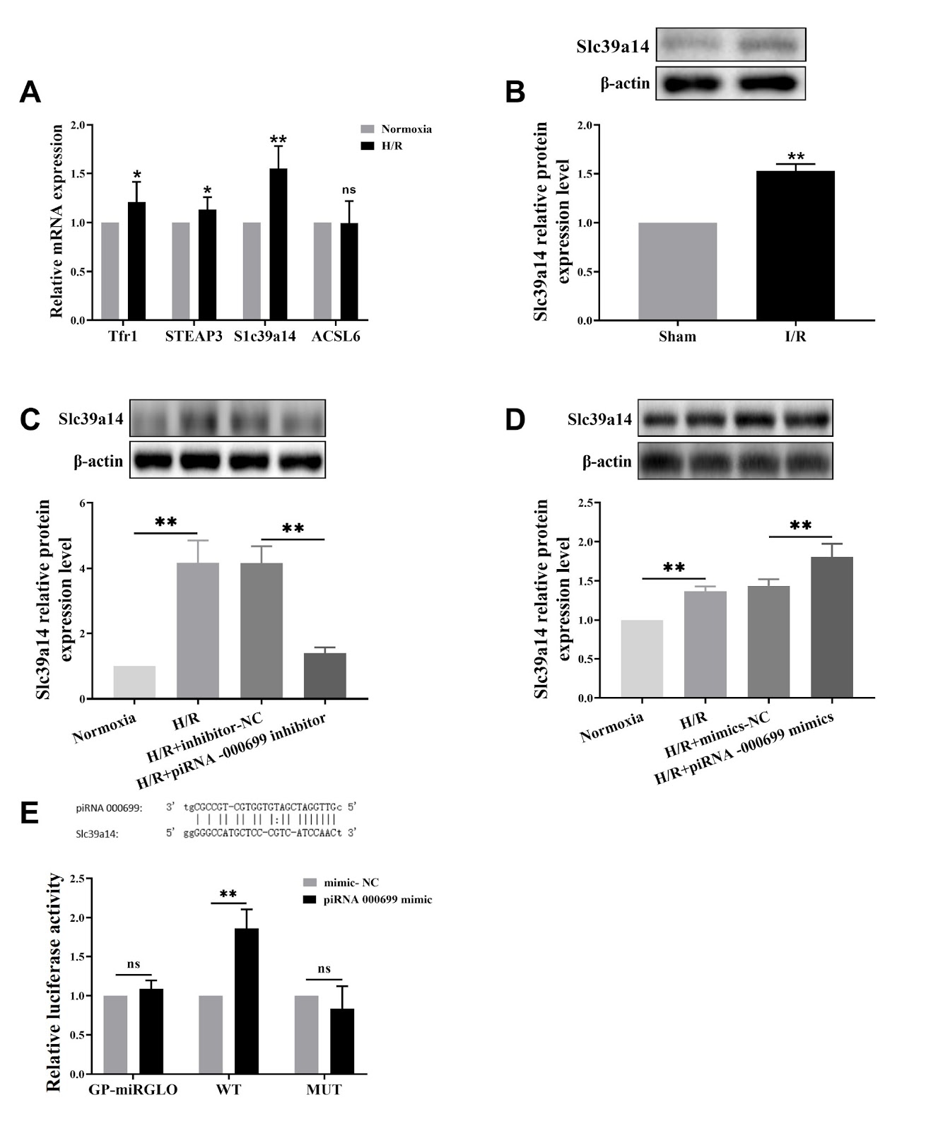
Myocardial ischemia/reperfusion (I/R) injury is a classic type of cardiovascular disease characterized by injury to cardiomyocytes leading to different types of cell death. The degree of irreversible myocardial damage is closely related to age, and ferroptosis is involved in cardiomyocyte damage. However, the mechanisms underlying ferroptosis regulation in aging myocardial I/R injury are still unclear. The present study aims to explore the underlying mechanism of piRNA regulation in ferroptosis. Using left anterior descending coronary artery ligation in an aging rat model and a D-galactose-induced rat cardiomyocyte line (H9C2) to construct an aging cardiomyocyte model, we investigate whether ferroptosis occurs after reperfusion injury in vitro and in vivo. This study focuses on the upregulation of piR-000699 after hypoxia/reoxygenation treatment in aging cardiomyocytes by observing hypoxia/reoxygenation (H/R) injury indicators and ferroptosis-related indicators and clarifying the role of piR-000699 in H/R injury caused by ferroptosis in aging cardiomyocytes. Bioinformatics analysis reveals that SLC39A14 is a gene that binds to piR-000699. Our data show that ferroptosis plays an important role in I/R injury both in vivo and in vitro. Furthermore, the results show the potential role of piR-000699 in regulating SLC39A14 in ferroptosis in aging cardiomyocytes under hypoxia/reoxygenation conditions. Together, our results reveal that the mechanism by which piR-000699 binds to SLC39A14 regulates ferroptosis in aging myocardial I/R injury.

Read Full Abstract10.3724/abbs.2024024
TRMT13 inhibits the growth of papillary thyroid cancer by targeting ANAPC4Graphical AbstractVerified
Acta Biochimica et Biophysica Sinica

TRMT13 inhibits the growth of papillary thyroid cancer by targeting ANAPC4

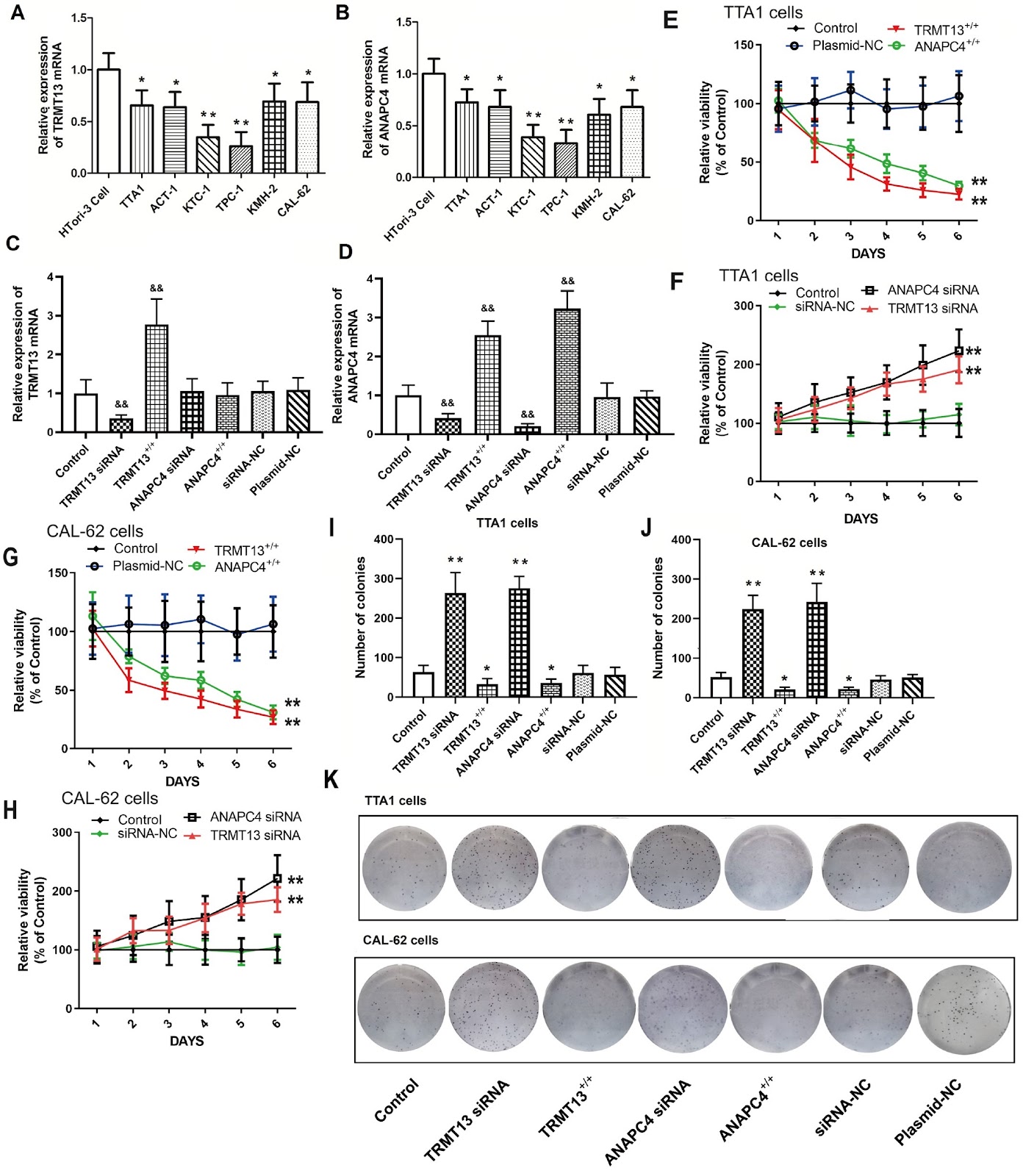
The recently discovered gene TRMT13 encodes a type of RNA methylase and is a member of the CCDC family (also called CCDC76). Here, we delineate its role in papillary thyroid cancer (PTC). Bioinformatics analysis shows significant TRMT13 and ANAPC4 downregulation in PTC and reveals that the expression levels of both genes are linearly correlated. Subsequent analyses confirm that both TRMT13 and ANAPC4 expressions are downregulated in PTC tissues and that this change in expression has a significant impact on cancer diagnosis. We conduct assays on PTC cells subjected to TRMT13 and ANAPC4 silencing or overexpression to assess the biological effects of these genes. We also perform rescue experiments to validate the regulatory effects of TRMT13 on ANAPC4. A nude mouse tumor model is used to evaluate the effects of TRMT13 and ANAPC4 on PTC tumorigenesis. TRMT13 expression is decreased in PTC tissues and cell lines and is positively correlated with that of ANAPC4. Cell assays reveal that TRMT13/ANAPC4 attenuates the malignancy of PTC cells by restraining cell proliferation, migration and invasion, while rescue experiments corroborate that ANAPC4 is a downstream target of TRMT13. In the nude mouse xenograft model, both TRMT13 and ANAPC4 inhibit tumor growth, and TRMT13 and ANAPC4 expression levels are significantly associated with survival. Taken together, these findings lead to the conclusion that TRMT13 inhibits PTC growth via ANAPC4, indicating a new role of TRMT13 and providing insights into the tRNA methyltransferase and coiled-coil domain-containing protein families.

Read Full Abstract10.3724/abbs.2024010
Proanthocyanidins isolated from lotus seed skin mitigate glycolipid metabolism disorder through the p38/Nrf2/NF-κB signaling pathwayGraphical AbstractVerified
Acta Biochimica et Biophysica Sinica

Proanthocyanidins isolated from lotus seed skin mitigate glycolipid metabolism disorder through the p38/Nrf2/NF-κB signaling pathway

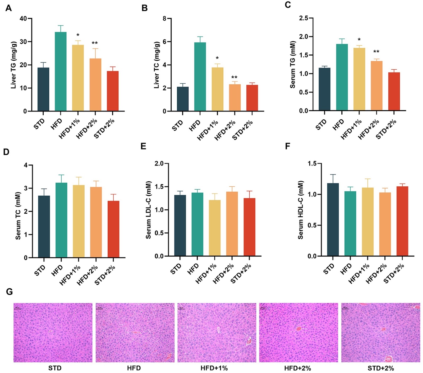
Lotus seed skin extract is rich in flavonoids, making it a promising candidate for developing health products. In a previous study, we found that proanthocyanidins from lotus seed skin, particularly proanthocyanidin B1 (PB1), can indirectly activate the Nrf2 signaling pathway, exerting an antioxidant effect. In this study, we isolate proanthocyanidins from lotus seed skin (PLS) using ethanol extraction and RP-HPLC identification, and investigate its effects on glycolipid metabolism both in vivo and in vitro. Our results demonstrate that PLS reduces body weight in high-fat diet (HFD) mice by decreasing feed efficiency. PLS also normalizes serum glucose, insulin secretion, glycosylated hemoglobin (HbA1c), and intraperitoneal glucose tolerance (IPGTT). Furthermore, PLS significantly improves blood lipid parameters and inhibits the expressions of six proinflammatory factors, including IL-1α, IL-1β, IL-3, IL-6, IFN-γ and TNF-α in HFD mice. Additionally, analysis of fresh liver tissues reveals that PLS and PB1 induce the expressions of antioxidant proteins such as HO-1 and NQO1 by activating the p38-Nrf2 signaling pathway and inhibiting the NF-κB signaling pathway. In conclusion, proanthocyanidins from lotus seed skin regulate glycolipid metabolism disorders by targeting the p38/Nrf2/NF-κB signaling pathway. Our study offers a new approach for the high-value comprehensive utilization of lotus seed skin by-products and precise dietary intervention for metabolic syndrome.

Read Full Abstract10.3724/abbs.2024042
The rod cell, a small form of Candida albicans, possesses superior fitness to the host gut and adaptation to commensalismGraphical AbstractVerified
Acta Biochimica et Biophysica Sinica

The rod cell, a small form of Candida albicans, possesses superior fitness to the host gut and adaptation to commensalism

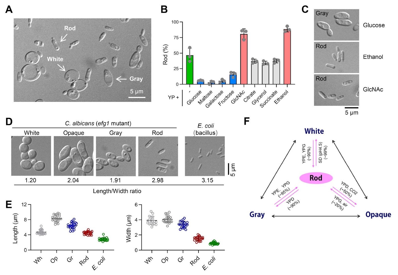
Candida albicans deploys various morphological forms through complex switching mechanisms, ensuring its survival and thriving as a commensal or pathogen in vastly different human niches. In this study, we demonstrate that a novel ''rod'' morphological form of C. albicans coexists and is interchangeable with previously reported white, gray, and opaque forms, constituting a tetra-stable phenotypic switching system. Rod cells arise from the efg1 mutant of SC5314 cells or from the clinical BJ1097 strain cultured under glucose-free conditions. They are characterized by a distinct gene expression profile and can be stably maintained through in vitro passaging or in vivo inhabitation of the gastrointestinal (GI) tract of mice. Remarkably, the majority of the efg1 mutant cells become rod cells in N-acetylglucosamine (GlcNAc)-containing medium, and the GlcNAc sensor Ngs1 is instrumental in converting the white or gray cells to the rod cells. Conversely, glucose inhibits rod cells through Cph1; consequently, the loss of Cph1 in the efg1 mutant cells permits their conversion to rod cells in glucose-replete media. Notably, rod cells of the efg1/cph1 mutant display superior adaptation and longer persistence in the murine GI environment than wild-type white cells. Taken together, these findings establish rod cells as a previously unappreciated form that is not only morphologically and transcriptionally distinguishable but also defined by specific genetic and environmental determinants, shedding light on complex fungus-host interactions.

Read Full Abstract10.3724/abbs.2024066
O-glycosylation of SARS-CoV-2 spike protein by host O-glycosyltransferase strengthens its trimeric structureGraphical AbstractVerified
Acta Biochimica et Biophysica Sinica

O-glycosylation of SARS-CoV-2 spike protein by host O-glycosyltransferase strengthens its trimeric structure

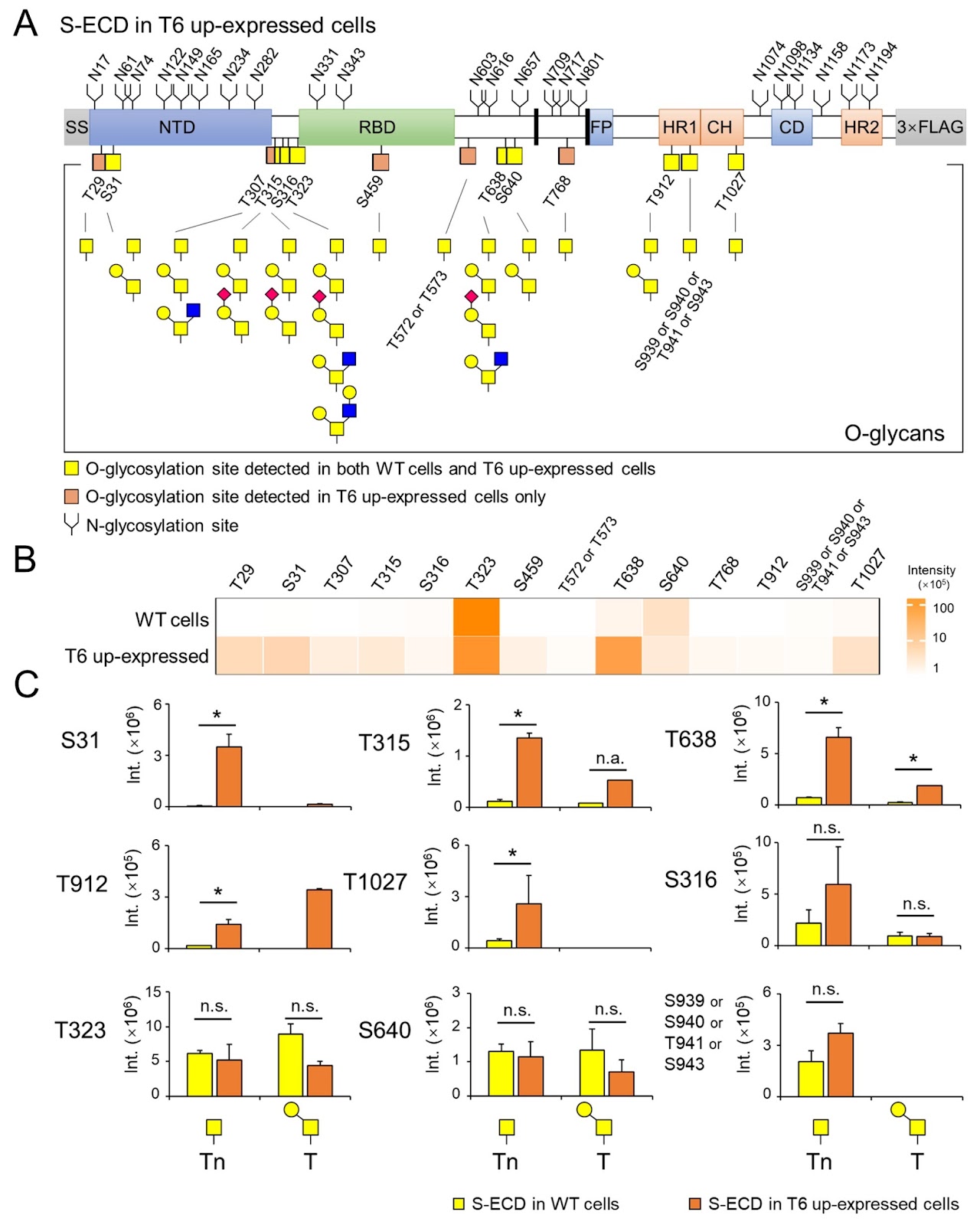
Protein O-glycosylation, also known as mucin-type O-glycosylation, is one of the most abundant glycosylation in mammalian cells. It is initially catalyzed by a family of polypeptide GalNAc transferases (ppGalNAc-Ts). The trimeric spike protein (S) of SARS-CoV-2 is highly glycosylated and facilitates the virus’s entry into host cells and membrane fusion of the virus. However, the functions and relationship between host ppGalNAc-Ts and O-glycosylation on the S protein remain unclear. Herein, we identify 15 O-glycosites and 10 distinct O-glycan structures on the S protein using an HCD-product-dependent triggered ETD mass spectrometric analysis. We observe that the isoenzyme T6 of ppGalNAc-Ts (ppGalNAc-T6) exhibits high O-glycosylation activity for the S protein, as demonstrated by an on-chip catalytic assay. Overexpression of ppGalNAc-T6 in HEK293 cells significantly enhances the O-glycosylation level of the S protein, not only by adding new O-glycosites but also by increasing O-glycan heterogeneity. Molecular dynamics simulations reveal that O-glycosylation on the protomer-interface regions, modified by ppGalNAc-T6, potentially stabilizes the trimeric S protein structure by establishing hydrogen bonds and non-polar interactions between adjacent protomers. Furthermore, mutation frequency analysis indicates that most O-glycosites of the S protein are conserved during the evolution of SARS-CoV-2 variants. Taken together, our finding demonstrate that host O-glycosyltransferases dynamically regulate the O-glycosylation of the S protein, which may influence the trimeric structural stability of the protein. This work provides structural insights into the functional role of specific host O-glycosyltransferases in regulating the O-glycosylation of viral envelope proteins.

Read Full Abstract10.3724/abbs.2024127
Macrophages exploit the mannose receptor and JAK-STAT1-MHC-II pathway to drive antigen presentation and the antimycobacterial immune response after BCG vaccinationGraphical AbstractVerified
Acta Biochimica et Biophysica Sinica

Macrophages exploit the mannose receptor and JAK-STAT1-MHC-II pathway to drive antigen presentation and the antimycobacterial immune response after BCG vaccination

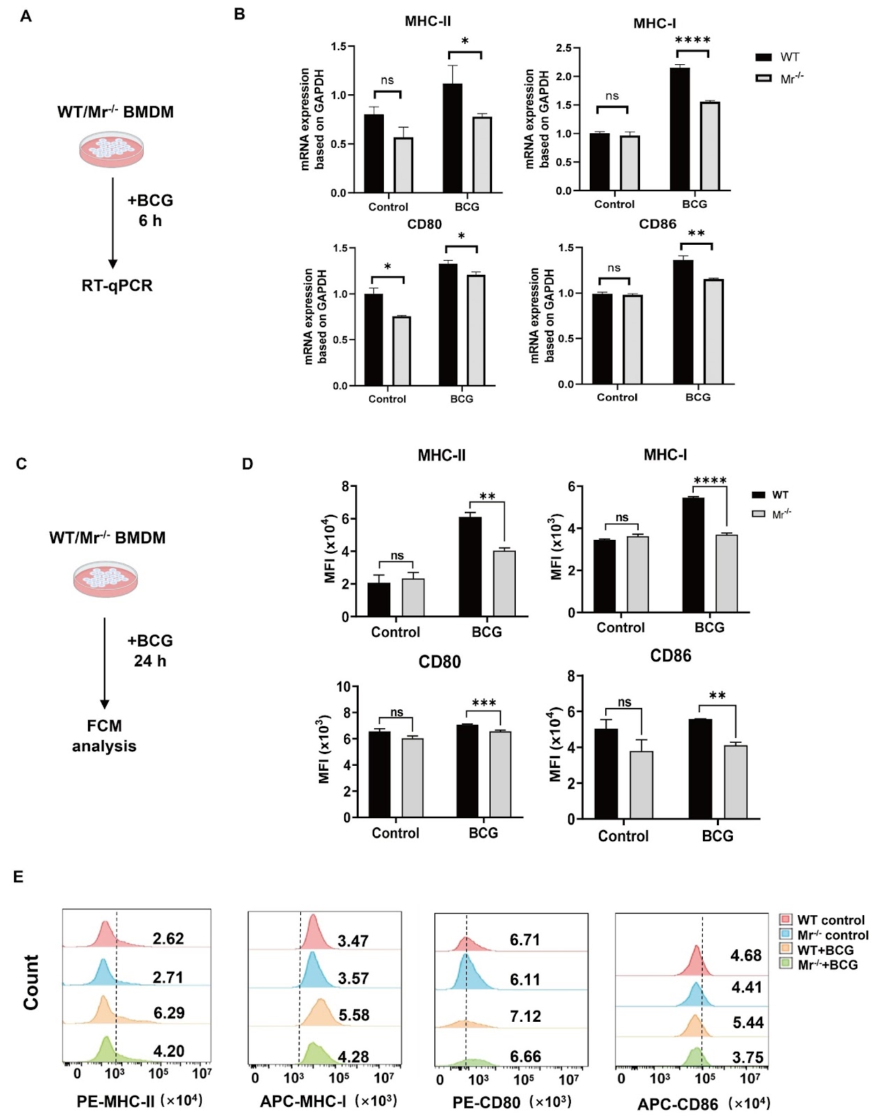
Tuberculosis (TB), caused by Mycobacterium tuberculosis (M.tb), remains one of the leading causes of fatal infectious diseases worldwide. The only licensed vaccine, Mycobacterium bovis Bacillus Calmette-Guérin (BCG), has variable efficacy against TB in adults. Insufficiency of immune cell function diminishes the protective effects of the BCG vaccine. It is critical to clarify the mechanism underlying the antimycobacterial immune response during BCG vaccination. Macrophage mannose receptor (MR) is important for enhancing the uptake and processing of glycoconjugated antigens from pathogens for presentation to T cells, but the roles of macrophage MR in the BCG-induced immune response against M.tb are not yet clear. Here, we discover that macrophage MR deficiency impairs the antimycobacterial immune response in BCG-vaccinated mice. Mechanistically, macrophage MR triggers JAK-STAT1 signaling, which promotes antigen presentation via upregulated MHC-II and induces IL-12 production by macrophages, contributing to CD4+ T cell activation and IFN-γ production. MR deficiency in macrophages reduces the vaccine efficacy of BCG and increases susceptibility to M.tb H37Ra challenge in mice. Our results suggest that MR is critical for macrophage antigen presentation and the antimycobacterial immune response to BCG vaccination and offer valuable guidance for the preventive strategy of BCG immunization.

Read Full Abstract10.3724/abbs.2024100
CD40 ligation-induced ERK activation leads to enhanced radiosensitivity in cervical carcinoma cells via promoting autophagyGraphical AbstractVerified
Acta Biochimica et Biophysica Sinica

CD40 ligation-induced ERK activation leads to enhanced radiosensitivity in cervical carcinoma cells via promoting autophagy

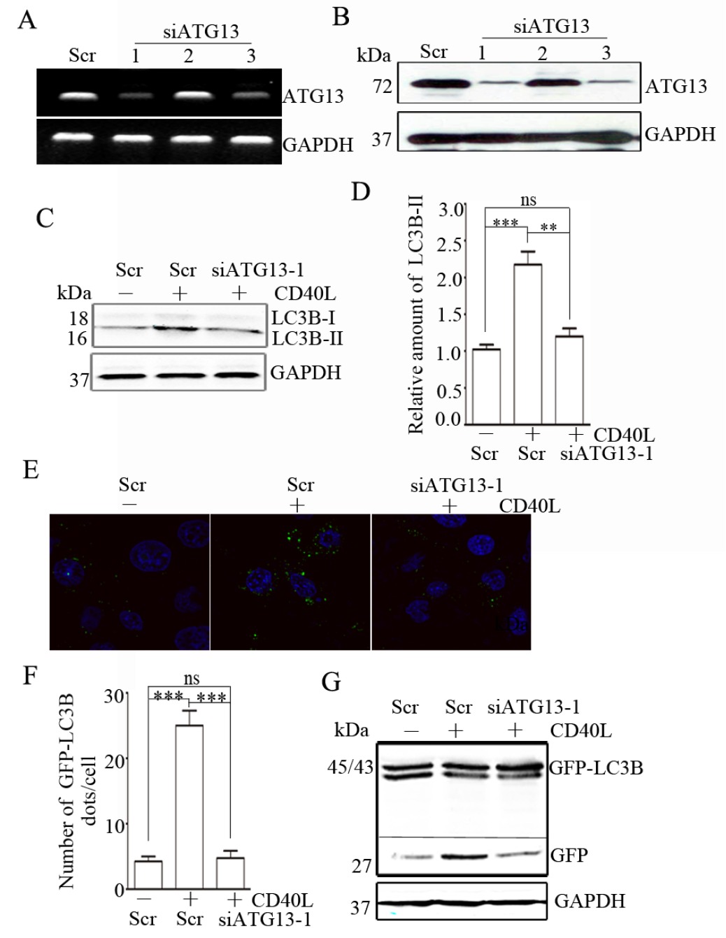
CD40, a member of the tumor necrosis factor (TNF) receptor superfamily, plays an important role not only in the immune system but also in tumor progression. CD40 ligation reportedly promotes autophagy in immune cells. However, the effects of CD40 ligation on autophagy and its mechanism in solid tumor cells are still unclear. In this study, we find that CD40 ligation promotes autophagosome formation and consequently promotes autophagic flux in cervical cancer cells. Mechanistically, this effect relies on ERK contributing to CD40 ligation-induced ATG13 upregulation by p53. Furthermore, we demonstrate that CD40 ligation-induced autophagy increases the radiosensitivity of cervical cancer cells. Taken together, our results provide new evidence for the involvement of the CD40 pathway in autophagy and radiotherapy in cervical cancer cells.

Read Full Abstract10.3724/abbs.2024229
Crystal structures of Kif2A complexed with WDR5 reveal the structural plasticity of WIN-S7 sitesGraphical AbstractVerified
Acta Biochimica et Biophysica Sinica

Crystal structures of Kif2A complexed with WDR5 reveal the structural plasticity of WIN-S7 sites

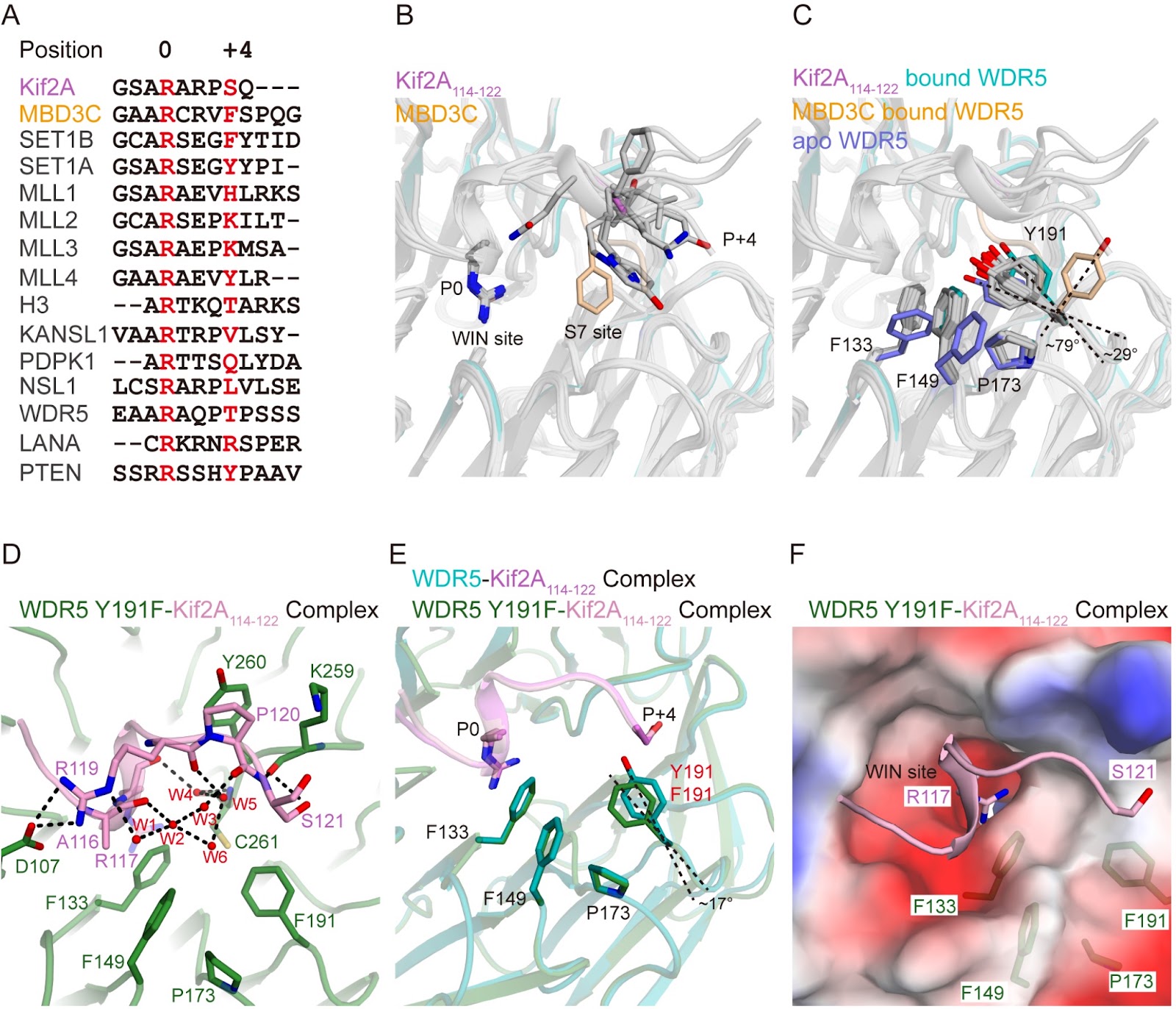
Chromosome congression and spindle assembly are essential for genomic stability and proper cell division, with deficiencies in these processes linked to tumorigenesis. WD repeat-containing protein 5 (WDR5), a core component of the mixed lineage leukemia (MLL) methyltransferase complex, directly binds to kinesin family member 2A (Kif2A) to regulate these mitotic events. Despite the importance of this interaction, its structural basis for Kif2A recognition by WDR5 remains unclear. Here, we determine the crystal structure of WDR5 in complex with a Kif2A-derived peptide (residues 114–122) at a resolution of 1.85 Å. Structural analysis reveals that Kif2A engages both the WIN and S7 sites of WDR5 via Arg117 and Ser121, with Ser121 forming hydrogen bonds with WDR5 Tyr191 and Lys259, driving Tyr191 rotation and opening the S7 pocket. Additional structures of WDR5 complexed with truncated or mutated Kif2A peptides and a WDR5 Y191F variant highlight the dynamic nature of Tyr191. Notably, anti-WDR5 compounds exhibit a similar binding mode at the WDR5 WIN-S7 site. The results of mutagenesis combined with isothermal titration calorimetry (ITC) assays underscore the critical roles of Arg117 and Ser121 in mediating the binding of Kif2A to WDR5. In summary, our findings provide atomic-level insights into the molecular mechanisms underlying the non-canonical mitotic function of the MLL/WDR5 complex and highlight WIN-S7 sites as promising therapeutic targets for diseases associated with chromosomal instability, such as cancers.

Read Full Abstract10.3724/abbs.2025066
Pyruvate dehydrogenase alleviates macrophage autophagy in Hcy-induced ApoE–/– miceGraphical AbstractVerified
Acta Biochimica et Biophysica Sinica

Pyruvate dehydrogenase alleviates macrophage autophagy in Hcy-induced ApoE–/– mice

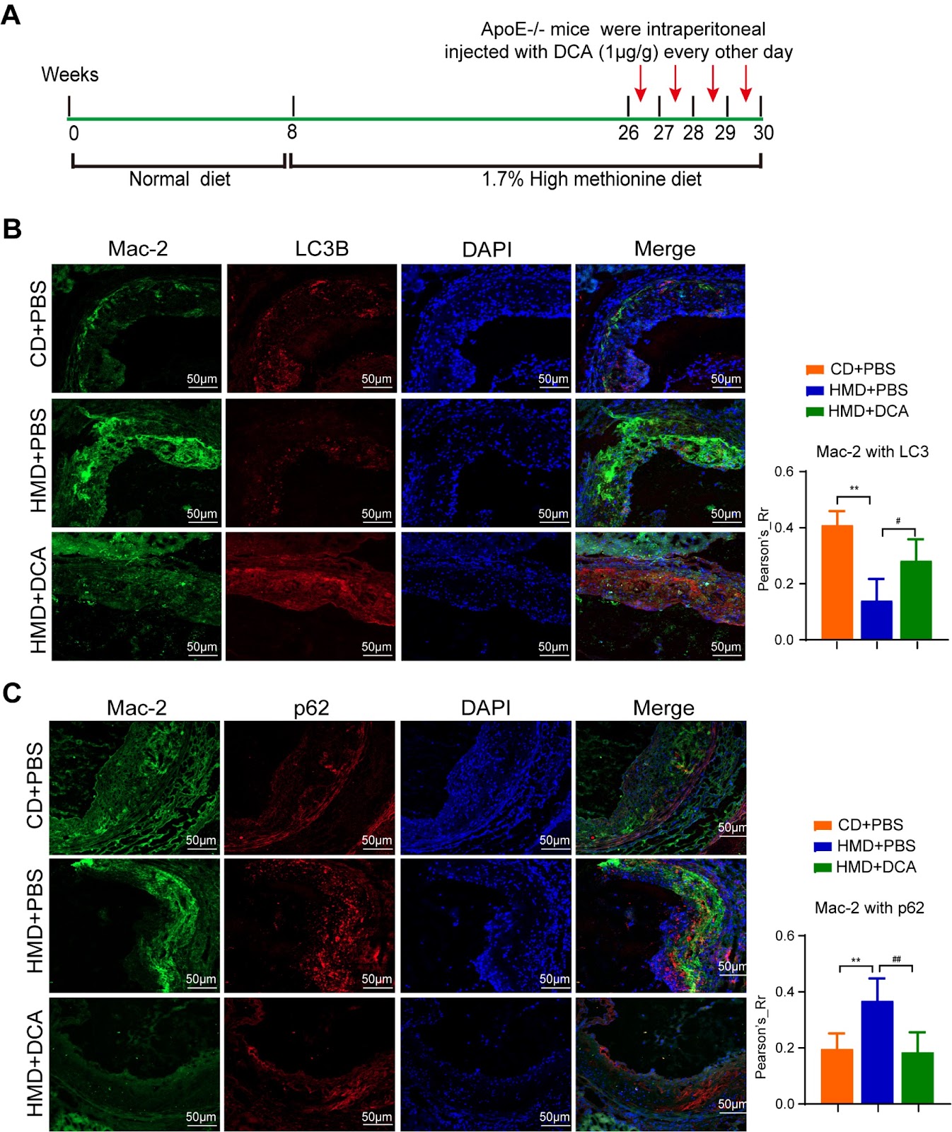
Macrophages play a protective role in atherosclerosis, whereas homocysteine (Hcy) is recognized as an independent risk factor for atherosclerosis. Defects in macrophage autophagy contribute to the formation of atherosclerotic plaques, and dysregulated energy metabolism is closely linked to the process of autophagy. However, the regulation of macrophage autophagy by pyruvate dehydrogenase (PDH), a key component of the PDH complex involved in energy and metabolic homeostasis, remains poorly understood in the context of atherosclerosis induced by Hcy. In our study, proteomic profiling identifies 748 upregulated proteins and 760 downregulated proteins in Hcy-treated macrophages. KEGG pathway analysis reveals significant enrichment of differentially expressed proteins in metabolism-related pathways, including those related to the biosynthesis of amino acids, carbon metabolism, and glycolysis/gluconeogenesis. Additionally, we explore the role of PDH in mediating Hcy-induced atherosclerosis in ApoE–/– mice. The results show a marked reduction in PDH expression and activity in Hcy-treated macrophages, leading to impaired autophagy. Notably, PDH activation enhances the assembly of the autophagy initiator ULK1-FIP200-Atg13 complex through the modulation of the AMPK/mTOR signaling pathway, suggesting a potential therapeutic target for Hcy-induced atherosclerosis.

Read Full Abstract10.3724/abbs.2025021
Deciphering disease through glycan codes: leveraging lectin microarrays for clinical insightsGraphical AbstractVerified
Acta Biochimica et Biophysica Sinica

Deciphering disease through glycan codes: leveraging lectin microarrays for clinical insights

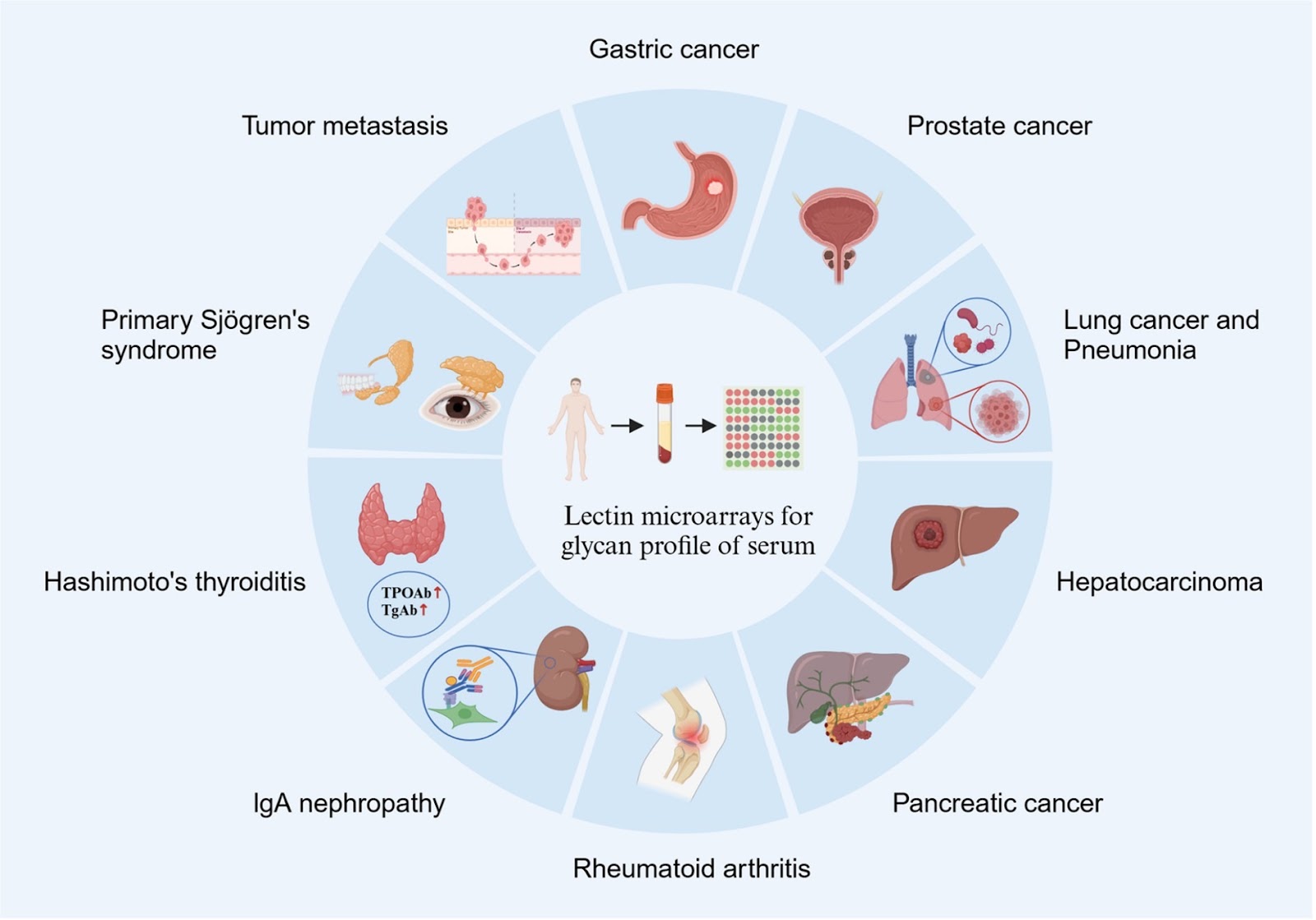
Glycosylation, a crucial posttranslational modification, plays a significant role in numerous physiological and pathological processes. Lectin microarrays, which leverage the high specificity of lectins for sugar binding, are ideally suited for profiling the glycan spectra of diverse and complex biological samples. In this review, we explore the evolution of lectin detection technologies, as well as the applications and challenges of lectin microarrays in analyzing the glycome profiles of various clinical samples, including serum, saliva, tissues, sperm, and urine. This review not only emphasizes significant advancements in the high-throughput analysis of polysaccharides but also provides insight into the potential of lectin microarrays for diagnosing and managing diseases such as tumors, autoimmune diseases, and chronic inflammation. We aim to provide a clear, concise, and comprehensive overview of the use of lectin microarrays in clinical settings, thereby assisting researchers in conducting clinical studies in glycobiology.

Read Full Abstract10.3724/abbs.2024123
Glycosylation in aging and neurodegenerative diseasesGraphical AbstractVerified
Acta Biochimica et Biophysica Sinica

Glycosylation in aging and neurodegenerative diseases

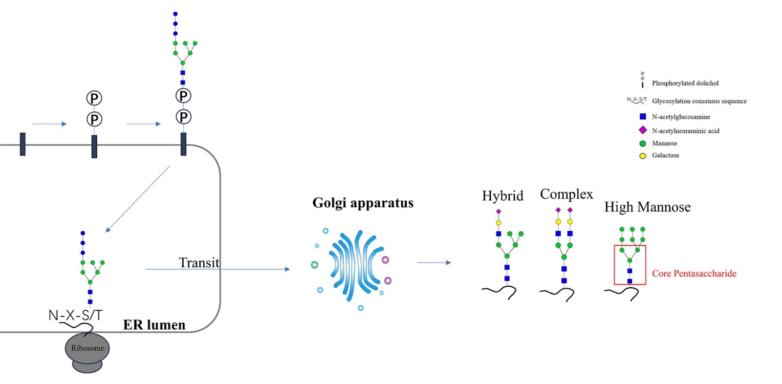
Aging, a complex biological process, involves the progressive decline of physiological functions across various systems, leading to increased susceptibility to neurodegenerative diseases. In society, demographic aging imposes significant economic and social burdens due to these conditions. This review specifically examines the association of protein glycosylation with aging and neurodegenerative diseases. Glycosylation, a critical post-translational modification, influences numerous aspects of protein function that are pivotal in aging and the pathophysiology of diseases such as Alzheimer’s disease, Parkinson’s disease, and other neurodegenerative conditions. We highlight the alterations in glycosylation patterns observed during aging, their implications in the onset and progression of neurodegenerative diseases, and the potential of glycosylation profiles as biomarkers for early detection, prognosis, and monitoring of these age-associated conditions, and delve into the mechanisms of glycosylation. Furthermore, this review explores their role in regulating protein function and mediating critical biological interactions in these diseases. By examining the changes in glycosylation profiles associated with each part, this review underscores the potential of glycosylation research as a tool to enhance our understanding of aging and its related diseases.

Read Full Abstract10.3724/abbs.2024136
CDC5L facilitates cardiomyocyte proliferation and ameliorates myocardial ischemia-reperfusion injury via modulation of the FGF10-YAP axisGraphical AbstractVerified
Acta Biochimica et Biophysica Sinica

CDC5L facilitates cardiomyocyte proliferation and ameliorates myocardial ischemia-reperfusion injury via modulation of the FGF10-YAP axis

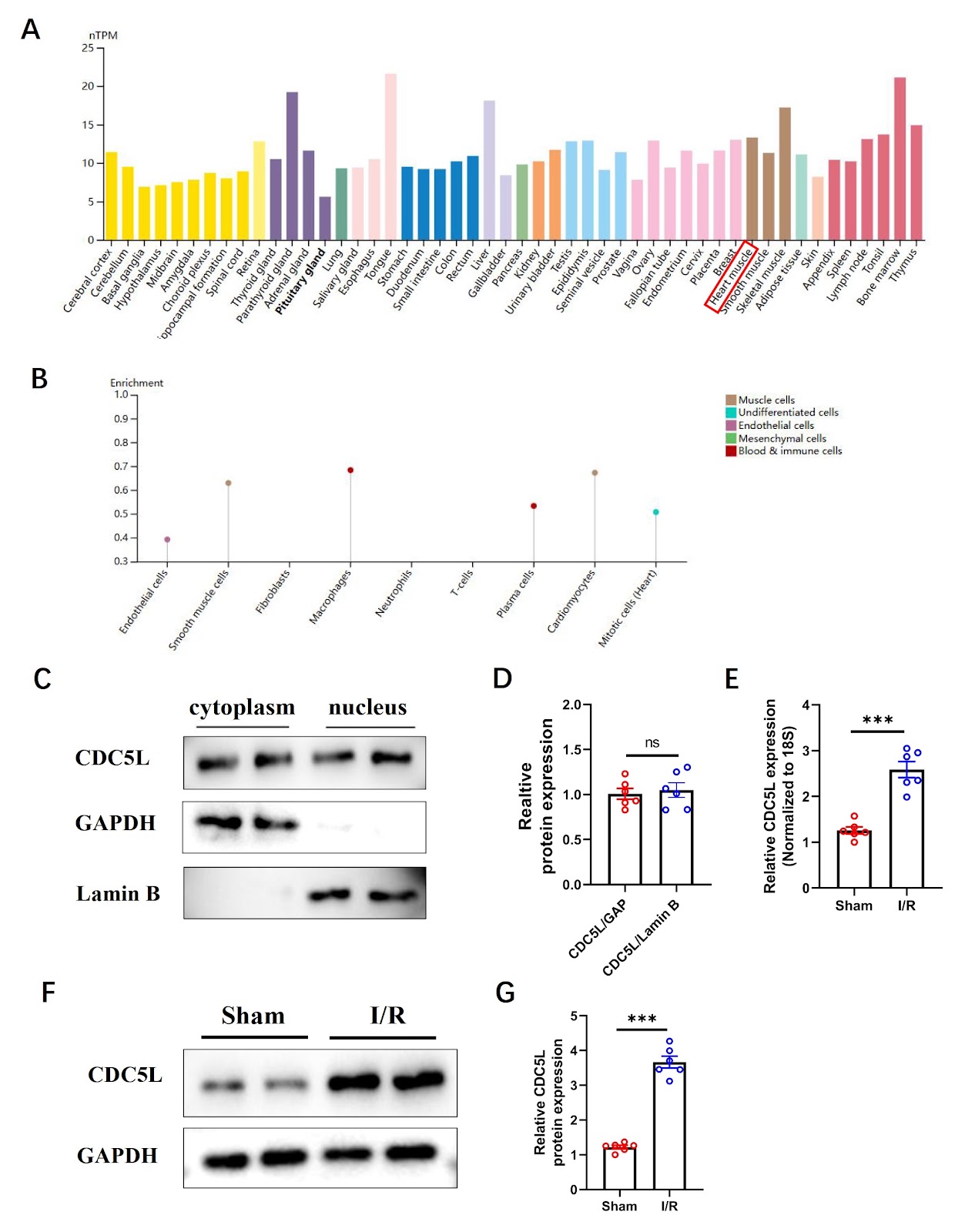
Myocardial infarction (MI) causes irreversible cardiomyocyte loss, creating a need for cardiac repair therapies. The role of cell division cycle 5-like (CDC5L), a cell cycle regulator, in cardiac repair is unknown. This study aims to define the role of CDC5L in mitigating ischemia-reperfusion (I/R) injury by assessing its impact on cardiomyocyte proliferation and apoptosis and to determine the mechanism involving the FGF10-YAP axis. We model cardiac injury using in vitro oxygen-glucose deprivation/reoxygenation (OGD/R) in neonatal mouse cardiomyocytes and in vivo I/R in adult mice. To investigate CDC5L function, we modulate its expression via adenoviral or AAV9-mediated overexpression or knockdown. Proliferation markers (EdU+, Ki67+, pH3+), apoptosis (TUNEL staining, Bax/Bcl-2 ratio), and cardiac function (echocardiography) are assessed. Through transcriptomic screening, we identify CDC5L downstream targets and validate their functional roles using FGF10 knockdown rescue assays. We find that CDC5L is upregulated in the post-I/R murine myocardium. Its overexpression enhances cardiomyocyte proliferation, preserves cardiac function, reduces apoptosis, and diminishes infarct size. Transcriptomic analysis identifies FGF10 as a key downstream effector, and we confirm that CDC5L upregulates FGF10 expression. Notably, FGF10 knockdown reverses the proliferative and anti-apoptotic effects of CDC5L. Moreover, the CDC5L-mediated reduction in YAP phosphorylation is also dependent on FGF10, as this effect is abolished upon FGF10 knockdown. In conclusion, CDC5L attenuates cardiac I/R injury by promoting cardiomyocyte proliferation and inhibiting apoptosis through the FGF10-YAP pathway. This CDC5L-FGF10-YAP axis represents a promising therapeutic target to improve myocardial regeneration and recovery after myocardial infarction.

Read Full Abstract10.3724/abbs.2025213
Inherited glycosylphosphatidylinositol deficiency: a review from molecular and clinical perspectivesGraphical AbstractVerified
Acta Biochimica et Biophysica Sinica

Inherited glycosylphosphatidylinositol deficiency: a review from molecular and clinical perspectives

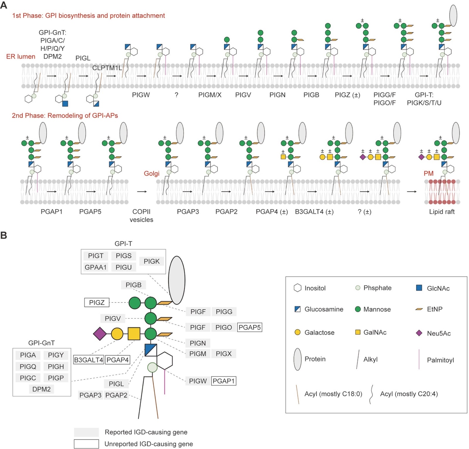
Glycosylphosphatidylinositol (GPI) is a highly conserved post-translational modification in eukaryotes, which is essential for anchoring various proteins to the cell surface. Dysfunction of GPI biogenesis leads to human diseases, such as inherited GPI deficiency (IGD) caused by germline mutations in GPI-related genes. With accumulating reports on individuals with IGD, there has been increasing interest and studies on disease mechanism, diagnosis, and therapy. This review outlines the biosynthetic pathway of GPI-anchored proteins (GPI-APs) and summarizes clinical IGD cases from a molecular perspective. We also review current diagnostic and therapeutic approaches for IGD. Finally, we discuss future research directions to facilitate the understanding and treatment of GPI-related disorders.

Read Full Abstract10.3724/abbs.2024128
Succinate accumulation induces pyroptosis and mitochondrial damage via the inhibition of ATP5F1D in HUVECsGraphical AbstractVerified
Acta Biochimica et Biophysica Sinica

Succinate accumulation induces pyroptosis and mitochondrial damage via the inhibition of ATP5F1D in HUVECs

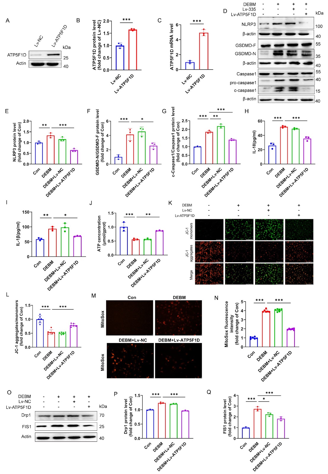
Atherosclerosis, a chronic inflammatory disorder, is pathophysiologically linked to endothelial cell (EC) pyroptosis. This study aims to elucidate the mechanisms by which succinate exacerbates EC pyroptosis through mitochondrial damage. Serum samples are collected from patients with coronary heart disease (CHD) and healthy controls (HCs), and the levels of succinate, interleukin (IL)-6, and IL-18 are quantified. To establish a succinate accumulation model, human umbilical vein endothelial cells (HUVECs) are treated with diethyl butyl malonate (DEBM), followed by analysis of inflammatory cytokines. The expression of pyroptosis-related proteins is assessed via western blot analysis. Morphological changes in pyroptotic vesicles and membrane pores are examined by scanning electron microscopy (SEM) and transmission electron microscopy (TEM). The mitochondrial membrane potential and reactive oxygen species (ROS) levels are evaluated via a JC-1 kit and MitoSOX, respectively. RNA sequencing (RNA-seq) is performed to identify potential target genes and regulatory pathways. To investigate the functional role of ATP5F1D, small interfering RNAs (siRNAs) are used to knockdown ATP5F1D, while lentiviral vectors are used to overexpress ATP5F1D in HUVECs. The results reveal significantly elevated levels of succinate, IL-6, and IL-18 in both CHD patients and DEBM-treated HUVECs. Succinate accumulation induced by DEBM triggers pyroptosis and mitochondrial damage in HUVECs, as evidenced by the upregulation of pyroptosis-related proteins and the impairment of mitochondrial structure and function. RNA sequencing analysis identifies ATP5F1D as a key downstream target of succinate accumulation. Downregulation of ATP5F1D promotes pyroptosis and mitochondrial injury in HUVECs, whereas restoration of ATP5F1D expression effectively mitigates these detrimental effects. Succinate-induced downregulation of ATP5F1D drives mitochondrial dysfunction and pyroptosis in HUVECs.

Read Full Abstract10.3724/abbs.2025116
LINC00114 promotes colorectal cancer metastasis by targeting HNRNPA1 to regulate glutamine metabolism reprogramming and angiogenesisGraphical AbstractVerified
Acta Biochimica et Biophysica Sinica

LINC00114 promotes colorectal cancer metastasis by targeting HNRNPA1 to regulate glutamine metabolism reprogramming and angiogenesis

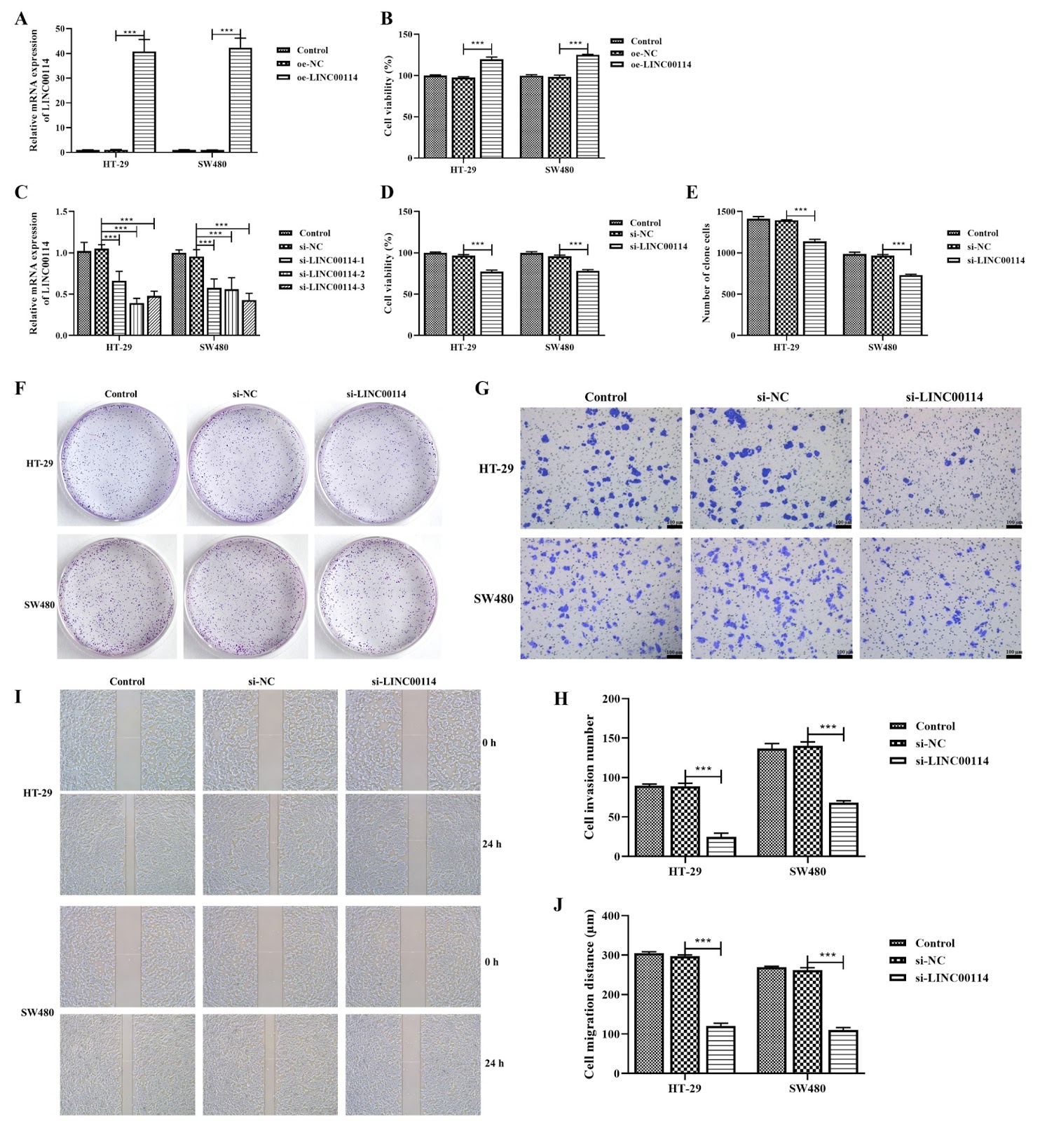
Colorectal cancer (CRC) is a common type of gastrointestinal malignancy, and it has a close connection with long noncoding RNAs (lncRNAs). This study aims to examine the involvement of long noncoding RNA LINC00114, which targets heterogeneous nuclear ribonucleoprotein A1 (HNRNPA1) in regulating glutamine metabolism and angiogenesis in the metastasis of colorectal cancer (CRC). LINC00114 and HNRNPA1 levels are measured in CRC tissues and cells to determine their expression levels. Then, siRNA targeting LINC00114 (si-LINC00114) is used to transfect CRC cells, and cell proliferation and metastasis are detected. The influence of exogenous glucose and glutamine supplementation on angiogenesis induced by LINC00114 in CRC is investigated in HUVECs. Glutamine metabolism in CRC cells is also detected. Furthermore, the role of LINC00114 in CRC xenograft tumors is studied in vivo. LINC00114 and HNRNPA1 are highly expressed in CRC and positively correlate with CD31. si-LINC00114 significantly inhibits proliferation, metastasis and HNRNPA1 expression in CRC cells. An RNA-binding-protein immunoprecipitation (RIP) assay confirms that LINC00114 can bind to HNRNPA1 and positively regulate its expression. Further experiments confirm that si-LINC00114 significantly inhibits cell proliferation and tubule formation in HUVECs. Exogenous glucose and glutamine supplementation significantly promotes the levels of LINC00114 and HNRNPA1 in CRC cells and promotes tubule formation in HUVECs. In addition, transfection of CRC cells with si-LINC00114 and/or oe-HNRNPA1 regulates glutamine metabolism in CRC cells. Animal studies confirm that intervention with LINC00114 represses the progression and vascular normalization of CRC and regulates glutamine metabolism. In conclusion, LINC00114 promotes CRC metastasis by targeting HNRNPA1 to regulate glutamine metabolic reprogramming and angiogenesis.

Read Full Abstract10.3724/abbs.2025141
SUN5 interacts with TRIM28, enhancing IκBα ubiquitination to promote glycolysis in colorectal cancer cellsGraphical AbstractVerified
Acta Biochimica et Biophysica Sinica

SUN5 interacts with TRIM28, enhancing IκBα ubiquitination to promote glycolysis in colorectal cancer cells

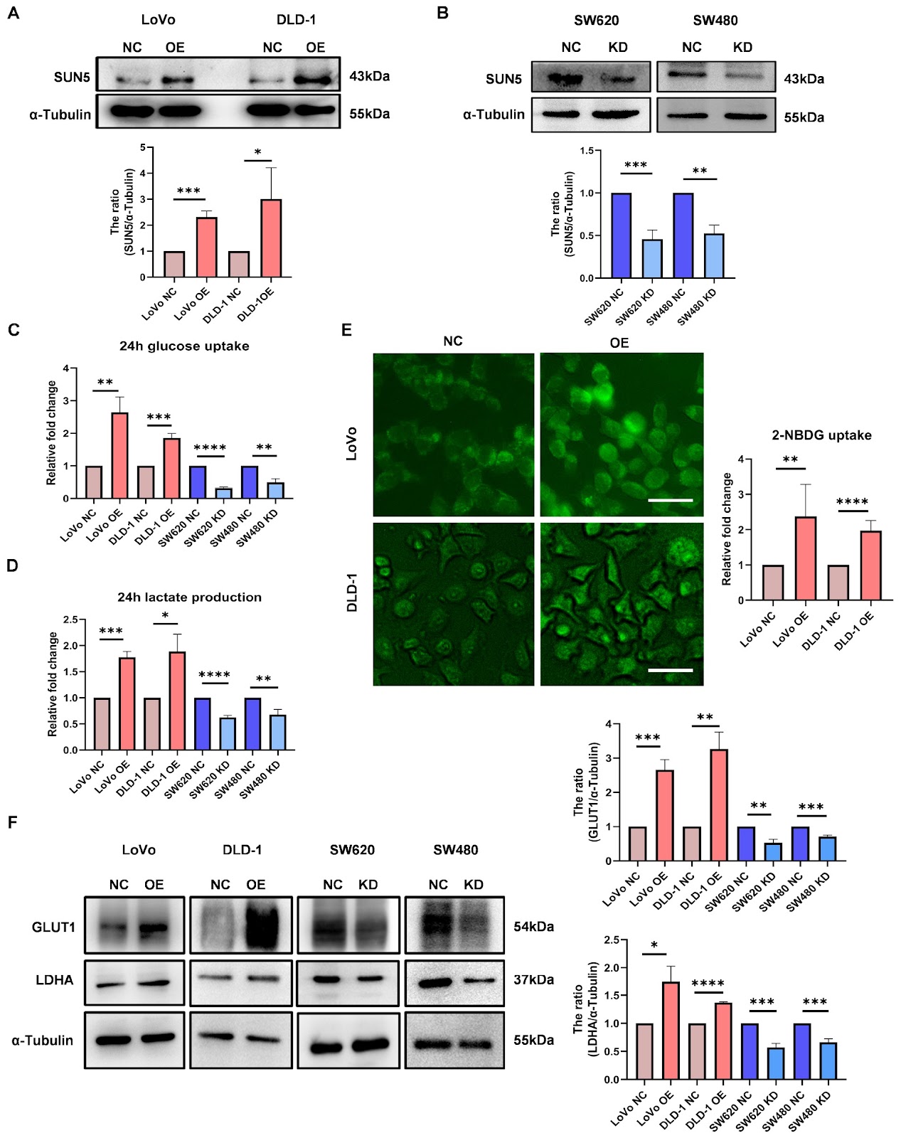
Glycolysis provides the main energy source for the rapid proliferation and migration of colorectal cancer (CRC) cells. In our previous studies, we reported that SUN5, a nuclear membrane protein, promotes proliferation and migration. However, whether SUN5 is involved in the process of glycolysis is unclear. Here, we demonstrate that overexpression of SUN5 enhances glucose uptake and lactate production in CRC cells, whereas the opposite results are observed in SUN5-knockdown cells. Mechanistically, SUN5 activates the NF-κB signaling pathway, which can be inhibited by the IKK inhibitor BAY11-7082. Further studies reveal that SUN5 interacts with TRIM28 to increase IκBα ubiquitination, leading to the nuclear translocation of phosphorylated P65 (phos-P65) and subsequent increases in the transcription of GLUT1 and LDHA, accelerating glycolysis. Moreover, xenograft transplantation experiments reveal that the knockdown of SUN5 inhibits glycolysis and tumorigenesis in vivo. Taken together, these findings indicate that SUN5 enhances the glycolysis and tumorigenesis of CRC cells via interaction with TRIM28, which provides a potential target for the diagnosis and treatment of CRC.

Read Full Abstract10.3724/abbs.2025201
Single-cell and bulk transcriptome analysis unveils a ligand-receptor-based signature for prognostication and reveals that TREM1 controls the malignant behaviors of hepatocellular carcinomaGraphical AbstractVerified
Acta Biochimica et Biophysica Sinica

Single-cell and bulk transcriptome analysis unveils a ligand-receptor-based signature for prognostication and reveals that TREM1 controls the malignant behaviors of hepatocellular carcinoma

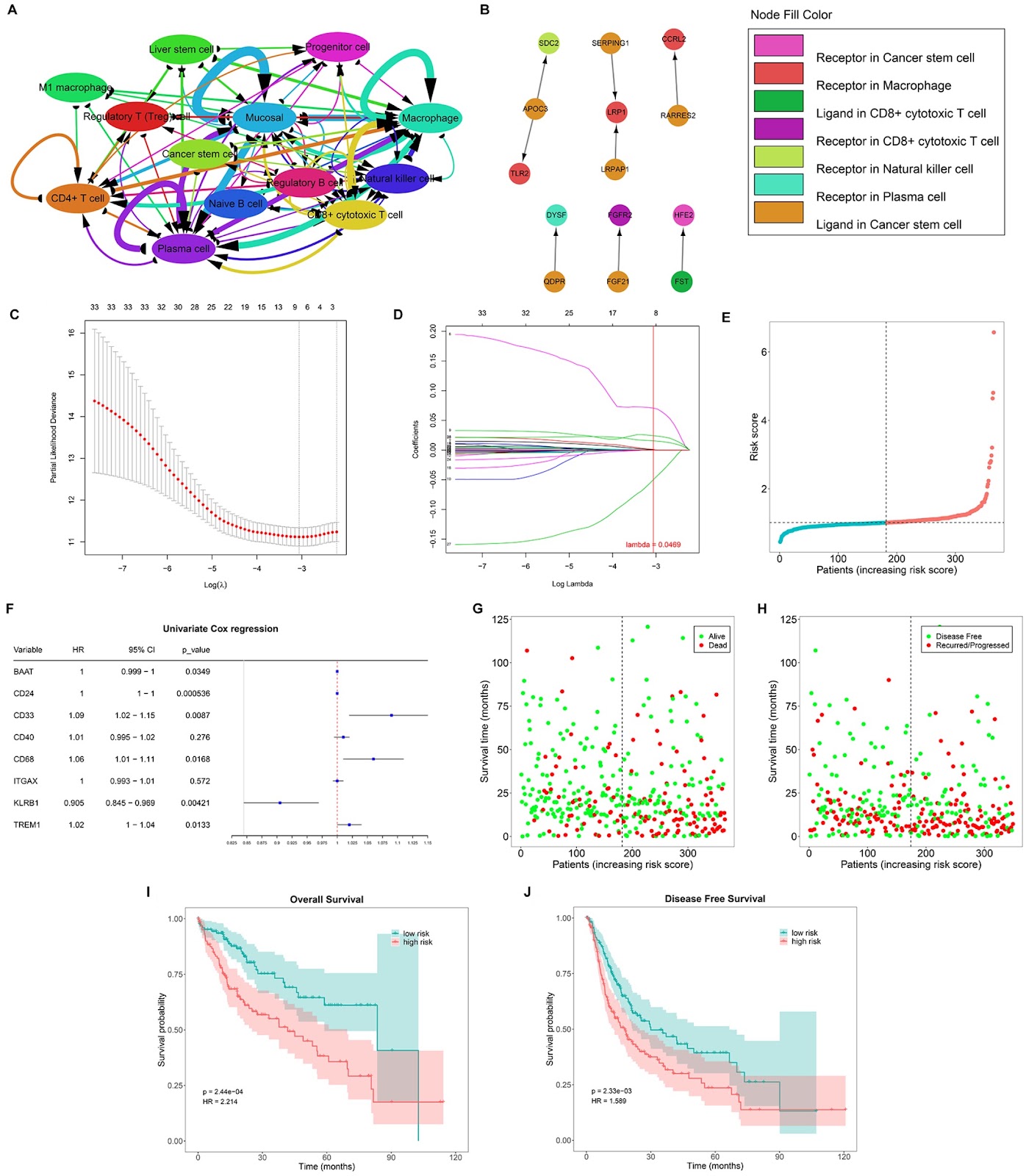
The transcriptional heterogeneity and cellular ecosystem diversity of HCC await further exploration. Single-cell and bulk RNA sequencing data from HCC cells are analyzed to generate a LASSO model for HCC prognostication. CCK-8, scratch assay, flow cytometry, and ROS assays are used to validate how TREM1 may affect HCC cell biological behaviors in vitro. qPCR, western blot analysis, immunohistochemistry, and flow cytometry are applied in a xenograft model to test the effects of TREM1 knockdown on carcinogenesis and the tumor microenvironment. A single-cell atlas of the multicellular ecosystem comprising 13 cell types in HCC is constructed. On the basis of ligand-receptor marker genes specifically extracted from the cell populations, a prognostic model is defined and subsequently validated in additional clinical cohorts. For the first time, a heterogeneous immune microenvironment is observed between low- and high-risk patients, primarily involving macrophages, CD4+ T cells, M1 macrophages, and regulatory T (Treg) cells. Sufficient evidence validates the positive effects of TREM1 on HCC cell proliferation, migration, and apoptosis. Additionally, TREM1 positively modulates the levels of the proinflammatory cytokines IL-1β, TNF-α, and MCP-1. TREM1 downregulation alters the proportions of M1 macrophages and Tregs in the tumor tissue from our HCC xenograft model. Eventually, the Nrf2/Keap1 signaling pathway, which is related to oxidative stress, is shown to be a key pathway downstream of TREM1 downregulation. In summary, we construct a novel prognostic model for HCC on the basis of ligand-receptor marker genes and investigate the role of TREM1 in HCC progression and its impact on the TME.

Read Full Abstract10.3724/abbs.2025059
Single-cell transcriptomic data reveal the cellular heterogeneity of glutamine metabolism in gastric premalignant lesions and early gastric cancerGraphical AbstractVerified
Acta Biochimica et Biophysica Sinica

Single-cell transcriptomic data reveal the cellular heterogeneity of glutamine metabolism in gastric premalignant lesions and early gastric cancer

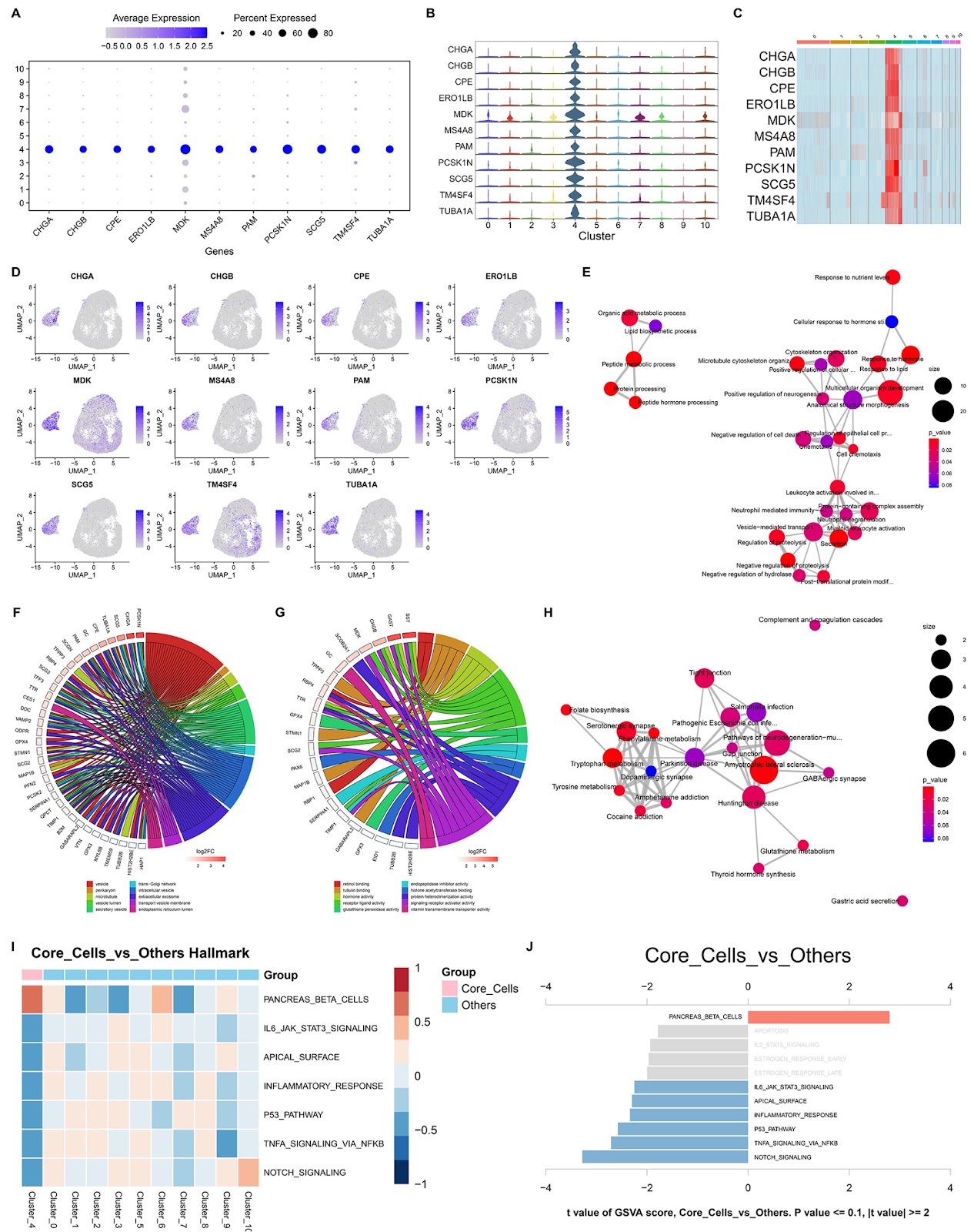
Glutamine metabolism is a hallmark of cancer metabolism. This study aims to perform a comprehensive and systematic single-cell profile of glutamine metabolism in premalignant and malignant gastric lesions. We use single-cell transcriptomics data from chronic atrophic gastritis (CAG) and early gastric cancer (EGC) lesions and investigate glutamine metabolism features at the single-cell level. Experiments are implemented to validate the expression and biological role of ERO1LB in gastric cancer (GC). A single-cell atlas based on 22511 cells from premalignant and early-malignant gastric lesions is established. Among these cells, epithelial cells constitute the dominant cell population in both CAG and EGC lesions. The activity of glutamine metabolism is higher in epithelial cells from EGC lesions than in those from CAG lesions. Among the epithelial cell subpopulations, glutamine metabolism is more active in the epithelial cell subpopulation cluster_4 in EGCs than in CAG lesions. As a key marker gene of this subpopulation, ERO1LB is experimentally proven to be overexpressed in human GC tissue lesions. In both in vitro and in vivo experiments, overexpression of ERO1LB in GC cells increases glutamine metabolism, facilitates cell growth and migration and prevents cell apoptosis, and vice versa. This study provides insight into the cellular heterogeneity of glutamine metabolism within the gastric mucosa in premalignant and malignant gastric lesions and identifies ERO1LB as a key orchestrator of glutamine metabolism, which may help to identify markers for GC prevention and contribute to our understanding of GC pathogenesis.

Read Full Abstract10.3724/abbs.2025061
CDDO-imidazolide ameliorates sepsis-induced ARDS by enhancing mitophagy via the Nrf2 pathway to prohibit alveolar macrophage pyroptosis and HMGB1 releaseGraphical AbstractVerified
Acta Biochimica et Biophysica Sinica

CDDO-imidazolide ameliorates sepsis-induced ARDS by enhancing mitophagy via the Nrf2 pathway to prohibit alveolar macrophage pyroptosis and HMGB1 release

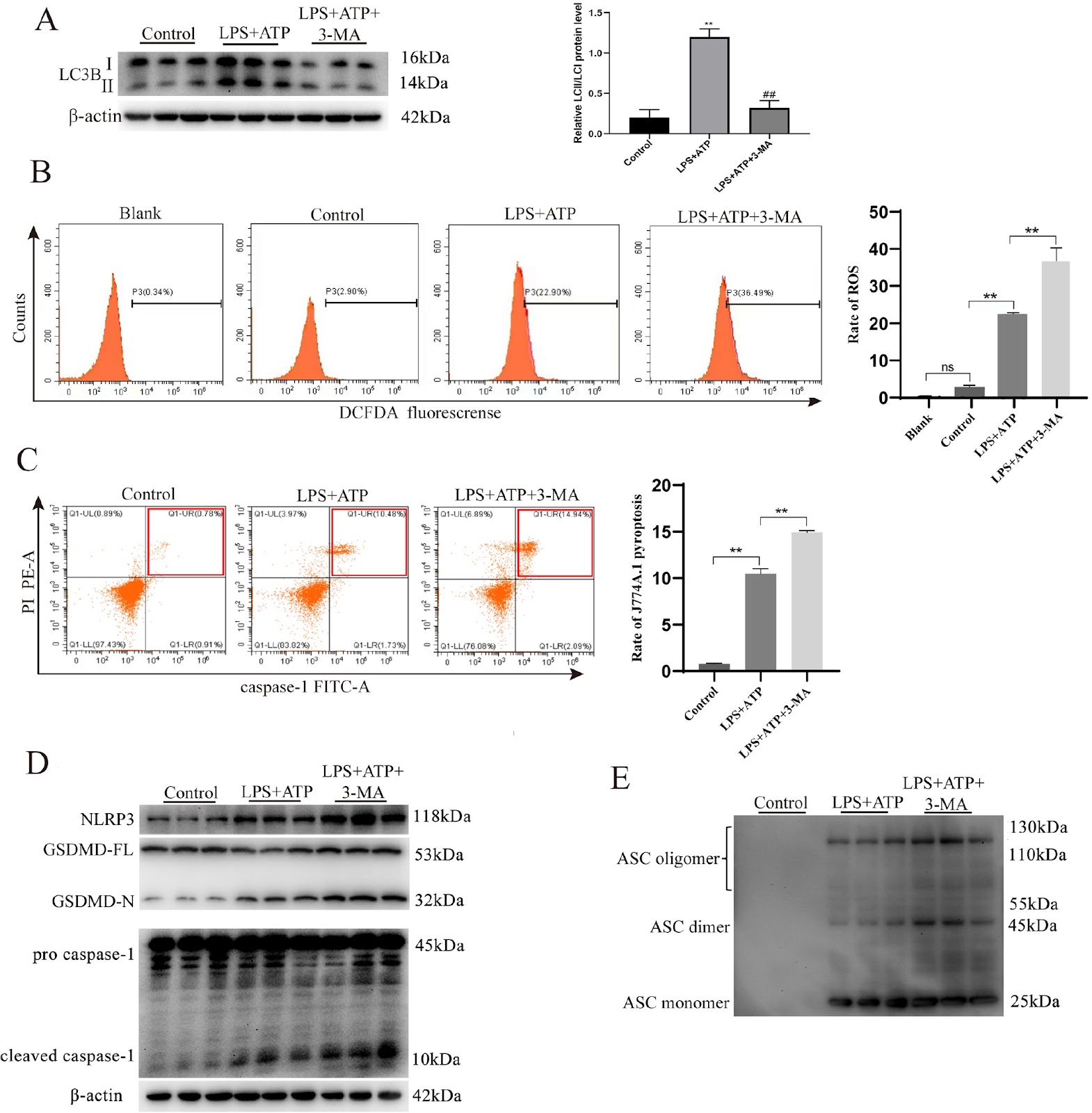
Accumulating evidence suggests that NLRP3-mediated alveolar macrophage (AM) pyroptosis and subsequent high mobility group box protein 1 (HMGB1) secretion play significant roles in the pathogenesis of acute respiratory distress syndrome (ARDS). Nrf2 has been shown to be individually involved in regulating pyroptosis. In this study, we investigate the ability of CDDO-imidazolide, a potent Nrf2 activator, to regulate AM pyroptosis and HMGB1 secretion in sepsis-associated ARDS, along with its underlying mechanism. The in vitro alveolar macrophage (AM) pyroptosis model, established by stimulating J774A.1 cells with LPS and ATP, was treated with CDDO-imidazolide or utilized Nrf2-knockout cells. The mice are intraperitoneally administered with CDDO-imidazolide before the in vivo sepsis-associated ARDS model is constructed via caecal ligation perforation and the Nrf2 inhibitor, ML385. In vitro studies reveal that the use of 3-MA to prohibit PINK1/Parkin-dependent mitophagy aggravates NLRP3-mediated pyroptosis and HMGB1 release in J774A.1 cells via LPS and ATP exposure. CDDO-imidazolide also significantly prevents NLRP3-mediated pyroptosis and HMGB1 release to increase PINK1/Parkin-dependent mitophagy, but these effects are not detected in Nrf2-knockout macrophages. Most importantly, CDDO-imidazolide significantly alleviates NLRP3 inflammasome protein expression in the lung tissues of septic mice and HMGB1 protein levels in the serum and bronchoalveolar lavage fluid (BALF), which can be reversed by ML385. Taken together, our results demonstrate that CDDO-imidazolide prominently protects the lungs by promoting Nrf2 activation and enhancing PINK1/Parkin mitophagy to inhibit AM pyroptosis and HMGB1 release. These findings provide novel insights for therapeutic strategies for sepsis-associated ARDS.

Read Full Abstract10.3724/abbs.2025092