🧬 SinoBioData Academic Portal
📚 Peer-Reviewed Translated Literature

All Biomedical & Clinical Articles (Page 52)

Browse complete peer-reviewed translations from top Chinese biomedical, oncology, and genomics journals. Read verified previews and download full authentic clinical reports.

Published Research Papers

Showing 24 of 1542 peer-reviewed translated articles (Page 52 of 65)

EEPD1 attenuates radiation-induced cardiac hypertrophy and apoptosis by degrading FOXO3A in cardiomyocytesGraphical AbstractVerified
Acta Biochimica et Biophysica Sinica

EEPD1 attenuates radiation-induced cardiac hypertrophy and apoptosis by degrading FOXO3A in cardiomyocytes

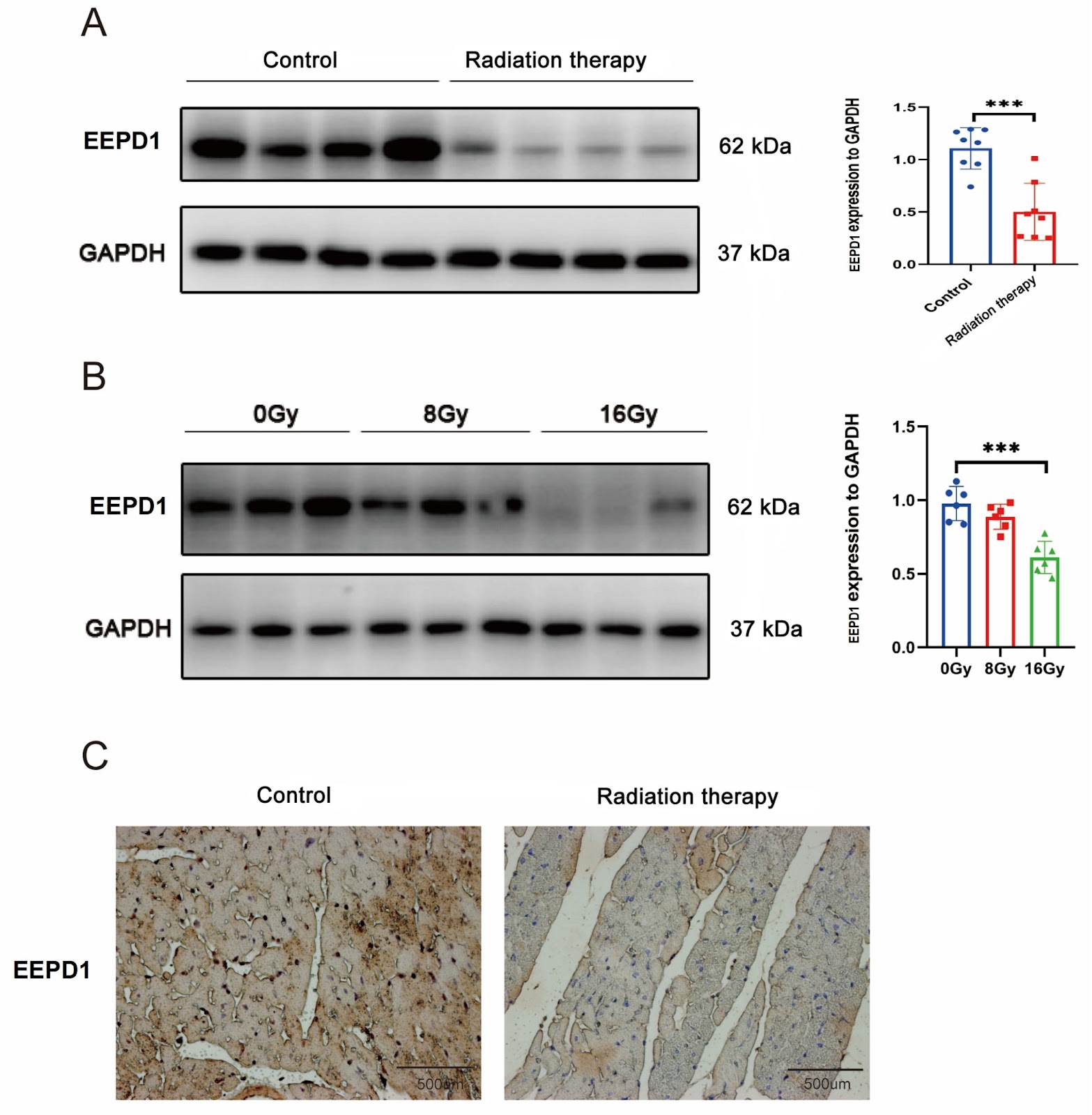
Radiation-induced heart disease (RIHD) is a severe delayed complication of thoracic irradiation (IR). Endonuclease/exonuclease/phosphatase family domain-containing 1 (EEPD1) plays an important role in DNA damage repair, but its role in RIHD is less known. In this study, EEPD1 global knockout mice, C57BL/6J mice, and C57BL/6J mice overexpressing EEPD1 are treated with radiation at a total dose of 20 Gy or 0 Gy. After 9 weeks, echocardiography is used to assess cardiac hypertrophy and apoptosis. The results show that EEPD1 deletion exacerbates radiation-induced cardiac hypertrophy and apoptosis, while EEPD1 overexpression has the opposite effect. Further mechanistic investigations reveal that EEPD1 interacts with FOXO3A and destabilizes it by catalyzing its deubiquitination. Inhibition of FOXO3A ameliorates cardiac hypertrophy and apoptosis after EEPD1 knockdown. Thus, EEPD1 protects against radiation-induced cardiac hypertrophy and apoptosis via destabilization of FOXO3A, which may offer new insight into therapeutic strategies for RIHD.

Read Full Abstract10.3724/abbs.2024130
PPIA as a central regulator in a novel cell death pathway activated by iron homeostasis and redox disruption in multiple myelomaGraphical AbstractVerified
Acta Biochimica et Biophysica Sinica

PPIA as a central regulator in a novel cell death pathway activated by iron homeostasis and redox disruption in multiple myeloma

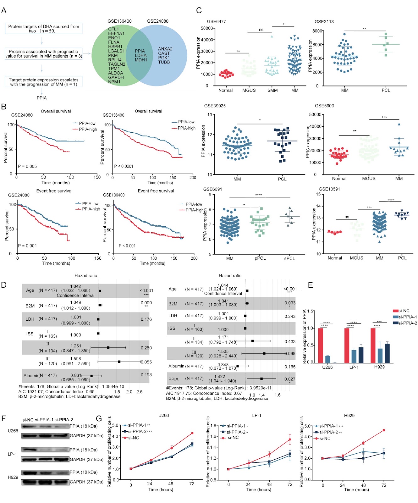
Multiple myeloma (MM) patients frequently experience relapse, disease progression, and drug resistance, necessitating novel therapeutic strategies. This study investigates the role of peptidylprolyl isomerase A (PPIA) in mediating dihydroartemisinin (DHA)-induced ferroptosis in MM. Building on our previous work establishing the prognostic relevance of ferroptosis in MM, we elucidate the mechanism by which DHA triggers this form of cell death through the disruption of iron metabolism and redox homeostasis. DHA significantly reduces the viability of MM cell lines and primary CD138+ cells derived from patient bone marrow samples and attenuates tumor burden in orthotopic MM models. Mechanistically, DHA upregulates the iron-regulatory genes transferrin receptor 1 (TFRC) and heme oxygenase-1 (HMOX1), thereby perturbing iron homeostasis and promoting ferroptosis. Crucially, DHA targets the oxidized form of PPIA, a redox-sensitive chaperone, binding to it and preventing its reduction, which elevates intracellular reactive oxygen species (ROS). Combined treatment with DHA and erastin, which concurrently disrupt iron and amino acid metabolism, exerts synergistic cytotoxicity and enhances MM inhibition. Furthermore, in a syngeneic mouse model, DHA promotes T-cell activation and augments tumor suppression. Collectively, these findings underscore PPIA’s pivotal role in a novel ferroptotic cell death pathway and reveal new therapeutic opportunities for MM.

Read Full Abstract10.3724/abbs.2026081
Sanguinarine exerts anti-hepatocellular carcinoma activity by targeting FDX1 to induce FDX1/LIAS/DLAT/HSP70 axis-dependent cuproptosisGraphical AbstractVerified
Acta Biochimica et Biophysica Sinica

Sanguinarine exerts anti-hepatocellular carcinoma activity by targeting FDX1 to induce FDX1/LIAS/DLAT/HSP70 axis-dependent cuproptosis

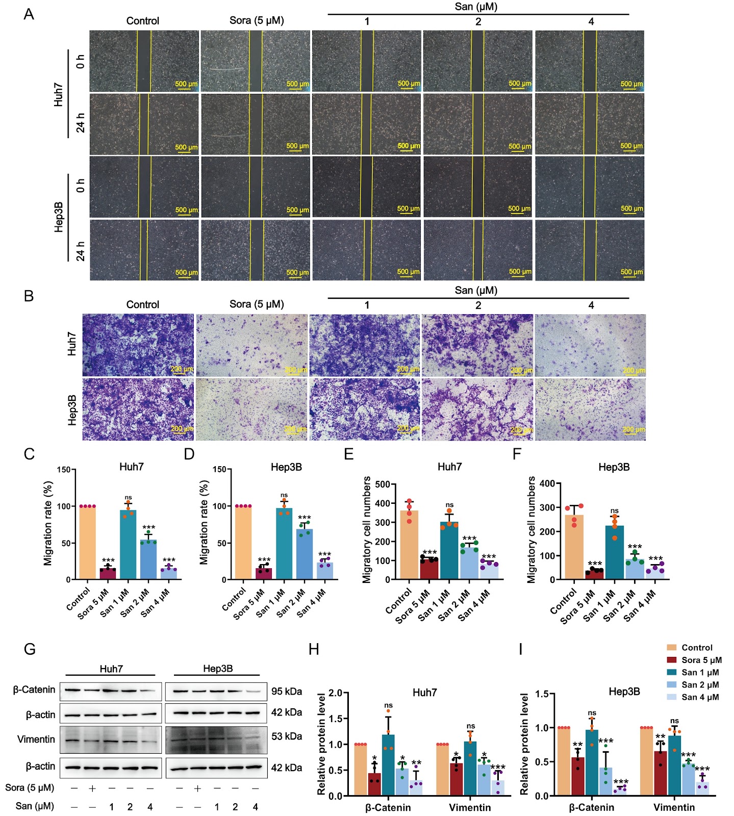
Hepatocellular carcinoma (HCC), the predominant type of primary liver cancer, represents an extremely aggressive malignancy. The induction of cuproptosis has developed into a favorable therapeutic direction for HCC, considering its strong association with HCC. Sanguinarine (San), a benzophenanthridine alkaloid derived from traditional herbs such as Chelidonium majus L., demonstrates broad-spectrum anticancer activities against various cancer cell types. However, the precise molecular mechanisms underlying its effects in the treatment of HCC remain largely undefined. This investigation seeks to examine the anti-HCC effects of San and to explore the mechanisms underlying these effects through the induction of cuproptosis. In vitro experiments demonstrate that San markedly inhibits the proliferation, movement, and epithelial-mesenchymal transition of HCC cells while enhancing their apoptosis. In vivo, San notably impedes tumor growth and upregulates the cuproptosis signature markers ferredoxin 1 (FDX1), oligomeric dihydrolipoamide S-acetyltransferase (DLAT), and heat shock protein 70 (HSP70) in HCC xenograft tumor models. Mechanistically, San induces proteotoxic stress and cuproptosis in HCC cells by increasing copper concentration, upregulating the expression of FDX1, lipoic acid synthetase (LIAS), HSP70, and lipoylated DLAT aggregation, and simultaneously reducing mitochondrial membrane potential and intracellular glutathione and pyruvate levels. Moreover, the combination of San with copper ionophores (Elesclomol-CuCl2) exhibits synergistic effects in promoting cuproptosis. FDX1 silencing markedly diminishes San-induced suppression of cell proliferation and FDX1 and HSP70 levels in HCC cells. Additionally, molecular docking analysis predicts that San exhibits the highest potential for binding with FDX1. Surface plasmon resonance experiments and cellular thermal shift assay confirm that San strongly interacts with FDX1 and markedly enhances the thermostability of FDX1. In conclusion, our findings indicate that San substantially inhibits the progression of HCC by targeting FDX1/LIAS/DLAT/HSP70 axis-dependent cuproptosis.

Read Full Abstract10.3724/abbs.2026025
OAZ1/CASP8AP2 double knockout enhances recombinant protein production in HEK293 cells through metabolic reprogramming and antiapoptotic effectsGraphical AbstractVerified
Acta Biochimica et Biophysica Sinica

OAZ1/CASP8AP2 double knockout enhances recombinant protein production in HEK293 cells through metabolic reprogramming and antiapoptotic effects

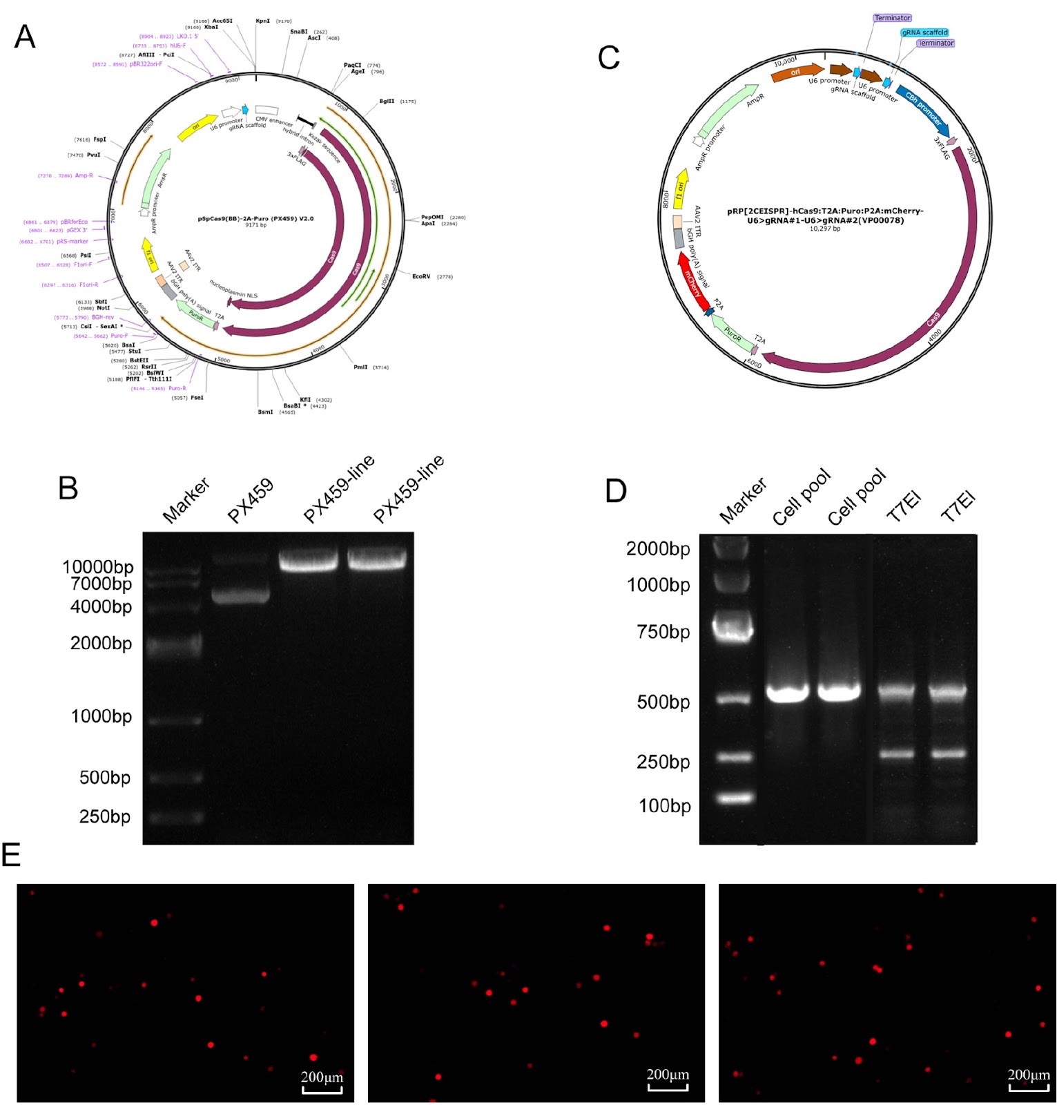
Human embryonic kidney (HEK) 293 cells are widely used for recombinant protein production because of their efficient posttranslational modification capabilities. However, their large-scale culture is often limited by metabolic stress and early apoptosis, leading to insufficient protein yields. In this study, we aim to increase protein expression through the coordinated modulation of metabolic and apoptotic pathways. Using CRISPR/Cas9 technology, we target and knockout the genes of ornithine decarboxylase antizyme 1 (OAZ1), which regulates polyamine metabolism, and caspase 8-associated protein 2 (CASP8AP2), an apoptosis-related protein. We successfully construct an OAZ1/CASP8AP2 double-knockout HEK293 cell line. Following transfection with the knockout vector and screening of single-cell clones, multiple levels of validation confirm the successful gene knockout. The results show that the double-knockout cells exhibit significantly reduced apoptosis rates. Furthermore, the production of recombinant secreted alkaline phosphatase (SEAP) and vitronectin (VN) increases by 2.1 folds and 2.9 folds, respectively, compared with those in wild-type cells. Metabolic profiling reveals that the cell cycle is arrested in the G1/G0 phase, accompanied by increased specific consumption and production rates of key metabolites. This study demonstrates that concurrent inhibition of apoptosis and optimization of metabolism effectively enhances recombinant protein production in HEK293 cells, suggesting a novel strategy for improving HEK293 cell-based expression.

Read Full Abstract10.3724/abbs.2025196
Vaccarin suppresses diabetic nephropathy through inhibiting the EGFR/ERK1/2 signaling pathwayGraphical AbstractVerified
Acta Biochimica et Biophysica Sinica

Vaccarin suppresses diabetic nephropathy through inhibiting the EGFR/ERK1/2 signaling pathway

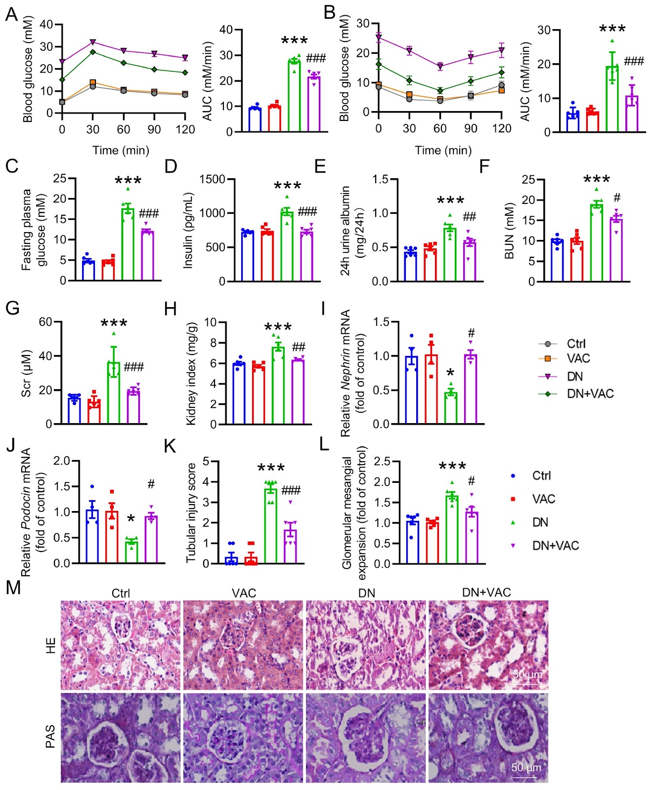
Diabetic nephropathy (DN) is recognized as one of the primary causes of chronic kidney disease and end-stage renal disease. Vaccarin (VAC) confers favorable effects on cardiovascular and metabolic diseases, including type 2 diabetes mellitus (T2DM). Nonetheless, the potential role and mechanism of VAC in the etiology of DN have yet to be completely elucidated. In this study, a classical mouse model of T2DM is experimentally induced via a high-fat diet (HFD)/streptozocin (STZ) regimen. Renal histological changes are assessed via H&E staining. Masson staining and immunohistochemistry (IHC) are employed to assess renal fibrosis. RT-PCR is utilized to quantify the mRNA levels of renal fibrosis, oxidative stress and inflammation markers. The levels of malondialdehyde (MDA) and reactive oxygen species (ROS), as well as the content of glutathione peroxidase (GSH-Px), are measured. The protein expressions of collagen I, TGF-β1, α-SMA, E-cadherin, Nrf2, catalase, SOD3, SOD2, SOD1, p-ERK, p-EGFR (Y845), p-EGFR (Y1173), p-NFκB P65, t-ERK, t-EGFR and t-NFκB P65 are detected by western blot analysis. Our results reveal that VAC has a beneficial effect on DN mice by improving renal function and mitigating histological damage. This is achieved through its inhibition of renal fibrosis, inflammatory cytokine overproduction, and ROS generation. Moreover, VAC treatment effectively suppresses the process of epithelial-mesenchymal transition (EMT), a crucial characteristic of renal fibrosis, in high glucose (HG)-induced HK-2 cells. Network pharmacology analysis and molecular docking identify epidermal growth factor receptor (EGFR) as a potential target for VAC. Amino acid site mutations reveal that Lys-879, Ile-918, and Ala-920 of EGFR may mediate the direct binding of VAC to EGFR. In support of these findings, VAC reduces the phosphorylation levels of both EGFR and its downstream mediator, extracellular signal-regulated kinase 1/2 (ERK1/2), in diabetic kidneys and HG-treated HK-2 cells. Notably, blocking either EGFR or ERK1/2 yields renal benefits similar to those observed with VAC treatment. Therefore, this study reveals that VAC attenuates renal damage via inactivation of the EGFR/ERK1/2 signaling axis in T2DM patients.

Read Full Abstract10.3724/abbs.2024141
A PCR-independent, annealing-free cloning method for the insertion of short DNA fragmentsGraphical AbstractVerified
Acta Biochimica et Biophysica Sinica

A PCR-independent, annealing-free cloning method for the insertion of short DNA fragments

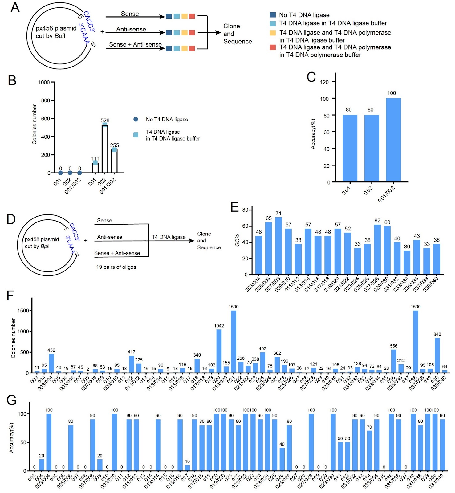
Cloning short DNA fragments, such as shRNA and sgRNA, is a routine but time-consuming task in molecular biology. Traditional methods require annealing of complementary oligos or PCR amplification, which are labor-intensive and time-consuming. Here, we report a novel PCR-independent, annealing-free cloning method that enables the insertion of short DNA fragments using a single oligo. The method relies on T4 DNA ligase for ligation and host cell DNA polymerase for complementary strand synthesis. We demonstrate that adding T4 DNA polymerase and dNTPs to the ligation mixture significantly improves cloning efficiency. This approach simplifies the cloning process, reduces time to less than 1 hour, and is compatible with standard laboratory reagents. Our method provides a rapid and efficient alternative for cloning short DNA fragments, with broad applications in gene knockdown and genome editing.

Read Full Abstract10.3724/abbs.2024088
PCIF1 modulates glioblastoma cell migration and invasion by altering PI(3,4)P2 levels through the PI5-phosphatase INPP5BGraphical AbstractVerified
Acta Biochimica et Biophysica Sinica

PCIF1 modulates glioblastoma cell migration and invasion by altering PI(3,4)P2 levels through the PI5-phosphatase INPP5B

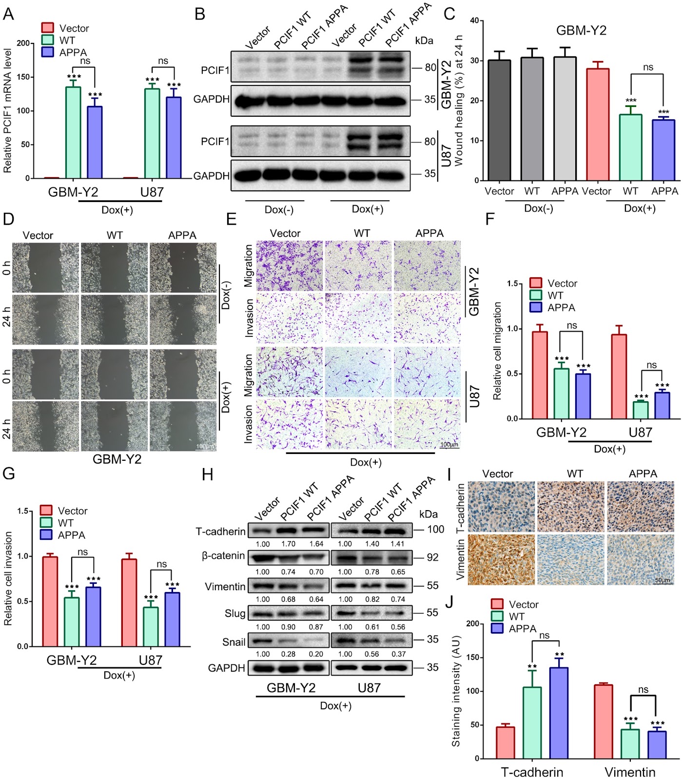
Phosphorylated CTD Interacting Factor 1 (PCIF1) is the exclusive methyltransferase responsible for the N6,2-O-dimethyladenosine (m6Am) modification in mammalian mRNA. Our previous research identified PCIF1 as a potent tumor suppressor in glioma, demonstrating its ability to impair cell proliferation, induce G2/M phase arrest, and promote apoptosis. However, its role in glioma cell migration and invasion remains unclear. In this study, we investigate how PCIF1 regulates glioma cell migration and invasion. Overexpression of PCIF1 inhibits migration and invasion, whereas PCIF1 knockdown enhances these behaviors. Corresponding changes are observed in mesenchymal markers (Vimentin, β-catenin, Snail, Slug) and the epithelial marker T-cadherin, indicating that PCIF1 suppresses epithelial-to-mesenchymal transition (EMT)-mediated glioma invasion. Mechanistically, PCIF1 modulates the AKT pathway by promoting proteasomal degradation of AKT while increasing phosphorylated AKT (p-AKT) levels, revealing a complex regulatory mechanism. PCIF1 knockdown upregulates INPP5B, a lipid phosphatase, causing accumulation of PI(3,4)P2 and enhanced AKT activation. Conversely, PCIF1 overexpression increases PI(3,4,5)P3 production, elevating p-AKT levels. This bidirectional regulation suggests that PCIF1 influences phosphoinositide signaling and AKT activation. Our findings highlight PCIF1 as a key modulator of glioblastoma cell migration and invasion through phosphoinositide signaling, positioning it as a potential biomarker and therapeutic target in glioma.

Read Full Abstract10.3724/abbs.2026027
P300-mediated H3K18 acetylation triggers necroptosis via modulation of KRT18 transcription in diabetic nephropathyGraphical AbstractVerified
Acta Biochimica et Biophysica Sinica

P300-mediated H3K18 acetylation triggers necroptosis via modulation of KRT18 transcription in diabetic nephropathy

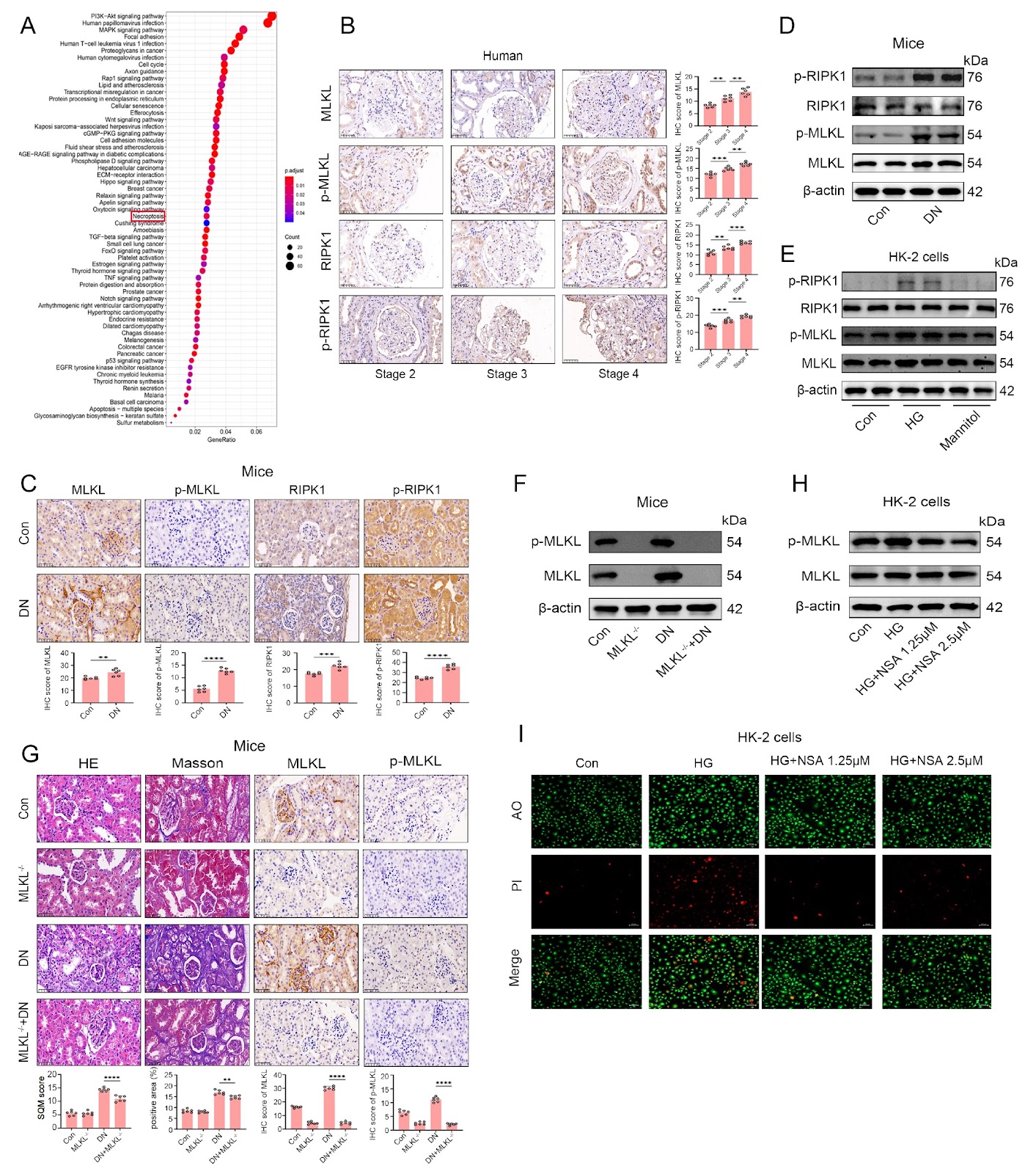
Diabetic nephropathy (DN) is a major cause of end-stage renal disease. While glomerular damage is a known aspect of its pathology, tubular epithelial cell necroptosis also plays a crucial role in disease progression. Epigenetic modifications, particularly histone acetylation, have garnered attention for their role in the regulation of kidney damage-related gene expression. This study explores whether the histone acetyltransferase P300 regulates KRT18 expression via histone H3 lysine 18 acetylation (H3K18ac), driving tubular epithelial cell necroptosis and accelerating DN progression. We establish an STZ-induced diabetic nephropathy mouse model and a high glucose-treated HK-2 cell model. Western blot analysis, qPCR, immunohistochemistry, and AO/PI staining are employed to assess the expression levels of P300, H3K18ac, KRT18, and necroptosis-related proteins (RIPK1 and MLKL). Functional validation of the P300-KRT18 axis is performed using shRNA interference, overexpression, and the small molecule inhibitor C646. Both in vivo and in vitro models show significant upregulation of P300, H3K18ac, and KRT18, coupled with RIPK1/MLKL pathway activation and increased cell death. P300 knockdown or C646 treatment effectively inhibits H3K18ac and KRT18 expression, reducing necroptosis; KRT18 knockdown also alleviates P300 overexpression-induced cell death. Co-transfection with P300 overexpression and KRT18 interference demonstrates that KRT18 is a key downstream effector of P300-mediated necroptosis. In conclusion, P300 upregulates KRT18 expression through H3K18 acetylation, subsequently activating the RIPK1/MLKL pathway and promoting tubular epithelial cell necroptosis. The P300-KRT18 axis may serve as a novel epigenetic therapeutic target for DN, suggesting that epigenetic regulation could be a viable intervention strategy to delay DN progression.

Read Full Abstract10.3724/abbs.2026015
Integrins and NAFLD-associated liver diseases: clinical associations, pathophysiological mechanisms and pharmacological implicationsGraphical AbstractVerified
Acta Biochimica et Biophysica Sinica

Integrins and NAFLD-associated liver diseases: clinical associations, pathophysiological mechanisms and pharmacological implications

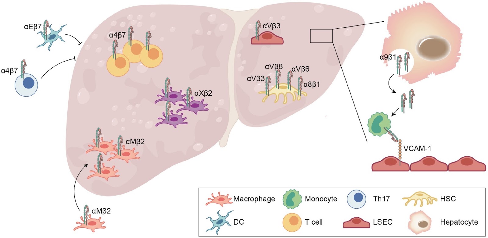
Nonalcoholic fatty liver disease (NAFLD) is a leading cause of chronic liver disease and poses a substantial health burden with increasing incidence globally. NAFLD encompasses a spectrum extending from hepatic steatosis to nonalcoholic steatohepatitis (NASH), with the possibility of progressing to cirrhosis or, in severe instances, hepatocellular carcinoma (HCC). NAFLD extends beyond simple metabolic disruption and involves multiple immune cell-mediated inflammatory processes. Integrins are a family of heterodimeric transmembrane cell adhesion receptors that regulate various aspects of NAFLD onset and progression, including hepatocellular steatosis, hepatic stellate cell (HSC) activation and immune cell infiltration. In this review, we comprehensively summarize the involvement of integrins in NAFLD, as well as the downstream signal transduction mediated by these receptors. Furthermore, we present the latest clinical and preclinical findings on drugs that target integrins for steatosis, inflammation, fibrosis and NAFLD-related HCC treatment.

Read Full Abstract10.3724/abbs.2024149
Acetyl-11-keto-β-boswellic acid restrains the progression of synovitis in osteoarthritis via the Nrf2/HO-1 pathwayGraphical AbstractVerified
Acta Biochimica et Biophysica Sinica

Acetyl-11-keto-β-boswellic acid restrains the progression of synovitis in osteoarthritis via the Nrf2/HO-1 pathway

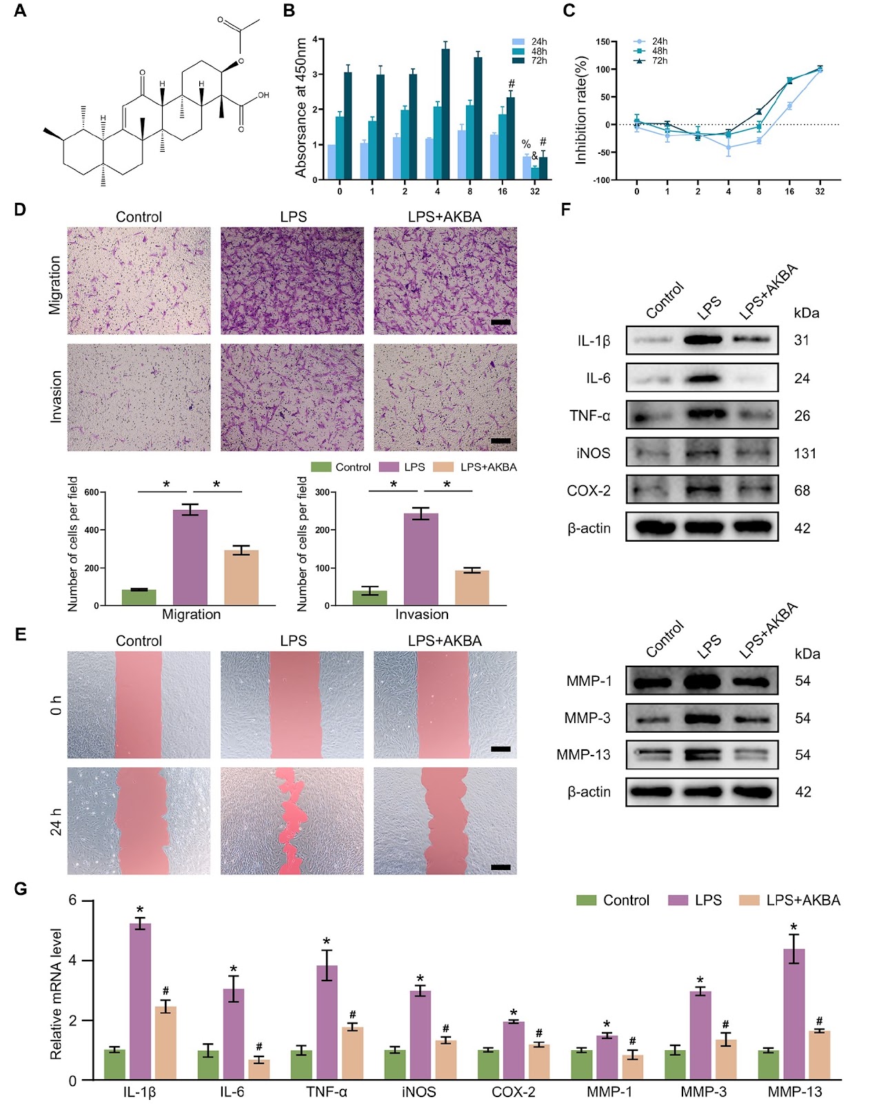
Synovial inflammation plays a key role in osteoarthritis (OA) pathogenesis. Fibroblast-like synoviocytes (FLSs) represent a distinct cell subpopulation within the synovium, and their unique phenotypic alterations are considered significant contributors to inflammation and fibrotic responses. The underlying mechanism by which acetyl-11-keto-β-boswellic acid (AKBA) modulates FLS activation remains unclear. This study aims to assess the beneficial effects of AKBA through both in vitro and in vivo investigations. Network pharmacology evaluation is used to identify potential targets of AKBA in OA. We evaluate the effects of AKBA on FLSs activation in vitro and the regulatory role of AKBA on the Nrf2/HO-1 signaling pathway. ML385 (an Nrf2 inhibitor) is used to verify the binding of AKBA to its target in FLSs. We validate the in vivo efficacy of AKBA in alleviating OA using anterior cruciate ligament transection and destabilization of the medial meniscus (ACLT+DMM) in a rat model. Network pharmacological analysis reveals the potential effect of AKBA on OA. AKBA effectively attenuates lipopolysaccharide (LPS)-induced abnormal migration and invasion and the production of inflammatory mediators, matrix metalloproteinases (MMPs), and reactive oxygen species (ROS) in FLSs, contributing to the restoration of the synovial microenvironment. After treatment with ML385, the effect of AKBA on FLSs is reversed. In vivo studies demonstrate that AKBA mitigates synovial inflammation and fibrotic responses induced by ACLT+DMM in rats via activation of the Nrf2/HO-1 axis. AKBA exhibits theoretical potential for alleviating OA progression through the Nrf2/HO-1 pathway and represents a viable therapeutic candidate for this patient population.

Read Full Abstract10.3724/abbs.2024102
Tryptophan-substituted antimicrobial peptide temporin-1CEb: in vitro and in vivo antibacterial activity against clinically isolated multidrug-resistant Klebsiella pneumoniaGraphical AbstractVerified
Acta Biochimica et Biophysica Sinica

Tryptophan-substituted antimicrobial peptide temporin-1CEb: in vitro and in vivo antibacterial activity against clinically isolated multidrug-resistant Klebsiella pneumonia

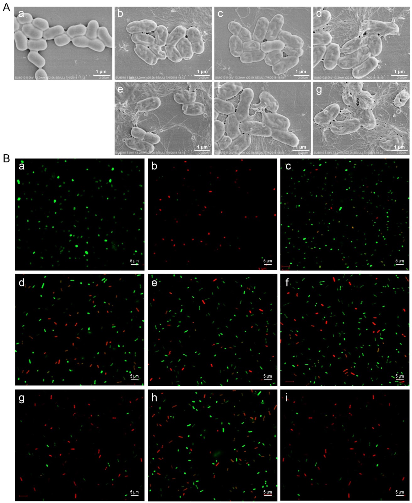
Tryptophan (Trp)-substituted antimicrobial peptides (AMPs) exhibit enhanced interactions with bacterial cell membranes, potentially improving their antimicrobial efficacy. Klebsiella pneumoniae (20.59% of 2054 ICU isolates) is resistant to multiple clinically used antibiotics and presents significant treatment challenges. In the present study, three Trp-modified peptides (I4W, L12W, and I1WL5W) are generated by substituting Ile or Leu residues in temporin-1CEb, a peptide derived from frog skin, with Trp at various sites to assess their antibacterial effects and mechanisms against K. pneumoniae. Compared with L12W, both I4W and I1WL5W display superior antimicrobial activity and lower cytotoxicity. Mechanistic studies reveal that AMPs exert antibacterial and bactericidal effects through bacterial surface charge neutralization, insertion into bacterial cell membranes, increase permeability of both the inner and outer membranes, and disruption of membrane integrity. Notably, I1WL5W exhibit the most potent membrane-disrupting activity. Assessment of the impact of Trp-containing peptides on bacterial biofilms reveals that these peptides not only inhibit exopolysaccharide production and biofilm formation but also degrade preformed biofilms. A murine lung infection model is established to investigate the therapeutic efficacy of I1WL5W against MDRKP 1203-induced lung infection in mice. Compared with the control treatment, treatment with I1WL5W results in reduced bacterial counts and levels of IL-6 and TNF-α in both the blood and lung tissues of MDRKP 1203-infected mice, and treatment with I1WL5W improves lung tissue structure. The present study provides valuable insights for designing Trp-containing peptides with potent antimicrobial properties by facilitating their penetration across bacterial membranes.

Read Full Abstract10.3724/abbs.2026074
Proximity-based proteomic profiling uncovers distinct interactome of human RAG1 and RAG2Graphical AbstractVerified
Acta Biochimica et Biophysica Sinica2026

Proximity-based proteomic profiling uncovers distinct interactome of human RAG1 and RAG2

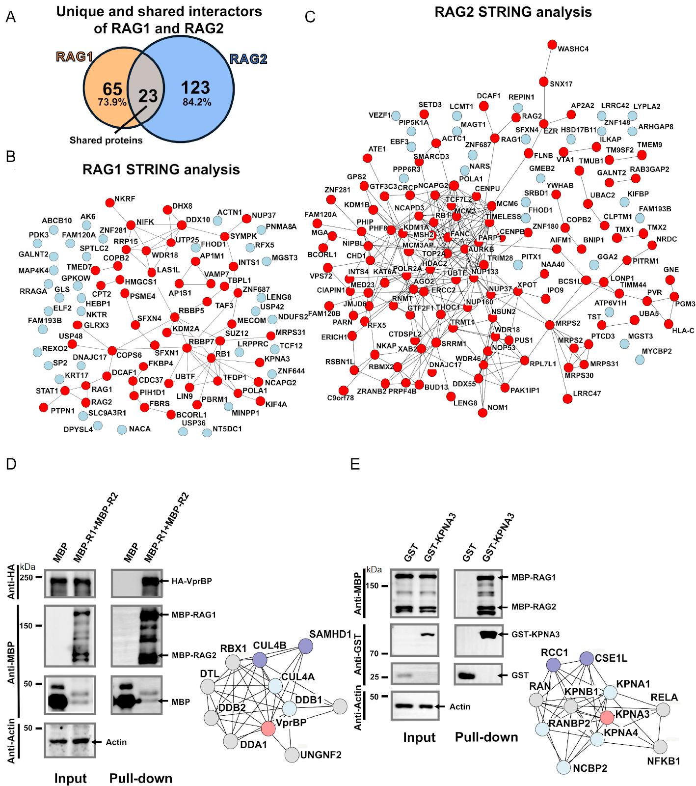
The recombination-activating gene (RAG) complex initiates adaptive immunity by catalyzing V(D)J recombination to generate diverse antigen receptors. While the catalytic function of the RAG core is well defined, its regulatory interactions and physiological roles remain poorly understood due to limited knowledge of RAG-associated proteins. The RAG complex forms a heterotetramer of two RAG1 and RAG2 subunits, yet the individual contributions of each subunit remain unclear. Here, we use TurboID-mediated proximity labelling to map the human RAG interactome. By fusing TurboID to RAG1 or RAG2, we identify 88 RAG1- and 146 RAG2-associated proteins, with only 23 shared proteins, indicating distinct sets of proximal proteins. Although RAG1 and RAG2 are thought to exert their physiological functions by forming a complex, they display distinct potential interaction networks, suggesting subunit-specific functions and revealing their spatial proximity to each subunit. These findings uncover distinct RAG1 and RAG2 interaction landscapes and establish a framework for exploring broader RAG functions in immunity.

Read Full Abstract10.3724/abbs.2025246
Paclitaxel Induces Neurotoxicity via Activating Ferroptosis by Suppressing Nrf2/SLC7A11/GSH/GPX4 SignalingGraphical AbstractVerified
Acta Biochimica et Biophysica Sinica2026

Paclitaxel Induces Neurotoxicity via Activating Ferroptosis by Suppressing Nrf2/SLC7A11/GSH/GPX4 Signaling

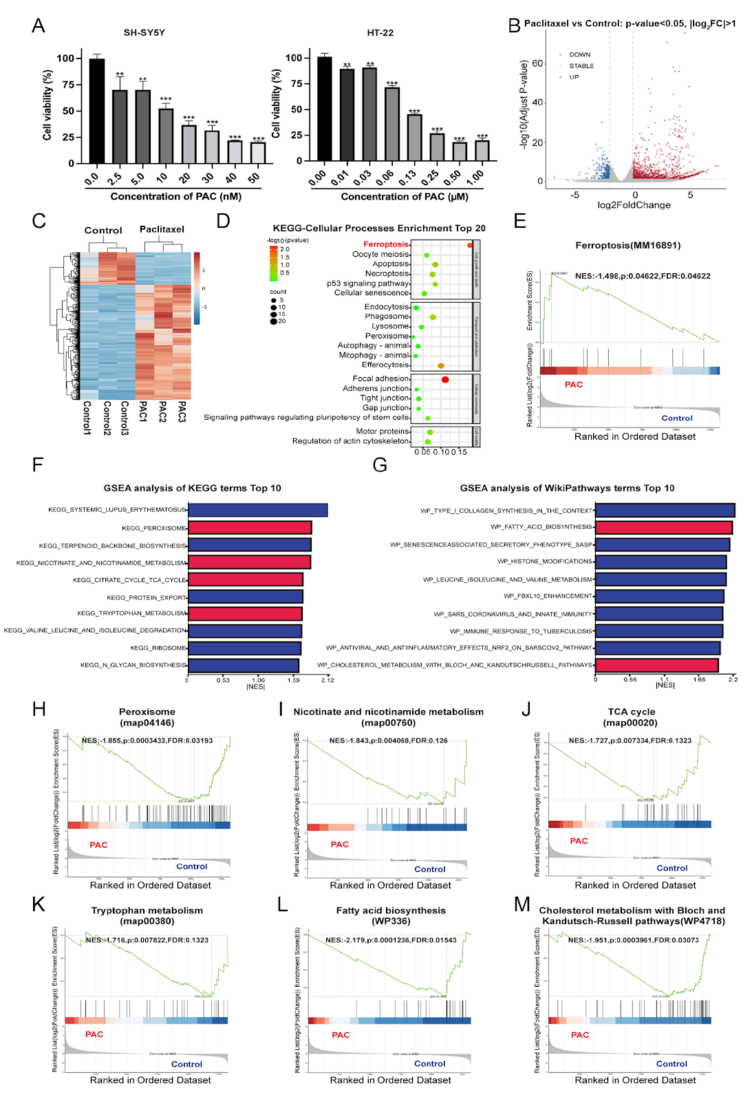
Chemotherapy-induced neurotoxicity (CIN) is a prevalent and debilitating side effect of cancer treatment, with paclitaxel being a classic anticancer drug that often causes significant neurotoxicity. However, effective interventions are lacking. This study aimed to elucidate the mechanisms underlying paclitaxel-induced neurotoxicity. We confirmed that paclitaxel exerts cytotoxic effects on SH-SY5Y and HT-22 neuronal cells. RNA-seq analysis revealed that ferroptosis is among the top enriched pathways in paclitaxel-treated neurons, and gene set enrichment analysis (GSEA) confirmed the enrichment of ferroptosis-related pathways. Transmission electron microscopy showed fragmented mitochondria with decreased cristae and increased membrane density, characteristic of ferroptosis. Paclitaxel dose-dependently increased intracellular reactive oxygen species (ROS) and iron levels while decreasing glutathione (GSH) levels. Mechanistically, paclitaxel suppressed the expression of Nrf2, SLC7A11, and GPX4, key components of the Nrf2/SLC7A11/GSH/GPX4 signaling pathway that protects against ferroptosis. Rescue experiments with ferroptosis inhibitors (liproxstatin-1) further confirmed the involvement of ferroptosis. These findings demonstrate that paclitaxel induces neurotoxicity by activating ferroptosis via suppression of the Nrf2/SLC7A11/GSH/GPX4 axis, providing potential therapeutic targets for preventing and treating paclitaxel-induced neurotoxicity.

Read Full Abstract10.3724/abbs.2026068
UHRF1 knockdown induces cell cycle arrest and apoptosis in breast cancer cells through the ZBTB16/ANXA7/Cyclin B1 axisGraphical AbstractVerified
Acta Biochimica et Biophysica Sinica

UHRF1 knockdown induces cell cycle arrest and apoptosis in breast cancer cells through the ZBTB16/ANXA7/Cyclin B1 axis

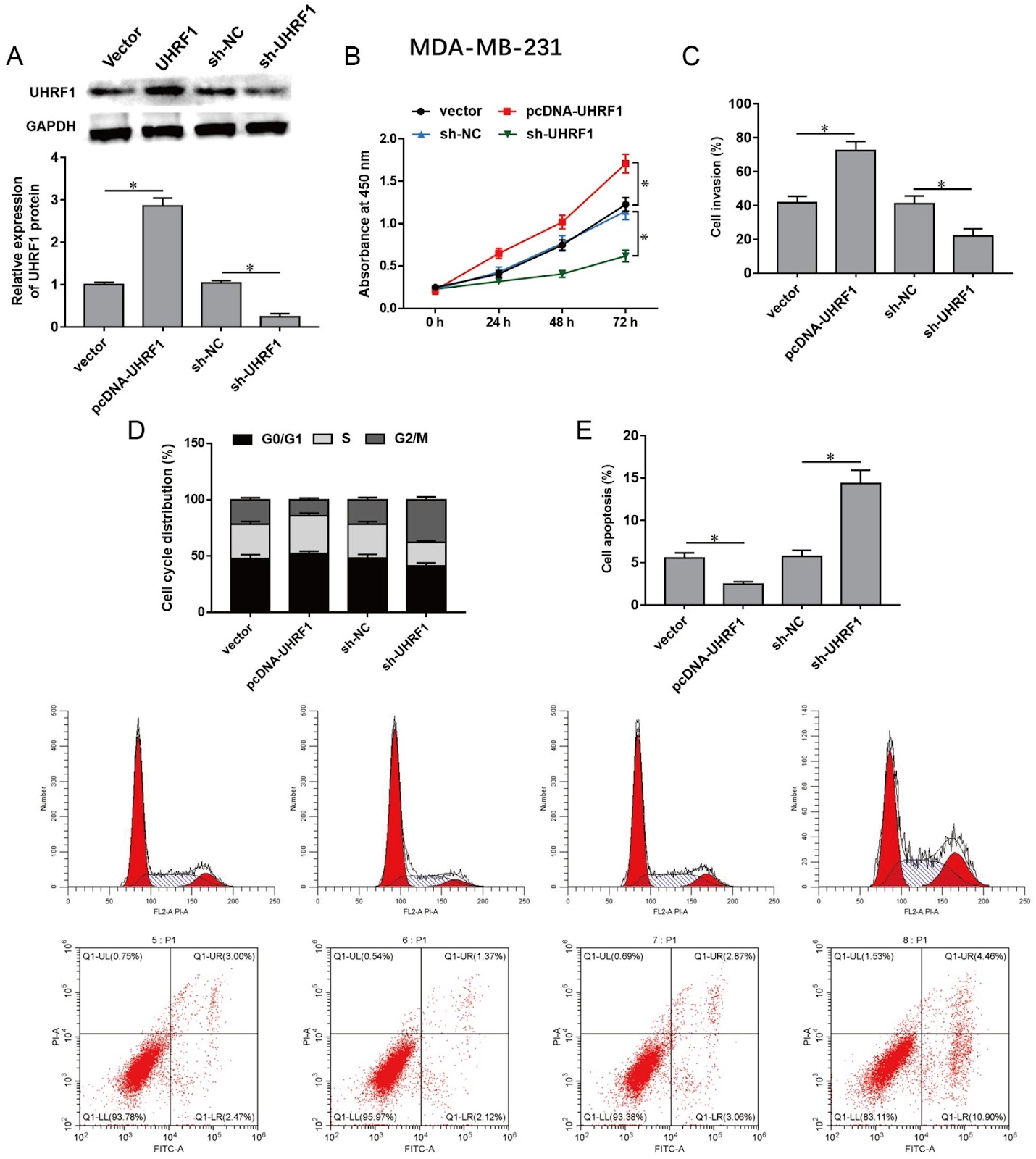
Ubiquitin-like containing PHD and RING finger domains 1 (UHRF1) is involved in tumorigenicity through DNA methylation in various cancers, including breast cancer. This study aims to investigate the regulatory mechanisms of UHRF1 in breast cancer progression. Herein, we show that UHRF1 is upregulated in breast cancer tissues and cell lines as measured by western blot analysis and immunohistochemistry. Breast cancer cells are transfected with a UHRF1 overexpression plasmid (pcDNA-UHRF1) or short hairpin RNA targeting UHRF1 (sh-UHRF1), followed by detection of cell proliferation, invasion, apoptosis, and cell cycle. UHRF1 overexpression promotes proliferation and invasion and attenuates cell cycle arrest and apoptosis in breast cancer cells, while UHRF1 knockdown shows the opposite effect. Moreover, methylation-specific PCR and ChIP assays indicate that UHRF1 inhibits zinc finger and BTB domain containing 16 (ZBTB16) expression by promoting ZBTB16 promoter methylation via the recruitment of DNA methyltransferase 1 (DNMT1). Then, a co-IP assay is used to verify the interaction between ZBTB16 and the annexin A7 (ANXA7) protein. ZBTB16 promotes ANXA7 expression and subsequently inhibits Cyclin B1 expression. Rescue experiments reveal that ZBTB16 knockdown reverses the inhibitory effects of UHRF1 knockdown on breast cancer cell malignancies and that ANXA7 knockdown abolishes the inhibitory effects of ZBTB16 overexpression on breast cancer cell malignancies. Additionally, UHRF1 knockdown significantly inhibits xenograft tumor growth in vivo. In conclusion, UHRF1 knockdown inhibits proliferation and invasion, induces cell cycle arrest and apoptosis in breast cancer cells via the ZBTB16/ANXA7/Cyclin B1 axis, and reduces xenograft tumor growth in vivo.

Read Full Abstract10.3724/abbs.2024148
SUN5, a testis-specific nuclear membrane protein, participates in recruitment and export of nuclear mRNA in spermatogenesisGraphical AbstractVerified
Acta Biochimica et Biophysica Sinica

SUN5, a testis-specific nuclear membrane protein, participates in recruitment and export of nuclear mRNA in spermatogenesis

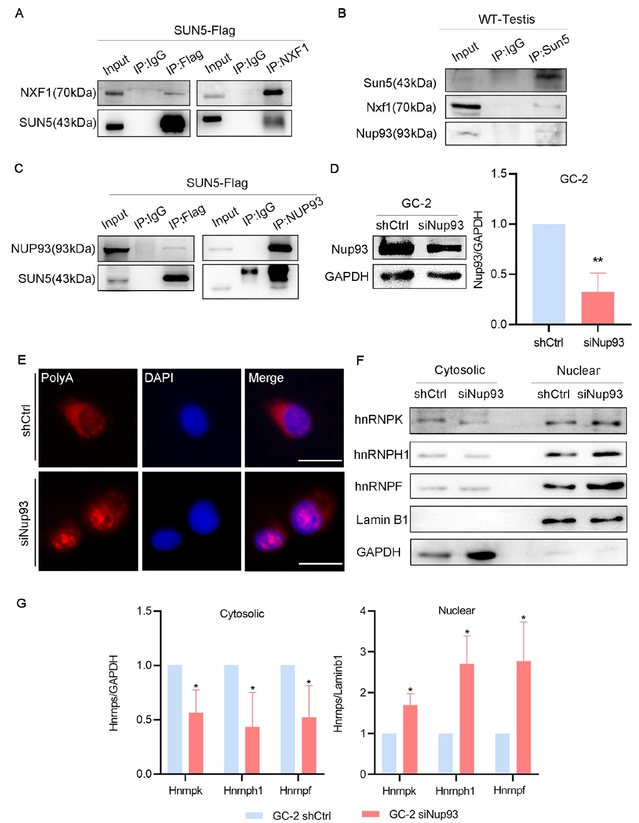
SUN5, a testis-specific gene, is associated with acephalic spermatozoa syndrome (ASS). Here, we demonstrate that SUN5 is involved in mRNA export. In Sun5-knockout mice (Sun5–/–), poly(A)+ RNA accumulates in the nuclei of germ cells, leading to reduced sperm counts, decreased sperm motility and disrupted sperm head-to-tail junctions. Additionally, in the GC-2 germ cell line with RNA interference of Sun5, heterogeneous nuclear ribonucleoproteins (hnRNPs) and poly (A)+ RNA (mainly mRNA) are retained in the nucleus. Further mechanistic studies reveal that SUN5 interacts with Nxf1 (nuclear RNA export factor 1) and nucleoporin 93 (Nup93). Interference with Nup93 inhibits mRNA export. Treatment with leptomycin B to block the CRM1 pathway indicates that Sun5 regulates mRNA export through an Nxf1-dependent pathway. In Sun5–/– mice, the binding of Nxf1 and Nup93 decreases due to loss of Sun5 function, and the process of submitting Nxf1-binding mRNPs to Nup93 is inhibited, resulting in abnormal spermatogenesis. Together, these data may elucidate a novel pathway for mRNA export in male germ cells.

Read Full Abstract10.3724/abbs.2024134
FTO-mediated m6A demethylation of ULK1 mRNA promotes autophagy and activation of hepatic stellate cells in liver fibrosisGraphical AbstractVerified
Acta Biochimica et Biophysica Sinica

FTO-mediated m6A demethylation of ULK1 mRNA promotes autophagy and activation of hepatic stellate cells in liver fibrosis

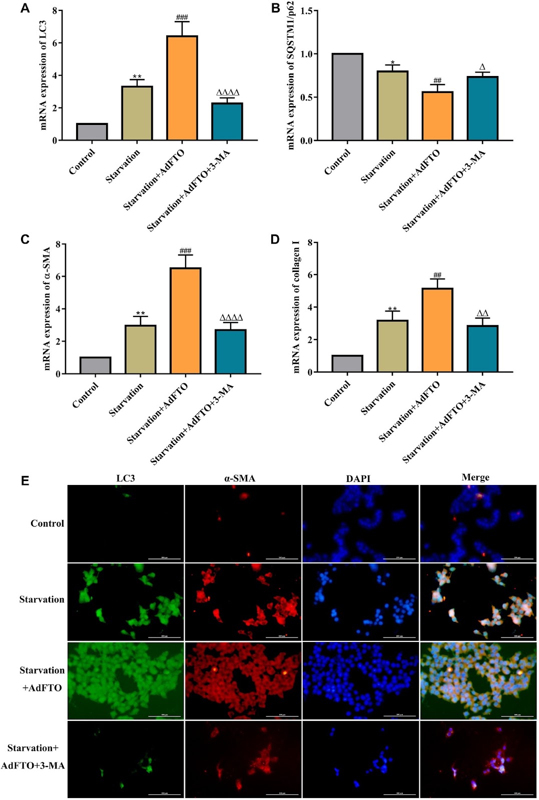
The activation of hepatic stellate cells (HSCs) is central to the occurrence and development of liver fibrosis. Our previous studies showed that autophagy promotes HSC activation and ultimately accelerates liver fibrosis. Unc-51-like autophagy activating kinase 1 (ULK1) is an autophagic initiator in mammals, and N6-methyladenosine (m6A) modification is closely related to autophagy. In this study, we find that the m6A demethylase fat mass and obesity-associated protein (FTO), which is the m6A methylase with the most significant difference in expression, is upregulated during HSC activation and bile duct ligation (BDL)-induced hepatic fibrosis. Importantly, we identify that FTO overexpression aggravates HSC activation and hepatic fibrosis via autophagy. Mechanistically, compared with other autophagy-related genes, ULK1 is a target of FTO because FTO mainly mediates the m6A demethylation of ULK1 and upregulates its expression, thereby enhancing autophagy and the activation of HSCs. Notably, the m6A reader YTH domain-containing protein 2 (YTHDC2) decreases ULK1 mRNA level by recognizing the m6A binding site and ultimately inhibiting autophagy and HSC activation. Taken together, our findings highlight m6A-dependent ULK1 as an essential regulator of HSC autophagy and reveal that ULK1 is a novel potential therapeutic target for hepatic fibrosis treatment.

Read Full Abstract10.3724/abbs.2024098
Up-regulation of miR-10a-5p expression inhibits the proliferation and differentiation of neural stem cells by targeting Chl1Graphical AbstractVerified
Acta Biochimica et Biophysica Sinica

Up-regulation of miR-10a-5p expression inhibits the proliferation and differentiation of neural stem cells by targeting Chl1

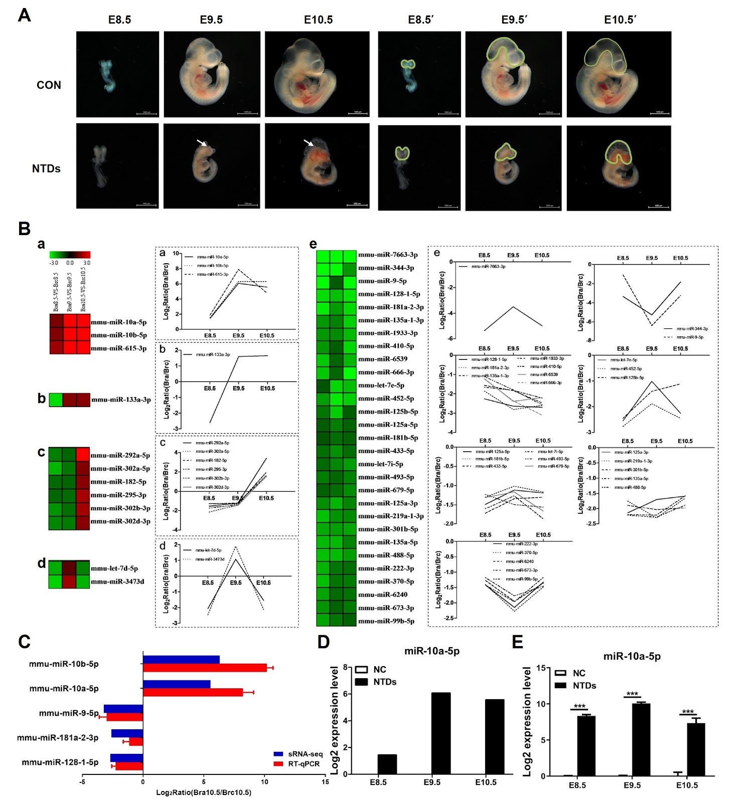
Neural tube defects (NTDs) are characterized by the failure of neural tube closure during embryogenesis and are considered the most common and severe central nervous system anomalies during early development. Recent microRNA (miRNA) expression profiling studies have revealed that the dysregulation of several miRNAs plays an important role in retinoic acid (RA)-induced NTDs. However, the molecular functions of these miRNAs in NTDs remain largely unidentified. Here, we show that miR-10a-5p is significantly upregulated in RA-induced NTDs and results in reduced cell growth due to cell cycle arrest and dysregulation of cell differentiation. Moreover, the cell adhesion molecule L1-like (Chl1) is identified as a direct target of miR-10a-5p in neural stem cells (NSCs) in vitro, and its expression is reduced in RA-induced NTDs. siRNA-mediated knockdown of intracellular Chl1 affects cell proliferation and differentiation similar to those of miR-10a-5p overexpression, which further leads to the inhibition of the expressions of downstream ERK1/2 MAPK signaling pathway proteins. These cellular responses are abrogated by either increased expression of the direct target of miR-10a-5p (Chl1) or an ERK agonist such as honokiol. Overall, our study demonstrates that miR-10a-5p plays a major role in the process of NSC growth and differentiation by directly targeting Chl1, which in turn induces the downregulation of the ERK1/2 cascade, suggesting that miR-10a-5p and Chl1 are critical for NTD formation in the development of embryos.

Read Full Abstract10.3724/abbs.2024078
miR-373-3p promotes aerobic glycolysis in colon cancer cells by targeting MFN2Graphical AbstractVerified
Acta Biochimica et Biophysica Sinica

miR-373-3p promotes aerobic glycolysis in colon cancer cells by targeting MFN2

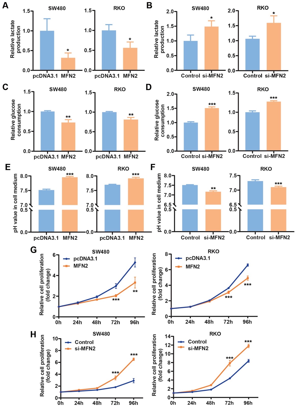
MicroRNAs (miRNAs) are implicated in the development of cancers and may serve as potential targets for therapy. However, the functions and underlying mechanisms of miRNAs in cancers are not well understood. This work aims to study the role of miR-373-3p in colon cancer cells. We find that the expression of miR-373-3p mimics promotes and the miR-373-3p inhibitor suppresses aerobic glycolysis and proliferation of colon cancer cells. Mechanistically, miR-373-3p inhibits the expression of MFN2, a gene that is known to suppress glycolysis, which leads to the activation of glycolysis and eventually the proliferation of cells. In a nude mouse tumor model, the expression of miR-373-3p in colon cancer cells promotes tumor growth by enhancing lactate formation, which is inhibited by the co-expression of MFN2 in the cells. Administration of the miR-373-3p antagomir blunts in vivo tumor growth by decreasing lactate production. In addition, in human colon cancers, the expression levels of miR-373-3p are increased, while those of MFN2 mRNA are decreased, and the increase of miR-373-3p is associated with the decrease of MFN2 mRNA. Our results reveal a previously unknown function and underlying mechanism of miR-373-3p in the regulation of glycolysis and proliferation in cancer cells and underscore the potential of targeting miR-373-3p for colon cancer treatment.

Read Full Abstract10.3724/abbs.2024090
Identification of RACK1 as a novel regulator of non-structural protein 4 of chikungunya virusGraphical AbstractVerified
Acta Biochimica et Biophysica Sinica

Identification of RACK1 as a novel regulator of non-structural protein 4 of chikungunya virus

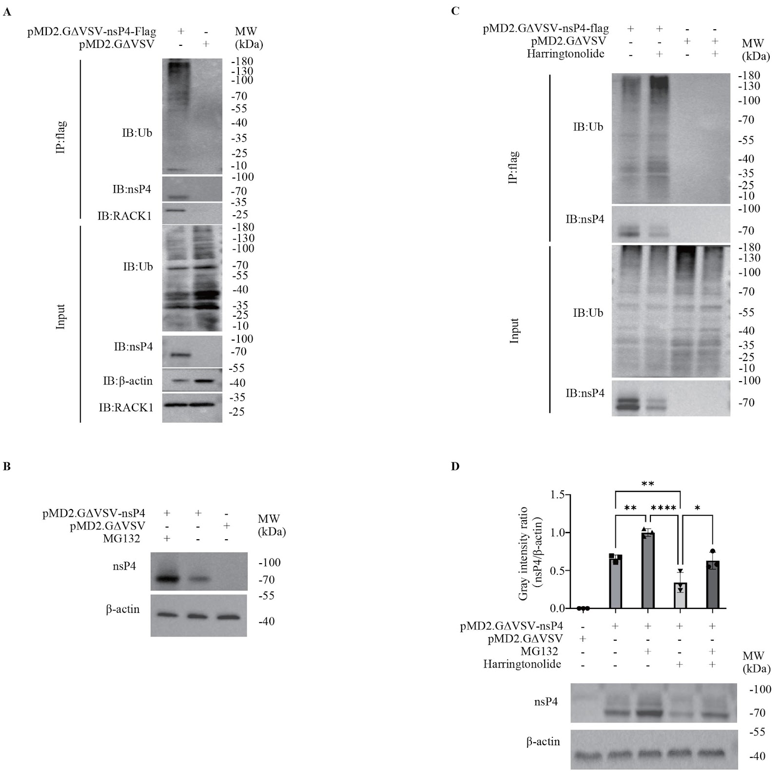
Chikungunya virus (CHIKV) is a neglected arthropod-borne and anthropogenic alphavirus. Over the past two decades, the CHIKV distribution has undergone significant changes worldwide, from the original tropics and subtropics regions to temperate regions, which has attracted global attention. However, the interactions between CHIKV and its host remain insufficiently understood, which dampens the need for the development of an anti-CHIKV strategy. In this study, on the basis of the optimal overexpression of non-structural protein 4 (nsP4), we explore host interactions of CHIKV nsP4 using mass spectrometry-based protein-protein interaction approaches. The results reveal that some cellular proteins that interact with nsP4 are enriched in the ubiquitin-proteasome pathway. Specifically, the scaffold protein receptor for activated C kinase 1 (RACK1) is identified as a novel host interactor and regulator of CHIKV nsP4. The inhibition of the interaction between RACK1 and nsP4 by harringtonolide results in the reduction of nsP4, which is caused by the promotion of degradation but not the inhibition of nsP4 translation. Furthermore, the decrease in nsP4 triggered by the RACK1 inhibitor can be reversed by the proteasome inhibitor MG132, suggesting that RACK1 can protect nsP4 from degradation through the ubiquitin-proteasome pathway. This study reveals a novel mechanism by which the host factor RACK1 regulates CHIKV nsP4, which could be a potential target for developing drugs against CHIKV.

Read Full Abstract10.3724/abbs.2024073
Exploring the mechanism of Panax notoginseng saponin in inhibiting the inflammatory response of microglia in cerebral ischemia based on network pharmacologyGraphical AbstractVerified
Acta Biochimica et Biophysica Sinica

Exploring the mechanism of Panax notoginseng saponin in inhibiting the inflammatory response of microglia in cerebral ischemia based on network pharmacology

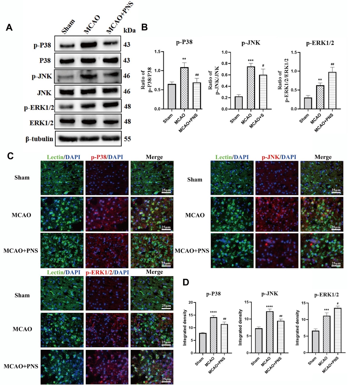
With the increasing global population and aging demographic, the incidence of stroke is rising. Among these, ischemic stroke (IS), also known as cerebral ischemia, constitutes over 80% of all stroke cases. This condition is characterized by an acute cerebrovascular disease caused by the blockage and interruption of the brain's blood supply, resulting in localized tissue ischemia, oxygen, and glucose deficiency, ultimately leading to the death of nerve cells and tissue necrosis [1,2]. "Vascular recanalization and the restoration of cerebral blood flow" are the primary clinical treatment objectives and are achieved through the intravenous administration of drugs such as tissue plasminogen activator or through surgical thrombectomy. These interventions not only restore the delivery of oxygen and glucose to the affected cerebral area but also help prevent the expansion of the infarcted region. However, the restoration of reperfusion cerebral blood flow similarly exposes the infarct area to peripheral immune cells, triggering the activation of the immune response and inflammation-induced injury [3]. Research indicates that IS elicits a robust inflammatory response, with neuroinflammation playing a crucial role in the secondary neurodegeneration process following stroke. Neuroinflammatory responses are initiated and perpetuated through injury cascades that include the release of inflammatory mediators, the migration and recruitment of white blood cells across the blood-brain barrier, and the impairment of endothelial nitric oxide synthase. These mechanisms collectively promote the activation of pro-inflammatory genes, which in turn activate microglia (MG) and exacerbate ischemic damage and neurological dysfunction [4]. MG are resident immune cells of the central nervous system (CNS). Its function is akin to that of macrophages, serving as the first line of defense against injuries within the central nervous system. Under typical conditions, brain microglia participate in immune surveillance and defense against infectious agents. However, in the pathogenesis of neurodegenerative diseases such as IS, MG are activated by various stimuli. Once activated, MG are known to release numerous proinflammatory or cytotoxic factors, such as inducible nitric oxide synthase (iNOS), tumor necrosis factor-α (TNF-α), interleukin-1β (IL-1β), and reactive oxygen species (ROS). These factors initiate the neuroinflammatory response, exacerbate inflammation, intensify damage to brain tissue and neurons, and significantly hinder the repair of brain injuries and neurogenesis [5,6]. Therefore, inhibiting the activation of microglia and reducing the inflammatory response in the central nervous system are crucial for minimizing brain damage caused by IS and are vital for developing effective prevention and treatment strategies. In recent years, certain natural compounds extracted from traditional drug formulations have shown high therapeutic potential in protecting the brain from cerebral ischemic injury. These compounds reduce the neuroinflammatory response and apoptosis following stroke. Traditional Chinese herbal medicine (TCHM) and its constituent herbs feature a multiplicity of components, targets, and pathways owing to their complex formulations and therapeutic principles, making them promising sources for developing effective treatments for IS. Panax notoginseng saponin (PNS), as the principal bioactive component of Panax notoginseng, is extensively utilized in the prevention and treatment of cardiovascular and cerebrovascular diseases. Its pharmacological benefits include dissipating blood stasis, promoting hemostasis, alleviating swelling and pain, regulating energy metabolism disorders, balancing ion metabolism, and reducing and accelerating the clearance of free radicals [7]. Research indicates that PNS mitigates apoptosis by maintaining mitochondrial homeostasis, enhancing the integrity of the blood‒brain barrier (BBB), augmenting cerebral blood supply, and fostering the differentiation of neural stem cells and proliferation of hippocampal neurons. In addition, PNS offers neuroprotection against focal cerebral I/R injury in rats by reducing brain edema, upregulating the expression of the heat shock protein HSP70, and downregulating the expression of transferrin [8,9]. Additionally, PNS has been reported to enhance the recovery of neurogenesis and neurological function in cerebral embolism induced by microspheres and to reduce sepsis-induced acute kidney injury by suppressing inflammation [10]. However, the mechanism by which PNS targets IS has not been fully elucidated. In this study, we investigated the anti-inflammatory effects of PNS on IS and identified potential target pathways that could inhibit microglia-mediated inflammatory response.
Read Full Abstract10.3724/abbs.2024114
Posttranslational regulatory mechanism of PD-L1 in cancers and associated opportunities for novel small-molecule therapeuticsGraphical AbstractVerified
Acta Biochimica et Biophysica Sinica

Posttranslational regulatory mechanism of PD-L1 in cancers and associated opportunities for novel small-molecule therapeutics

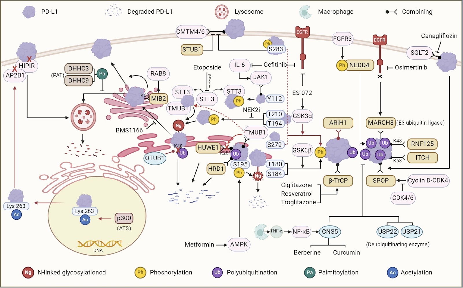
Despite the tremendous progress in cancer research over the past few decades, effective therapeutic strategies are still urgently needed. Accumulating evidence suggests that immune checkpoints are the cause of tumor immune escape. PD-1/PD-L1 are among them. Posttranslational modification is the most critical step for protein function, and the regulation of PD-L1 by small molecules through posttranslational modification is highly valuable. In this review, we discuss the mechanisms of tumor cell immune escape and several posttranslational modifications associated with PD-L1 and describe examples in which small molecules can regulate PD-L1 through posttranslational modifications. Herein, we propose that the use of small molecule compounds that act by inhibiting PD-L1 through posttranslational modifications is a promising therapeutic approach with the potential to improve clinical outcomes for cancer patients.

Read Full Abstract10.3724/abbs.2024085
Ginsenoside Rh2 alleviates osteoporosis by attenuating oxidative stress-induced osteoblast dysfunction via the FoxO1/β-catenin pathwayGraphical AbstractVerified
Acta Biochimica et Biophysica Sinica

Ginsenoside Rh2 alleviates osteoporosis by attenuating oxidative stress-induced osteoblast dysfunction via the FoxO1/β-catenin pathway

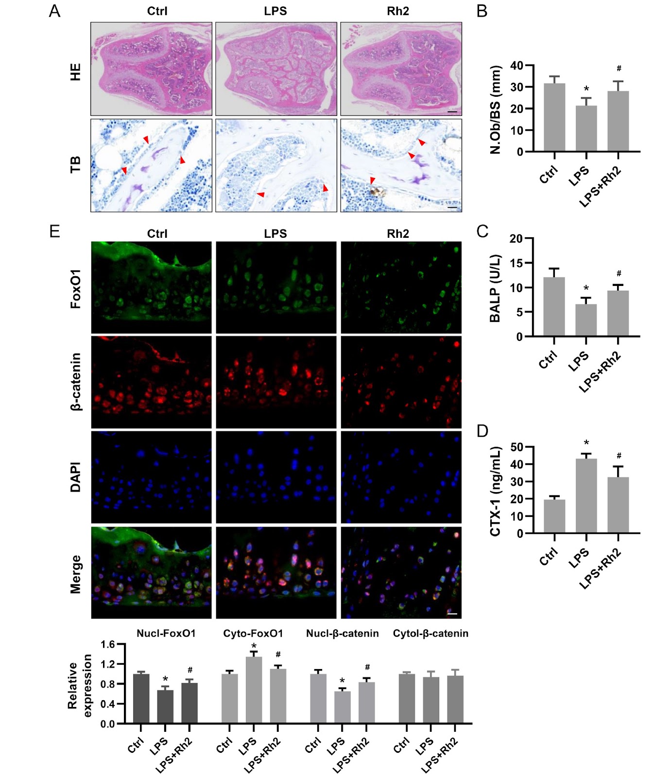
The degree of oxidative stress decreases osteoblast function with age, which leads to a decline in bone compressive capacity. Ginsenoside Rh2 is a known clinical or adjuvant therapy for various tissues. In this study, we investigate the pharmacological effects of Rh2 against oxidative stress-induced osteoblasts. Osteoblasts are pretreated with Rh2 for 48 h and then exposed to hydrogen peroxide (H2O2), which results in significantly decreased ROS levels, increased antioxidant enzyme activity, and enhanced mitochondrial function. Functionally, Rh2 increases alkaline phosphatase (ALP) expression, together with enhanced mineralization and expression of osteogenesis-associated genes. Rh2 also promotes the nuclear translocation of FoxO1 and β-catenin, whereas it does not reverse reduced mineralization caused by decreased FoxO1 or β-catenin activity, indicating that its effect is mediated through the functional interaction between FoxO1 and β-catenin. In a mouse model of lipopolysaccharide (LPS)-induced bone loss, Rh2 administration improves trabecular microstructure, increases osteoblast numbers, and upregulates serum metabolites associated with bone formation. Immunofluorescence analysis further reveals that Rh2 promotes the nuclear co-localization of FoxO1 and β-catenin in femurs, indicating their coordinated action within this signaling axis. These findings indicate that Rh2 mitigates oxidative stress-induced osteoblast dysfunction via the FoxO1/β-catenin pathway, highlighting the pivotal role of redox balance in bone remodeling and suggesting a promising therapeutic strategy for osteoporosis.

Read Full Abstract10.3724/abbs.2026065
Molecular insight into the potential functional role of pseudoenzyme GFOD1 via interaction with NKIRAS2Graphical AbstractVerified
Acta Biochimica et Biophysica Sinica

Molecular insight into the potential functional role of pseudoenzyme GFOD1 via interaction with NKIRAS2

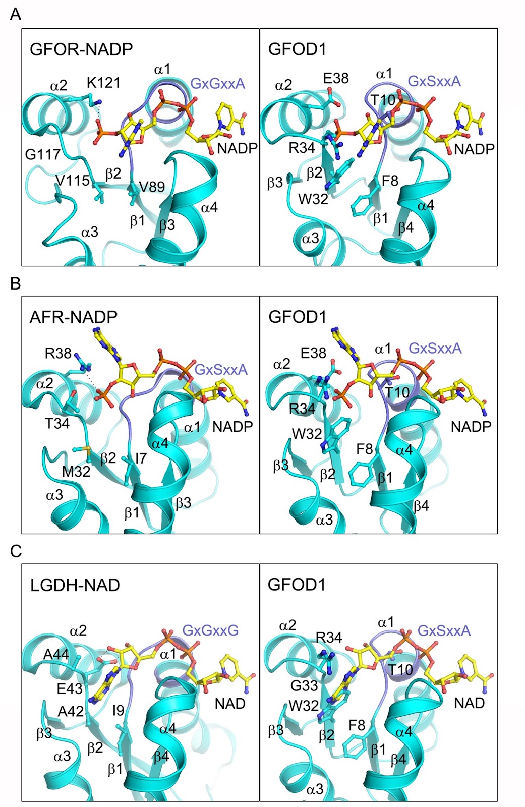
The glucose-fructose oxidoreductase/inositol dehydrogenase/rhizopine catabolism protein (Gfo/Idh/MocA) family includes a variety of oxidoreductases with a wide range of substrates that utilize NAD or NADP as redox cofactor. Human contains two members of this family, namely glucose-fructose oxidoreductase domain-containing protein 1 and 2 (GFOD1 and GFOD2). While GFOD1 exhibits low tissue specificity, it is notably expressed in the brain, potentially linked to psychiatric disorders and severe diseases. Nevertheless, the specific function, cofactor preference, and enzymatic activity of GFOD1 remain largely unknown. In this work, we find that GFOD1 does not bind to either NAD or NADP. Crystal structure analysis unveils that GFOD1 exists as a typical homodimer resembling other family members, but lacks essential residues required for cofactor binding, suggesting that it may function as a pseudoenzyme. Exploration of GFOD1-interacting partners in proteomic database identifies NF-κB inhibitor-interacting Ras-like 2 (NKIRAS2) as one potential candidate. Co-immunoprecipitation (co-IP) analysis indicates that GFOD1 interacts with both GTP- and GDP-bound forms of NKIRAS2. The predicted structural model of the GFOD1-NKIRAS2 complex is validated in cells using point mutants and shows that GFOD1 selectively recognizes the interswitch region of NKIRAS2. These findings reveal the distinct structural properties of GFOD1 and shed light on its potential functional role in cellular processes.

Read Full Abstract10.3724/abbs.2024105
Lactate activates CCL18 expression via H3K18 lactylation in macrophages to promote tumorigenesis of ovarian cancerGraphical AbstractVerified
Acta Biochimica et Biophysica Sinica

Lactate activates CCL18 expression via H3K18 lactylation in macrophages to promote tumorigenesis of ovarian cancer

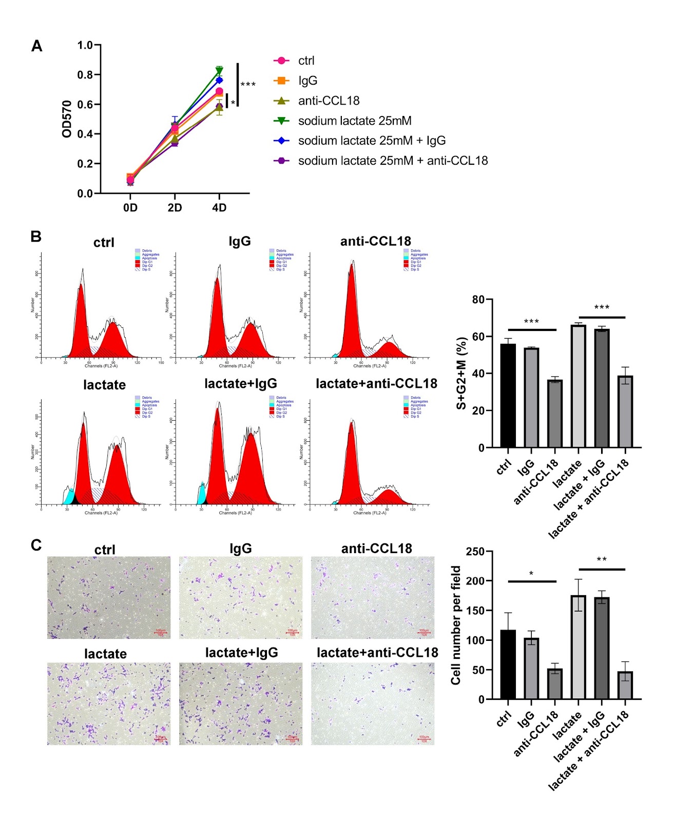
This study investigates the role of lactate in the genesis and progression of ovarian cancer (OV) and explores the underlying mechanisms. Serum lactate levels show a positive correlation with tumor grade and poor prognosis in patients with OV. Bioinformatics analysis identifies CCL18 as a lactate-related gene in OV. CCL18 is up-regulated in cancerous tissues and positively related to serum lactate levels in OV patients. THP-1 cells are exposed to phorbol-12-myristate-13-acetate for M0 macrophage induction. The results of RT-qPCR and ELISA for M1/M2 macrophage-related markers and inflammatory cytokines show that the exposure of lactate to macrophages induces M2 polarization. Based on the coculture of OV cells with macrophages, lactate-treated macrophages induces a significant increase in the proliferation and migration of OV cells. However, these effects can be reversed by silencing of Gpr132 in macrophages or treatment with anti-CCL18 antibody. Experiments using the xenograft model verify that the oncogenic role of lactate in tumor growth and metastasis relies on Gpr132 and CCL18. ChIP-qPCR and luciferase reporter assays reveal that lactate regulates CCL18 expression via H3K18 lactylation. In conclusion, lactate is a potential therapeutic target for OV. It is involved in tumorigenesis by activating CCL18 expression via H3K18 lactylation in macrophages.

Read Full Abstract10.3724/abbs.2024111