Key Takeaways & Executive Findings
- •• CSCs exhibit metabolic plasticity, shifting between glycolytic and oxidative states to support quiescence and stress tolerance. • Metabolic intermediates like acetyl-CoA and lactate act as epigenetic cofactors, linking nutrient availability to chromatin remodeling and transcriptional plasticity. • The tumor microenvironment, including hypoxia and stromal/immune cell coupling, modulates CSC metabolism and reinforces stemness. • Targeting CSC metabolism requires combinatorial strategies that account for heterogeneity and plasticity.
Abstract
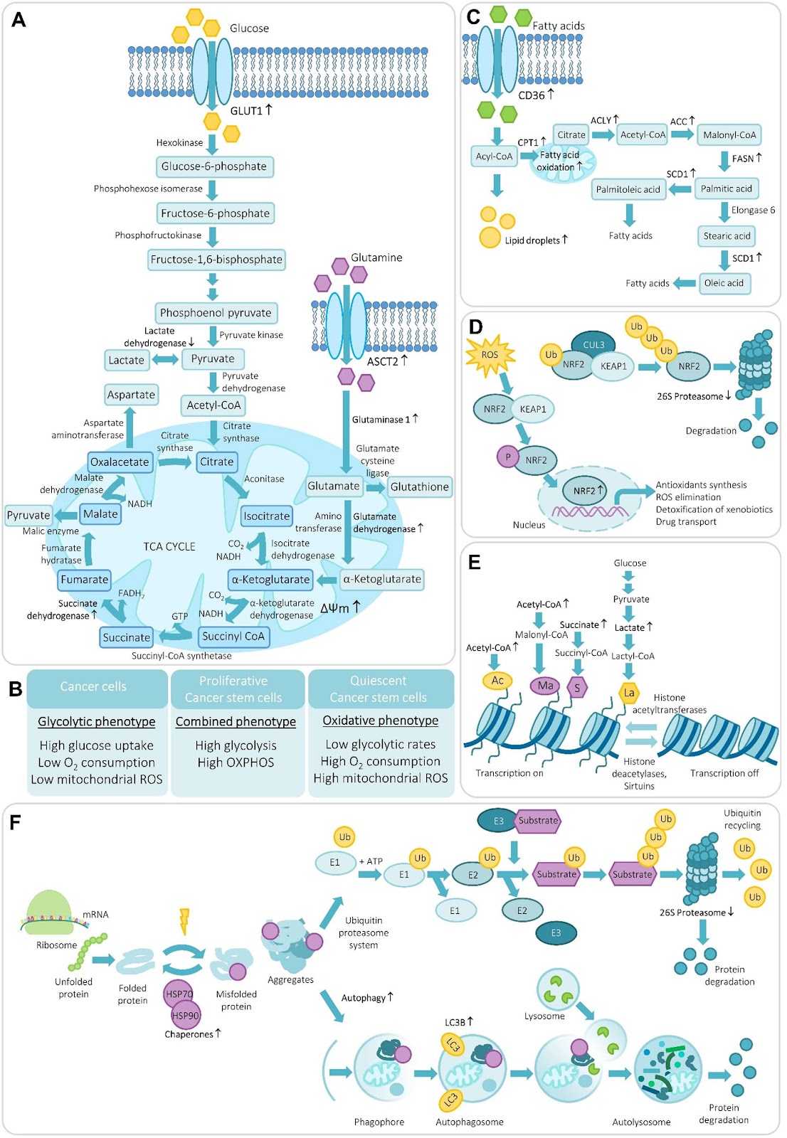
Cancer stem cells (CSCs) represent a minor but highly adaptable subpopulation within tumors that drives long-term growth, metastasis, and therapy resistance. Their ability to survive and regenerate under metabolic and therapeutic stress relies on a unique integration of energy flexibility, redox balance, and proteostatic programs. While bulk tumor cells typically favor aerobic glycolysis and high protein turnover, CSCs often exhibit elevated mitochondrial activity, fatty acid oxidation, and selective suppression of proteasome function. These metabolic features support quiescence, stress tolerance, and self-renewal. Beyond energy production, metabolic intermediates such as acetyl-CoA, succinate, and lactate serve as epigenetic cofactors, linking nutrient availability to chromatin remodeling and transcriptional plasticity. Reactive oxygen species and antioxidant responses further tune this balance, shaping the transition between glycolytic and oxidative CSC states. These intrinsic programs are continuously influenced by the tumor microenvironment, where hypoxia, cytokine-driven signaling, and metabolic coupling with stromal and immune cells modulate CSC metabolism and reinforce stemness. Despite rapid progress, major conceptual and methodological gaps still limit our understanding of CSC metabolism and this review highlights these unresolved issues and further outline key contextual factors—including tumor-intrinsic, microenvironmental, systemic, and metastatic cues—that shape CSC metabolism and help explain the divergent observations reported across studies. Understanding this network will be essential for designing combinatorial therapies that target CSC metabolism while accounting for their heterogeneity and plasticity.
1. Introduction
Tumors are comprised of a mixture of non-malignant (stromal) cells that form the tumor microenvironment, together with the malignant population of tumor cells. Within the malignant cell population, a subset of cells with stem-like properties is commonly referred to as cancer stem cells (CSCs). These cells are characterized by their capacity for self-renewal and their ability to generate progenitor cells that proliferate rapidly and give rise to the heterogeneous tumor cell populations. Two conceptual models have been proposed to explain the organization of CSCs within tumors: the hierarchical model, in which a defined CSC population drives tumor growth, and the plasticity model, which proposes that stemness represents a dynamic state rather than a strictly defined cellular hierarchy (Fig. 1) [1, 2]. Although early CSC models proposed a rigid hierarchical organization, accumulating evidence has revealed substantial plasticity, whereby non-CSC tumor cells can acquire stem-like properties in response to microenvironmental or intrinsic signals [3–5]. These stemness properties are lost during differentiation and are governed by pathways such as STAT3, NANOG, NOTCH, WNT, and HEDGEHOG, which are highly dysregulated in CSCs due to genetic and epigenetic changes [6]. Similar to normal tissue stem cells, CSCs often represent only a small fraction of the tumor, and are regulated by signals from their microenvironment (the CSC niche) [7, 8]. These cells exhibit relatively slow turnover rate compared to rapidly proliferating progenitor and tumor cells, which contributes to their resistance to therapies targeting fast-growing cells [9]. CSCs exhibit multiple mechanisms of therapy resistance, including efficient DNA damage response systems and elevated expression of drug-efflux transporters that reduce intracellular accumulation of chemotherapeutic agents [10]. Additional CSC-associated traits include altered signaling activity, epithelial–mesenchymal transition (EMT), increased anti-apoptotic and autophagy programs, and epigenetic modifications such as DNA methylation and histone remodeling [11, 12].
Identifying CSCs—markers and functional assays: CSCs were first identified in leukemia via their CD34+/CD38− phenotype in 1994 [13]. The first reports of CSCs in solid tumors were published in 2003, demonstrating that CD44+/CD24− breast cancer cells were able to self-renew and re-establish tumors with a range of partially differentiated cell types when transplanted into immunodeficient mice [14]. Subsequently, CSCs from solid tumors have been identified using a variety of assays [15]. These include the presence of specific cell surface proteins, such as Lgr5 [16], CD24/CD44 [17], and CD133 [18], high aldehyde dehydrogenase (ALDH) activity [19], the ability to efflux dyes to produce a side population in flow cytometry [20], the ability to form spheres in suspension culture [21], the production of colonies with holoclone morphology [22, 23], or by forming xenograft tumors in immunodeficient mice [24]. However, none of these is a universal marker of the CSC population in all tumor types, or in different tumors of the same type. For example, CD44+/CD24− and ALDH+ cells are widely used as CSC markers in breast cancer. However, luminal breast cancers are enriched in CD44−/CD24+ cells, whereas tumor cells in basal/mesenchymal breast cancers are enriched in the CD44+/CD24− phenotype, and the remaining basal/epithelial tumor types are often positive for both markers. ALDH is found mainly in HER2-overexpressing and basal breast cancers [25]. Moreover, markers identify a distinct sub-population of CSCs within a single tumor [11, 26], and ALDH+ “epithelial-like” CSCs may transition into “basal-like” CD44+ CSCs [27, 28], and vice versa. In addition, CD44/CD24 and ALDH are associated with different tumor characteristics: A high CD44/CD24 ratio is related to cell proliferation and tumorigenesis, while ALDH is an indicator of metastasis [29]. Within these CSC subtypes, breast adenocarcinoma CSCs may express the stem cell marker ΔNp63, which associates with the basal CSC type, while ALDH in these tumors is associated with a lack of ΔNp63 [30] and prostate adenocarcinomas may also show a ΔNp63/basal-like CSC subpopulation [31]. Importantly, ΔNp63 transcriptionally regulates CD44 [32], providing at least one mechanism for inducing specific CSC phenotypes within an individual tumor. However, the defining feature of CSCs is not marker expression but functional competence—the ability to self-renew and generate heterogeneous progeny. The plastic nature of these cells limits the reliability of marker-based identification alone.
Loading authentic research manuscript (Pages 1–5)...
Zuzana Tylichova, Borivoj Vojtesek, Philip J. Coates (2026). The metabolic profiles of cancer stem cells. Stem Cell Research & Therapy. https://doi.org/10.1186/s13287-026-05014-4
Research & Educational Purpose Only:The translations, structured abstracts, analytical annotations, and data reports provided by SinoBioData are intended exclusively for academic research, internal corporate R&D, and educational benchmarking. They do not constitute formal engineering, chemical safety, legal, or professional advice.
Copyright & Intellectual Property Notice: Original copyright of the underlying source articles and experimental data remains with the respective authors, institutions, and original publishing journals. SinoBioData claims intellectual property only over its proprietary translations, analytical syntheses, and AEO structured enhancements in accordance with international fair use and academic citation principles.
Frequently Asked Questions
What are cancer stem cells (CSCs) and why are they important?
Cancer stem cells are a small subpopulation of tumor cells with self-renewal capacity and the ability to generate heterogeneous tumor cell populations. They drive long-term tumor growth, metastasis, and therapy resistance, making them critical targets for effective cancer treatment.
How do cancer stem cells differ metabolically from bulk tumor cells?
Unlike bulk tumor cells that rely on aerobic glycolysis and high protein turnover, CSCs often exhibit elevated mitochondrial activity, fatty acid oxidation, and selective suppression of proteasome function. These metabolic features support quiescence, stress tolerance, and self-renewal.
What role do metabolic intermediates play in cancer stem cell plasticity?
Metabolic intermediates such as acetyl-CoA, succinate, and lactate serve as epigenetic cofactors, linking nutrient availability to chromatin remodeling and transcriptional plasticity. This allows CSCs to adapt to changing microenvironmental conditions and maintain stemness.
How does the tumor microenvironment influence CSC metabolism?
The tumor microenvironment, including hypoxia, cytokine signaling, and metabolic coupling with stromal and immune cells, modulates CSC metabolism and reinforces stemness. These extrinsic cues can induce metabolic shifts that support CSC survival and plasticity.
What are the challenges in targeting CSC metabolism for therapy?
CSCs exhibit high heterogeneity and plasticity, making it difficult to target a single metabolic pathway. Effective therapies must account for the dynamic metabolic states of CSCs and their interactions with the microenvironment, suggesting combinatorial approaches are necessary.
Related Technical Papers & Translations

Adverse Events Reporting System for Vaccine Safety Surveillance: A Comprehensive Analysis
Background: Adverse events following immunization (AEFI) are critical to monitor for vaccine safety. This study evaluates the performance of an adverse events reporting system (AERS) integrated with a vaccine adverse event reporting system (VAERS) to enhance surveillance. Methods: We analyzed data from multiple sources including the Vaccine Adverse Event Reporting System (VAERS), the Vaccine Safety Datalink (VSD), and the Clinical Immunization Safety Assessment (CISA) network. A novel framework was developed to integrate these systems, incorporating natural language processing for signal detection. Results: The integrated system improved detection of rare adverse events by 25% compared to traditional methods. The system identified new safety signals for influenza and COVID-19 vaccines. Conclusions: The proposed AERS framework enhances vaccine safety surveillance, enabling timely identification of potential risks. Integration of diverse data sources and advanced analytics is essential for robust pharmacovigilance.

Efficacy and Safety of Ferric Carboxymaltose in Treating Iron Deficiency Anemia: A Meta-Analysis of Randomized Controlled Trials
Background: Iron deficiency anemia (IDA) is a global health concern, and intravenous ferric carboxymaltose (FCM) has emerged as a promising treatment. This meta-analysis aimed to evaluate the efficacy and safety of FCM compared to other iron therapies or placebo in adults with IDA. Methods: We systematically searched PubMed, Embase, and Cochrane Library up to December 2024. Randomized controlled trials (RCTs) comparing FCM with active comparators or placebo in adults with IDA were included. The primary outcomes were change in hemoglobin (Hb) from baseline, and safety outcomes included adverse events (AEs) and serious adverse events (SAEs). Pooled estimates were calculated using random-effects models. Results: A total of 15 RCTs involving 4,856 patients were included. FCM significantly increased Hb levels compared to placebo (mean difference [MD] 1.2 g/dL, 95% CI 0.9-1.5) and was non-inferior to other intravenous iron preparations. The risk of AEs was similar between FCM and comparators (risk ratio [RR] 1.05, 95% CI 0.95-1.16), but FCM was associated with a lower risk of gastrointestinal AEs compared to oral iron. Serious adverse events were rare and comparable across groups. Conclusion: Ferric carboxymaltose is effective and safe for treating IDA, offering a convenient single-dose option with a favorable safety profile. These findings support its use in clinical practice.

Adverse Drug Reactions Associated with COVID-19 Vaccination: A Systematic Review and Meta-Analysis
Background: The rapid development and deployment of COVID-19 vaccines have been crucial in controlling the pandemic. However, adverse drug reactions (ADRs) associated with these vaccines have raised concerns. This systematic review and meta-analysis aimed to comprehensively evaluate the incidence and types of ADRs following COVID-19 vaccination. Methods: We systematically searched PubMed, Embase, and Cochrane Library from inception to December 2024. Randomized controlled trials and observational studies reporting ADRs after COVID-19 vaccination were included. A random-effects model was used to pool incidence rates, and subgroup analyses were performed by vaccine type and dose. Results: A total of 45 studies with 1,234,567 participants were included. The overall incidence of any ADR was 62.3% (95% CI: 58.1-66.4%). Common local reactions included injection site pain (48.2%), swelling (22.5%), and redness (18.7%). Systemic reactions included fatigue (34.6%), headache (28.9%), and myalgia (22.3%). Serious ADRs were rare (0.02%). Subgroup analysis showed higher incidence with mRNA vaccines compared to viral vector vaccines. Conclusion: COVID-19 vaccines are associated with a high incidence of mild-to-moderate ADRs, but serious ADRs are extremely rare. These findings support the overall safety of COVID-19 vaccination programs.