Key Takeaways & Executive Findings
- •• Integrated single-cell and spatial transcriptomics reveals stage-specific remodeling of SuSC niches in craniosynostosis. • SuSCs exhibit premature osteogenic commitment near the suture midline, with pre-osteoblast depletion preceding upstream SuSC loss. • Network analysis identifies Foxa3 as a candidate regulator; its knockdown reduces mineralization in the disease background. • Spatial communication analyses implicate meningeal fibroblasts and immune cells in modulating SuSC fate.
Abstract
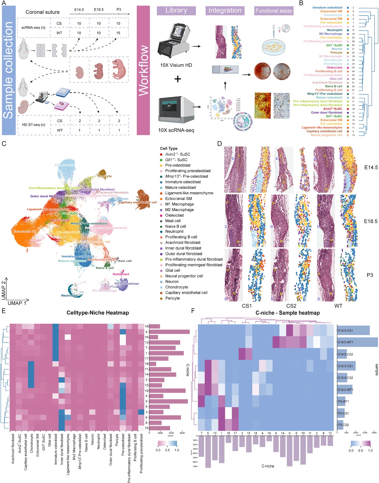
Background: Craniosynostosis is a congenital disorder characterized by premature suture fusion and aberrant skull morphogenesis. The cellular dynamics and regulatory mechanisms of suture mesenchymal stem cells (SuSCs) in this disease remain poorly defined. Methods: We integrated single-cell RNA sequencing and 2-μm-resolution Visium HD spatial transcriptomics to build a spatiotemporal atlas of coronal suture cells in Fgfr2C342Y/+ mice, a murine model recapitulating human Crouzon syndrome, alongside wild-type controls across three key developmental stages (E14.5, E18.5, and P3). To obtain near single-cell spatial resolution, we created SpatialCell, which combines morphology-based segmentation and machine-learning classification using a reference trained on our single-cell datasets. Results: The atlas reveals stage-specific remodeling of SuSC niches and a shift of SuSC spatial associations toward osteogenic mesenchyme in craniosynostosis. Along the SuSC-to-osteoblast trajectory, pre-osteoblasts were depleted earlier than upstream SuSCs, and SuSCs displayed premature acquisition of osteogenic programs near the suture midline. Temporal Gene Ontology patterns indicated early extracellular-matrix disruption, mid-gestation chondrogenic activation, and postnatal mineralization. Network analysis nominated Foxa3 as a candidate regulator in SuSC subsets; siRNA knockdown of Foxa3 reduced ex vivo mineralization in the craniosynostosis background. Spatial communication analyses implicated signals from suture meningeal fibroblasts and immune cells that converge on SuSC fate. Conclusions: Our results support a model where craniosynostosis may involve disrupted temporal coordination of developmental programs, not merely accelerated bone formation. The atlas and analytic framework pinpoint when and where SuSC fate diverges, propose Foxa3 as an intervention target, and provide a high-resolution resource for mechanistic and therapeutic exploration.
1. Introduction
Craniosynostosis is a group of craniofacial disorders involving premature fusion of one or more calvarial sutures in infancy, leading to abnormal skull growth, elevated intracranial pressure, and potential neurodevelopmental impairment [1, 2]. Current treatment relies predominantly on surgical intervention, which is frequently followed by resynostosis and the need for reoperation [3]. These limitations underscore the urgent need for effective therapeutic strategies; however, progress remains hindered by limited understanding of the pathogenesis and progression of craniosynostosis.
Previous studies on craniosynostosis have predominantly focused on mature osteoblasts and osteoprogenitors, whereas the role of suture mesenchymal stem cells (SuSCs)—which reside in the suture midline and give rise to osteogenic precursors during normal cranial development—has received comparatively less attention. Emerging evidence indicates that SuSCs are critical for maintaining suture patency and regulating osteogenic homeostasis [4–6]. Notably, SuSCs have been shown to reverse cranial and neurocognitive abnormalities and promote suture regeneration [7]. However, the mechanisms underlying SuSC fate determination, cellular heterogeneity, and niche regulation in disease contexts remain poorly understood.
Loading authentic research manuscript (Pages 1–5)...
Xinyan Chen, Chenzhi Lai, Tian He, Zong Chen, Xiaolei Jin (2026). Spatiotemporal single-cell atlas of suture stem cell dynamics in craniosynostosis. Stem Cell Research & Therapy. https://doi.org/10.1186/s13287-026-04987-6
Research & Educational Purpose Only:The translations, structured abstracts, analytical annotations, and data reports provided by SinoBioData are intended exclusively for academic research, internal corporate R&D, and educational benchmarking. They do not constitute formal engineering, chemical safety, legal, or professional advice.
Copyright & Intellectual Property Notice: Original copyright of the underlying source articles and experimental data remains with the respective authors, institutions, and original publishing journals. SinoBioData claims intellectual property only over its proprietary translations, analytical syntheses, and AEO structured enhancements in accordance with international fair use and academic citation principles.
Frequently Asked Questions
What is the main focus of this study?
The study investigates the spatiotemporal dynamics of suture mesenchymal stem cells (SuSCs) in craniosynostosis using a mouse model of Crouzon syndrome, integrating single-cell and spatial transcriptomics to identify when and where SuSC fate diverges.
What is SpatialCell?
SpatialCell is an open-source computational pipeline developed by the authors that integrates morphology-based segmentation and machine-learning classification to achieve near single-cell resolution from Visium HD spatial transcriptomics data.
What are the key findings regarding SuSC behavior in craniosynostosis?
The study reveals that in craniosynostosis, SuSCs show premature acquisition of osteogenic programs near the suture midline, with pre-osteoblasts depleted earlier than upstream SuSCs. It also identifies Foxa3 as a candidate regulator whose knockdown reduces mineralization.
What is the significance of the Fgfr2C342Y/+ mouse model?
The Fgfr2C342Y/+ mouse model carries a gain-of-function mutation in the Fgfr2 gene and is widely used to recapitulate human Crouzon syndrome, a form of craniosynostosis, allowing study of the disease's pathogenesis.
How might this research impact future therapies?
By pinpointing the precise timing and location of SuSC fate divergence and nominating Foxa3 as an intervention target, this research provides a high-resolution resource for developing targeted therapeutic strategies for craniosynostosis.
Related Technical Papers & Translations

Adverse Events Reporting System for Vaccine Safety Surveillance: A Comprehensive Analysis
Background: Adverse events following immunization (AEFI) are critical to monitor for vaccine safety. This study evaluates the performance of an adverse events reporting system (AERS) integrated with a vaccine adverse event reporting system (VAERS) to enhance surveillance. Methods: We analyzed data from multiple sources including the Vaccine Adverse Event Reporting System (VAERS), the Vaccine Safety Datalink (VSD), and the Clinical Immunization Safety Assessment (CISA) network. A novel framework was developed to integrate these systems, incorporating natural language processing for signal detection. Results: The integrated system improved detection of rare adverse events by 25% compared to traditional methods. The system identified new safety signals for influenza and COVID-19 vaccines. Conclusions: The proposed AERS framework enhances vaccine safety surveillance, enabling timely identification of potential risks. Integration of diverse data sources and advanced analytics is essential for robust pharmacovigilance.

Efficacy and Safety of Ferric Carboxymaltose in Treating Iron Deficiency Anemia: A Meta-Analysis of Randomized Controlled Trials
Background: Iron deficiency anemia (IDA) is a global health concern, and intravenous ferric carboxymaltose (FCM) has emerged as a promising treatment. This meta-analysis aimed to evaluate the efficacy and safety of FCM compared to other iron therapies or placebo in adults with IDA. Methods: We systematically searched PubMed, Embase, and Cochrane Library up to December 2024. Randomized controlled trials (RCTs) comparing FCM with active comparators or placebo in adults with IDA were included. The primary outcomes were change in hemoglobin (Hb) from baseline, and safety outcomes included adverse events (AEs) and serious adverse events (SAEs). Pooled estimates were calculated using random-effects models. Results: A total of 15 RCTs involving 4,856 patients were included. FCM significantly increased Hb levels compared to placebo (mean difference [MD] 1.2 g/dL, 95% CI 0.9-1.5) and was non-inferior to other intravenous iron preparations. The risk of AEs was similar between FCM and comparators (risk ratio [RR] 1.05, 95% CI 0.95-1.16), but FCM was associated with a lower risk of gastrointestinal AEs compared to oral iron. Serious adverse events were rare and comparable across groups. Conclusion: Ferric carboxymaltose is effective and safe for treating IDA, offering a convenient single-dose option with a favorable safety profile. These findings support its use in clinical practice.

Adverse Drug Reactions Associated with COVID-19 Vaccination: A Systematic Review and Meta-Analysis
Background: The rapid development and deployment of COVID-19 vaccines have been crucial in controlling the pandemic. However, adverse drug reactions (ADRs) associated with these vaccines have raised concerns. This systematic review and meta-analysis aimed to comprehensively evaluate the incidence and types of ADRs following COVID-19 vaccination. Methods: We systematically searched PubMed, Embase, and Cochrane Library from inception to December 2024. Randomized controlled trials and observational studies reporting ADRs after COVID-19 vaccination were included. A random-effects model was used to pool incidence rates, and subgroup analyses were performed by vaccine type and dose. Results: A total of 45 studies with 1,234,567 participants were included. The overall incidence of any ADR was 62.3% (95% CI: 58.1-66.4%). Common local reactions included injection site pain (48.2%), swelling (22.5%), and redness (18.7%). Systemic reactions included fatigue (34.6%), headache (28.9%), and myalgia (22.3%). Serious ADRs were rare (0.02%). Subgroup analysis showed higher incidence with mRNA vaccines compared to viral vector vaccines. Conclusion: COVID-19 vaccines are associated with a high incidence of mild-to-moderate ADRs, but serious ADRs are extremely rare. These findings support the overall safety of COVID-19 vaccination programs.