🧬 SinoBioData Academic Portal
🏛️ Indexed Academic JournalOriginal: 中国组织工程研究

Chinese Journal of Tissue Engineering Research

Premier Chinese Biomedical Journal indexed in SinoBioData: Chinese Journal of Tissue Engineering Research (中国组织工程研究).

Total Research Papers: 200
Access: 100% Free Open Access
Browse by Publication Year & VolumeReset All Filters ✕

Published Research PapersFiltered: Year 2026 • Vol 1898

Showing 31 of 200 peer-reviewed papers with full Graphical Abstracts.

Original ResearchVol 1898, Issue 26 • pp. 100-112DOI: 10.12307/2026.21286Jan 15, 2026

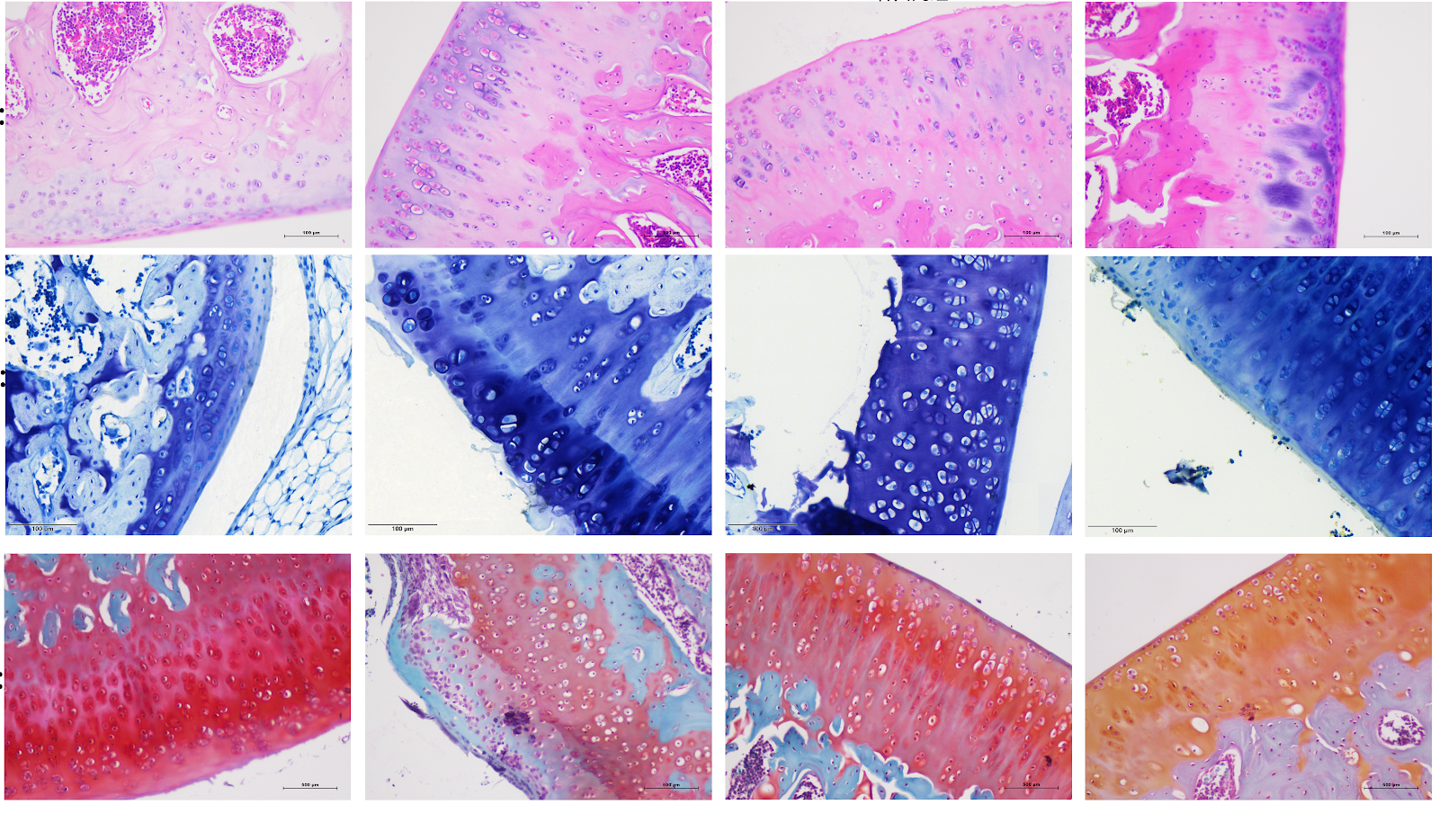
Mechanism by which Yougui Pill inhibits pyroptosis of chondrocytes in rats with knee osteoarthritis

Authors: FAN Yuanhe, YANG Yongju, HE Fanyu, QIAO Long, ZHANG Yu, ZHANG Shuai, LI Zhiwen, GUAN Xuefeng

BACKGROUND: Yougui Pill is derived from Jingyue Quanshu. Studies have confirmed that Yougui Pill is highly effective in treating patients with knee osteoarthritis, but its mechanism of action remains unclear. OBJECTIVE: To explore the potential molecular mechanism of Yougui Pill in improving knee osteoarthritis in rats. METHODS: Forty-eight SPF-grade Sprague-Dawley rats were randomly divided into four groups: blank control group, model group, Yougui Pill group and celecoxib group. The latter three groups were subjected to modified Hulth method for surgical modeling of knee osteoarthritis. After wound healing, rats were driven for 8 weeks. After modeling, the celecoxib group was given celecoxib suspension by gavage, the Yougui Pill group was given Yougui Pill decoction by gavage, and the sham operation group and model group were given equal volume of normal saline, once daily for 4 weeks. Hematoxylin-eosin staining, toluidine blue staining, and safranin O-fast green staining were used to observe the pathological changes of rat cartilage tissue; transmission electron microscopy was used to observe the ultrastructure of rat chondrocytes; ELISA was used to detect the levels of interleukin-18, interleukin-1β, and tumor necrosis factor-α in rat serum; western blot was used to detect the protein expression of PI3K, AKT, NF-κB, p-PI3K, p-AKT, p-P65, NLRP3, GSDMD, GSDMD-N, Caspase1, Cleaved-Caspase1, interleukin-18, and interleukin-1β in rat cartilage tissue. RESULTS AND CONCLUSION: Compared with the blank group, the model group showed severe destruction of cartilage edge, cartilage tissue defect, thinning and disordered arrangement of cartilage layer cells, subchondral bone hyperplasia, disordered tide line, severe structural damage of chondrocytes, formation of pyroptotic bodies, significantly increased serum levels of tumor necrosis factor-α, interleukin-18, and interleukin-1β (P < 0.05), and significantly increased protein expression of p-PI3K, p-AKT, p-P65, NLRP3, GSDMD-N, Cleaved-Caspase1, interleukin-18, and interleukin-1β in cartilage tissue (P < 0.01). Compared with the model group, the cartilage structure of rats in the Yougui Pill group and celecoxib group tended to be normal, with deeper cartilage staining, thicker cartilage, more complete chondrocyte membrane, significantly decreased serum levels of tumor necrosis factor-α, interleukin-18, and interleukin-1β (P < 0.05), and significantly decreased protein expression of p-PI3K, p-AKT, p-P65, NLRP3, GSDMD-N, Cleaved-Caspase1, interleukin-18, and interleukin-1β in cartilage tissue (P < 0.01). Compared with the celecoxib group, the Yougui Pill group showed more regular arrangement of chondrocytes, smoother articular cartilage surface, significantly thickened cartilage layer, relatively complete tide line, relatively complete chondrocyte membrane, significantly decreased serum levels of tumor necrosis factor-α, interleukin-18, and interleukin-1β (P < 0.05), and significantly decreased protein expression of p-PI3K, p-AKT, p-P65, NLRP3, GSDMD-N, Cleaved-Caspase1, interleukin-18, and interleukin-1β in cartilage tissue (P < 0.01). These results indicate that Yougui Pill can improve the inflammatory response of chondrocytes in rats with knee osteoarthritis, and the mechanism may be related to inhibiting the activation of PI3K/AKT/NF-κB pathway, thereby regulating NLRP3/Caspase1/GSDMD pathway-mediated pyroptosis.

Mechanism by which Yougui Pill inhibits pyroptosis of chondrocytes in rats with knee osteoarthritis
Graphical Abstract
Original ResearchVol 1898, Issue 26 • pp. 100-112DOI: 10.12307/2026.21311Jan 15, 2026

m6A-related ferroptosis gene expression and its association with immune infiltration in Alzheimer’s disease: machine learning and molecular biology validation

Authors: XU Dongfang, ZHAO Kun, LU Changzhu, WANG Yuge, BAI Lianjie, MENG Fanmou, WANG Yang, YAO Hongbo

BACKGROUND: Alzheimer’s disease (AD) is a neurodegenerative disorder. Although β-amyloid and Tau proteins are core biomarkers for AD diagnosis, their heterogeneity and diagnostic limitations necessitate the exploration of novel biomarkers for disease diagnosis and treatment. OBJECTIVE: To analyze the interaction between N6-methyladenosine (m6A) epitranscriptomic modifications and ferroptosis genes in AD using machine learning, bioinformatics analysis, and experimental validation, to identify characteristic genes for AD pathogenesis, and to reveal their association with immune microenvironment regulation, thereby providing novel biomarkers for early diagnosis and precise treatment of AD. METHODS: Genomic data of human hippocampal tissues from GSE5281, GSE48350 (training sets), and GSE33000 (validation set) in the GEO database were integrated. Differentially expressed m6A regulators in AD were screened in the training sets, and the correlation between m6A and ferroptosis genes was assessed to identify ferroptosis-related differentially expressed genes associated with m6A. Support vector machine recursive feature elimination combined with Boruta feature selection was used to determine AD characteristic genes. Gene set enrichment analysis was performed to dissect functional modules of characteristic genes. A logistic regression model combined with receiver operating characteristic curves was constructed to evaluate the diagnostic efficacy of characteristic genes in the validation set. Single-sample gene set enrichment analysis was applied to quantify immune cell infiltration levels and analyze their regulatory association with characteristic genes. Transcription factor/miRNA-mRNA regulatory networks were predicted using ENCORI, miRWalk 3.0, and NetworkAnalyst databases. Potential therapeutic compounds were screened via the CTD database. qRT-PCR and western blotting were used to validate characteristic genes in hippocampal tissues of APP/PS1 double-transgenic mice. RESULTS AND CONCLUSION: (1) Two significantly differentially expressed m6A regulators, Wilms tumor 1 associated protein (WTAP) and methyltransferase-like protein 14 (METTL14), were identified, with 16 ferroptosis-related genes associated with them. (2) Machine learning identified five core characteristic genes: fumarate hydratase (FH), aspartate aminotransferase (GOT1), HRas proto-oncogene (HRAS), metallothionein 3 (MT3), and SET domain containing 1B (SETD1B). (3) Characteristic genes were functionally enriched in oxidative phosphorylation, Huntington disease, Parkinson disease, fatty acid degradation and metabolism, and proteasome signaling pathways. (4) The logistic regression diagnostic model achieved area under the curve values of 0.873 and 0.904 in the training and validation sets, respectively, indicating excellent diagnostic efficacy. (5) Immune microenvironment analysis showed that HRAS was significantly correlated with chemokine receptor family and plasmacytoid dendritic cell infiltration levels. (6) A regulatory network comprising 5 mRNAs, 37 miRNAs, and 142 transcription factors was constructed, and 71 potential therapeutic drugs were predicted. (7) Experimental validation showed that mRNA and protein expression of GOT1, HRAS, and SETD1B in the hippocampus of APP/PS1 mice were significantly different (P < 0.05 or P < 0.01), consistent with bioinformatics analysis. (8) The results reveal that FH, GOT1, HRAS, MT3, and SETD1B can serve as characteristic genes for AD; immune infiltration correlation analysis suggests that HRAS may serve as a potential immunotherapeutic marker for AD, providing a theoretical basis for early diagnosis and targeted therapy.

m6A-related ferroptosis gene expression and its association with immune infiltration in Alzheimer’s disease: machine learning and molecular biology validation
Graphical Abstract
Original ResearchVol 1898, Issue 26 • pp. 100-112DOI: 10.12307/2026.21309Jan 15, 2026

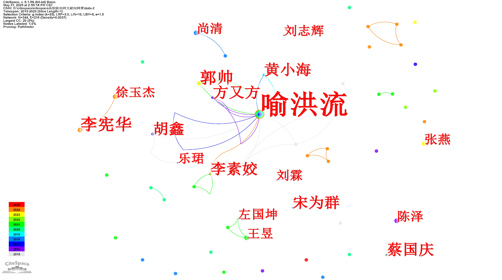
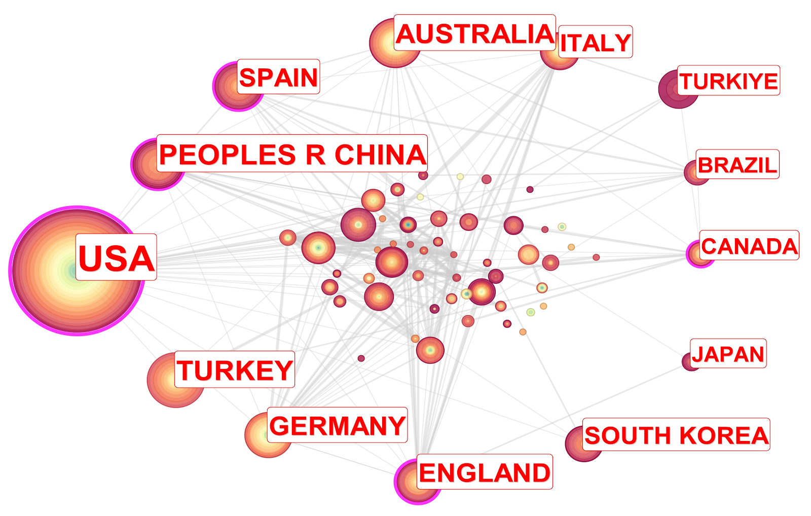
Scientometric deconstruction of developmental dynamics in upper-limb rehabilitation robotics: evidence network analysis via CiteSpace

Authors: Wang Feifei, Wang Zhennan

BACKGROUND: Upper limb rehabilitation robots have emerged as an indispensable component in global healthcare systems. Despite the extensive body of research in this field, which primarily focuses on areas such as development and application, there remains a significant need for comprehensive, systematic literature analysis to thoroughly examine the current research landscape, identify emerging hotspots, and predict future trends in this domain. OBJECTIVE: To conduct a visual analysis of research status, hotspots, and trends in the field of upper limb rehabilitation robots over the past decade using CiteSpace software, with the aim of identifying key research directions and providing intuitive references for researchers. METHODS: This study systematically retrieved literature related to upper-limb rehabilitation robotics from CNKI and the Web of Science Core Collection published between January 1, 2015, and March 13, 2025. CiteSpace 6.1.R1 software was employed for visualized analysis of included literature, covering key dimensions such as publication volume, authors, institutions, keywords, clusters, and bursts. RESULTS AND CONCLUSION: (1) A total of 1 054 articles were included, involving 659 authors. The United States held the highest overall ranking in terms of both research publication volume and centrality in this field. Among the contributing institutions, Northeast University and Univ Shanghai Sci&Technol ranked the highest in publication volume. Visualization analysis indicated an overall upward trend in upper limb rehabilitation robotics research, yet revealed limited collaboration among researchers and institutions. Main research directions included core technical methods, robot design and optimization, control strategies and algorithms, clinical application and evaluation, and emerging technologies and interdisciplinary integration. (2) Future research could focus on technological integration and intelligent upgrading, interdisciplinary collaborative innovation, and improvement of clinical applications to promote the continuous development and refinement of the upper limb rehabilitation robot field.

Scientometric deconstruction of developmental dynamics in upper-limb rehabilitation robotics: evidence network analysis via CiteSpace
Graphical Abstract
Original ResearchVol 1898, Issue 26 • pp. 100-112DOI: 10.12307/2026.21308Jan 15, 2026

Keloid pathogenesis is correlated with fibroblast heterogeneity genes: single-cell transcriptomic analysis based on GEO database

Authors: Guo Tao, Liu Yuxin, Yan Meirong, Wang Xiaoni

BACKGROUND: Keloid is a chronic fibrotic skin disorder driven by abnormal fibroblast activation and dysregulated immune responses. However, its underlying molecular mechanisms remain largely unclear. With the advancement of single-cell transcriptomic technologies, integrating public databases with systematic bioinformatics analyses offers new opportunities to identify diagnostic biomarkers and therapeutic targets. OBJECTIVE: To identify key biomarkers associated with fibroblast heterogeneity and immune cell interactions in the pathogenesis of keloids. METHODS: Single-cell transcriptomic dataset GSE181297 and bulk transcriptomic dataset GSE14572 were retrieved from the Gene Expression Omnibus (GEO) public database. Cell subtypes were annotated and analyzed for changes in cellular composition. Pseudotime analysis was applied to infer differentiation trajectories of various cell populations. Weighted gene co-expression network analysis and differential gene expression analysis were conducted to identify fibroblast-related differentially expressed genes. Functional enrichment analyses, including Gene Ontology and Kyoto Encyclopedia of Genes and Genomes, were used to determine the involved biological processes and pathways. Protein-protein interaction network analysis, combined with three machine learning algorithms, was employed to identify hub genes. Receiver operating characteristic curve analysis was conducted to assess the diagnostic value of the candidate biomarkers. The expression patterns and correlation of hub genes in immune cells were evaluated. RESULTS AND CONCLUSION: Single-cell transcriptomic analysis revealed significantly increased proportions of endothelial cells, fibroblasts, smooth muscle cells, T cells, mast cells, macrophages, and lymphatic endothelial cells in keloid tissues, with fibroblasts being the predominant cell type. Pseudotime analysis showed that fibroblasts were mainly distributed in states 1, 2, and 3, at the initial stage of differentiation, with high developmental potential. Differential analysis identified 80 fibroblast-related differentially expressed genes, mainly enriched in regionalization, skeletal system morphogenesis, and embryonic skeletal development pathways. Integrating protein-protein interaction network and multiple machine learning models, HOXC4 was finally identified as a key biomarker. Receiver operating characteristic curve analysis indicated that HOXC4 had good diagnostic performance and was highly expressed in keloid tissues. Correlation analysis showed that HOXC4 was significantly positively correlated with resting natural killer cells, while negatively correlated with activated dendritic cells and activated natural killer cells. This study systematically revealed the heterogeneity of keloid fibroblasts and their association with the immune microenvironment. HOXC4 was identified as a key biomarker related to fibroblast functional status and immune regulation, providing a potential new target for early diagnosis and targeted therapy of keloids.

Keloid pathogenesis is correlated with fibroblast heterogeneity genes: single-cell transcriptomic analysis based on GEO database
Graphical Abstract
Original ResearchVol 1898, Issue 26 • pp. 100-112DOI: 10.12307/2026.21307Jan 15, 2026

Multi-omics approach unveils novel therapeutic targets for osteoporosis: integrated analysis of Asian and European gene-tissue expression consortium data

Authors: Chen Yongxi

BACKGROUND: With the acceleration of China's aging population, the number of osteoporosis patients has been increasing significantly. Recent advancements in genome-wide association studies and single-cell transcriptomic sequencing have empowered researchers to identify novel osteoporosis-associated genes through integrative multi-omics analyses. OBJECTIVE: To identify potential therapeutic targets for osteoporosis using summary data-based Mendelian randomization approaches that integrate genome-wide association studies and transcriptomic data from Asian and European populations. METHODS: By integrating cis-expression quantitative trait loci (cis-eQTL) and protein quantitative trait loci (pQTL) datasets from multiple tissues (blood and muscle-bone) with osteoporosis genome-wide association study data (the 2021 European population osteoporosis GWAS data from FinnGen and the 2020 East Asian population GWAS from Biobank Japan), we employed summary data-based Mendelian randomization (SMR) to identify osteoporosis-associated genes. Colocalization analysis, single-cell sequencing, and enrichment analysis were performed for further validation. All data were obtained from published studies or publicly available databases with ethical approval and informed consent. RESULTS AND CONCLUSION: SMR analysis identified 64 genes significantly associated with osteoporosis (after removing duplicates), among which HLA-DQA1, HLA-DQA2, HLA-DQB1, HLA-DQB2, and HLA-DRB5 were validated in both outcome datasets. Colocalization analysis provided evidence for HLA-DQA2 and HLA-DQB1 (posterior probability PPH4 > 0.8). Plasma levels of HLA-DQA2 were associated with reduced osteoporosis risk. Single-cell analysis revealed increased abundance of dendritic cells, B cells, macrophages, and neutrophils in the osteoporotic immune microenvironment. Enrichment analysis showed that identified genes were enriched in MHC class II antigen presentation pathway. This study identified several previously unreported osteoporosis-associated genes through bioinformatics integration of Asian and European GWAS data, warranting further exploration as potential therapeutic targets.

Multi-omics approach unveils novel therapeutic targets for osteoporosis: integrated analysis of Asian and European gene-tissue expression consortium data
Graphical Abstract
Original ResearchVol 1898, Issue 26 • pp. 100-112DOI: 10.12307/2026.21310Jan 15, 2026

Visual analysis of the research literature on plantar fasciitis

Authors: Wang Fei, Wang Hangping, Dai Rongjuan, Gong Lingxuan

BACKGROUND: Existing research on plantar fasciitis predominantly focuses on single techniques, short-term efficacy, pathogenesis, and diagnostic/evaluation studies. Systematic integration of global research trends and core hotspots is lacking. OBJECTIVE: To analyze the current status, hotspots, and trends in research on plantar fasciitis. METHODS: Search terms and free-text keywords related to plantar fasciitis were retrieved from PubMed to construct a search strategy. Relevant literature published between 1996 and 2025 was retrieved from the Web of Science Core Collection database. CiteSpace software, Excel, and Sci Explorer were used to perform co-occurrence analysis, salience analysis, and clustering analysis on countries, authors, institutions, disciplines, journals, keywords, and cited literature within this field. RESULTS AND CONCLUSION: A total of 1 606 articles were included in this visual analysis. The number of publications in plantar fasciitis research showed a fluctuating upward trend from 1996 to 2025. The United States was the most prolific country, contributing 423 articles (27.10% of the total). The institution with the highest output was Harvard University (USA) with 36 publications. The most prolific author was Landorf KB from La Trobe University with 33 publications. The most influential journal was Foot & Ankle International, with 112 articles and 950 total citations. The most influential paper was 'Shock Wave Therapy for Chronic Proximal Plantar Fasciitis'. In the field of plantar fasciitis, major treatment hotspots include extracorporeal shock wave therapy, platelet-rich plasma, and corticosteroid injections, but the long-term effectiveness, safety, cost, and standardized treatment protocols remain controversial and are current clinical research hotspots. In diagnosis, shear wave elastography is a hotspot for diagnostic evaluation, and more objective and quantitative diagnostic criteria are current research focuses. In mechanism exploration, research includes the biomechanical chain of vertical pressure transmission from hip-knee-ankle-foot and its influence on the occurrence of plantar fasciitis.

Visual analysis of the research literature on plantar fasciitis
Graphical Abstract
Original ResearchVol 1898, Issue 26 • pp. 100-112DOI: 10.12307/2026.21300Jan 15, 2026

Traditional Chinese Medicine compound formula and effective ingredients for treating intervertebral disc degeneration: a multi-targeted and holistic regulation concept

Authors: ZHONG Haotian, LI Jianwen, HAN Weichao, LI Songbo

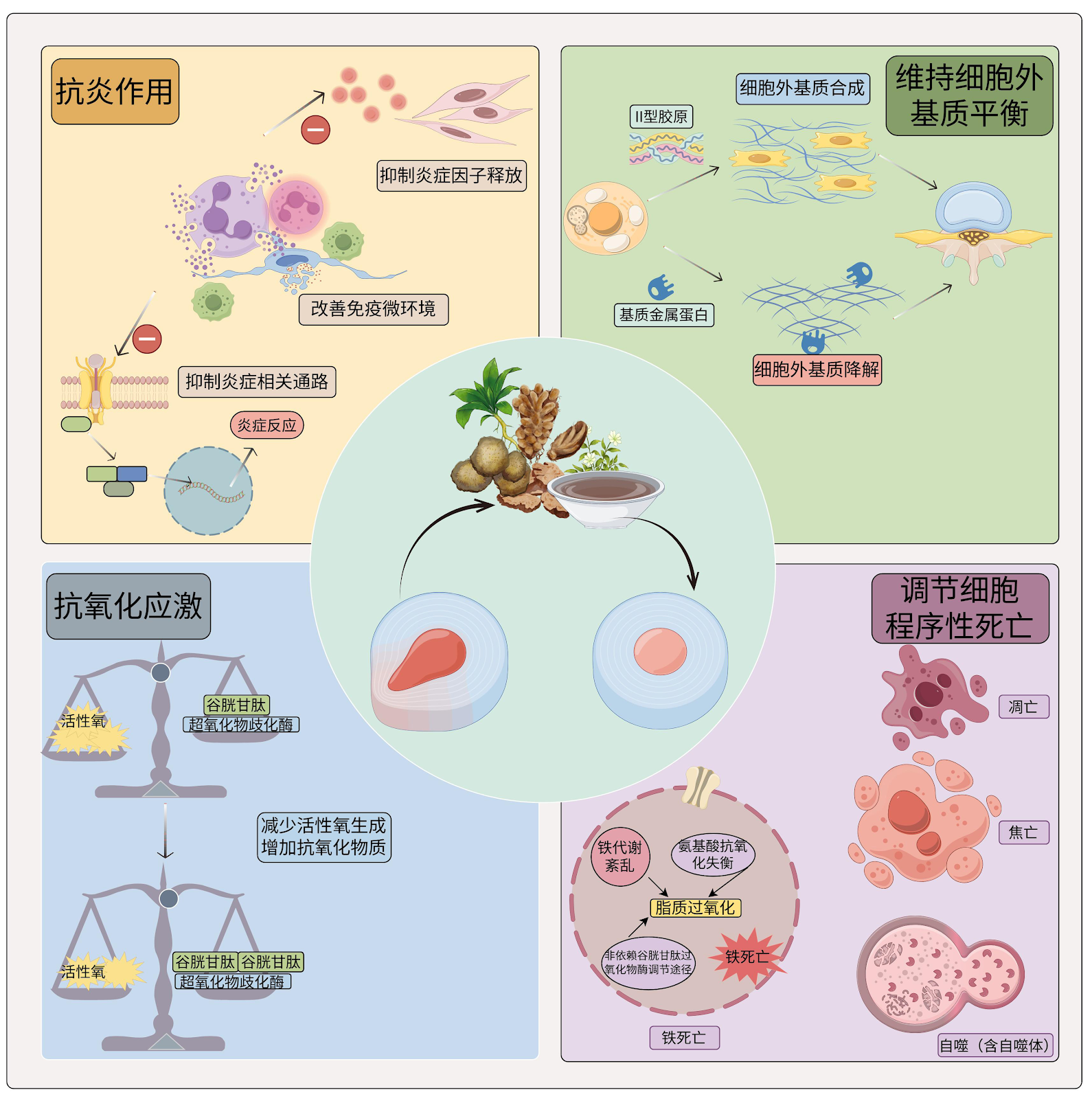
BACKGROUND: Traditional Chinese Medicine (TCM) has significant potential and benefits in the treatment of intervertebral disc degeneration. However, reviews on the mechanisms by which TCM ameliorates disc degeneration are relatively limited. OBJECTIVE: To review how TCM compounds and their active ingredients improve intervertebral disc degeneration through mechanisms such as anti-inflammation, antioxidative stress, maintenance of extracellular matrix balance and regulation of programmed cell death. METHODS: CNKI, WanFang, PubMed, and Web of Science were retrieved for relevant literature published from January 2001 to June 2025. The Chinese search terms were “Chinese herb, compound, traditional Chinese medicine, herbal medicine, soup, formula, degenerative disc disease, disc degeneration, disc injury, annulus fibrosus, cartilage endplates, nucleus pulposus, research, progress, review, experiment, inflammation, oxidative stress, extracellular matrix, programmed cell death, apoptosis, pyroptosis, ferroptosis, autophagy, immune cells, mechanism”, and English search terms were “Traditional Chinese medicine, herbal medicine, herbal formula, Intervertebral Disc Degeneration, Intervertebral Disc, disc degeneration, nucleus pulposus, annulus fibrosus, cartilage endplate, Oxidative Stress, Extracellular Matrix, Inflammation, Programmed Cell Death, Apoptosis, Pyroptosis, Ferroptosis, Autophagy, review, progress, experiment”. According to inclusion and exclusion criteria, 104 articles were finally included for review. RESULTS AND CONCLUSION: TCM can effectively inhibit the release of inflammatory factors and activation of inflammatory pathways through multiple mechanisms, improving the local inflammatory state of the intervertebral disc, but the specific mechanisms in immune cell-mediated inflammatory responses remain unclear. TCM shows strong multi-target regulatory ability in alleviating oxidative stress in the intervertebral disc, especially in enhancing antioxidant enzyme activity, stabilizing mitochondrial function, and inhibiting reactive oxygen species accumulation. However, systematic comparisons of antioxidant effects of different active ingredients in different disc cell types are lacking, and the crosstalk between related signaling pathways needs further exploration. TCM can effectively alleviate disc structural disruption by regulating extracellular matrix synthesis and degradative enzyme activity. Apoptosis, pyroptosis, ferroptosis, and autophagy collectively participate in the development of intervertebral disc degeneration; they are not isolated but interact through complex signaling networks. Therefore, the multi-target and holistic regulatory advantages of TCM are expected to simultaneously regulate multiple programmed cell death processes, protecting disc cells from multiple levels and delaying degeneration.

Traditional Chinese Medicine compound formula and effective ingredients for treating intervertebral disc degeneration: a multi-targeted and holistic regulation concept
Graphical Abstract
Original ResearchVol 1898, Issue 26 • pp. 100-112DOI: 10.12307/2026.21303Jan 15, 2026

Effects of antioxidant pretreatment on skeletal muscle damage and oxidative stress following acute high-intensity exercise: a meta-analysis

Authors: Xia Caigui, Li Wei, Su Yuying, Shi Yu, Yang Zhonghe

OBJECTIVE: Current evidence indicates that exercise-induced oxidative stress involves a dual role of reactive oxygen species, which participate in exercise adaptation while potentially causing tissue damage, highlighting the necessity for precise regulation of antioxidant dosage and timing. This study employs a Meta-analytic approach to systematically evaluate the effects of antioxidant pretreatment on biomarkers of skeletal muscle oxidative stress injury following acute strenuous exercise, and to explore the moderating effects of dosage, intervention duration, and training status. METHODS: A systematic search was conducted for randomized controlled trials that investigated the effects of antioxidant pretreatment on exercise-induced oxidative stress in PubMed, Web of Science, EBSCO, CNKI, VIP and WanFang databases from inception to February 2025. Literature quality was assessed using the physiotherapy evidence database scale. Data analysis was performed using RevMan 5.4 and Stata statistical software. RESULTS: (1) This meta-analysis included 16 studies from 12 publications, comprising 264 athletes and regularly exercising individuals. (2) The physiotherapy evidence database scale scores ranged from 6-8 (7 studies) to 9 (5 studies), indicating overall high methodological quality. (3) Meta analysis results showed that antioxidant pretreatment significantly decreased post-exercise serum creatine kinase [standardized mean difference (SMD)=-0.31, 95% confidence interval (CI) (-0.63, 0.00), P=0.05], interleukin-6 [SMD=-0.66, 95%CI (-1.03, -0.29), P=0.0005], and malondialdehyde levels [SMD=-1.10, 95%CI (-1.96, -0.23), P=0.01], and increased glutathione peroxidase activity [SMD=1.33, 95%CI (0.87, 1.78), P < 0.00001] and total antioxidant capacity [MD=4.77, 95%CI (3.87, 5.67), P < 0.00001]. Subgroup analysis showed that low-dose (≤500 mg/d) short-term (≤14 d) intervention had a more significant inhibitory effect on malondialdehyde levels (SMD=-1.15), while high-dose long-term intervention may inhibit exercise adaptation. Training status significantly moderated the effect size, with general athletes showing greater reduction in malondialdehyde levels than elite athletes (P < 0.05). CONCLUSION: Antioxidant pretreatment can effectively alleviate oxidative stress damage induced by acute strenuous exercise, but its effect is influenced by dosage, intervention duration, and training status. Short-term high-dose supplementation is suitable for rapid recovery during competition periods, while long-term application requires weighing antioxidant benefits against the risk of adaptation inhibition.

Effects of antioxidant pretreatment on skeletal muscle damage and oxidative stress following acute high-intensity exercise: a meta-analysis
Graphical Abstract
Original ResearchVol 1898, Issue 26 • pp. 100-112DOI: 10.12307/2026.21304Jan 15, 2026

Traditional Chinese sports intervene in sarcopenia and its complications in the elderly: a meta-analysis on improving muscle strength, mass, and physical function

Authors: Chu Rui, Li Mingming, Xie Yeshou, Ni Tao, Du Yinuo

OBJECTIVE: Muscle function decline and physical impairment caused by sarcopenia and its complications increase the risk of adverse health outcomes in the elderly. However, systematic evidence on the efficacy of traditional Chinese sport interventions for these conditions remains insufficient. Therefore, this study employs meta-analysis to systematically evaluate the effectiveness of traditional Chinese sports in treating sarcopenia and its complications. METHODS: Following the Preferred Reporting Items for Systematic reviews and Meta-Analyses (PRISMA) guidelines, we systematically searched PubMed, Web of Science, Cochrane Library, CNKI, and Wanfang databases to choose randomized controlled trials evaluating traditional Chinese sport interventions for sarcopenia and its complications. Experimental group received one or more traditional Chinese sports, including Yijinjing, Tai Chi, and Baduanjin, while control group received health education, usual care, or remote Tai Chi instruction. Outcome measures included handgrip strength, knee muscle strength, appendicular skeletal muscle mass index, sit-to-stand test, gait speed, timed up-and-go test, and short physical performance battery. Meta-analysis was performed using Revman 5.4 and Stata 15.1 software, and subgroup analyses were conducted to identify optimal intervention parameters. RESULTS: A total of 21 studies (n=1,313) were included. Meta-analysis showed that traditional Chinese sports significantly improved handgrip strength (MD=1.83, 95%CI: 1.65, 2.01, P < 0.00001), knee muscle strength (MD=5.98, 95%CI: 3.85, 8.11, P < 0.00001), appendicular skeletal muscle mass index (MD=0.22, 95%CI: 0.10, 0.34, P=0.0004), gait speed (MD=0.12, 95%CI: 0.09, 0.16, P < 0.00001), sit-to-stand test performance (MD=1.92, 95%CI: 1.41, 2.43, P < 0.00001), timed up-and-go test performance (SMD=-1.00, 95%CI: -1.25, -0.74, P < 0.00001), and short physical performance battery score (MD=1.12, 95%CI: 0.77, 1.46, P < 0.00001). Subgroup analyses revealed that for improving handgrip strength, Yijinjing (intervention >12 weeks, >3 times/week, ≤30 min/session) was most effective; for improving appendicular skeletal muscle mass index, Baduanjin (12 weeks, ≤3 times/week, >30 min/session) was optimal; for improving gait speed, both Tai Chi (>12 weeks, >3 times/week, ≤30 min/session) and Baduanjin (12 weeks, 3 times/week, >30 min/session) were effective; for improving sit-to-stand test performance, Yijinjing (8 weeks, 5 times/week, ≤40 min/session) was best; and for improving timed up-and-go test performance, Baduanjin (>12 weeks, ≤3 times/week, >30 and ≤40 min/session) was most significant. CONCLUSION: Traditional Chinese sports can effectively improve handgrip strength, knee muscle strength, appendicular skeletal muscle mass, and physical function in elderly patients with sarcopenia and its complications, with specific effects under different intervention parameters. It is recommended to prescribe personalized exercise programs based on target outcomes.

Traditional Chinese sports intervene in sarcopenia and its complications in the elderly: a meta-analysis on improving muscle strength, mass, and physical function
Graphical Abstract
Original ResearchVol 1898, Issue 26 • pp. 100-112DOI: 10.12307/2026.21305Jan 15, 2026

Network meta-analysis of different virtual reality devices for treating upper limb motor dysfunction after stroke

Authors: NIE Yue, SONG Shuhua, ZHAO Shengting, DONG Yangyang, YANG Bingxin

OBJECTIVE: Upper limb dysfunction after stroke is a common complication that seriously affects the quality of life and daily activity of patients. Virtual reality technology, as an emerging rehabilitation method, can effectively promote neural remodeling and functional recovery. This study will systematically evaluate the therapeutic effects of different virtual reality devices on upper limb motor dysfunction in patients with stroke. METHODS: The China National Knowledge Infrastructure (CNKI), WanFang Database, VIP website, PubMed, Web of Science, Embase, and the Cochrane Library were searched to retrieve relevant literature. Relevant data were extracted, and their quality was assessed. The control group received conventional rehabilitation treatment, while the experimental group received virtual reality rehabilitation training in addition to the treatment provided to the control group. Statistical analysis was performed using RevMan 5.4 and Stata 18.0 software. RESULTS: (1) A total of 12 articles and 571 patients were included in the meta-analysis. (2) Meta-analysis results showed that the Fugl-Meyer score of the upper limb in the virtual reality group was 7.29 times that of the conventional group (MD=7.29, 95%CI: 5.60-8.98, P < 0.05); the Action Research Arm Test score in the virtual reality group was 10.69 times that of the conventional group (MD=10.69, 95%CI: 4.96-16.43, P < 0.05); the modified Barthel index score in the virtual reality group was 8.25 times that of the conventional group (MD=8.25, 95%CI: 3.38-13.12, P < 0.05). (3) Subgroup analysis showed that patients aged 50-59 years had better improvement in upper limb Fugl-Meyer score; patients with disease duration within 3 months had better improvement; intervention duration ≥4 weeks had the best improvement. (4) Network meta-analysis showed that smart glove intervention [MD=-1.05, 95%CI(-1.85, -0.24), P < 0.05] was most effective for improving upper limb motor function; Armeo Spring intervention [MD=-1.19, 95%CI(-1.87, -0.51), P < 0.05] was most effective for improving upper limb coordination; Kinect intervention [MD=-0.59, 95%CI(-1.13, -0.06), P < 0.05] was most effective for improving hand dexterity; VREX intervention [MD=-0.76, 95%CI(-1.28, -0.23), P < 0.05] was most effective for improving activities of daily living. CONCLUSION: For improving upper limb motor function, the smart glove system is the first choice; for improving upper limb coordination, the Armeo Spring system is the first choice; for improving hand dexterity, the Kinect system is the first choice; for improving activities of daily living, the VREX system is the first choice. This study has certain limitations, and the above conclusions should be interpreted with caution.

Network meta-analysis of different virtual reality devices for treating upper limb motor dysfunction after stroke
Graphical Abstract
Original ResearchVol 1898, Issue 26 • pp. 100-112DOI: 10.12307/2026.21302Jan 15, 2026

Exercise-intestinal flora and aging

Authors: WANG Yinfeng, YAO Lijuan, MA Zhennan, CHEN Leqin

BACKGROUND: The benefits of exercise as a classical intervention for aging have been widely recognized. The homeostatic balance of a wide range of microorganisms in the intestinal flora indirectly regulates aging, and the bidirectional association between exercise and the intestinal flora can collectively influence the process of aging. OBJECTIVE: To sort out the effects of exercise, intestinal flora and their interactions on aging, and to explore the specific physiological mechanisms involved. METHODS: A computer-based search in CNKI, WanFang, VIP, PubMed, MedReading, and Web of Science, with the time limit of 1976-01-01/2025-02-28, was conducted to collect the relevant studies on the effects of exercise and intestinal flora on aging. The search terms were “intestinal flora, gut microbiota, physical exercise, age, aerobic exercise, resistance exercise, low intensity exercise, moderate intensity exercise, high intensity exercise” in Chinese and English. RESULTS AND CONCLUSION: (1) Exercise and intestinal flora are both means to intervene in aging, and the combined benefits of exercise and gut microbiota in intervening in aging are even more pronounced. (2) Exercise changes the composition and function of intestinal flora, stimulates intestinal production of short-chain fatty acids, regulates host metabolism and immune function, reduces inflammatory response, and promotes the synthesis of vitamins and neurotransmitters. (3) The specific manifestations of aging, when the intestinal flora is regulated via different exercise modes, are different. (4) Different gut-organ axis regulated by exercise has different effects on aging.

Exercise-intestinal flora and aging
Graphical Abstract
Original ResearchVol 1898, Issue 26 • pp. 100-112DOI: 10.12307/2026.21297Jan 15, 2026

The interaction and balance between cellular senescence and tissue repair

Authors: Kan Weiyi, Wang Linrong, Cheng Leping

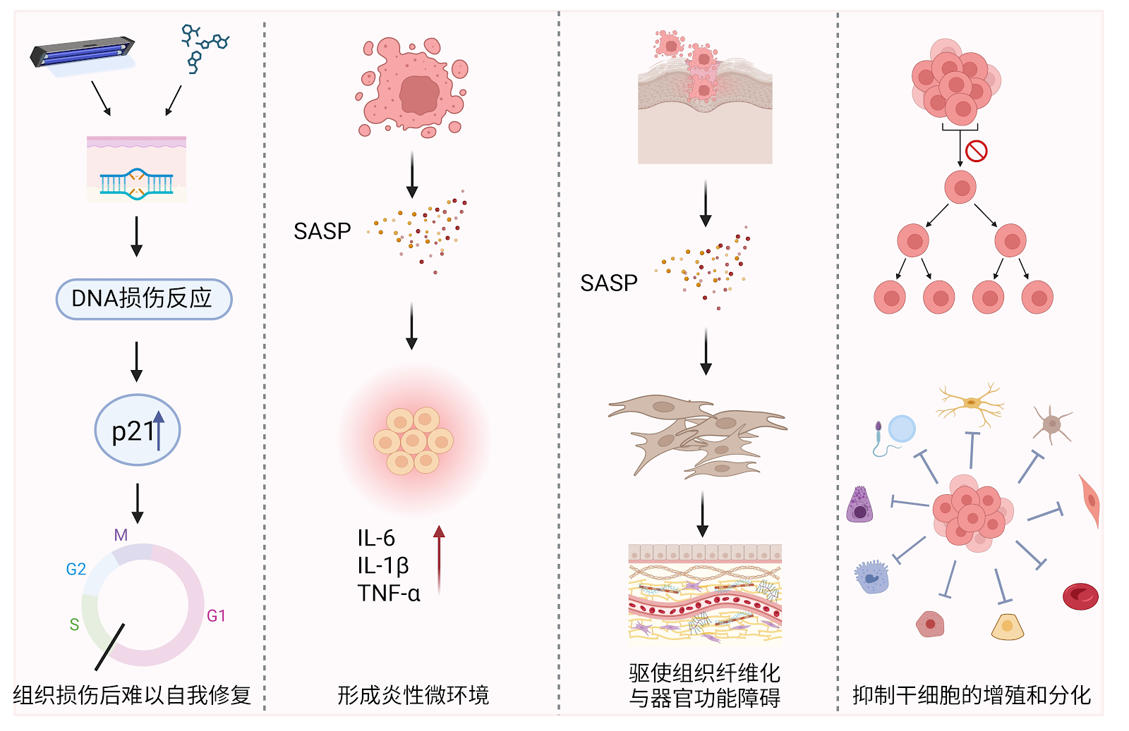
Background: The role of cellular senescence in tissue injury repair has attracted increasing attention. The accumulation of senescent cells not only affects normal physiological functions but also exacerbates inflammatory responses and fibrosis, thereby accelerating the aging process. In recent years, the potential therapeutic value of clearing senescent cells in tissue repair has become a research hotspot. Objective: To review the dual role of cellular senescence in tissue injury repair and explore the application prospects of senescent cell clearance strategies in delaying aging and promoting tissue repair. Methods: The authors searched PubMed for relevant literature up to May 2025 using English search terms "Senolytics, aging, senescence, treatment, clearance, tissue injury". After initial screening by reading titles and abstracts, and excluding irrelevant, outdated, or repetitive studies, 137 articles were selected for analysis. Results and Conclusion: Cellular senescence is an important hallmark of organismal aging. Senescent cells secrete senescence-associated secretory phenotype (SASP) factors, affecting local tissue repair and regeneration. Clearing senescent cells can alleviate chronic inflammation, promote tissue functional recovery, and delay the onset of aging-related diseases. Current strategies for clearing senescent cells include non-targeted and targeted approaches. Non-targeted strategies delay aging through lifestyle modifications, while targeted strategies selectively eliminate senescent cells via drugs, transgenic techniques, gene editing, reprogramming, and immunotherapy, thereby delaying the aging process.

The interaction and balance between cellular senescence and tissue repair
Graphical Abstract
Original ResearchVol 1898, Issue 26 • pp. 100-112DOI: 10.12307/2026.21301Jan 15, 2026

Mechanisms by which exercise regulates gut microbiota in the prevention and treatment of non-alcoholic fatty liver disease

Authors: WANG Wei, CHEN Jun, JIA Shaohui, XUE Xinxuan, DONG Kunwei

BACKGROUND: The pathogenesis of non-alcoholic fatty liver disease is closely associated with gut microbiota dysbiosis. In recent years, accumulating evidence has indicated that exercise may exert beneficial effects on host metabolic homeostasis by modulating the composition and function of the gut microbiota, thereby playing a positive role in the prevention and treatment of non-alcoholic fatty liver disease. OBJECTIVE: To systematically summarize current research progress on the interplay between gut microbiota and non-alcoholic fatty liver disease, to further elucidate the regulatory effects of exercise on gut microbiota, and to explore in depth the potential mechanisms by which exercise intervention may prevent or ameliorate non-alcoholic fatty liver disease via the "gut–liver axis." METHODS: Search terms included "non-alcoholic fatty liver disease," "gut microbiota," "exercise," "bile acid," "short-chain fatty acid," "lipopolysaccharide," "trimethylamine oxide," and "indole" in Chinese and English, respectively. China National Knowledge Infrastructure (CNKI), and WanFang Database were searched for relevant studies published up to March 2025. A total of 85 core studies were identified based on the inclusion and exclusion criteria. RESULTS AND CONCLUSION: (1) The composition of gut microbiota in patients with non-alcoholic fatty liver disease is significantly abnormal, with increased abundance of pro-inflammatory bacteria (such as Proteobacteria, Escherichia coli, and Streptococcus) and pathogenic bacteria (such as Enterobacteriaceae), while the abundance of anti-inflammatory and homeostatic bacteria (such as Ruminococcus and Faecalibacterium) is decreased. These adverse changes in microbial composition may promote the entry of metabolites into the liver by increasing intestinal permeability, activating inflammatory pathways, and increasing endogenous ethanol production, thereby driving the pathological progression of non-alcoholic fatty liver disease. (2) Modulating gut microbiota through probiotic supplementation or fecal microbiota transplantation can effectively reduce transaminase levels and chronic inflammation in patients with non-alcoholic fatty liver disease, suggesting that gut microbiota may be an important target for the prevention and treatment of non-alcoholic fatty liver disease. (3) Exercise can regulate gut microbiota composition, increase the abundance of beneficial bacteria, reduce the abundance of pro-inflammatory bacteria, and promote the activation of key metabolic pathways, thereby improving host metabolic health. However, current research on the effects of exercise on gut microbiota in patients with non-alcoholic fatty liver disease remains relatively limited, especially the effects and mechanisms of different exercise types, intensities, and durations on gut microbiota and host metabolism are still unclear. (4) Exercise may regulate gut microbiota and increase short-chain fatty acid production to activate G protein-coupled receptors 41/43 and AMP-activated protein kinase pathways, inhibit histone deacetylase activity, thereby reducing hepatic fat accumulation, alleviating liver inflammation, and decreasing insulin resistance; exercise may regulate the gut microbiota-bile acid axis to improve bile acid metabolism, thereby mediating the farnesoid X receptor/G protein-coupled bile acid receptor 5 signaling pathway to prevent and treat non-alcoholic fatty liver disease; exercise may reshape gut microbiota to reduce the abundance of lipopolysaccharide-containing Gram-negative bacteria and improve intestinal barrier function to reduce lipopolysaccharide production and translocation, preventing and treating non-alcoholic fatty liver disease; exercise may regulate gut microbiota composition to enhance the synthesis of indole and its derivatives, while inhibiting the production of ethanol and trimethylamine oxide, thereby enhancing liver metabolic capacity, improving intestinal barrier function, and reducing liver inflammation, playing a positive role in the prevention and treatment of non-alcoholic fatty liver disease.

Mechanisms by which exercise regulates gut microbiota in the prevention and treatment of non-alcoholic fatty liver disease
Graphical Abstract
Original ResearchVol 1898, Issue 26 • pp. 100-112DOI: 10.12307/2026.21306Jan 15, 2026

Non-invasive brain stimulation for core symptoms in children with autism spectrum disorder: a network meta-analysis

Authors: Fang Enhui, Guan Hui, Ma Lihong

OBJECTIVE: Non-invasive brain stimulation has been shown to improve restricted, repetitive behaviors and social deficits in children with autism; however, the efficacy of different stimulation protocols varies. This study systematically evaluated the efficacy of non-invasive brain stimulation on core symptoms in children with autism and compared the efficacy of different stimulation protocols. METHODS: Comprehensive electronic searches were conducted across CNKI, VIP, WanFang, CBM, PubMed, Embase, Cochrane Library, and Web of Science from database inception through March 2025 to identify randomized controlled trials evaluating non-invasive brain stimulation protocols targeting core symptoms in children with autism spectrum disorder. Two independent reviewers performed dual-phase screening, data extraction, and methodological quality assessment using the Cochrane Risk of Bias Tool version 2.0. Both conventional and network meta-analyses were implemented through Revman 5.4 and Stata 17.0. RESULTS: A total of 27 studies were finally included for review, involving 10 stimulation protocols of non-invasive brain stimulation and including 1 701 children with autism. (1) The results of conventional Meta-analysis showed that non-invasive brain stimulation was more effective than conventional rehabilitation in lowering the scores of Childhood Autism Rating Scale, Autism Behavior Checklist, Autism Treatment Evaluation Checklist, and Repetitive Behavior Scale-Revised. (2) The network meta-analysis showed that compared with conventional rehabilitation, high-frequency repetitive transcranial magnetic stimulation over the dorsolateral prefrontal cortex [MD=-6.00, 95%CI(-8.68, -3.33), P < 0.05, SUCRA=89.5%] was most effective in improving Childhood Autism Rating Scale scores, while high-frequency repetitive transcranial magnetic stimulation over Broca's area [MD=-15.11, 95%CI(-18.28, -11.95), P < 0.05, SUCRA=91.1%] was most effective in improving Autism Behavior Checklist scores. CONCLUSION: Current evidence indicates that high-frequency repetitive transcranial magnetic stimulation is most effective in improving core symptoms in children with autism. Due to the dual limitations of methodological heterogeneity and limited sample size in the included studies, large-sample, methodologically rigorous randomized controlled trials are urgently needed to further verify the reliability of these conclusions.

Non-invasive brain stimulation for core symptoms in children with autism spectrum disorder: a network meta-analysis
Graphical Abstract
Original ResearchVol 1898, Issue 26 • pp. 100-112DOI: 10.12307/2026.21299Jan 15, 2026

Animal models of neurogenic heterotopic ossification: key disease progression and pathogenesis

Authors: Ren Qingsong, Xie Yulei, Liu Jingjing, Lin Jingyi, Long Danlei, Zhang Chunyu, Xie Liang, Zheng Kaiyuan, Wang Yinxu

BACKGROUND: Neurogenic heterotopic ossification frequently occurs within 1 to 3 months following spinal cord injury or traumatic brain injury, characterized by abnormal bone formation in periarticular soft tissues. The precise pathogenesis remains unclear, underscoring the urgent need for systematic research to inform clinical management. OBJECTIVE: To summarize recent advances in animal models of neurogenic heterotopic ossification and elucidate its underlying mechanisms, with a particular focus on the pathological differentiation of osteogenic precursor cells, remodeling of the local tissue microenvironment, and the interplay between neural regulation and neurogenic heterotopic ossification formation. METHODS: PubMed, CNKI, and SinoMed were searched from inception to January 2025. Chinese search terms included 'neurogenic heterotopic ossification, spinal cord injury, traumatic brain injury, heterotopic ossification'; English search terms included 'Neurogenic Heterotopic Ossification, spinal cord injury, Traumatic brain injury, ossification, heterotopic, Central nervous system'. Literature related to animal models and mechanisms of neurogenic heterotopic ossification was included to summarize key pathogenic processes. RESULTS AND CONCLUSION: The recruitment and aberrant osteogenic differentiation of osteogenic precursor cells (mainly fibro-adipogenic progenitors) are regulated by local microenvironmental factors such as hypoxia, inflammation, and angiogenesis. Neurotrophic factors, calcitonin gene-related peptide, and substance P promote aberrant ossification through neuro-immune interactions. Future research should construct a systematic molecular map, explore core signaling pathways, and develop novel targeted interventions to achieve early identification and individualized treatment of neurogenic heterotopic ossification, thereby improving patient outcomes.

Original ResearchVol 1898, Issue 26 • pp. 100-112DOI: 10.12307/2026.21296Jan 15, 2026

Mechanisms of miRNAs involved in cartilage development: new strategies and targets

Authors: WANG Zhengye, LIU Wanlin, ZHAO Zhenqun

BACKGROUND: The molecular regulation of cartilage development is one of the key scientific issues in the field of orthopedics. MicroRNAs (miRNAs), as important regulators of gene expression, regulate post-transcriptional silencing or translation inhibition by binding to the 3' untranslated region (UTR) of target mRNAs, and are involved in the regulation of cell differentiation, proliferation, and metabolic homeostasis. In recent years, studies have found that miRNAs play an important role in cartilage development and various cartilage-related diseases, and their abnormal expression is closely related to diseases such as skeletal dysplasia and osteoarthritis. In-depth research on the mechanisms of miRNAs in cartilage development is of great significance for understanding the fate determination of chondrocytes and the pathogenesis of related diseases. OBJECTIVE: To comprehensively summarize the mechanisms of miRNAs in cartilage development, explore their regulatory roles in chondrocyte differentiation, proliferation, and apoptosis, and their functions in disease states such as osteoarthritis and chondrodysplasia, providing a theoretical basis for the prevention and treatment of related diseases. METHODS: PubMed, CNKI, Wanfang, and VIP databases were searched from inception to June 2025, supplemented by manual retrieval of relevant books. High-quality literature on miRNA regulation of cartilage development was screened, prioritizing studies published within the last 10 years. A total of 99 articles (95 in English, 4 in Chinese) were included for systematic analysis and summary. RESULTS AND CONCLUSION: Studies have shown that miRNAs precisely regulate key transcription factors, signaling pathways, and epigenetic modifications, thereby influencing chondrocyte differentiation, proliferation, and apoptosis. In disease states, abnormal miRNA expression is closely related to various cartilage diseases. These findings not only reveal the direct regulatory role of miRNAs in cartilage development but also provide new strategies and targets for the diagnosis, prognosis, and treatment of related diseases. With the continuous development of cutting-edge technologies such as gene editing, single-cell sequencing, and bioinformatics, the application of miRNA research in cartilage development and diseases has made significant progress, promising new means for early diagnosis, precise treatment, and prognosis assessment of skeletal diseases.

Mechanisms of miRNAs involved in cartilage development: new strategies and targets
Graphical Abstract
Original ResearchVol 1898, Issue 26 • pp. 100-112DOI: 10.12307/2026.21284Jan 15, 2026

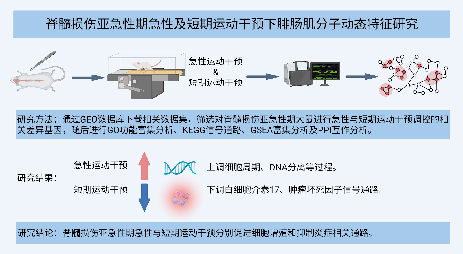
Molecular dynamic characteristics of rat gastrocnemius muscle under acute and short-term exercise intervention during the subacute phase of spinal cord injury

Authors: Wei Xinyi, Zheng Yan, Chen Qian, Ren Jiajia, Li Jian

BACKGROUND: Spinal cord injury triggers a cascade of neuro-muscular system damage, involving central pattern generator dysfunction and peripheral muscle molecular network disruptions. Exercise can regulate key genes and promote the recovery of spinal cord injury. OBJECTIVE: To identify exercise-regulated key genes through bioinformatics analysis and explore the mechanisms by which exercise intervention facilitates the recovery of spinal cord injury. METHODS: The GSE45550 dataset based on the GPL1355 platform was obtained from the Gene Expression Omnibus (GEO) database. Differentially expressed genes regulated by acute and short-term exercise interventions during the subacute phase of spinal cord injury in rats were identified. Gene Ontology functional enrichment analysis, Kyoto Encyclopedia of Genes and Genomes pathway analysis, and Gene Set Enrichment Analysis were performed on these differentially expressed genes, and a protein-protein interaction network was constructed. RESULTS AND CONCLUSION: (1) After acute exercise intervention in the subacute phase of spinal cord injury, 106 genes were upregulated and 97 genes were downregulated in the gastrocnemius muscle, whereas short-term exercise intervention resulted in 138 upregulated and 105 downregulated genes. (2) Gene Ontology analysis showed that acute exercise mainly enriched genes related to chromosome segregation, while short-term exercise mainly promoted signal transduction processes. (3) KEGG analysis indicated that acute exercise was mainly associated with gastric acid secretion and upregulation of motor protein pathways, whereas short-term exercise mainly involved upregulation of neuroactive ligand-receptor interaction and downregulation of some inflammatory signaling pathways. (4) GSEA analysis revealed that acute exercise mainly upregulated cell cycle and DNA separation, while short-term exercise mainly downregulated interleukin-17 signaling and tumor necrosis factor signaling pathways. (5) Protein-protein interaction network showed that acute and short-term exercise interventions formed 2 and 3 central modules, respectively. In summary, acute exercise intervention significantly activates cell proliferation-related pathways (such as cell cycle and mitosis), upregulating pro-proliferative genes like Top2a and Sele; whereas short-term intervention exerts anti-inflammatory effects by downregulating inflammatory pathways such as interleukin-17 and tumor necrosis factor, and downregulating factors like COMP. These findings provide a reference for the molecular mechanism research of spinal cord injury rehabilitation.

Molecular dynamic characteristics of rat gastrocnemius muscle under acute and short-term exercise intervention during the subacute phase of spinal cord injury
Graphical Abstract
Original ResearchVol 1898, Issue 26 • pp. 100-112DOI: 10.12307/2026.21287Jan 15, 2026

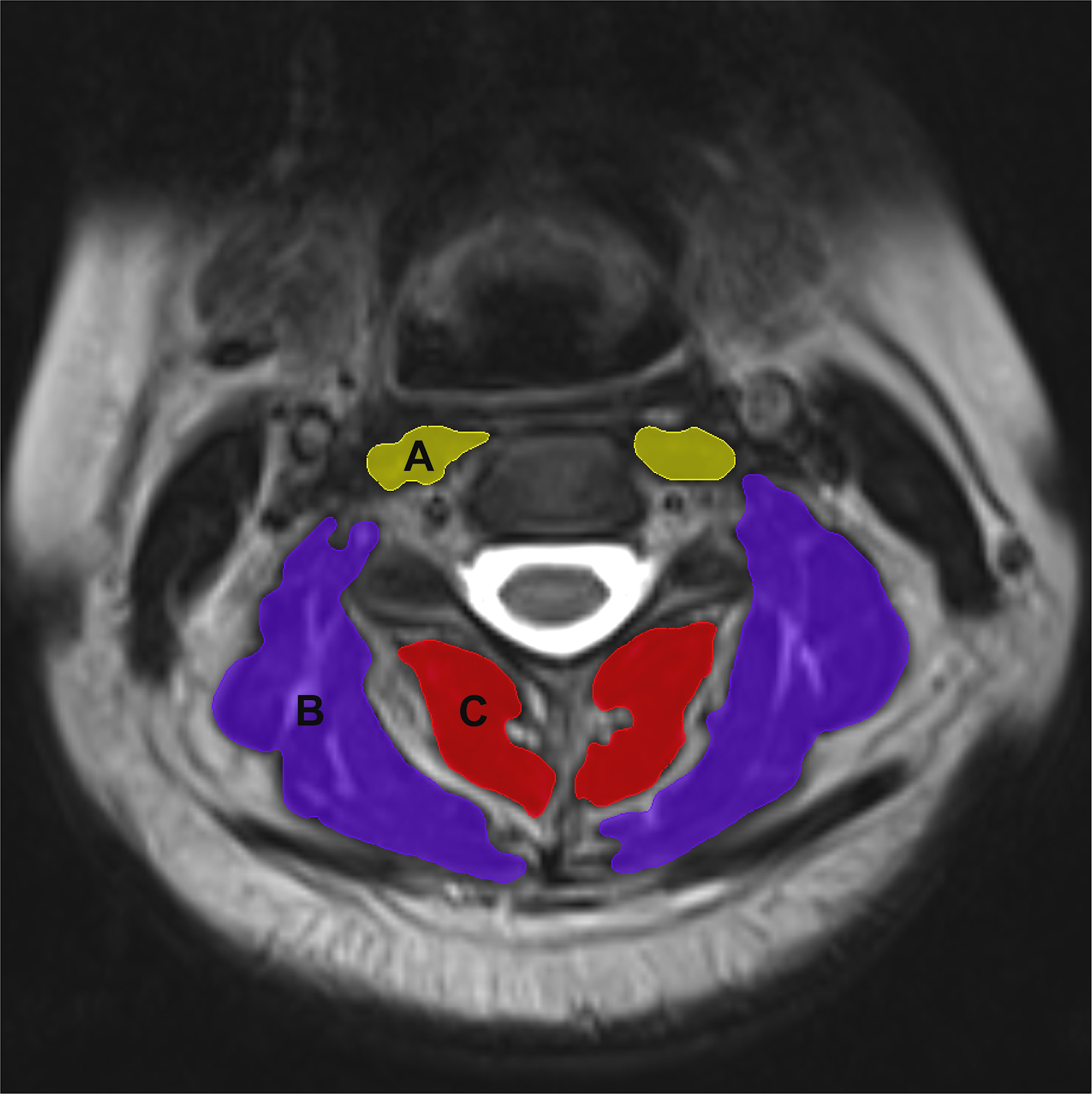
Correlation between cervical instability and neck muscle changes in middle-aged and young adults

Authors: Lu Guangqi, Li Jing, Sun Xinyue, Liang Long, Liu Guangwei, Zhou Shuaiqi, Mao Hanze, Ma Mingming, Cui Ying, Liu Yakun, Hu Jiaming, Zhu Liguo, Yu Jie, Zhuang Minghui

BACKGROUND: The onset of cervical instability in middle-aged and young adults often begins with neck muscle injuries. A deeper understanding of changes in neck muscles during cervical instability and their correlation can provide valuable data to support the prevention and treatment of cervical instability in this population. OBJECTIVE: To explore the correlation between cervical instability and neck muscle changes in middle-aged and young adults. METHODS: A total of 98 patients with cervical C4/5 instability and 88 healthy subjects, aged 18-45 years, were enrolled through recruitment advertisements and the Department of Spine, Wangjing Hospital, China Academy of Traditional Chinese Medicine. Cervical X-rays were collected to measure cervical curvature and C4/5 vertebral angular displacement. Cervical magnetic resonance imaging was taken to obtain data on C4/5 intervertebral disc signal intensity, as well as the relative cross-sectional area and fat ratio of neck muscles, including prevertebral muscles, deep posterior cervical muscles, and superficial muscles. A univariate intergroup comparison of X-ray and magnetic resonance imaging data was conducted between cervical instability subjects and healthy controls, along with Spearman correlation analysis between C4/5 angular displacement and disc signal intensity, relative cross-sectional area of neck muscles and fat percentage at the C4/5 level in cervical instability patients. RESULTS AND CONCLUSION: The cervical instability group had significantly greater age, C4/5 horizontal displacement, C4/5 angular displacement, and fat ratio of deep posterior cervical muscles than the healthy group (P < 0.05), while cervical curvature and relative cross-sectional area of deep posterior cervical muscles were significantly smaller (P < 0.05). Spearman correlation analysis showed a negative correlation between C4/5 angular displacement and relative cross-sectional area of deep posterior cervical muscles (P < 0.05). These findings suggest that changes in deep posterior cervical muscles may be closely related to the occurrence of cervical instability in middle-aged and young adults.

Correlation between cervical instability and neck muscle changes in middle-aged and young adults
Graphical Abstract
Original ResearchVol 1898, Issue 26 • pp. 100-112DOI: 10.12307/2026.21292Jan 15, 2026

Stroboscopic visual interference combined with balance training improves the balance ability of older adults

Authors: HU Zhe, SUN Yuxiang, HAN Xiao, LIU Yabin, SI Luyao, LI Zhouyu, JIA Yi

BACKGROUND: Numerous studies have indicated that stroboscopic visual interference combined with balance training can promote postural stability by reducing visual compensation in the central nervous system and increasing residual proprioceptive and vestibular input during training. OBJECTIVE: To clarify the effects of stroboscopic visual interference combined with balance training on improving balance ability of older adults by comparing the effects of balance training under different visual conditions. METHODS: Forty-three older adults were recruited and randomized into a normal balance training group (n=23) and a strobe vision training group (n=20). Among them, the balance training content was the same, and the training was conducted 3 times per week for 8 weeks. For the strobe vision training group, strobe glasses were worn during the balance training, and the strobe difficulty level (levels 1-8) was adjusted adaptively. Indicator tests were conducted at weeks 0, 4, 8, and 10 to assess the dynamic and static postural stability and the scores on the Berg Balance Scale of all subjects. RESULTS AND CONCLUSION: (1) Static postural stability: In the flat-surface eyes-closed single-leg stance test, both groups showed a significant time main effect (P < 0.001), but the group effect (P=0.530) and group×time interaction effect (P=0.780) were not significant. In the foam-surface eyes-closed single-leg stance test, both groups showed significant time (P < 0.001) and group (P=0.024) effects, but no group×time interaction effect (P=0.063). Compared with before training, both groups showed significant improvements in static postural stability after 8 weeks (P=0.034, P < 0.001) and 10 weeks (P=0.003, P < 0.001). (2) Dynamic postural stability: There was a significant group×time interaction effect (P < 0.001), while the main effects of group (Timed Up and Go and 3-m heel-to-toe walk: P=0.461, P=0.926) and time (P=0.120, P=0.937) were not statistically significant. Compared with before training, both groups showed significant improvements in dynamic postural stability after 4, 8, and 10 weeks of training (all P < 0.05). (3) Berg Balance Scale scores: There was a significant time effect (P < 0.001); group (P=0.420) and group×time (P=0.239) factors had no statistical significance on Berg Balance Scale scores. Compared with before training, both groups had higher Berg Balance Scale scores after 4 weeks (P=0.025), 8 weeks (P < 0.001), and 10 weeks (P=0.003). (4) It is suggested that both traditional balance training and strobe training can significantly improve the dynamic and static stability of older adults. Compared with traditional balance training, balance training based on stroboscopic visual interference can significantly improve the dynamic balance ability of older adults.

Stroboscopic visual interference combined with balance training improves the balance ability of older adults
Graphical Abstract
Original ResearchVol 1898, Issue 26 • pp. 100-112DOI: 10.12307/2026.21285Jan 15, 2026

Differences in angiogenesis and osteogenic effects between autogenous bone and mixed bone in guided bone regeneration

Authors: YANG Ning, HAN Zekui, WANG Xinyu, HUANG Yiping, HAN Jiaqi, WANG Yu, DUAN Feng

BACKGROUND: Angiogenesis is a key factor in the success of guided bone regeneration, but the impact of different bone graft materials on angiogenesis and osteogenic effects remains unclear. OBJECTIVE: To compare the angiogenesis and osteogenic effects of autologous bone powder and the mixture of autologous bone powder and artificial bone powder during guided bone regeneration. METHODS: Preoperative scanning of the jawbone data of New Zealand white rabbits using cone beam CT was performed to extract a rabbit jawbone model, which was then 3D printed to create a bone cutting guide plate. Twenty-one New Zealand white rabbits were used to construct a rabbit mandibular bone defect model. Each rabbit had two bone defect areas on both sides, one side was the autologous bone powder + artificial bone powder group, implanted with a mixture of autologous bone powder and artificial bone powder at a volume ratio of 1:1; the other side was the autologous bone powder group, implanted with autologous bone powder only. Angiogenesis and osteogenesis in the bone defect area were detected at 2, 5, 7, 14 days and 4, 8, 12 weeks after modeling. RESULTS AND CONCLUSION: (1) At 12 weeks postoperatively, the tissue morphology and angiogenesis of the autologous bone powder group and the autologous bone powder + artificial bone powder group were similar; (2) The number of vascular cross-sections in the autologous bone powder group showed a more significant upward trend at 2-7 days, while the trends of the two groups were similar in other periods; (3) At 8 weeks postoperatively, the distribution range of new bone in the autologous bone powder + artificial bone powder group was not as large as that in the autologous bone powder group. At 12 weeks, the continuity and density of bone tissue in both groups were better, close to normal bone tissue morphology; (4) At 8 weeks, the bone volume fraction of the autologous bone powder group was significantly higher than that of the autologous bone powder + artificial bone powder group (P < 0.05). At 12 weeks, there was no significant difference in bone volume parameters between the two groups (P > 0.05); (5) The results indicate that early angiogenesis and bone regeneration were more obvious with autologous bone powder implantation, but by 12 weeks postoperatively, the effect of mixed implantation of autologous bone powder and artificial bone powder was similar to that of autologous bone powder alone in repairing rabbit mandibular defects, indicating that with the progress of bone remodeling, the comprehensive effects of the two materials tend to be consistent.

Differences in angiogenesis and osteogenic effects between autogenous bone and mixed bone in guided bone regeneration
Graphical Abstract
Original ResearchVol 1898, Issue 26 • pp. 100-112DOI: 10.12307/2026.21283Jan 15, 2026

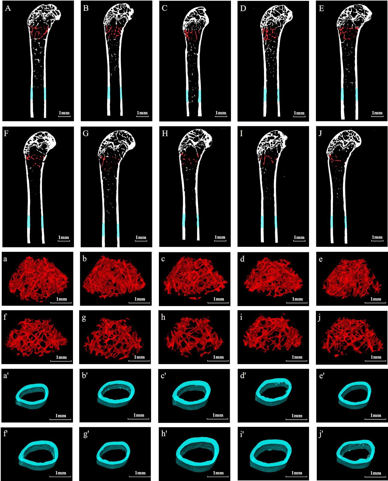
Serum remnant cholesterol reduces bone quality in obese mice

Authors: HOU Xiao-li, CAO Fu-yuan, GAO Jing-yuan, XING Lei, LIU Ning, ZHANG Nan, FAN Xin-hao, CAO Guo-long, TIAN Fa-ming

BACKGROUND: High cumulative remnant cholesterol levels are associated with the risk of various metabolic diseases, but their impact on bone quality remains to be explored. OBJECTIVE: To investigate the effects of high cumulative remnant cholesterol levels on bone mass, microstructure, and biomechanical properties in high-fat diet treated mice. METHODS: Ten healthy male SPF-grade C57BL6 mice were randomly allocated into normal control group and high-fat diet group. The normal control group was fed a normal diet for 20 weeks, while the high-fat diet group was fed a high-fat, high-cholesterol diet for 20 weeks. Mouse body mass was detected every week. After 20 weeks of feeding, serum levels of total cholesterol, remnant cholesterol, low-density lipoprotein cholesterol, high-density lipoprotein cholesterol, type I collagen carboxy-terminal cross-linked telopeptide, and type I procollagen amino-terminal propeptide were measured. Micro-CT was used to assess the microstructure of femoral cancellous and cortical bone. Three-point bending test was performed to measure the elastic modulus and maximum stress of the femur. RT-qPCR was used to detect the mRNA expression of RUNT-related transcription factor 2, type I collagen, osteocalcin, alkaline phosphatase, osteoprotegerin, receptor activator of nuclear factor-κB ligand, nuclear factor of activated T cells 1, and cathepsin K in the tibia. Pearson correlation analysis was used to analyze the correlation between remnant cholesterol, total cholesterol, high-density lipoprotein cholesterol, and low-density lipoprotein cholesterol levels and bone mineral density, as well as the correlation between remnant cholesterol levels and bone volume fraction, trabecular number, structure model index, and trabecular separation. RESULTS AND CONCLUSION: (1) From the 6th week of feeding, the body mass of mice in the high-fat diet group was higher than that in the control group (P < 0.05). The serum levels of total cholesterol, remnant cholesterol, and type I collagen carboxy-terminal cross-linked telopeptide in the high-fat diet group were higher than those in the control group (P < 0.05). (2) Micro-CT detection showed that compared with the control group, the bone microstructure of mice in the high-fat diet group underwent obvious degeneration, specifically manifested as significantly decreased bone mineral density, bone volume fraction, trabecular connectivity density, and trabecular number in cancellous bone, and significantly increased trabecular separation, structure model index, and trabecular pattern factor (P < 0.05). There were no significant changes in cortical bone thickness, volume, and area (P > 0.05). There was no significant difference in elastic modulus and maximum stress of the femur between the two groups (P > 0.05). (3) RT-qPCR detection showed that the mRNA expression of RUNT-related transcription factor 2, type I collagen, osteocalcin, alkaline phosphatase, and osteoprotegerin in the high-fat diet group was lower than that in the control group (P < 0.05), while the mRNA expression of nuclear factor of activated T cells 1 and cathepsin K was higher than that in the control group (P < 0.05). (4) Pearson analysis showed that remnant cholesterol and total cholesterol levels were significantly negatively correlated with bone mineral density (P < 0.05), and remnant cholesterol was significantly negatively correlated with bone volume fraction and trabecular number (P < 0.05), and significantly positively correlated with structure model index and trabecular separation (P < 0.05). These results indicate that remnant cholesterol may reduce bone quality by affecting the balance of bone turnover.

Serum remnant cholesterol reduces bone quality in obese mice
Graphical Abstract
Original ResearchVol 1898, Issue 26 • pp. 100-112DOI: 10.12307/2026.21290Jan 15, 2026

Methylation alterations of Fbln1 gene in the hippocampus of PSEN1/PSEN2 double knockout and APP/PS1 transgenic mice

Authors: Ruan Sibei, Li Li, Jian Yue, Ling Feng, Tang Mingxi

BACKGROUND: Alzheimer's disease (AD) is a progressive neurodegenerative disorder characterized by abnormal deposition of β-amyloid (Aβ) and neurofibrillary tangles of tau protein. Current medications only alleviate some symptoms, and despite extensive efforts to develop new therapies, such as anti-Aβ immunotherapy and β-secretase inhibitors, clinical trials have not been successful. OBJECTIVE: To investigate the methylation changes of the Fbln1 gene in the hippocampus of PSEN1/PSEN2 double knockout (dKO) mice, which lack Aβ deposition, and APP/PS1 double transgenic (DTG) mice, which exhibit Aβ deposition, to explore non-Aβ-related mechanisms and potential targets in AD. METHODS: Hippocampal tissues were collected from female dKO mice at 7 months (early AD) and 12 months (mid-stage AD) of age, with age-matched wild-type (WT) mice as controls. Epigenetic reduced representation bisulfite sequencing (RRBS) was used to screen for aberrantly methylated genes, identifying Fbln1. Bisulfite sequencing PCR (BSP) was performed to validate the methylation status of Fbln1 in mid-stage dKO mice. RT-PCR and western blot were used to measure Fbln1 mRNA and protein expression in early and mid-stage dKO mice, as well as in 12-month-old DTG mice. Finally, the expression levels of Fbln1 and Aβ were compared between dKO and DTG mice, with age-matched WT mice as controls. RESULTS AND CONCLUSION: RRBS showed that Fbln1 was hypomethylated in the hippocampus of mid-stage dKO mice (P < 0.05), while early-stage dKO mice showed a trend of hypomethylation but without statistical significance (P > 0.05). BSP confirmed the abnormal hypomethylation of Fbln1 in mid-stage dKO mice. In early-stage dKO mice, Fbln1 mRNA and protein levels were not significantly different from WT (P > 0.05). In mid-stage dKO mice, Fbln1 mRNA and protein levels were significantly higher than in WT (t=5.336, P < 0.01; t=8.985, P < 0.01). Similarly, mid-stage DTG mice showed significantly higher Fbln1 mRNA and protein levels than WT (t=4.151, P < 0.01; t=8.392, P < 0.01), but there was no significant difference between the two AD models (P > 0.05). In mid-stage dKO mice, there was no significant difference between Fbln1 and Aβ protein levels (P > 0.05), whereas in DTG mice, the difference was significant (t=6.348, P < 0.01), indicating that Fbln1 plays a role in both Aβ-dependent and Aβ-independent mechanisms. These findings suggest that Fbln1 methylation changes may contribute to age-dependent neurodegeneration in dKO mice and may be involved in both Aβ and non-Aβ pathways in AD, providing new insights and potential targets for non-Aβ-related mechanisms. Fbln1, as an aging-related factor, holds promise as a novel target.

Methylation alterations of Fbln1 gene in the hippocampus of PSEN1/PSEN2 double knockout and APP/PS1 transgenic mice
Graphical Abstract
Original ResearchVol 1898, Issue 26 • pp. 100-112DOI: 10.12307/2026.21293Jan 15, 2026

Roles of pregnane X receptor in sodium arsenite-induced oxidative stress and inflammatory injury in human normal hepatocytes

Authors: ZHANG Xiaoxu, TIAN Zhenli, XIE Tingting

BACKGROUND: As the primary organ for arsenic metabolism in the body, the liver has become a focal point for research on the mechanisms of arsenic toxicity. OBJECTIVE: To investigate the role of pregnane X receptor in sodium arsenite-induced oxidative stress and inflammatory injury in human normal hepatocytes. METHODS: Human normal hepatocyte MIHA cells were exposed to 0 (control), 10, 20, 30 μmol/L sodium arsenite for 48 hours. Changes in cell morphology were observed. Cell viability was measured via the cell counting kit-8 assay. Intracellular reactive oxygen species levels were detected using fluorescence probe staining combined with a microplate reader. Malondialdehyde levels were measured by thiobarbituric acid method. Glutathione reductase activity was detected by NADPH method. Total superoxide dismutase activity was measured by WST-8 method. Levels of interleukin-6, interleukin-1β, and tumor necrosis factor-α in cell supernatant were detected by ELISA. mRNA expression of pregnane X receptor and cytochrome P450 3A4 enzyme was detected by qRT-PCR. Protein expression of pregnane X receptor, cytochrome P450 3A4, nuclear factor-κB p65, nuclear factor-κB p-p65, proliferating cell nuclear antigen, interleukin-6, interleukin-1β, tumor necrosis factor-α, nuclear factor-κB inhibitor protein α, cyclooxygenase-2, p-nuclear factor-κB inhibitor protein α, nuclear factor erythroid 2-related factor 2, Keap1, and p-nuclear factor erythroid 2-related factor 2 was detected by western blot. RESULTS AND CONCLUSION: Compared with the control group, cells in all sodium arsenite groups showed unclear cell membrane boundaries, reduced cytoplasm, decreased cell fusion rate, and widened intercellular spaces. Compared with the control group, intracellular reactive oxygen species and malondialdehyde levels were increased (P < 0.05), levels of interleukin-6, interleukin-1β, and tumor necrosis factor-α in cell supernatant were increased (P < 0.05), protein expression of p-nuclear factor-κB inhibitor protein α, nuclear factor-κB p-p65, nuclear factor-κB p65, tumor necrosis factor-α, and interleukin-1β were increased (P < 0.05), cell viability was decreased (P < 0.05), protein expression of proliferating cell nuclear antigen, nuclear factor erythroid 2-related factor 2, p-nuclear factor erythroid 2-related factor 2 and total superoxide dismutase activity were decreased (P < 0.05), and mRNA and protein expression of pregnane X receptor and cytochrome P450 3A4 enzyme were decreased (P < 0.05). Compared with the control group, glutathione reductase activity was decreased in 20 and 30 μmol/L sodium arsenite groups (P < 0.05), and protein expression of Keap1, interleukin-6, and cyclooxygenase-2 was increased (P < 0.05). These results indicate that sodium arsenite may induce oxidative stress and inflammatory injury in hepatocytes by downregulating pregnane X receptor expression, inhibiting the nuclear factor erythroid 2-related factor 2 antioxidant pathway, and activating the nuclear factor-κB inflammatory pathway, while also inhibiting the expression of the drug-metabolizing enzyme cytochrome P450 3A4.

Roles of pregnane X receptor in sodium arsenite-induced oxidative stress and inflammatory injury in human normal hepatocytes
Graphical Abstract
Original ResearchVol 1898, Issue 26 • pp. 100-112DOI: 10.12307/2026.21282Jan 15, 2026

Mechanical loading of the patellofemoral joint and its correlation with various body mass indices in older adults practicing Tai Chi exercises

Authors: Li Guangmin, Wang Jiangna

BACKGROUND: As the aging population grows in China, overweight and obesity are increasingly leading to lower limb joint injuries in older adults. OBJECTIVE: To compare the biomechanical data of the lower limb patellofemoral joint during Tai Chi movements and normal walking among older adults with different body mass indices, analyze the correlation between body mass index and biomechanical data of the lower limb patellofemoral joint, and investigate the impact of different body mass indices on the mechanical loading of the patellofemoral joint in older adults. METHODS: The older adults with obesity (n=17), overweight (n=17), and normal body mass index (n=19) who had been practicing Tai Chi for an extended period were recruited based on body mass index classification. The VCION 3D infrared motion capture system and Kistler force measurement system were used to collect kinematic and dynamic data during typical Tai Chi movements and normal walking. A biomechanical model of the patellofemoral joint was established to calculate biomechanical parameters of the patellofemoral joint. The effects of body mass index on the mechanical characteristics of the patellofemoral joint during walking and Tai Chi in the older adults were quantified using analysis of variance and correlation analysis. RESULTS AND CONCLUSION: (1) Compared with the normal group, patellofemoral joint force and contact area peak during normal walking were significantly lower in the overweight and obese groups (P < 0.05); during Tai Chi movements, knee flexion moment, patellofemoral joint force, and quadriceps muscle force were significantly lower (P < 0.05). (2) Body mass index was significantly negatively correlated with the maximum knee flexion angle, maximum and minimum patellofemoral joint contact area during the Tai Chi movement 'Reversing the Wind' (P < 0.05); and with the maximum knee extension moment, maximum and minimum patellofemoral joint reaction force, maximum quadriceps muscle force, maximum knee angle, and maximum and minimum patellofemoral joint contact area during the 'Brush Knee and Twist Step' movement (P < 0.05). These results indicate that as body mass index increases, the dynamic load on the patellofemoral joint during Tai Chi movements decreases; compared with normal-weight individuals, overweight and obese older adults show significantly lower maximum patellofemoral joint force during the 'Brush Knee and Twist Step' movement, indicating lower dynamic load. Therefore, Tai Chi is recommended as a fitness exercise for overweight and obese older adults to avoid sports injuries caused by high joint loads.

Mechanical loading of the patellofemoral joint and its correlation with various body mass indices in older adults practicing Tai Chi exercises
Graphical Abstract
Original ResearchVol 1898, Issue 26 • pp. 100-112DOI: 10.12307/2026.21291Jan 15, 2026

Role and mechanism of ABL1 in myocardial necroptosis and cardiac ischemia/reperfusion injury

Authors: Yuan Min, Han Yu, Liu Jinhong, Zhang Jingyu, Cao Jimin, Sun Teng

BACKGROUND: ABL1 is involved in the regulation of multiple cellular processes, yet its functions within the cardiovascular system remains largely unexplored. In particular, its role in cardiac ischemia/reperfusion injury and necroptosis has not been reported. OBJECTIVE: To investigate the role of ABL1 in cardiac ischemia/reperfusion injury and myocardial necroptosis, as well as the underlying molecular mechanisms. METHODS: (1) Animal experiment: C57BL/6J mice were randomly divided into four groups: sham surgery group, ischemia/reperfusion group, ABL1 knockdown + ischemia/reperfusion group, and ABL1 negative control + ischemia/reperfusion group. Lentiviral vectors targeting ABL1 were injected in situ into the myocardium. One week later, ischemia/reperfusion injury was induced by ligation of the left anterior descending coronary artery followed by reperfusion. ABL1 protein expression, cardiac function, myocardial fibrosis, and cardiomyocyte surface area were assessed. (2) Cell experiment: H9c2 cells were divided into four groups: negative control cell line + PBS, ABL1 knockdown cell line + PBS, negative control cell line + H2O2 500 µmol/L, and ABL1 knockdown cell line + H2O2 500 µmol/L. Additionally, H9c2 cells were divided into five groups: negative control cell line + PBS, negative control cell line + H2O2 500 µmol/L, ABL1 knockdown cell line + H2O2 500 µmol/L, ABL1 knockdown cell line + Parkin overexpression adenovirus + H2O2 500 µmol/L, and ABL1 knockdown cell line + Parkin negative control adenovirus + H2O2 500 µmol/L. Cell viability, necroptosis, reactive oxygen species levels, and mitochondrial membrane potential were measured. Expression of ABL1, Parkin, and cyclophilin D was detected, and the interaction between ABL1 and Parkin was examined. RESULTS AND CONCLUSION: (1) ABL1 protein expression was significantly downregulated in the mouse cardiac ischemia/reperfusion model. (2) Knockdown of ABL1 exacerbated ischemia/reperfusion-induced cardiac dysfunction, as evidenced by decreased left ventricular ejection fraction and fractional shortening, and increased left ventricular end-systolic and end-diastolic diameters. (3) Knockdown of ABL1 promoted ischemia/reperfusion-induced myocardial fibrosis and aggravated ventricular remodeling. (4) ABL1 protein expression was significantly downregulated in the cardiomyocyte oxidative stress model. (5) Knockdown of ABL1 exacerbated oxidative stress-induced cell viability loss, necroptosis, and reactive oxygen species accumulation. (6) ABL1 regulated mitochondrial membrane permeability, modulated the expression of Parkin and cyclophilin D, and regulated cellular oxidative stress levels by targeting Parkin. (7) These results indicate that ABL1 expression is significantly downregulated in both in vivo ischemia/reperfusion and in vitro oxidative stress models, and knockdown of ABL1 aggravates cardiac ischemia/reperfusion injury and cardiomyocyte oxidative stress injury, acting through the Parkin-CypD pathway.

Role and mechanism of ABL1 in myocardial necroptosis and cardiac ischemia/reperfusion injury
Graphical Abstract
Original ResearchVol 1898, Issue 26 • pp. 100-112DOI: 10.12307/2026.21295Jan 15, 2026

Role of chondrocyte ferroptosis in the pathogenesis of osteoarthritis

Authors: Su Jiemao, Qi Yansong, Kong Keyu, Zhai Zanjing, Xu Yongsheng

BACKGROUND: As a programmed cell death, ferroptosis relies on lipid peroxidation triggered by iron overload, leading to the death of chondrocytes and the exacerbation of joint degeneration. OBJECTIVE: To summarize the molecular mechanism of chondrocyte ferroptosis in the progression of osteoarthritis, and intervention strategies for this mechanism. METHODS: A literature search was conducted in CNKI, PubMed, and Web of Science databases using Chinese and English search terms including 'cartilage, ferroptosis, Nrf2, NF-E2-related factor 2, SIRT, Sirtuin, PINK1, PTEN induced putative kinase 1, stromal cell-derived factor 1, SDF1, nanoparticle, mitophagy, hydrogel' etc. According to inclusion criteria, 69 articles were finally included for review. RESULTS AND CONCLUSION: The mechanism of ferroptosis involves iron deposition, lipid peroxidation, and abnormal amino acid metabolism, among which reactive oxygen species accumulation and lipid peroxidation are necessary conditions for generating ferroptosis signals. In osteoarthritis, chondrocyte ferroptosis is one of the key pathological mechanisms leading to cartilage degeneration. Current research on the mechanism of chondrocyte ferroptosis and related materials mainly focuses on antioxidant pathways. Multiple studies have attempted to construct materials with antioxidant properties or regulate metal ion distribution to alleviate cartilage damage caused by osteoarthritis and improve disease progression. In addition, developing materials that interfere with ferroptosis through other pathways is also a focus of current research on osteoarthritis treatment. However, these research results have only produced effective therapeutic effects in cell and animal experiments, and clinical studies have not yet been conducted. Future research needs to further explore ferroptosis-related signaling networks and the interaction between materials and biological interfaces to develop more efficient and safe treatment strategies.

Role of chondrocyte ferroptosis in the pathogenesis of osteoarthritis
Graphical Abstract
Original ResearchVol 1898, Issue 26 • pp. 100-112DOI: 10.12307/2026.21288Jan 15, 2026

Mechanical differences between medial collateral ligament and lateral collateral ligament and influence of elastin degradation

Authors: Xu Hongzhang, Huang Bo, Zhao Dongliang, Hu Ying, Qiao Dan, Deng Yuping

BACKGROUND: As crucial stabilizers of the knee joint, the medial collateral ligament and lateral collateral ligament play essential roles in restricting valgus and varus movements, respectively. However, the mechanical differences between the medial collateral ligament and lateral collateral ligament, the microstructure characteristics, and the effect of elastin degradation on their mechanical properties remain poorly understood. OBJECTIVE: To compare the mechanical differences between the medial collateral ligament and lateral collateral ligament, quantify the structural characteristics of the collagen fiber alignment, and investigate the effect of elastin degradation on the mechanical properties of both ligaments. METHODS: Left medial collateral ligaments and lateral collateral ligaments were harvested from adult pigs, frozen, and thawed. Quasi-static uniaxial tensile tests were performed to measure the mechanical properties of the medial collateral ligament and lateral collateral ligament, and the effects of repeated stretching on their mechanical properties were compared. Second harmonic generation imaging using a two-photon microscope was used to quantify the collagen fiber structure of the medial collateral ligament and lateral collateral ligament. After repeated stretching, the medial collateral ligament and lateral collateral ligament were incubated in elastase solution for 12 hours, followed by uniaxial tensile tests to determine the effect of elastin treatment on ligament mechanical properties. RESULTS AND CONCLUSION: (1) Quasi-static uniaxial tensile tests showed that the high-tension elastic modulus of the medial collateral ligament was higher than that of the lateral collateral ligament (P < 0.05), while there was no significant difference in the low-tension elastic modulus between the two groups (P > 0.05). Repeated stretching significantly reduced the low-tension elastic modulus of both the medial collateral ligament and lateral collateral ligament. (2) Elastase treatment significantly reduced the low-tension and high-tension elastic moduli of both the medial collateral ligament and lateral collateral ligament, and the decrease in the high-tension elastic modulus of the lateral collateral ligament was greater than that of the medial collateral ligament. After elastase treatment, both the low-tension and high-tension elastic moduli of the medial collateral ligament were higher than those of the lateral collateral ligament (P < 0.05). (3) Two-photon imaging showed that the collagen fibers of the medial collateral ligament maintained a crimped structure, and its fiber waviness was significantly higher than that of the lateral collateral ligament. (4) These results indicate that the medial collateral ligament has stronger elastic properties than the lateral collateral ligament, and elastase treatment has a greater effect on the mechanical properties of the lateral collateral ligament. These mechanical results may be related to the more crimped collagen fiber arrangement in the medial collateral ligament.

Mechanical differences between medial collateral ligament and lateral collateral ligament and influence of elastin degradation
Graphical Abstract
Original ResearchVol 1898, Issue 26 • pp. 100-112DOI: 10.12307/2026.21289Jan 15, 2026

Constructing an in vitro model of ulcerative colitis in mice based on organoid technology

Authors: Zhou Li, Li Rui, Chen Hao, Chen Jiaqi, Liu Yuhong, Wu Na

BACKGROUND: The pathogenesis of ulcerative colitis is highly complex, necessitating the development of models that more closely mimic human physiological and pathological responses to study the mechanisms underlying its onset and progression. OBJECTIVE: To establish a mouse ulcerative colitis organoid model. METHODS: Colon organoids of C57BL/6J mice were extracted, cultured and passaged in vitro. Colon organoids from mice after three generations of passage were taken and incubated in lipopolysaccharide at varying concentrations [0 (control), 150, 175, 200, 225, 250, 275, 300, 325, and 350 μg/mL] to induce inflammation for 24 hours. The morphology of mouse colon organoids was observed under a microscope, and changes in proliferation viability were assessed using the cell counting kit-8 assay. After 24 hours of incubation with 0, 225, 250, 275 μg/mL lipopolysaccharide, the levels of tumor necrosis factor α, interleukin-6, interleukin-9, and interleukin-10 were measured by ELISA. After 24 hours of incubation with 0 and 275 μg/mL lipopolysaccharide, the expression of occludin and zonula occludens-1 was detected by immunofluorescence staining, and the mRNA expression of tumor necrosis factor α, interleukin-6, interleukin-9, occludin, and zonula occludens-1 was detected by q-PCR. RESULTS AND CONCLUSION: (1) Under the microscope, colon organoids in the 150-275 μg/mL lipopolysaccharide group showed varying degrees of swelling, while those in the 300-350 μg/mL lipopolysaccharide group had inhibited growth and swelling. CCK-8 assay showed that 150-350 μg/mL lipopolysaccharide reduced the proliferation viability of mouse colon organoids, with 225-350 μg/mL having a more pronounced effect. Based on cell morphology and proliferation viability results, 225, 250, and 275 μg/mL lipopolysaccharide were selected for ELISA. (2) Compared with the control group, the levels of interleukin-6 and tumor necrosis factor α were increased in the 225, 250, and 275 μg/mL lipopolysaccharide groups (P < 0.05), and the level of interleukin-9 was increased in the 275 μg/mL lipopolysaccharide group (P < 0.05). (3) Immunofluorescence staining showed that compared with the control group, the expression of occludin and zonula occludens-1 was decreased in the 275 μg/mL lipopolysaccharide group. q-PCR detection showed that compared with the control group, the mRNA expression of interleukin-6 and tumor necrosis factor α was increased (P < 0.05), occludin mRNA expression was decreased (P < 0.05), and there was no significant difference in the expression of interleukin-9 and zonula occludens-1 (P > 0.05). (4) These results indicate that an in vitro mouse ulcerative colitis model based on organoids was successfully constructed, providing a powerful tool for studying the mechanisms of ulcerative colitis and screening effective drugs.

Constructing an in vitro model of ulcerative colitis in mice based on organoid technology
Graphical Abstract
Original ResearchVol 1898, Issue 26 • pp. 100-112DOI: 10.12307/2026.21294Jan 15, 2026

Mechanism of cuproptosis in the diagnosis and treatment of orthopedic-related diseases

Authors: Li Huaying, Li Hao, Peng Wuxun, Dong Wentao

BACKGROUND: Studies have shown that cuproptosis plays a critical role in the pathogenesis and treatment of orthopedic diseases. However, the regulatory roles and mechanisms of cuproptosis in orthopedic-related diseases remain unclear. OBJECTIVE: To review the roles and mechanisms of cuproptosis in orthopedic-related diseases. METHODS: A literature search was conducted in the PubMed database using the following English keywords: "cuproptosis," "copper steady state," "osteoarthritis," "osteoporosis," "rheumatoid arthritis," "osteosarcoma," and "oxidative stress." The search included publications up to March 2025. According to the inclusion criteria, 55 articles were finally included for review. RESULTS AND CONCLUSION: In osteoarthritis, excessive copper ions induce the expression of metal-regulatory transcription factor 1, indirectly activating matrix metalloproteinases and leading to cartilage degradation. Cuproptosis disrupts the tricarboxylic acid cycle and inhibits glutamine metabolism, triggering oxidative stress and accelerating chondrocyte death. In osteoporosis, cuproptosis suppresses glutamine metabolism and mineralization in osteoblasts while promoting osteoclast differentiation, thereby disrupting the balance between bone formation and resorption. In rheumatoid arthritis, copper ions activate the phosphatidylinositol 3-kinase/protein kinase B/mitogen-activated protein kinase signaling pathway, promoting synovial cell abnormal activation and inflammatory factor release, exacerbating joint destruction. In osteosarcoma, high concentrations of copper ions selectively kill tumor cells by targeting ferredoxin 1, inducing mitochondrial dysfunction and proteotoxic stress. The mechanisms of cuproptosis in orthopedic diseases are complex and diverse, involving multiple cell types and signaling pathways. Targeting cuproptosis or its related pathways may provide new strategies for the treatment of orthopedic diseases, such as the application of copper chelators or copper ion carriers, which hold significant clinical potential. Future research should further explore the specific regulatory mechanisms of cuproptosis and its therapeutic value in disease treatment.

Mechanism of cuproptosis in the diagnosis and treatment of orthopedic-related diseases
Graphical Abstract
Original ResearchVol 1898, Issue 26 • pp. 100-112DOI: 10.12307/2026.21280Jan 15, 2026

Bushen Tongshi Pills improves osteogenic disorders in alcoholic femoral head necrosis rats

Authors: WANG Weiwei, DING Qiang, RONG Xiangbin, GUO Liang, ZHAO Canbin, TAO Hongcheng, NIU Chicheng, LIU Jinfu, ZENG Ping

BACKGROUND: Bushen Tongshi Pills has been proven to delay the progression of collapse in alcoholic femoral head necrosis, but the mechanism is still unclear. OBJECTIVE: To explore the mechanism by which Bushen Tongshi Pills improve osteogenic disorders of alcoholic femoral head necrosis. METHODS: Fifty male Sprague-Dawley rats were randomly divided into a control group, a model group, and low, medium, and high dose groups of Bushen Tongshi Pills, with 10 rats in each group. Except for the control group, other groups were fed with ethanol-containing Lieber-DeCarli liquid diet for 8 weeks to construct alcoholic femoral head necrosis models, while the low, medium, and high dose groups were given 1.05, 2.1, 4.2 g/kg Bushen Tongshi Pills by gavage daily. After 8 weeks, Micro-CT was used to observe the gross morphology of the femoral head, hematoxylin-eosin staining was used to observe the pathological morphology of the femoral head, ELISA was used to detect serum levels of interleukin-1β and interleukin-18, immunohistochemical staining and western blot were used to detect the expression of NOD-like receptor protein 3, Caspase-1, gasdermin D, Runt-related transcription factor 2, osteocalcin, and type I collagen in rat femoral head tissues, and RT-qPCR was used to detect the mRNA expression of these genes. RESULTS AND CONCLUSION: (1) Micro-CT and hematoxylin-eosin staining results showed that the medium and high dose groups of Bushen Tongshi Pills significantly improved bone loss and pathological morphological development in the femoral head of alcoholic femoral head necrosis model rats. (2) ELISA results showed that compared with the control group, serum levels of interleukin-1β and interleukin-18 in the model group were significantly increased (P < 0.05); compared with the model group, serum levels of interleukin-1β and interleukin-18 in all Bushen Tongshi Pills dose groups were significantly decreased (P < 0.05), in a dose-dependent manner, with the most significant decrease in the high dose group (P < 0.05). (3) Immunohistochemistry, western blot, and RT-qPCR results showed that compared with the control group, the protein and mRNA expression of NOD-like receptor protein 3, Caspase-1, and gasdermin D in the femoral head of model rats were significantly upregulated (P < 0.05), while the protein and mRNA expression of osteogenic factors Runt-related transcription factor 2, osteocalcin, and type I collagen were significantly downregulated (P < 0.05); compared with the model group, the protein and mRNA expression of NOD-like receptor protein 3, Caspase-1, and gasdermin D in the femoral head of rats in all Bushen Tongshi Pills dose groups were downregulated in a dose-dependent manner, while the protein and mRNA expression of Runt-related transcription factor 2, osteocalcin, and type I collagen were upregulated in a dose-dependent manner, with the most significant upregulation in the high dose group (P < 0.05). (4) Western blot results showed that compared with the control group, the expression of cleaved-Caspase-1, gasdermin D-N protein, cleaved-Caspase-1/Caspase-1 ratio, and gasdermin D-N/gasdermin D ratio in the femoral head of model rats were significantly upregulated (P < 0.05); compared with the model group, the expression of cleaved-Caspase-1, gasdermin D-N protein, and gasdermin D-N/gasdermin D ratio in all Bushen Tongshi Pills dose groups were significantly downregulated (P < 0.05), and the cleaved-Caspase-1/Caspase-1 ratio in the medium and high dose groups was significantly downregulated (P < 0.05). (5) These results indicate that Bushen Tongshi Pills may inhibit the release of inflammatory factors in alcoholic femoral head necrosis model rats through the NOD-like receptor protein 3/Caspase-1/gasdermin D pyroptosis pathway, promote osteogenic differentiation, and repair necrotic bone tissue.

Bushen Tongshi Pills improves osteogenic disorders in alcoholic femoral head necrosis rats
Graphical Abstract
Original ResearchVol 1898, Issue 26 • pp. 100-112DOI: 10.12307/2026.21281Jan 15, 2026

Characteristics of lower limb muscle motor activation in patients with unilateral knee osteoarthritis

Authors: Shao Yunbo, Guo Jiayi, Li Feng

BACKGROUND: There are mechanical changes in the lower limb muscles in patients with knee osteoarthritis. These changes can be objectively reflected through surface electromyography. Currently, there is limited research on abnormal electrophysiological signals of lower limb muscles, especially of calf muscles, during walking in patients with unilateral knee osteoarthritis. OBJECTIVE: To investigate the motor activation status and activation mode of the medial thigh muscle, lateral thigh muscle, tibialis anterior muscle, and gastrocnemius muscle during daily walking in patients with unilateral knee osteoarthritis using surface electromyography. METHODS: Fifty subjects with unilateral knee osteoarthritis who met the inclusion criteria underwent surface electromyography walking tests. The target muscles were the medial thigh muscle, lateral thigh muscle, anterior tibialis muscle, and gastrocnemius muscle. The walking time was 60 seconds. Surface electromyography signals were collected from both healthy and affected sides. The main observation indicators were root mean square value, integrated electromyography value, median frequency, and muscle contraction ratio. Whether there is an abnormal activation status and its characteristics of lower limb muscles in unilateral knee osteoarthritis patients were investigated by surface electromyography. The abnormal movement activation of lower limb muscles in unilateral knee osteoarthritis patients was explored from an electrophysiological perspective. RESULTS AND CONCLUSION: (1) The integrated electromyography value and root mean square value of the medial and lateral thigh muscles on the affected side were significantly lower than those on the healthy side (P < 0.001); the median frequency values of the medial and lateral thigh muscles on the affected side were significantly lower than those on the healthy side (P < 0.001), with a more significant shift to low frequency, indicating a higher fatigue risk. (2) The integrated electromyography value and root mean square value of the tibialis anterior muscle on the affected side were significantly higher than those on the healthy side (P < 0.001); the median frequency value of the tibialis anterior muscle on the affected side was significantly lower than that on the healthy side (P < 0.001), with a more significant shift to low frequency, indicating a significantly increased fatigue risk. (3) The integrated electromyography value and root mean square value of the gastrocnemius muscle on the affected side were significantly higher than those on the healthy side (P < 0.001); the median frequency value of the gastrocnemius muscle on the affected side did not show a significant excessive shift compared with the healthy side (P > 0.05), indicating similar fatigue levels. (4) The muscle activation ratio on the affected side was significantly lower than that on the healthy side (P < 0.001), and the activation of the medial and lateral thigh muscles on the affected side was significantly unbalanced compared with the healthy side. These results indicate that during daily walking, patients with unilateral knee osteoarthritis exhibit insufficient activation and reduced activation efficiency of the medial and lateral thigh muscles, with high fatigue levels and risk, and unbalanced activation of these muscles, which is detrimental to knee joint stability and balance. The tibialis anterior and gastrocnemius muscles on the affected side show compensatory activation and abnormal co-activation levels, which are important factors aggravating joint stiffness, with the most obvious compensation in the tibialis anterior muscle, and there is a risk of fatigue-induced muscle atrophy.

Characteristics of lower limb muscle motor activation in patients with unilateral knee osteoarthritis
Graphical Abstract