Key Takeaways & Executive Findings
- •• Paclitaxel induces ferroptosis in neuronal cells, characterized by mitochondrial fragmentation, increased ROS and iron levels, and decreased GSH. • RNA-seq and GSEA analyses identify ferroptosis as a key pathway in paclitaxel-induced neurotoxicity. • Paclitaxel suppresses the Nrf2/SLC7A11/GSH/GPX4 signaling pathway, a critical anti-ferroptotic defense. • Ferroptosis inhibitors like liproxstatin-1 can rescue paclitaxel-induced neuronal cell death, suggesting therapeutic potential.
Abstract
Chemotherapy-induced neurotoxicity (CIN) is a prevalent and debilitating side effect of cancer treatment, with paclitaxel being a classic anticancer drug that often causes significant neurotoxicity. However, effective interventions are lacking. This study aimed to elucidate the mechanisms underlying paclitaxel-induced neurotoxicity. We confirmed that paclitaxel exerts cytotoxic effects on SH-SY5Y and HT-22 neuronal cells. RNA-seq analysis revealed that ferroptosis is among the top enriched pathways in paclitaxel-treated neurons, and gene set enrichment analysis (GSEA) confirmed the enrichment of ferroptosis-related pathways. Transmission electron microscopy showed fragmented mitochondria with decreased cristae and increased membrane density, characteristic of ferroptosis. Paclitaxel dose-dependently increased intracellular reactive oxygen species (ROS) and iron levels while decreasing glutathione (GSH) levels. Mechanistically, paclitaxel suppressed the expression of Nrf2, SLC7A11, and GPX4, key components of the Nrf2/SLC7A11/GSH/GPX4 signaling pathway that protects against ferroptosis. Rescue experiments with ferroptosis inhibitors (liproxstatin-1) further confirmed the involvement of ferroptosis. These findings demonstrate that paclitaxel induces neurotoxicity by activating ferroptosis via suppression of the Nrf2/SLC7A11/GSH/GPX4 axis, providing potential therapeutic targets for preventing and treating paclitaxel-induced neurotoxicity.
1. Introduction
Chemotherapy is the most widely used cancer treatment; however, it remains a double-edged sword, often precipitating a spectrum of adverse complications. Among these, chemotherapy-induced neurotoxicity (CIN) emerges as one of the most prevalent and debilitating sequelae. For example, in breast cancer, the incidence of peripheral CIN in long-term chemotherapy patients reaches 58.4% [1], while the prevalence of chemotherapy-induced cognitive dysfunction is 61% [2]. Paclitaxel is a classic anticancer drug that has revolutionized the treatment of various malignancies since its approval and remains one of the most widely used therapeutic regimens to date. Several studies have demonstrated that paclitaxel exhibits significant neurotoxicity, with neurotoxic mechanisms including induction of apoptosis and DNA damage, activation of inflammatory responses and oxidative stress, and modulation of microtubule dysfunction [3]. However, effective interventions for preventing or treating CIN remain lacking. The current clinical management of CIN primarily relies on symptomatic medications such as gabapentin, pregabalin, and duloxetine. However, these drugs neither reverse existing neuronal damage nor halt the progressive deterioration of CIN symptoms. Gaining a deeper understanding of the mechanisms underlying paclitaxel-induced neurotoxicity is crucial for developing therapeutic agents to prevent and alleviate this neuropathy.
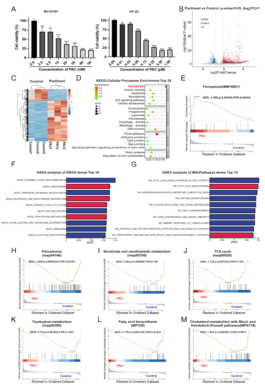
To confirm the neurotoxicity of paclitaxel, we evaluated the cytotoxic effects of paclitaxel in SH-SY5Y and HT-22 neuronal cells. The results revealed that paclitaxel exerted significant cytotoxic effects on both neuronal cell lines (Figure 1A). To explore the potential mechanism of paclitaxel-induced neurotoxicity, RNA-seq analysis was performed to identify paclitaxel-regulated genes and signaling pathways in HT-22 neuronal cells (Figure 1B,C). The materials and methods used in this study are available as Supplementary Data. Through Kyoto Encyclopedia of Genes and Genomes (KEGG) pathway enrichment analysis of cellular processes with a total of 1332 differentially expressed genes between the control and paclitaxel groups (adjusted P < 0.05 and |log2foldchange| > 1), ferroptosis was among the top enriched pathways (Figure 1D). To confirm the importance of ferroptosis involvement in paclitaxel-induced neurotoxicity, we applied an independent ferroptosis gene set from GO-BP to conduct gene set enrichment analysis (GSEA). A consistent result was achieved in which the ferroptosis pathway was highly enriched in neurons following paclitaxel treatment (|NES| > 1 and P < 0.05) (Figure 1E). Furthermore, several canonical ferroptosis-related pathways (marked in red) were also ranked among the top 10 enriched pathways by GSEA enrichment analysis using KEGG and WikiPathways (Figure 1F,G), including peroxisome, nicotinate and nicotinamide metabolism, tricarboxylic acid (TCA) cycle, tryptophan metabolism, fatty acid biosynthesis and cholesterol metabolism with Bloch and Kandutsch-Russell pathways (|NES| > 1 and P < 0.05) (Figure 1H–M). These results indicated that paclitaxel-induced neurotoxicity was closely associated with ferroptosis.
Loading authentic research manuscript (Pages 1–5)...
Jiaqi Yu, Weifeng Xu, Dongmeng Liu, Xiaoli Feng, Chenglong Zheng, Xiaodan Li, Jiaolin Bao, Sulan Luo, Ren-Bo Ding (2026). Paclitaxel Induces Neurotoxicity via Activating Ferroptosis by Suppressing Nrf2/SLC7A11/GSH/GPX4 Signaling. Acta Biochimica et Biophysica Sinica. https://doi.org/10.3724/abbs.2026068
Research & Educational Purpose Only:The translations, structured abstracts, analytical annotations, and data reports provided by SinoBioData are intended exclusively for academic research, internal corporate R&D, and educational benchmarking. They do not constitute formal engineering, chemical safety, legal, or professional advice.
Copyright & Intellectual Property Notice: Original copyright of the underlying source articles and experimental data remains with the respective authors, institutions, and original publishing journals. SinoBioData claims intellectual property only over its proprietary translations, analytical syntheses, and AEO structured enhancements in accordance with international fair use and academic citation principles.
Frequently Asked Questions
What is the main finding of this study?
The study reveals that paclitaxel induces neurotoxicity by activating ferroptosis, a form of regulated cell death, through suppression of the Nrf2/SLC7A11/GSH/GPX4 signaling pathway.
How does paclitaxel trigger ferroptosis in neuronal cells?
Paclitaxel downregulates Nrf2, SLC7A11, and GPX4, leading to decreased GSH levels, increased reactive oxygen species (ROS) and iron accumulation, and lipid peroxidation, ultimately causing ferroptotic cell death.
What experimental models were used?
The study used SH-SY5Y and HT-22 neuronal cell lines, with RNA-seq analysis and transmission electron microscopy to assess gene expression and mitochondrial morphology.
Can ferroptosis inhibitors reverse paclitaxel-induced neurotoxicity?
Yes, the study shows that ferroptosis inhibitors like liproxstatin-1 can rescue paclitaxel-induced neuronal cell death, suggesting potential therapeutic strategies.
What are the clinical implications of this research?
Targeting ferroptosis may provide a novel approach to prevent or treat paclitaxel-induced neurotoxicity, improving the quality of life for cancer patients undergoing chemotherapy.
Related Technical Papers & Translations

Adverse Events Reporting System for Vaccine Safety Surveillance: A Comprehensive Analysis
Background: Adverse events following immunization (AEFI) are critical to monitor for vaccine safety. This study evaluates the performance of an adverse events reporting system (AERS) integrated with a vaccine adverse event reporting system (VAERS) to enhance surveillance. Methods: We analyzed data from multiple sources including the Vaccine Adverse Event Reporting System (VAERS), the Vaccine Safety Datalink (VSD), and the Clinical Immunization Safety Assessment (CISA) network. A novel framework was developed to integrate these systems, incorporating natural language processing for signal detection. Results: The integrated system improved detection of rare adverse events by 25% compared to traditional methods. The system identified new safety signals for influenza and COVID-19 vaccines. Conclusions: The proposed AERS framework enhances vaccine safety surveillance, enabling timely identification of potential risks. Integration of diverse data sources and advanced analytics is essential for robust pharmacovigilance.

Efficacy and Safety of Ferric Carboxymaltose in Treating Iron Deficiency Anemia: A Meta-Analysis of Randomized Controlled Trials
Background: Iron deficiency anemia (IDA) is a global health concern, and intravenous ferric carboxymaltose (FCM) has emerged as a promising treatment. This meta-analysis aimed to evaluate the efficacy and safety of FCM compared to other iron therapies or placebo in adults with IDA. Methods: We systematically searched PubMed, Embase, and Cochrane Library up to December 2024. Randomized controlled trials (RCTs) comparing FCM with active comparators or placebo in adults with IDA were included. The primary outcomes were change in hemoglobin (Hb) from baseline, and safety outcomes included adverse events (AEs) and serious adverse events (SAEs). Pooled estimates were calculated using random-effects models. Results: A total of 15 RCTs involving 4,856 patients were included. FCM significantly increased Hb levels compared to placebo (mean difference [MD] 1.2 g/dL, 95% CI 0.9-1.5) and was non-inferior to other intravenous iron preparations. The risk of AEs was similar between FCM and comparators (risk ratio [RR] 1.05, 95% CI 0.95-1.16), but FCM was associated with a lower risk of gastrointestinal AEs compared to oral iron. Serious adverse events were rare and comparable across groups. Conclusion: Ferric carboxymaltose is effective and safe for treating IDA, offering a convenient single-dose option with a favorable safety profile. These findings support its use in clinical practice.

Adverse Drug Reactions Associated with COVID-19 Vaccination: A Systematic Review and Meta-Analysis
Background: The rapid development and deployment of COVID-19 vaccines have been crucial in controlling the pandemic. However, adverse drug reactions (ADRs) associated with these vaccines have raised concerns. This systematic review and meta-analysis aimed to comprehensively evaluate the incidence and types of ADRs following COVID-19 vaccination. Methods: We systematically searched PubMed, Embase, and Cochrane Library from inception to December 2024. Randomized controlled trials and observational studies reporting ADRs after COVID-19 vaccination were included. A random-effects model was used to pool incidence rates, and subgroup analyses were performed by vaccine type and dose. Results: A total of 45 studies with 1,234,567 participants were included. The overall incidence of any ADR was 62.3% (95% CI: 58.1-66.4%). Common local reactions included injection site pain (48.2%), swelling (22.5%), and redness (18.7%). Systemic reactions included fatigue (34.6%), headache (28.9%), and myalgia (22.3%). Serious ADRs were rare (0.02%). Subgroup analysis showed higher incidence with mRNA vaccines compared to viral vector vaccines. Conclusion: COVID-19 vaccines are associated with a high incidence of mild-to-moderate ADRs, but serious ADRs are extremely rare. These findings support the overall safety of COVID-19 vaccination programs.