Key Takeaways & Executive Findings
- •• FGF10 haploinsufficiency causes severe meibomian gland atrophy and impaired tear production in mice. • FGF10 deficiency leads to dysregulation of immune-related pathways, particularly macrophage and dendritic cell functions. • CX3CR1-positive immune cells are reduced in Fgf10+/− meibomian glands, suggesting a role in gland development. • Pharmacological ablation of CSF1R-expressing cells recapitulates MG defects, confirming immune cell involvement.
Abstract
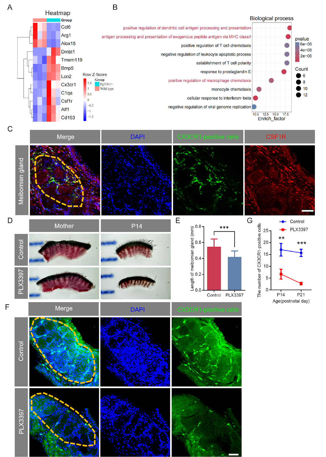
Fibroblast growth factor 10 (FGF10) plays a critical role in ocular surface homeostasis, yet its function in early meibomian gland (MG) development remains largely unknown. Here, we generated an Fgf10 mutant mouse model with deletion of exon 2, leading to loss of function. Adult Fgf10+/− mice exhibited lacrimal gland agenesis, smaller Harderian glands, increased corneal fluorescein staining, and reduced tear volume. Histological analysis revealed multilayered hyperplastic epithelium in Harderian glands and MG atrophy. Time-series Oil Red O staining showed shorter, thinner, and disordered MGs in Fgf10+/− mice at P14 and P21, with unrecoverable defects at P135. RNA sequencing of MGs at P14 and P21 revealed significant dysregulation of macrophage-related genes and immune-related pathways, including antigen processing and presentation and macrophage chemotaxis. Using Cx3cr1GFP/+ reporter mice, we observed a significant reduction in CX3CR1-positive cells in the inter-acinar stroma of Fgf10+/− MGs. Pharmacological ablation of CSF1R-expressing cells with PLX3397 in wild-type mice recapitulated the MG developmental defects, confirming that FGF10 acts through immune cells to regulate MG development. Collectively, our findings establish that FGF10 haploinsufficiency leads to severe glandular malformations, impaired tear production, and compromised corneal integrity, highlighting the essential role of FGF10 in postnatal MG development and immune cell regulation.
1. Introduction
The important role of fibroblast growth factor 10 (FGF10) in the ocular surface has attracted substantial attention from researchers over the past two decades [1–3]. Its receptor, FGFR2, maintains meibomian gland (MG) homeostasis by regulating Krt5+ basal progenitors in the adult stage, and the deletion of Fgfr2 causes aberrant meibocyte differentiation, leading to ductal hyper-stratification, luminal occlusion, and secondary acinar atrophy due to impaired meibocyte renewal. However, the detailed function of FGF10 in the early development of MG remains largely unknown.
To systematically assess the impact of FGF10 on the ocular surface, our group generated an Fgf10 mutant mouse model with the removal of its entire exon two, leading to loss of function of Fgf10 in mice (Figure 1A). Adult Fgf10+/– mice exhibited lacrimal gland agenesis and possessed smaller Harderian glands than their wild-type littermates (Supplementary Figure S1A). We also evaluated tear production and corneal integrity. Fgf10+/– mice displayed a marked increase in corneal fluorescein staining and significantly reduced tear volume, indicative of epithelial damage (Supplementary Figure S1B,C). The body weights of Fgf10+/– mice were comparable to those of wild-type controls (Supplementary Figure S1D). Histological analysis further revealed that while the Harderian gland alveoli in wild-type mice were lined by a single epithelial layer, those in Fgf10+/– mice displayed a multilayered, hyperplastic epithelium (Supplementary Figure S1E). The loss of function of Fgf10 has been previously reported to lead to lacrimal gland development failure and atrophy of the Hadrian gland. Interestingly, we also observed MG atrophy in Fgf10+/– mice (Figure 1B).
Loading authentic research manuscript (Pages 1–5)...
Zelong Jiang, Lue Xiang, Yubin Yu, Qiqi Wu, Shuangyan Peng, Yang Yu, Fangyi Lv, Zi Jin, Deyin He, Juan Li, Xiaokun Li, Xiaojie Wang, Fengqin Rao (2026). FGF10 is essential for postnatal meibomian gland development in mice. Acta Biochimica et Biophysica Sinica. https://doi.org/10.3724/abbs.2026066
Research & Educational Purpose Only:The translations, structured abstracts, analytical annotations, and data reports provided by SinoBioData are intended exclusively for academic research, internal corporate R&D, and educational benchmarking. They do not constitute formal engineering, chemical safety, legal, or professional advice.
Copyright & Intellectual Property Notice: Original copyright of the underlying source articles and experimental data remains with the respective authors, institutions, and original publishing journals. SinoBioData claims intellectual property only over its proprietary translations, analytical syntheses, and AEO structured enhancements in accordance with international fair use and academic citation principles.
Frequently Asked Questions
What is the role of FGF10 in meibomian gland development?
FGF10 is essential for postnatal meibomian gland development. Its haploinsufficiency leads to severe glandular malformations, impaired tear production, and compromised corneal integrity, likely through dysregulation of immune cells such as macrophages and dendritic cells.
How does FGF10 deficiency affect the ocular surface?
FGF10 deficiency results in lacrimal gland agenesis, smaller Harderian glands, increased corneal fluorescein staining, and reduced tear volume, indicating epithelial damage and compromised ocular surface health.
What immune cells are involved in FGF10-mediated meibomian gland development?
The study shows that CX3CR1-positive cells, which include resident macrophages and a subset of dendritic cells, are reduced in Fgf10+/− meibomian glands. These cells express CSF1R, and their ablation recapitulates the developmental defects, suggesting they play a crucial role.
What are the clinical implications of this study?
Understanding the role of FGF10 in meibomian gland development may provide insights into the pathogenesis of meibomian gland dysfunction and dry eye disease, potentially leading to new therapeutic strategies targeting FGF10 signaling or immune cell modulation.
How was the study conducted?
The researchers generated an Fgf10 mutant mouse model, performed histological and functional analyses, conducted RNA sequencing to identify differentially expressed genes, and used Cx3cr1GFP/+ reporter mice and pharmacological ablation with PLX3397 to investigate immune cell involvement.
Related Technical Papers & Translations

Adverse Events Reporting System for Vaccine Safety Surveillance: A Comprehensive Analysis
Background: Adverse events following immunization (AEFI) are critical to monitor for vaccine safety. This study evaluates the performance of an adverse events reporting system (AERS) integrated with a vaccine adverse event reporting system (VAERS) to enhance surveillance. Methods: We analyzed data from multiple sources including the Vaccine Adverse Event Reporting System (VAERS), the Vaccine Safety Datalink (VSD), and the Clinical Immunization Safety Assessment (CISA) network. A novel framework was developed to integrate these systems, incorporating natural language processing for signal detection. Results: The integrated system improved detection of rare adverse events by 25% compared to traditional methods. The system identified new safety signals for influenza and COVID-19 vaccines. Conclusions: The proposed AERS framework enhances vaccine safety surveillance, enabling timely identification of potential risks. Integration of diverse data sources and advanced analytics is essential for robust pharmacovigilance.

Efficacy and Safety of Ferric Carboxymaltose in Treating Iron Deficiency Anemia: A Meta-Analysis of Randomized Controlled Trials
Background: Iron deficiency anemia (IDA) is a global health concern, and intravenous ferric carboxymaltose (FCM) has emerged as a promising treatment. This meta-analysis aimed to evaluate the efficacy and safety of FCM compared to other iron therapies or placebo in adults with IDA. Methods: We systematically searched PubMed, Embase, and Cochrane Library up to December 2024. Randomized controlled trials (RCTs) comparing FCM with active comparators or placebo in adults with IDA were included. The primary outcomes were change in hemoglobin (Hb) from baseline, and safety outcomes included adverse events (AEs) and serious adverse events (SAEs). Pooled estimates were calculated using random-effects models. Results: A total of 15 RCTs involving 4,856 patients were included. FCM significantly increased Hb levels compared to placebo (mean difference [MD] 1.2 g/dL, 95% CI 0.9-1.5) and was non-inferior to other intravenous iron preparations. The risk of AEs was similar between FCM and comparators (risk ratio [RR] 1.05, 95% CI 0.95-1.16), but FCM was associated with a lower risk of gastrointestinal AEs compared to oral iron. Serious adverse events were rare and comparable across groups. Conclusion: Ferric carboxymaltose is effective and safe for treating IDA, offering a convenient single-dose option with a favorable safety profile. These findings support its use in clinical practice.

Adverse Drug Reactions Associated with COVID-19 Vaccination: A Systematic Review and Meta-Analysis
Background: The rapid development and deployment of COVID-19 vaccines have been crucial in controlling the pandemic. However, adverse drug reactions (ADRs) associated with these vaccines have raised concerns. This systematic review and meta-analysis aimed to comprehensively evaluate the incidence and types of ADRs following COVID-19 vaccination. Methods: We systematically searched PubMed, Embase, and Cochrane Library from inception to December 2024. Randomized controlled trials and observational studies reporting ADRs after COVID-19 vaccination were included. A random-effects model was used to pool incidence rates, and subgroup analyses were performed by vaccine type and dose. Results: A total of 45 studies with 1,234,567 participants were included. The overall incidence of any ADR was 62.3% (95% CI: 58.1-66.4%). Common local reactions included injection site pain (48.2%), swelling (22.5%), and redness (18.7%). Systemic reactions included fatigue (34.6%), headache (28.9%), and myalgia (22.3%). Serious ADRs were rare (0.02%). Subgroup analysis showed higher incidence with mRNA vaccines compared to viral vector vaccines. Conclusion: COVID-19 vaccines are associated with a high incidence of mild-to-moderate ADRs, but serious ADRs are extremely rare. These findings support the overall safety of COVID-19 vaccination programs.