Key Takeaways & Executive Findings
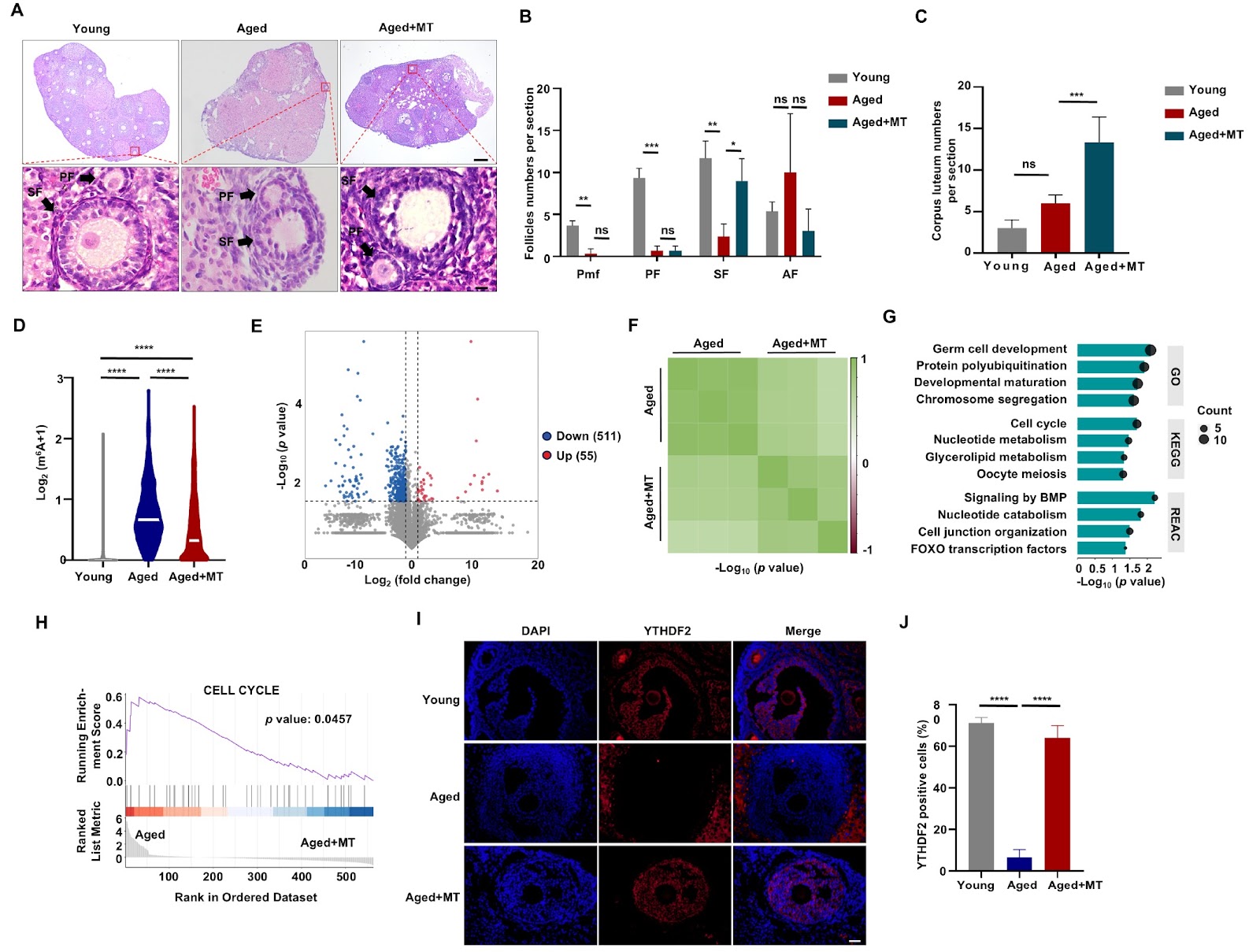
- •• Melatonin mitigates ovarian aging in murine models by significantly decreasing m6A methylation levels. • YTHDF2 expression is markedly increased in ovarian granulosa cells upon melatonin treatment, with differentially methylated genes enriched in the polyubiquitination pathway. • YTHDF2 enhances UBE3C expression by modulating m6A methylation of UBE3C mRNA, reducing P53 senescence factor and alleviating ovarian aging. • This study reveals a novel epigenetic mechanism (YTHDF2/m6A/UBE3C axis) underlying melatonin's anti-aging effects on the ovary, offering potential therapeutic targets for infertility.
Abstract
Ovarian aging is a natural process characterized by a decline in both the quantity and quality of oocytes, which subsequently leads to diminished fertility, particularly in women over the age of 35. Given the societal trend toward postponing childbirth, it is imperative to understand the molecular mechanisms that underpin ovarian aging to address infertility issues. Melatonin (MT) is recognized for its therapeutic potential in mitigating ovarian aging; however, the specific epigenetic mechanisms involved, particularly concerning m6A methylation, remain inadequately defined. Our investigation demonstrates that MT mitigates ovarian aging in murine models, significantly decreasing m6A methylation levels. In vitro analyses of ovarian granulosa (KGN) cells reveals a marked increase in YTHDF2 expression, with differentially methylated genes being notably enriched in the polyubiquitination pathway. Further examination shows that YTHDF2 enhances the expression of the E3 ligase UBE3C by modulating the m6A methylation of UBE3C mRNA, thereby reducing the expression of the P53 senescence factor and alleviating the effects of ovarian aging.
1. Introduction
The ovary is one of the first organs to manifest signs of aging in humans. Ovarian aging is a natural and physiological process characterized by a gradual decline in ovarian function over time. This process involves a reduction in both the quantity and quality of oocytes, as well as a decrease in the overall ovarian reserve [1]. Females are born with a finite number of oocytes, and as they age, the number of follicles diminishes [2]. Typically, female fertility begins to decline after the age of 30, with significant decreases in the success rates of natural conception and assisted reproductive technologies after the age of 35 [3]. In light of evolving social, economic, and demographic trends, more individuals are choosing to delay childbirth, resulting in a higher incidence of infertility among older women. Consequently, investigating and elucidating the molecular mechanisms associated with ovarian aging are essential to facilitate the development of effective interventions for infertility in women.
RNA N6-methyladenosine (m6A) methylation has emerged as a critical posttranscriptional modification that regulates gene expression and RNA metabolism. m6A is the most prevalent internal modification of mRNAs and noncoding RNAs and plays essential roles in diverse biological processes, including embryonic development [4–6], cell differentiation [7,8], stress responses [9,10], and aging-related disease [11]. The dynamic regulation of m6A is mediated by “writers” (methyltransferases such as METTL3 and METTL14) [12], “erasers” (demethylases such as FTO and ALKBH5) [13,14], and “readers” (m6A-binding proteins such as YTHDF2 and YTHDF3) [15,16], which together orchestrate the fate of m6A-modified RNAs. Specifically, m6A modifications are crucial for oocyte maturation, folliculogenesis, and maintenance of the ovarian reserve [17,18]. FTO delays ovarian aging in human granulosa cells (GCs) in a FOS-dependent manner [18]. The YTH family comprises YTHDC1/2 and the cytoplasmic YTHDF family (YTHDF1–3), all of which possess a highly conserved C-terminal YTH domain that facilitates m6A binding. YTHDF2, in particular, promotes the degradation of m6A-modified mRNAs [19]. Research indicates that YTHDF2 is essential for male fertility, with its deficiency resulting in oligoasthenoteratozoospermia [20,21]. Furthermore, maternal YTHDF2 is critical for female fertility and early embryonic development [22]. The low-complexity region at the N-terminus of YTHDF2 enables the protein to undergo phase separation, which leads to the aggregation of YTHDF2 and impairs mitochondrial function in the aging brain [23,24]. However, current investigations into the role of YTHDF family-mediated m6A in ovarian aging are limited.
Melatonin (MT) is a hormone primarily associated with circadian rhythm regulation, which alleviates neural tube defects by modulating the Src/PI3K/ERK signaling pathway [25]. MT is present in both oocytes and GCs, and long-term melatonin treatment has been shown to effectively mitigate ovarian aging [26,27]. It has been demonstrated to reduce oxidative stress and improve mitochondrial function, thereby protecting ovarian cells from age-related damage [27–29]. The capacity of MT to modulate m6A methylation introduces an additional layer of complexity to its protective effects on the ovary. For example, MT treatment has been reported to upregulate the expression of YTHDF2, a key m6A reader, in oocytes and GCs, indicating a potential mechanism through which MT exerts its antiaging effects [30]. Nevertheless, the specific mechanisms by which MT
Loading authentic research manuscript (Pages 1–5)...
Wenjuan Xia, Xin Wang, Jincheng Li, Ming Zhang, Jiafeng Lu, Hong Li, Quanze He, Qingxia Meng, Boxian Huang (2026). Melatonin mitigates ovarian aging through regulation of the YTHDF2/m6A/UBE3C axis. Acta Biochimica et Biophysica Sinica. https://doi.org/10.3724/abbs.2025090
Research & Educational Purpose Only:The translations, structured abstracts, analytical annotations, and data reports provided by SinoBioData are intended exclusively for academic research, internal corporate R&D, and educational benchmarking. They do not constitute formal engineering, chemical safety, legal, or professional advice.
Copyright & Intellectual Property Notice: Original copyright of the underlying source articles and experimental data remains with the respective authors, institutions, and original publishing journals. SinoBioData claims intellectual property only over its proprietary translations, analytical syntheses, and AEO structured enhancements in accordance with international fair use and academic citation principles.
Frequently Asked Questions
What is the role of melatonin in ovarian aging?
Melatonin mitigates ovarian aging by decreasing m6A methylation levels and modulating the YTHDF2/m6A/UBE3C axis, which reduces the expression of the P53 senescence factor.
How does YTHDF2 affect UBE3C expression?
YTHDF2 enhances UBE3C expression by binding to m6A-methylated UBE3C mRNA, likely promoting its stability or translation, thereby reducing P53 and alleviating ovarian aging.
What is the significance of the YTHDF2/m6A/UBE3C axis?
This axis represents a novel epigenetic mechanism through which melatonin exerts its anti-aging effects on the ovary, providing potential therapeutic targets for infertility.
What methods were used in this study?
The study used murine models and in vitro KGN cells, analyzing m6A methylation levels, YTHDF2 expression, and enrichment of differentially methylated genes in the polyubiquitination pathway.
What are the implications for infertility treatment?
Understanding this mechanism may lead to new interventions for ovarian aging and infertility, particularly for women over 35, by targeting the YTHDF2/m6A/UBE3C pathway.
Related Technical Papers & Translations

Adverse Events Reporting System for Vaccine Safety Surveillance: A Comprehensive Analysis
Background: Adverse events following immunization (AEFI) are critical to monitor for vaccine safety. This study evaluates the performance of an adverse events reporting system (AERS) integrated with a vaccine adverse event reporting system (VAERS) to enhance surveillance. Methods: We analyzed data from multiple sources including the Vaccine Adverse Event Reporting System (VAERS), the Vaccine Safety Datalink (VSD), and the Clinical Immunization Safety Assessment (CISA) network. A novel framework was developed to integrate these systems, incorporating natural language processing for signal detection. Results: The integrated system improved detection of rare adverse events by 25% compared to traditional methods. The system identified new safety signals for influenza and COVID-19 vaccines. Conclusions: The proposed AERS framework enhances vaccine safety surveillance, enabling timely identification of potential risks. Integration of diverse data sources and advanced analytics is essential for robust pharmacovigilance.

Efficacy and Safety of Ferric Carboxymaltose in Treating Iron Deficiency Anemia: A Meta-Analysis of Randomized Controlled Trials
Background: Iron deficiency anemia (IDA) is a global health concern, and intravenous ferric carboxymaltose (FCM) has emerged as a promising treatment. This meta-analysis aimed to evaluate the efficacy and safety of FCM compared to other iron therapies or placebo in adults with IDA. Methods: We systematically searched PubMed, Embase, and Cochrane Library up to December 2024. Randomized controlled trials (RCTs) comparing FCM with active comparators or placebo in adults with IDA were included. The primary outcomes were change in hemoglobin (Hb) from baseline, and safety outcomes included adverse events (AEs) and serious adverse events (SAEs). Pooled estimates were calculated using random-effects models. Results: A total of 15 RCTs involving 4,856 patients were included. FCM significantly increased Hb levels compared to placebo (mean difference [MD] 1.2 g/dL, 95% CI 0.9-1.5) and was non-inferior to other intravenous iron preparations. The risk of AEs was similar between FCM and comparators (risk ratio [RR] 1.05, 95% CI 0.95-1.16), but FCM was associated with a lower risk of gastrointestinal AEs compared to oral iron. Serious adverse events were rare and comparable across groups. Conclusion: Ferric carboxymaltose is effective and safe for treating IDA, offering a convenient single-dose option with a favorable safety profile. These findings support its use in clinical practice.

Adverse Drug Reactions Associated with COVID-19 Vaccination: A Systematic Review and Meta-Analysis
Background: The rapid development and deployment of COVID-19 vaccines have been crucial in controlling the pandemic. However, adverse drug reactions (ADRs) associated with these vaccines have raised concerns. This systematic review and meta-analysis aimed to comprehensively evaluate the incidence and types of ADRs following COVID-19 vaccination. Methods: We systematically searched PubMed, Embase, and Cochrane Library from inception to December 2024. Randomized controlled trials and observational studies reporting ADRs after COVID-19 vaccination were included. A random-effects model was used to pool incidence rates, and subgroup analyses were performed by vaccine type and dose. Results: A total of 45 studies with 1,234,567 participants were included. The overall incidence of any ADR was 62.3% (95% CI: 58.1-66.4%). Common local reactions included injection site pain (48.2%), swelling (22.5%), and redness (18.7%). Systemic reactions included fatigue (34.6%), headache (28.9%), and myalgia (22.3%). Serious ADRs were rare (0.02%). Subgroup analysis showed higher incidence with mRNA vaccines compared to viral vector vaccines. Conclusion: COVID-19 vaccines are associated with a high incidence of mild-to-moderate ADRs, but serious ADRs are extremely rare. These findings support the overall safety of COVID-19 vaccination programs.