Key Takeaways & Executive Findings
- •• Substrate stiffness modulates Aβ42-induced neurotoxicity: stiff substrates exacerbate cell death, synaptic loss, and impaired Ca2+ oscillations, while soft substrates confer protection. • Aβ42 disrupts spontaneous Ca2+ oscillations and synaptic transmission more severely on stiff ECM, mimicking the stiffening observed in AD brains. • Findings suggest that ECM stiffness is a critical biophysical factor in AD pathology, offering a potential target for therapeutic intervention. • The study provides a novel in vitro model to investigate the interplay between mechanical microenvironment and neurodegenerative processes.
Abstract
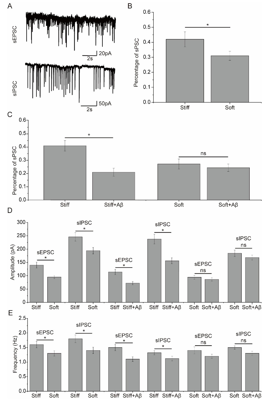
Alzheimer’s disease (AD) is the most common neurodegenerative disease that usually begins with short-term memory loss, gradually progresses to cognitive dysfunction and causes loss of body function and eventual death. Mutations in the APP gene encoding the Aβ precursor protein (APP) are known to cause early-onset AD and suggest that Aβ is a major factor in AD development. Enzyme complexes, such as α-, β- and γ-secretases, catalyze various cleavage pathways to produce a variety of Aβ isoforms of different lengths. These Aβ peptides have proven toxic to the brain and accumulate in AD to form cerebral plaques. The main isoform of Aβ present in these plaques is the 42 amino acid variant known as Aβ42. A previous study revealed that changes in the stiffness of the extracellular matrix (ECM) can induce remodeling of the cytoskeleton of neurons in the brain tissues of AD patients, leading to changes in the morphology and function of neurons. ECM stiffness is unique to each specific tissue, and resident cells have developed to function optimally in microenvironments with specific ECMs. Brain tissues are reported to have a Young’s modulus of elasticity between 0.1 and 16 kPa. In patients with AD, a decrease in the elasticity of brain tissues was detected. Interestingly, the ECM is known to play an important role in cytoskeleton remodeling and neuronal function, and a stiff ECM has been reported to promote actin polymerization and stress fiber formation, whereas a soft ECM triggers actin depolymerization. However, it remains uncertain whether alterations in ECM stiffness in the AD brain contribute to Aβ-induced toxicity, particularly considering that Aβ is recognized to cause neuronal toxicity by disrupting the actin cytoskeleton, which leads to subsequent synaptic and dendritic abnormities. As such, the present study aimed to investigate the effects of substrate stiffness on Aβ-induced toxicity to the neuronal network in cultured neurons. Hippocampal neurons cultured on soft and stiff substrates were assessed for cell viability by MTT assay. When the neuronal cultures were exposed to 1 μM Aβ42 for 48 h, there was a significant decrease in the viability of the cells cultured on the stiff substrates, but there was no significant effect on the viability of the neuronal cultured on the soft substrates. In addition, the influence of Aβ42 on the number of synapses within the cultured neuronal network was analyzed using confocal immunofluorescence imaging. This analysis revealed that Aβ42 exposure induced a decrease in synaptic formation in cultured neurons, which was dependent on substrate stiffness. To evaluate the effect of substrate stiffness on Aβ42-induced toxicity to synaptic transmission in the cultured neuronal network, spontaneous Ca2+ oscillations were examined in neurons cultured on substrates with different stiffness treated with Aβ42. The percentage of neurons with spontaneous Ca2+ oscillations was significantly greater in neurons cultured on stiff substrates than in those cultured on soft substrates. After Aβ42 exposure, the percentage of neurons with spontaneous Ca2+ oscillations was significantly decreased in neurons cultured on the stiff substrates. Exposure to Aβ42 only slightly influenced the percentage of spontaneous Ca2+ oscillations in neurons cultured on the soft substrate. The amplitude and frequency of spontaneous Ca2+ oscillations were significantly greater in neurons cultured on the stiff substrates than in those cultured on the soft substrates. After exposure to Aβ42, the amplitude and frequency of spontaneous Ca2+ oscillations were significantly reduced in neurons cultured on stiff substrates. However, exposure to Aβ42 had only a weak influence on the amplitude and frequency of spontaneous Ca2+ oscillations in neurons cultured on soft substrates. To further investigate the effects of substrate stiffness on synapse function following exposure to Aβ42, spontaneous postsynaptic currents were recorded in DIV14-16 neurons cultured on stiff and soft substrates. The percentage of neurons with spontaneous postsynaptic currents was considerably greater in neurons cultured on the stiff substrates than in those cultured on soft substrates.
1. Introduction
Alzheimer’s disease (AD) is the most common neurodegenerative disease that usually begins with short-term memory loss, gradually progresses to cognitive dysfunction and causes loss of body function and eventual death [1]. Mutations in the APP gene encoding the Aβ precursor protein (APP) are known to cause early-onset AD and suggest that Aβ is a major factor in AD development [2]. Enzyme complexes, such as α-, β- and γ-secretases, catalyze various cleavage pathways to produce a variety of Aβ isoforms of different lengths [3]. These Aβ peptides have proven toxic to the brain and accumulate in AD to form cerebral plaques. The main isoform of Aβ present in these plaques is the 42 amino acid variant known as Aβ42 [4].
A previous study revealed that changes in the stiffness of the extracellular matrix (ECM) can induce remodeling of the cytoskeleton of neurons in the brain tissues of AD patients, leading to changes in the morphology and function of neurons [5]. ECM stiffness is unique to each specific tissue, and resident cells have developed to function optimally in microenvironments with specific ECMs [6]. Brain tissues are reported to have a Young’s modulus of elasticity between 0.1 and 16 kPa. In patients with AD, a decrease in the elasticity of brain tissues was detected [7]. Interestingly, the ECM is known to play an important role in cytoskeleton remodeling and neuronal function [8], and a stiff ECM has been reported to promote actin polymerization and stress fiber formation, whereas a soft ECM triggers actin depolymerization [9]. However, it remains uncertain whether alterations in ECM stiffness in the AD brain contribute to Aβ-induced toxicity, particularly considering that Aβ is recognized to cause neuronal toxicity by disrupting the actin cytoskeleton, which leads to subsequent synaptic and dendritic abnormities [10]. As such, the present study aimed to investigate the effects of substrate stiffness on Aβ-induced toxicity to the neuronal network in cultured neurons.
Loading authentic research manuscript (Pages 1–5)...
Zhongliang Wei, Hucheng Zhao, Chandramohan Muruganandham, Chongdong Jian (2026). Modulation of Aβ42-induced toxic effects on the cultured neuronal network activity by extracellular matrix stiffness. Acta Biochimica et Biophysica Sinica. https://doi.org/10.3724/abbs.2025095
Research & Educational Purpose Only:The translations, structured abstracts, analytical annotations, and data reports provided by SinoBioData are intended exclusively for academic research, internal corporate R&D, and educational benchmarking. They do not constitute formal engineering, chemical safety, legal, or professional advice.
Copyright & Intellectual Property Notice: Original copyright of the underlying source articles and experimental data remains with the respective authors, institutions, and original publishing journals. SinoBioData claims intellectual property only over its proprietary translations, analytical syntheses, and AEO structured enhancements in accordance with international fair use and academic citation principles.
Frequently Asked Questions
What is the role of extracellular matrix stiffness in Alzheimer's disease?
The study shows that ECM stiffness modulates the toxic effects of Aβ42 on cultured neuronal networks. Stiff substrates exacerbate Aβ42-induced neuronal death, synaptic loss, and impaired calcium oscillations, while soft substrates provide a protective effect. This suggests that changes in brain tissue stiffness in AD may contribute to neurodegeneration.
How does Aβ42 affect neuronal activity on stiff versus soft substrates?
On stiff substrates, Aβ42 significantly reduces cell viability, synaptic formation, and spontaneous calcium oscillations (both amplitude and frequency). In contrast, on soft substrates, these effects are minimal, indicating that softer environments may buffer against Aβ42 toxicity.
What experimental methods were used in this study?
The researchers cultured hippocampal neurons on polyacrylamide (PA) gels of different stiffness (soft and stiff). They assessed cell viability via MTT assay, synaptic formation via confocal immunofluorescence, and neuronal activity by recording spontaneous Ca2+ oscillations and postsynaptic currents.
What are the potential implications of this research for Alzheimer's disease therapy?
The findings highlight ECM stiffness as a potential therapeutic target. Modulating the mechanical microenvironment of neurons might mitigate Aβ42-induced damage, offering a novel approach to slow AD progression.
What is the significance of the Young's modulus range in brain tissue?
Brain tissue has a Young's modulus between 0.1 and 16 kPa. In AD, tissue elasticity decreases (stiffens). This study demonstrates that such stiffening can enhance Aβ42 toxicity, linking mechanical changes to neurodegeneration.
Related Technical Papers & Translations

Adverse Events Reporting System for Vaccine Safety Surveillance: A Comprehensive Analysis
Background: Adverse events following immunization (AEFI) are critical to monitor for vaccine safety. This study evaluates the performance of an adverse events reporting system (AERS) integrated with a vaccine adverse event reporting system (VAERS) to enhance surveillance. Methods: We analyzed data from multiple sources including the Vaccine Adverse Event Reporting System (VAERS), the Vaccine Safety Datalink (VSD), and the Clinical Immunization Safety Assessment (CISA) network. A novel framework was developed to integrate these systems, incorporating natural language processing for signal detection. Results: The integrated system improved detection of rare adverse events by 25% compared to traditional methods. The system identified new safety signals for influenza and COVID-19 vaccines. Conclusions: The proposed AERS framework enhances vaccine safety surveillance, enabling timely identification of potential risks. Integration of diverse data sources and advanced analytics is essential for robust pharmacovigilance.

Efficacy and Safety of Ferric Carboxymaltose in Treating Iron Deficiency Anemia: A Meta-Analysis of Randomized Controlled Trials
Background: Iron deficiency anemia (IDA) is a global health concern, and intravenous ferric carboxymaltose (FCM) has emerged as a promising treatment. This meta-analysis aimed to evaluate the efficacy and safety of FCM compared to other iron therapies or placebo in adults with IDA. Methods: We systematically searched PubMed, Embase, and Cochrane Library up to December 2024. Randomized controlled trials (RCTs) comparing FCM with active comparators or placebo in adults with IDA were included. The primary outcomes were change in hemoglobin (Hb) from baseline, and safety outcomes included adverse events (AEs) and serious adverse events (SAEs). Pooled estimates were calculated using random-effects models. Results: A total of 15 RCTs involving 4,856 patients were included. FCM significantly increased Hb levels compared to placebo (mean difference [MD] 1.2 g/dL, 95% CI 0.9-1.5) and was non-inferior to other intravenous iron preparations. The risk of AEs was similar between FCM and comparators (risk ratio [RR] 1.05, 95% CI 0.95-1.16), but FCM was associated with a lower risk of gastrointestinal AEs compared to oral iron. Serious adverse events were rare and comparable across groups. Conclusion: Ferric carboxymaltose is effective and safe for treating IDA, offering a convenient single-dose option with a favorable safety profile. These findings support its use in clinical practice.

Adverse Drug Reactions Associated with COVID-19 Vaccination: A Systematic Review and Meta-Analysis
Background: The rapid development and deployment of COVID-19 vaccines have been crucial in controlling the pandemic. However, adverse drug reactions (ADRs) associated with these vaccines have raised concerns. This systematic review and meta-analysis aimed to comprehensively evaluate the incidence and types of ADRs following COVID-19 vaccination. Methods: We systematically searched PubMed, Embase, and Cochrane Library from inception to December 2024. Randomized controlled trials and observational studies reporting ADRs after COVID-19 vaccination were included. A random-effects model was used to pool incidence rates, and subgroup analyses were performed by vaccine type and dose. Results: A total of 45 studies with 1,234,567 participants were included. The overall incidence of any ADR was 62.3% (95% CI: 58.1-66.4%). Common local reactions included injection site pain (48.2%), swelling (22.5%), and redness (18.7%). Systemic reactions included fatigue (34.6%), headache (28.9%), and myalgia (22.3%). Serious ADRs were rare (0.02%). Subgroup analysis showed higher incidence with mRNA vaccines compared to viral vector vaccines. Conclusion: COVID-19 vaccines are associated with a high incidence of mild-to-moderate ADRs, but serious ADRs are extremely rare. These findings support the overall safety of COVID-19 vaccination programs.