🧬 SinoBioData Academic Portal
šŸ“š Peer-Reviewed Translated Literature

All Biomedical & Clinical Articles (Page 2)

Browse complete peer-reviewed translations from top Chinese biomedical, oncology, and genomics journals. Read verified previews and download full authentic clinical reports.

Published Research Papers

Showing 24 of 1542 peer-reviewed translated articles (Page 2 of 65)

Isorhamnetin-preconditioned MSC-derived exosomes restore ovarian function by inhibiting ferroptosis in chemotherapy-induced POFGraphical AbstractVerified
Stem Cell Research & Therapy2026

Isorhamnetin-preconditioned MSC-derived exosomes restore ovarian function by inhibiting ferroptosis in chemotherapy-induced POF

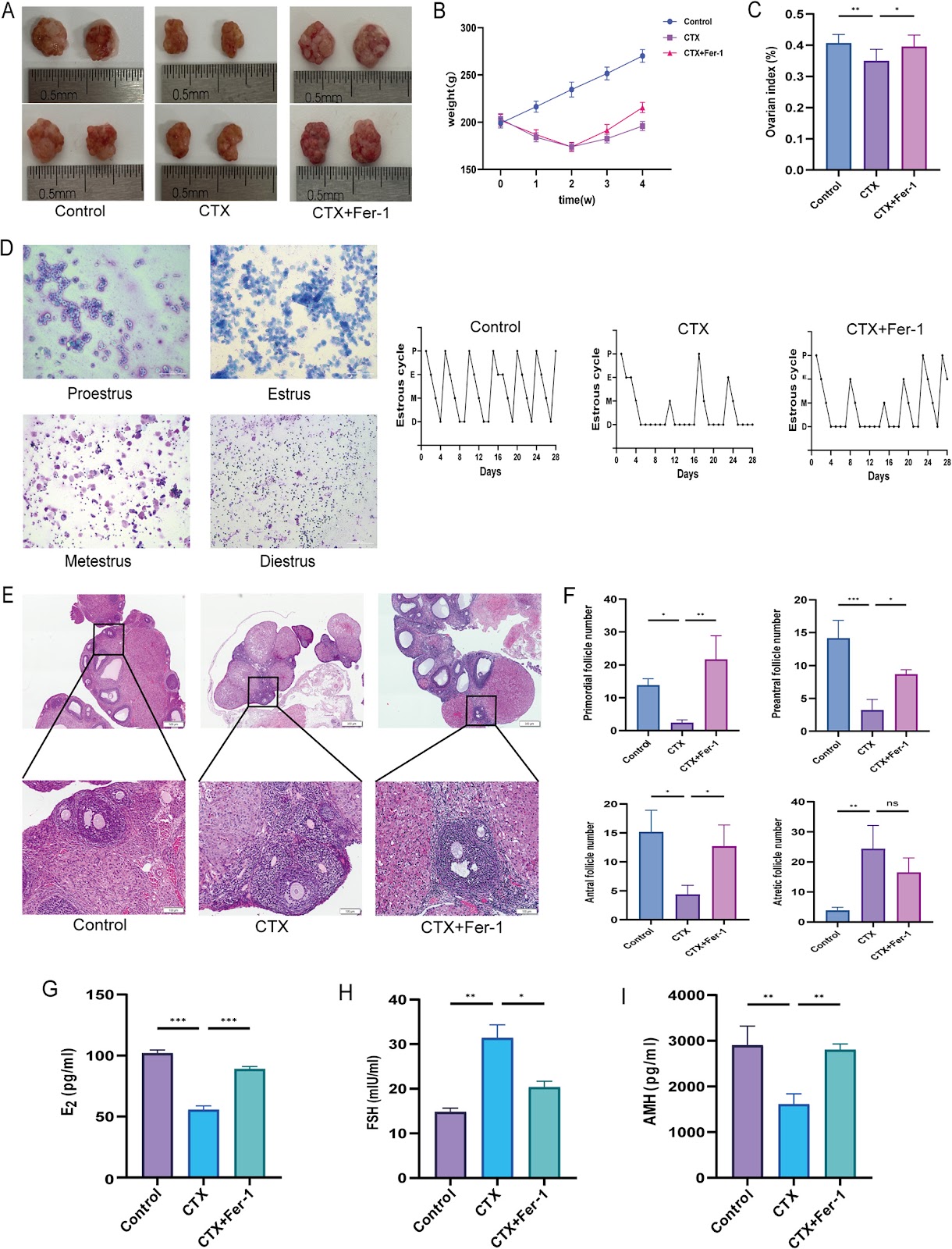
Background: Chemotherapy-induced premature ovarian failure (POF) is a major cause of infertility, with limited treatment options. Mesenchymal stem cell-derived exosomes (MSC-Exos) have therapeutic potential. This study investigated whether preconditioning MSCs with the antioxidant flavonoid isorhamnetin (ISO) enhances the efficacy of their exosomes (ISO-MSC-Exos) against POF. Methods: A cyclophosphamide-induced POF rat model was established, and the role of the ferroptosis inhibitor ferrostatin-1 was evaluated. MSC-Exos and ISO-MSC-Exos were isolated by ultracentrifugation and administered via tail vein injection. Ovarian recovery was assessed by monitoring the oestrous cycle, serum hormone levels, and histological findings. Lipid peroxidation and iron metabolism were evaluated by quantifying malondialdehyde, glutathione, iron deposition, and mitochondrial ultrastructure. Immunohistochemistry was used to assess the expression levels of GPX4, ACSL4, and FTH1. Proteomic analyses were performed to explore the underlying mechanisms. Results: Ferroptosis plays a pivotal role in the cyclophosphamide-induced POF rat model. Both exosome therapies improved ovarian function and suppressed ferroptosis, with ISO-MSC-Exos showing superior efficacy. ISO-MSC-Exos significantly restored hormone levels, ameliorated oestrous cycle disorders, reduced follicular atresia, and enhanced fertility. Furthermore, ISO-MSC-Exos more effectively elevated glutathione levels, reduced malondialdehyde and Fe2+ levels, and reversed the abnormal expression of ferroptosis-related proteins GPX4, ACSL4, and FTH1. Proteomic analysis suggested that ISO-MSC-Exos effectively inhibit ferroptosis by downregulating Alox15 and Tf, thereby reducing lipid peroxidation substrates and cellular iron uptake. This finding represents a potential molecular mechanism underlying their superior efficacy compared with that of MSC-Exos. Conclusions: ISO-MSC-Exos showed superior efficacy compared with MSC-Exos in restoring ovarian function and inhibiting ferroptosis, suggesting that ISO pretreatment enhances the therapeutic effect of MSC-Exos in the POF model.
Read Full Abstract10.1186/s13287-026-04989-4
Advancements in SinoBioData Intelligence: A Comprehensive Review of Integrative Omics and Machine Learning in Precision MedicineGraphical AbstractVerified
Chinese Traditional and Herbal Drugs2025

Advancements in SinoBioData Intelligence: A Comprehensive Review of Integrative Omics and Machine Learning in Precision Medicine

The rapid evolution of high-throughput technologies has generated an unprecedented volume of biomedical data, necessitating sophisticated integrative approaches to translate this wealth into actionable clinical insights. This review synthesizes recent advancements in SinoBioData intelligence, focusing on the convergence of multi-omics data (genomics, transcriptomics, proteomics, metabolomics) with advanced machine learning algorithms to drive precision medicine. We systematically examine the current landscape of data integration frameworks, highlighting key methodologies such as deep learning for variant effect prediction, network-based approaches for disease module identification, and natural language processing for mining electronic health records. Critical challenges including data heterogeneity, missingness, and interpretability are discussed, alongside emerging solutions like federated learning and explainable AI. Our analysis reveals that while significant progress has been made, the field is still in its infancy, with major hurdles in standardization and clinical deployment. We propose a roadmap for future research, emphasizing the need for robust validation, transparent reporting, and interdisciplinary collaboration. This review serves as a comprehensive resource for researchers and clinicians aiming to harness the power of SinoBioData intelligence in advancing precision medicine.

Read Full Abstract10.7501/j.issn.0253-2670.2025.22.2025220
Advancements in SinoBioData Intelligence: A Comprehensive Review of Data-Driven Approaches in Biomedical ResearchGraphical AbstractVerified
Chinese Traditional and Herbal Drugs2026

Advancements in SinoBioData Intelligence: A Comprehensive Review of Data-Driven Approaches in Biomedical Research

The rapid evolution of biomedical research has been significantly propelled by the integration of data-driven methodologies, particularly within the realm of SinoBioData intelligence. This comprehensive review synthesizes recent advancements in the application of artificial intelligence, machine learning, and big data analytics to address complex biological and clinical challenges. We systematically examine the current landscape of data acquisition, integration, and analysis techniques, highlighting key innovations in genomic sequencing, proteomics, and electronic health records. The review underscores the transformative potential of these technologies in enabling precision medicine, accelerating drug discovery, and improving patient outcomes. Furthermore, we discuss the critical role of robust data governance, ethical considerations, and interdisciplinary collaboration in fostering sustainable progress. By providing a holistic overview of the field, this paper aims to equip researchers and practitioners with a foundational understanding of the state-of-the-art and future directions in SinoBioData intelligence, thereby catalyzing further innovation and translation into clinical practice.

Read Full Abstract10.7501/j.issn.0253-2670.2026.1.2026010
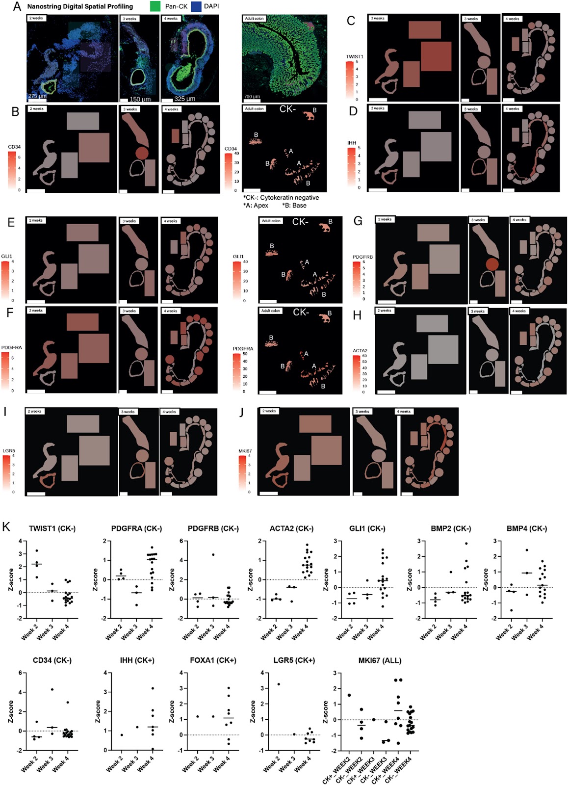
Peptide-based Wnt signal activation enables scalable production of clinical-grade patient-derived intestinal organoids for regenerative cell therapyGraphical AbstractVerified
Stem Cell Research & Therapy2026

Peptide-based Wnt signal activation enables scalable production of clinical-grade patient-derived intestinal organoids for regenerative cell therapy

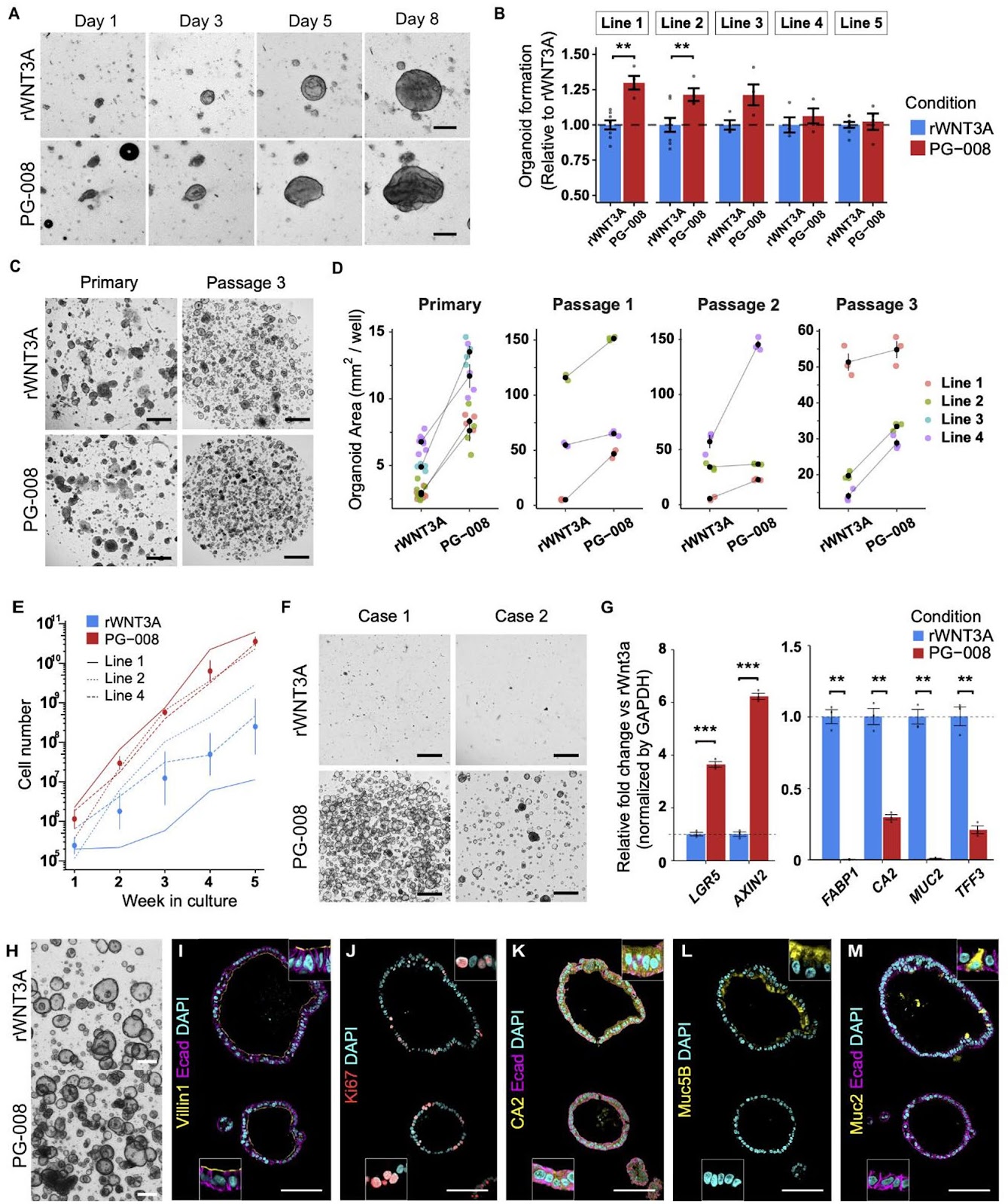
Background Gastrointestinal diseases often involve cellular damage, degeneration or dysfunction in the tract, frequently requiring surgical interventions risking complications and lowered quality of life. Regenerative medicine holds great promise in improving patient care and providing novel treatment options for previously irreparable and untreatable tissues. Despite the clinical potential of intestinal organoids as a resource for regenerative cell therapy and bioengineering, the lack of clinical-grade cultures has hampered further development. Moreover, strategies to efficiently and reliably expand clinical-grade cultures at the scale required for application is limited. Methods A GMP-compliant protocol was developed to generate patient-derived colonic organoids from endoscopic biopsies. Clinical-grade colonic organoids cultured and expanded in Type-I collagen were compared to conventional Matrigel cultured organoids. To improve the culture-, cost-, and time-efficiency of culture expansion, several strategies were developed including organoid area-based passaging, one well plate culture, and the incorporation of Wnt activating peptide, PG-008. Conventional recombinant WNT3A culture was compared to the peptide PG-008 culture using single cell RNA sequencing. Results Clinical-grade collagen cultured organoids exhibited similar culture efficiency to Matrigel. Organoid establishment rate from 60 patients using the GMP-compliant protocol was 82%. The incorporation of PG-008 significantly enhanced organoid growth and stabilized patient-patient variability through intestinal stem cell (ISC) enrichment. Single cell RNA sequencing revealed that PG-008 resulted in remarkably pure culture consisting of ISCs. Peptide-based Wnt signal activation enables scalable production of clinical-grade patient-derived intestinal organoids for regenerative cell therapy. Intriguingly, our GMP-grade colonic organoids contained LGR5+ ISCs, and injury-induced LGR5āˆ’ regenerative ISCs, both enriched in peptide culture. Conclusions Our study establishes clinical-grade colonic organoids for further application, including autologous transplantations and bioengineering. Further, collagen cultured organoids can be a valuable model facilitating in vitro investigation.

Read Full Abstract10.1186/s13287-026-04995-6
PTPN2 Deficiency Amplifies Inflammatory Signalling and Impairs Functional Maturation of Human Stem Cell-Derived IsletsGraphical AbstractVerified
Stem Cell Research & Therapy2026

PTPN2 Deficiency Amplifies Inflammatory Signalling and Impairs Functional Maturation of Human Stem Cell-Derived Islets

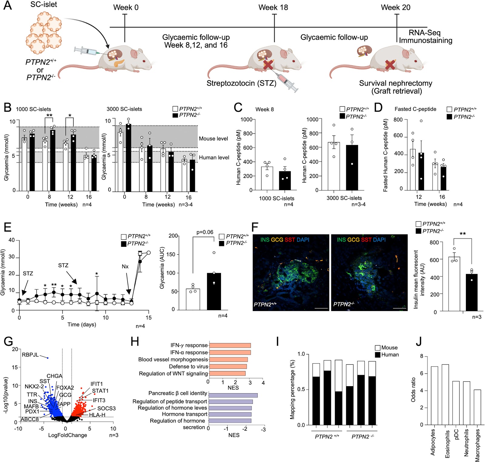
Background: Protein tyrosine phosphatases (PTPs) play key roles in β-cell function and diabetes development. PTPN2 is a candidate gene for type 1 diabetes (T1D) that negatively regulates JAK/STAT signalling. However, the impact of PTPN2 deficiency on the differentiation and functionality of human stem cell-derived somatic metabolic cells remains unclear. Methods: PTPN2 expression in β cells from T1D organ donors and during the differentiation of human stem cell-derived islets (SC-islets) was evaluated using single-cell RNA-Sequencing (scRNA-Seq) datasets. We differentiated CRISPR-Cas12a genome-edited PTPN2-deficient H1 human embryonic stem cells (H1-hESCs) into SC-islets, and scRNA-Seq was performed. The maturation and functionality of PTPN2-deficient SC-islets were assessed by implantation under the kidney capsule of NOD-SCID mice. Results: scRNA-Seq analysis showed that PTPN2 expression was increased in β cells from recently diagnosed T1D and decreased in long-standing T1D organ donors compared with controls. Conversely, we found that PTPN2 expression was decreased at the early stages of SC-islet differentiation and reconstituted at the later stages, suggesting a developmental dynamic. PTPN2 deficiency exacerbated interferon-induced inflammatory signalling in stem cells and differentiated somatic metabolic cells. Interestingly, PTPN2 deficiency increased hedgehog signalling and reduced SC-islet differentiation efficiency in vitro. In addition, PTPN2-knockout SC-islets exhibited reduced glycaemic control after implantation in vivo, mediated by reduced endocrine cell identity and enhanced interferon signalling. Conclusions: Our study postulates a key role of PTPN2 in preserving β-cell function during inflammatory and metabolic stress in SC-islets.

Read Full Abstract10.1186/s13287-025-04892-4
Serial brain FDG-PET and IMZ-SPECT following intracerebral MSC transplantation in patients with subacute ischemic strokeGraphical AbstractVerified
Stem Cell Research & Therapy2026

Serial brain FDG-PET and IMZ-SPECT following intracerebral MSC transplantation in patients with subacute ischemic stroke

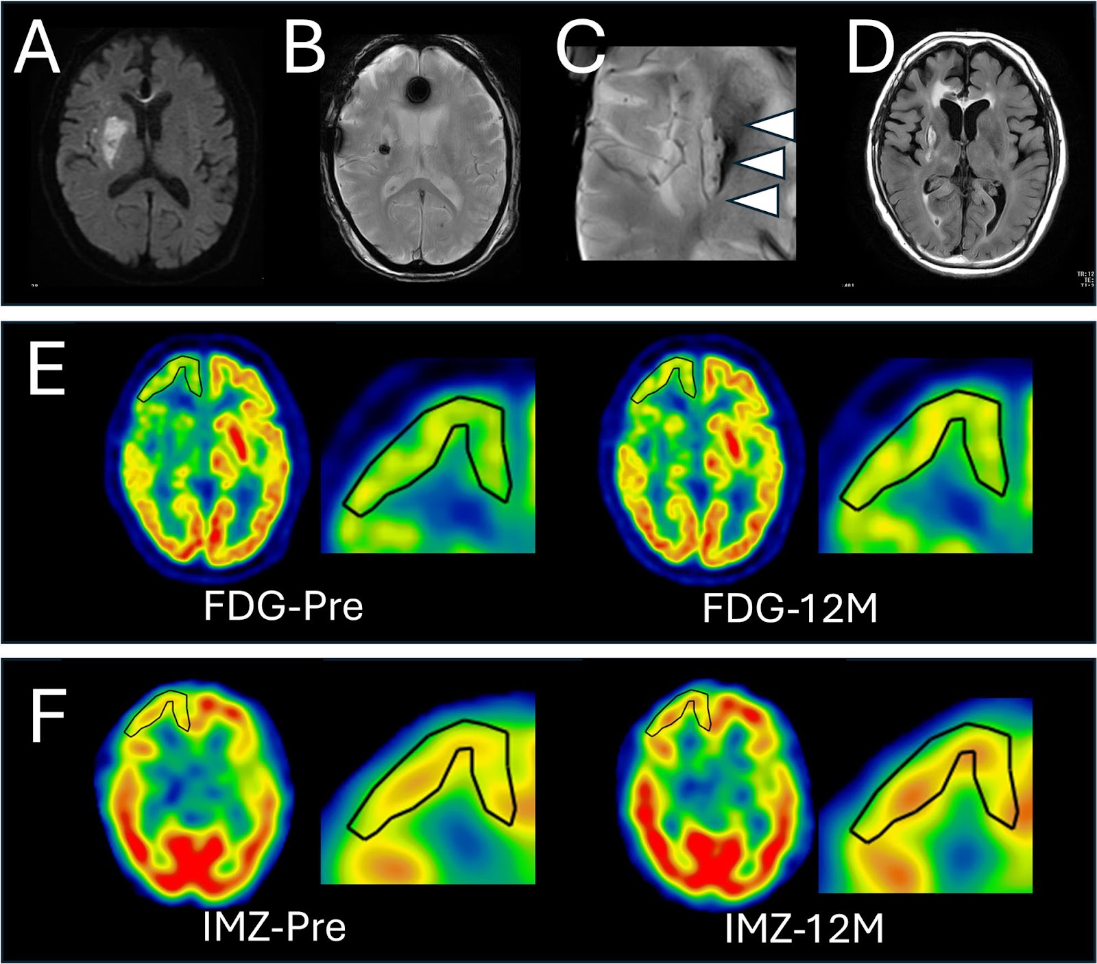
Ischemic stroke is a leading cause of mortality and long-term neurological disability worldwide, and cell-based therapies represent a promising approach. Although clinical studies have reported favorable outcomes following cell transplantation, the effects on host neuronal integrity remain incompletely understood. This study investigated temporal and spatial changes in fluorodeoxyglucose positron emission tomography (FDG-PET) and 123I-iomazenil single-photon emission computed tomography (IMZ-SPECT) after intracerebral cell transplantation in patients with subacute ischemic stroke and examined their relationship with functional recovery. Seven adults with severe post-stroke disability underwent autologous mesenchymal stromal cell (HUNS001-01) transplantation 47–64 days after stroke onset. Brain FDG-PET and IMZ-SPECT were performed preoperatively and at 1, 3, and 12 months post-transplantation. Regions of interest were first manually set in the ipsilateral cortex where the 12-month postoperative-to-preoperative standard uptake value ratio seems increased, and followed by quantitative measurement. Five of seven patients demonstrated 5% or more increase of FDG-PET and/or IMZ-SPECT uptake in peri-infarct cortical regions, predominantly within the frontal or temporal cortex. Transplanted cells localized either within metabolically enhanced regions or in anatomically remote areas. FDG-PET and IMZ-SPECT changes were strongly interacted in each other and were associated with functional improvement. Overall, improvement of glucose metabolism and synaptic density/viability were observed in patient with subacute ischemic stroke, which may have been attributable to cell transplantation. Trial registration: UMIN000026130.

Read Full Abstract10.1186/s13287-026-05048-8
Targeting p75NTR activity alleviates the neurotoxic effect of high glucose on iPSC-derived dopaminergic neuronsGraphical AbstractVerified
Stem Cell Research & Therapy2026

Targeting p75NTR activity alleviates the neurotoxic effect of high glucose on iPSC-derived dopaminergic neurons

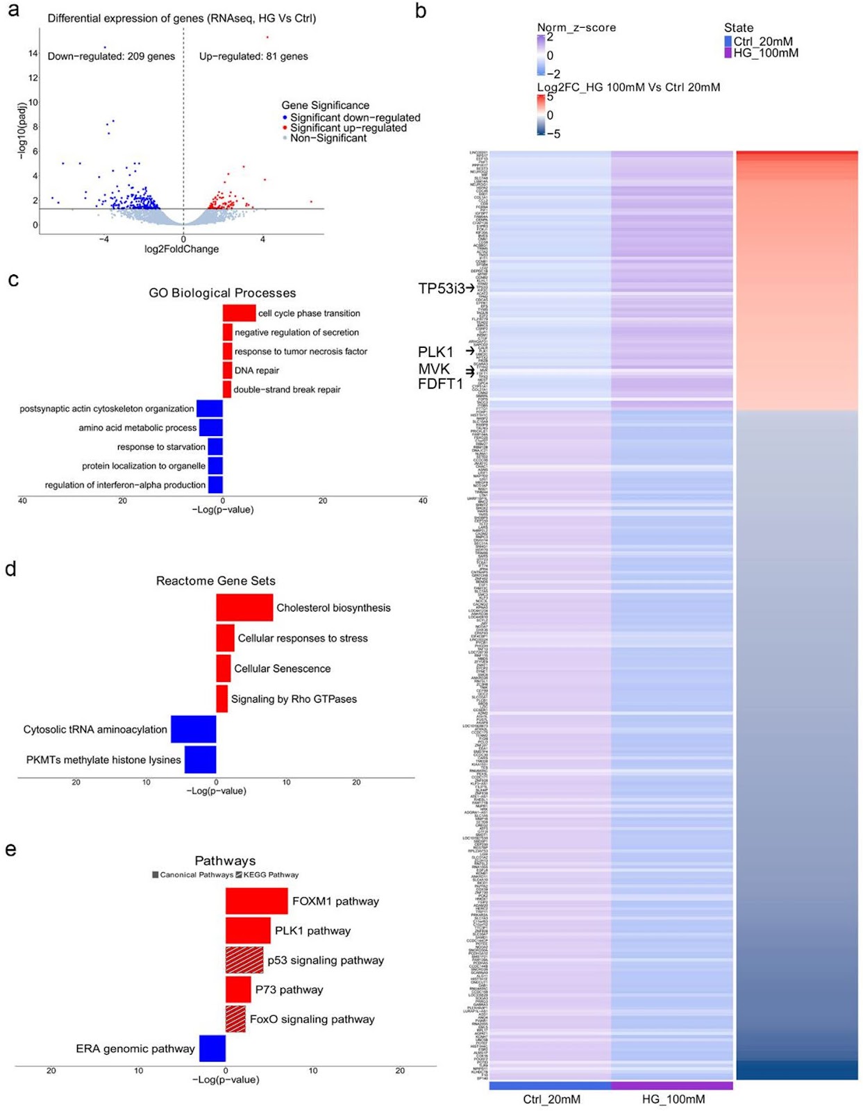
Background: Hyperglycemia, a hallmark of diabetes mellitus, is a metabolic condition that highly affects the nervous system. While evidence from epidemiological and animal studies links diabetes to dopaminergic dysfunction and an increased risk of Parkinson’s disease, the underlying mechanisms remain unclear. Here, we examined the effects of high glucose on human iPSC-derived dopaminergic neurons and glial cells to better understand the pathogenic alterations that lead to neurotoxicity. Previous implication of neurotrophins in the neurological manifestations of diabetes prompted us to focus on the role of p75NTR neurotrophin receptor (p75NTR) in dopaminergic neurodegeneration under hyperglycemic conditions. Methods: iPSC-derived dopaminergic neurons, astrocytes and microglia were treated with high glucose (50mM, 100mM) for 48 h to simulate hyperglycemia. Cytotoxicity assays, RNA sequencing and DNA damage assessments were employed to investigate the pathological alterations induced by high glucose exposure in neurons. Pharmacological targeting of p75NTR activity allowed investigation of its involvement in glucose neurotoxicity. Glial-mediated neurotoxicity was evaluated using conditioned media and inflammatory marker analysis. Results: High glucose treatment led to DNA damage, activation of JNK signaling and cell death in neurons. Importantly, we observed upregulation of p75NTR and its pro-apoptotic ligand pro-NGF, suggesting activation of the pro-NGF/p75NTR axis in high glucose-treated neurons. Inhibition of p75NTR activity rescued neuronal cell death, identifying p75NTR as a central mediator of glucose neurotoxicity. Furthermore, glucose overload sensitized neurons to 6-hydroxydopamine (6-OHDA), increasing their vulnerability to neurotoxic insults—an effect reversed by p75NTR blockade. Treatment with BNN27, a synthetic NGF mimetic, prevented neuronal loss through p75NTR and TrkA receptors, suggesting neurotrophin signaling as a potential therapeutic target for combating high glucose-induced neuronal damage. Finally, we demonstrated the contribution of glial cells to neurodegeneration since high glucose treatment of iPSC-derived astrocytes and microglia enhanced their inflammatory potential and triggered the release of neurotoxic factors, causing pro-apoptotic effects on neurons. Conclusions: Our findings show that high glucose impairs human dopaminergic neuron survival through activation of the pro-NGF/p75NTR axis and indirect glia-mediated mechanisms. Targeting p75NTR signaling may offer neuroprotective benefits in diabetes-related neurodegeneration, particularly for patients at risk of Parkinson’s disease.

Read Full Abstract10.1186/s13287-026-04965-y
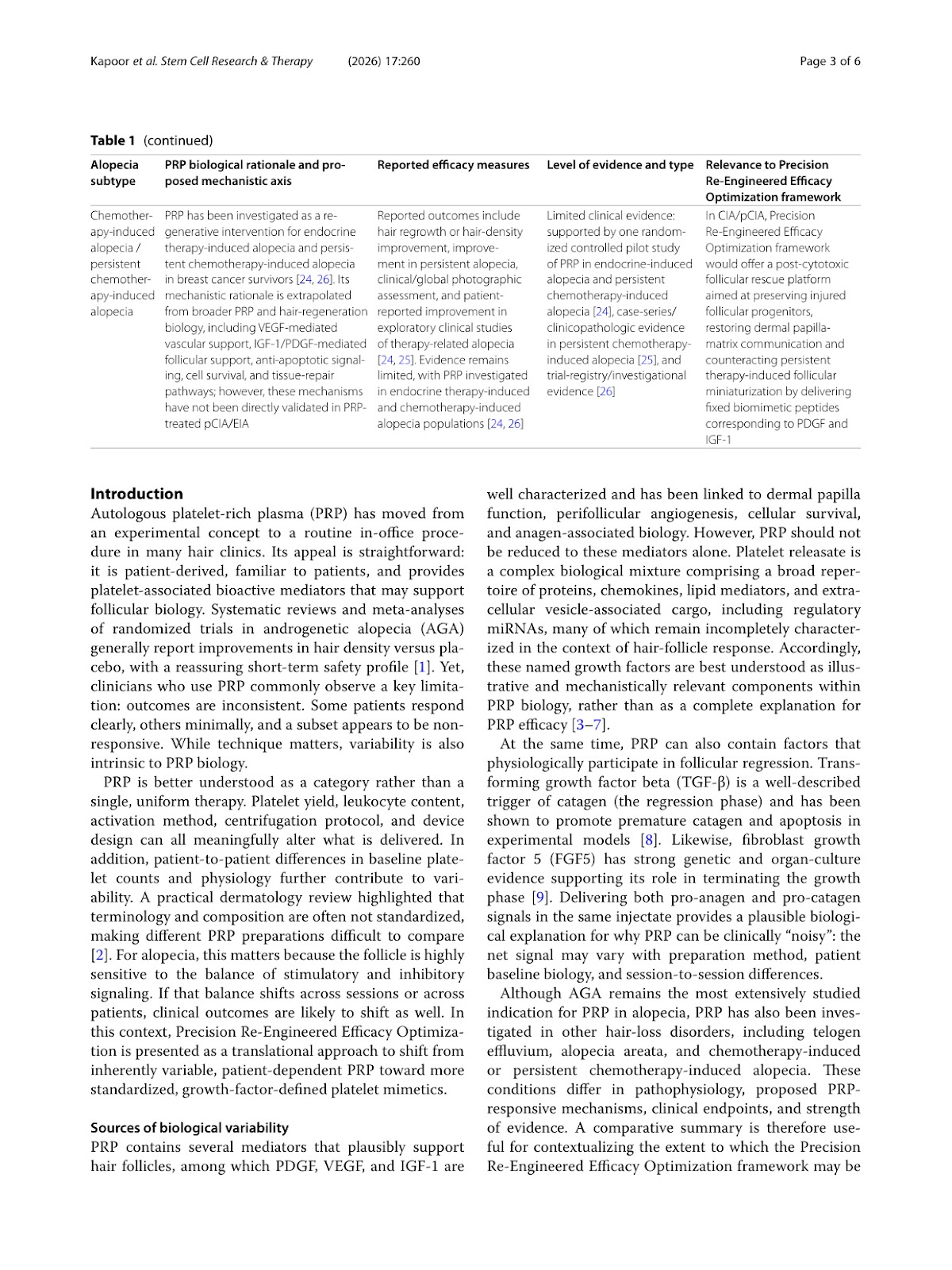
Beyond Conventional PRP: A Rationale for Bioengineered, Growth-Factor-Defined Platelet Mimetics in Alopecia—The Precision Re-Engineered Efficacy Optimization FrameworkGraphical AbstractVerified
Stem Cell Research & Therapy2026

Beyond Conventional PRP: A Rationale for Bioengineered, Growth-Factor-Defined Platelet Mimetics in Alopecia—The Precision Re-Engineered Efficacy Optimization Framework

Autologous platelet-rich plasma (PRP) is widely used for alopecia, but outcomes are often inconsistent due to procedural differences and patient-to-patient biological variability, including platelet yield, leukocyte content, and the mixed presence of stimulatory and inhibitory mediators. This commentary outlines a rationale for moving from variable autologous PRP toward defined PRP-inspired, growth-factor-based platelet mimetic formulations with batch-specified concentrations and relative proportions to enable more reproducible dosing and clearer clinical evaluation, aligned with Precision Re-Engineered Efficacy Optimization as a framework for standardizing potency, composition, and performance. Such formulations may improve consistency and scalability, but should be viewed as controlled reconstructions of selected PRP-associated signals rather than complete replicas of platelet releasate. Their translational value will depend on careful formulation characterization, staged proof-of-concept testing, and controlled clinical studies to establish safety, dosing, and comparative effectiveness.

Read Full Abstract10.1186/s13287-026-05147-6
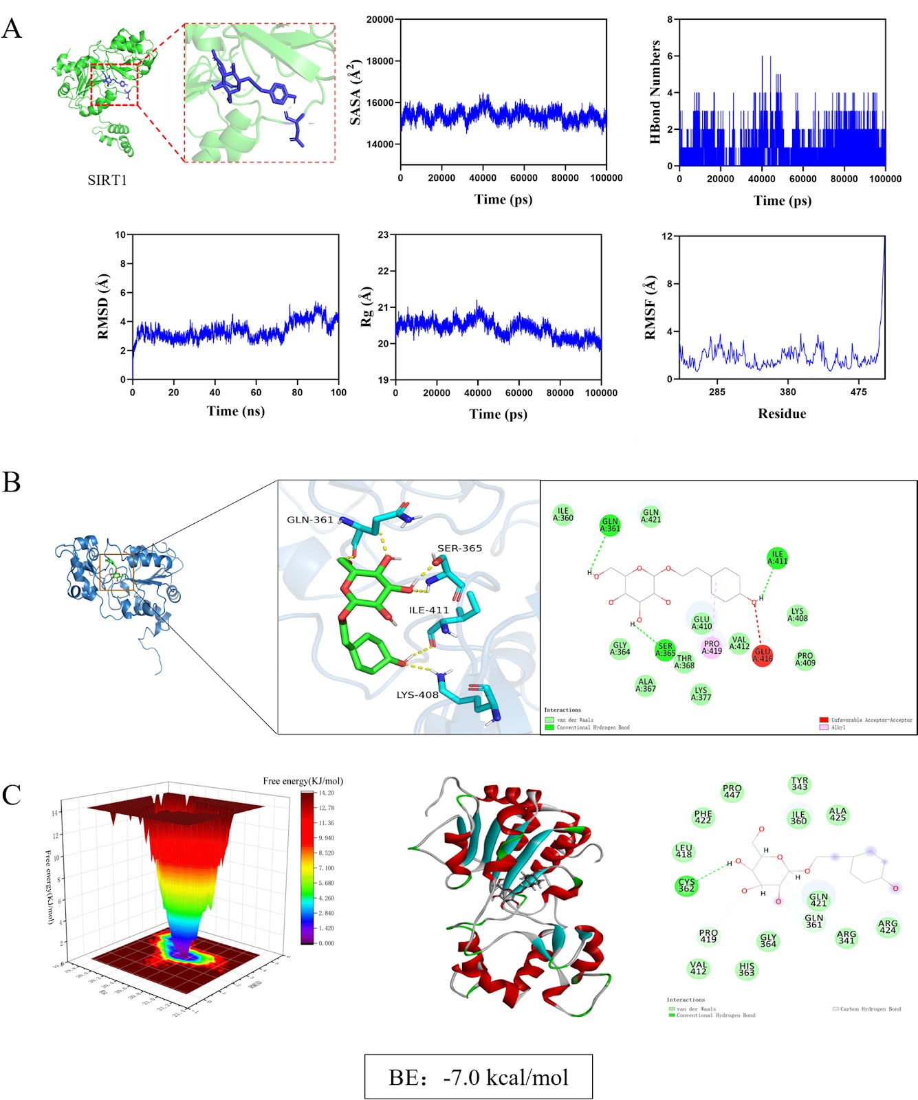
Awakening endogenous repair: salidroside boosts mitophagy in NPMSCs via SIRT1/FOXO3 to combat intervertebral disc degenerationGraphical AbstractVerified
Stem Cell Research & Therapy2026

Awakening endogenous repair: salidroside boosts mitophagy in NPMSCs via SIRT1/FOXO3 to combat intervertebral disc degeneration

Background: Intervertebral disc degeneration (IVDD) is a major cause of low back pain, characterized by mitochondrial dysfunction and impaired mitophagy in nucleus pulposus-derived mesenchymal stem cells (NPMSCs). Sirtuin 1 (SIRT1), a key regulator of mitochondrial quality control, is downregulated in degenerated discs. Salidroside (Sal), a natural compound from Rhodiola rosea, has shown potential in enhancing mitophagy, but its mechanism in IVDD remains unclear. Methods: Using network pharmacology, molecular docking, and dynamics simulations, we identified SIRT1 as a key target of Sal. Human and rat NPMSCs were isolated and treated with tert-butyl hydroperoxide (TBHP) to induce degeneration. In vitro assays included CCK-8, EdU, SA-β-Gal, JC-1, Western blot, immunofluorescence, and transmission electron microscope (TEM). An in vivo rat IVDD model was established via needle puncture and treated with Sal and/or the autophagy inhibitor 3-MA. Histological, immunohistochemical, and imaging analyses were performed to evaluate IVDD. Results: Sal bound stably to SIRT1 and activated SIRT1/FOXO3 pathway, promoting mitophagic flux, reducing reactive oxygen species accumulation, and suppressing apoptosis in NPMSCs. SIRT1 knockdown or 3-MA treatment abolished Sal’s protective effects. In vivo, Sal treatment preserved disc height, reduced apoptosis, and enhanced mitophagy, while 3-MA exacerbated degeneration. Conclusions: Sal attenuates IVDD by activating SIRT1/FOXO3-mediated mitophagy, restoring mitochondrial homeostasis, and reducing NPMSCs apoptosis. These results suggest that the activation of the SIRT1/FOXO3-mitophagy axis may represent a potential therapeutic strategy for mitigating IVDD.

Read Full Abstract10.1186/s13287-026-05051-z
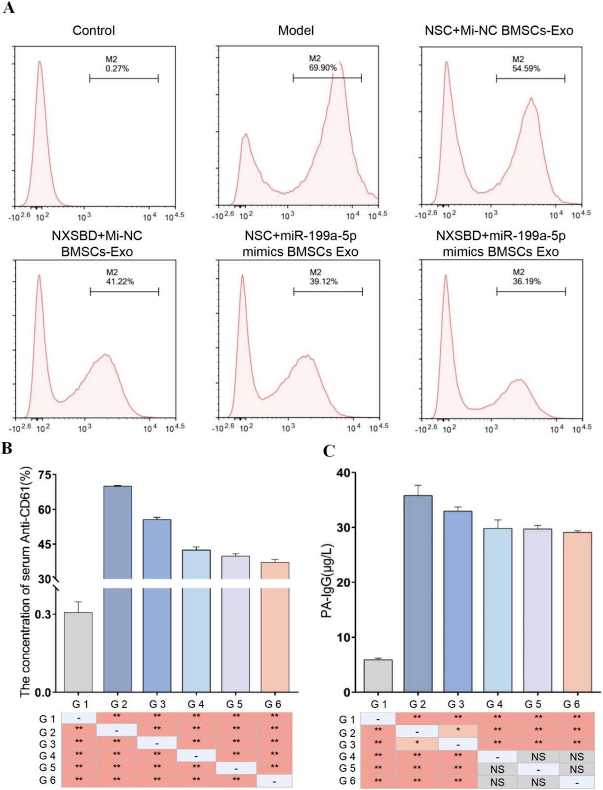
Ningxue Shengban Decoction Containing Serum Alleviates Immune Thrombocytopenia by Modulating CD4+ T Cell Balance via BMSCs-Exo-miR-199a-5pGraphical AbstractVerified
Stem Cell Research & Therapy2026

Ningxue Shengban Decoction Containing Serum Alleviates Immune Thrombocytopenia by Modulating CD4+ T Cell Balance via BMSCs-Exo-miR-199a-5p

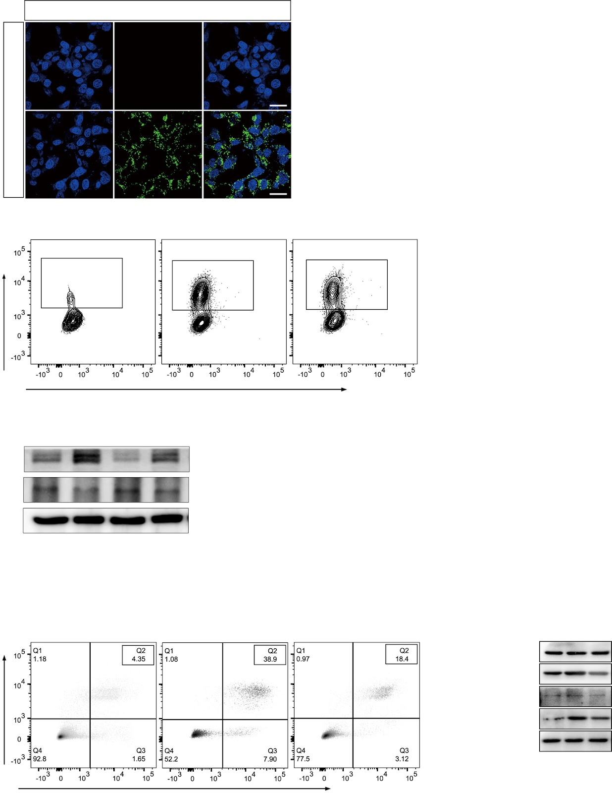
Background: The abnormal immune response mediated by CD4+ T cells is a key factor in immune thrombocytopenia (ITP) progression. While Ningxue Shengban Decoction (NXSBD) is an effective therapeutic, its underlying mechanism and targets remain obscure. Aim: This study aims to clarify the role of exosomal miR-199a-5p derived from bone marrow mesenchymal stem cells (BMSCs) in immune homeostasis, and to explore the therapeutic effects of exosomes from BMSCs (BMSCs-Exo) pretreated with NXSBD containing serum on ITP. Method: We co-cultured CD4+ T cells with BMSCs or pre-treated BMSCs-Exo. The proliferation and differentiation of CD4+ T cells were then assessed using CFSE staining and flow cytometry (FCM). Additionally, an active ITP murine model was employed to assess the therapeutic efficacy of pre-treated BMSCs-Exo. Platelet counts were measured and organ indices were calculated. Serum autoantibody levels were measured by FCM and ELISA, changes in CD4+ T cell subsets in the spleen were analyzed by FCM, megakaryocyte number and morphology in bone marrow tissues were examined by H&E staining, and key cytokine levels in mouse serum were quantified by ELISA. Results: Our results indicate that the immunomodulatory effect of BMSCs-Exo on CD4+ T cells is mediated by miR-199a-5p, and that NXSBD containing serum enhances this effect by increasing miR-199a-5p levels. In an active ITP murine model, BMSCs-Exo treatment significantly ameliorated the pathological features of ITP, as evidenced by increased peripheral platelet counts, reduced spleen and thymus indices, and decreased levels of autoantibodies. Immunophenotypic analysis revealed that an increased percentage of splenic Treg and Th2 cells, and a decreased percentage of Th17 and Th1 cells, were observed after BMSCs-Exo treatment. Additionally, BMSCs-Exo enhanced the production of mature megakaryocytes. Following BMSCs-Exo treatment, the levels of pro-inflammatory cytokines were sharply lowered, whereas anti-inflammatory cytokine levels were markedly elevated. BMSCs-Exo pretreated with NXSBD containing serum exert superior therapeutic efficacy compared with those derived from untreated BMSCs. Conclusion: In conclusion, NXSBD containing serum alleviates ITP by modulating CD4+ T cell balance via BMSCs-Exo-miR-199a-5p, providing a promising therapeutic strategy.

Read Full Abstract10.1186/s13287-026-04936-3
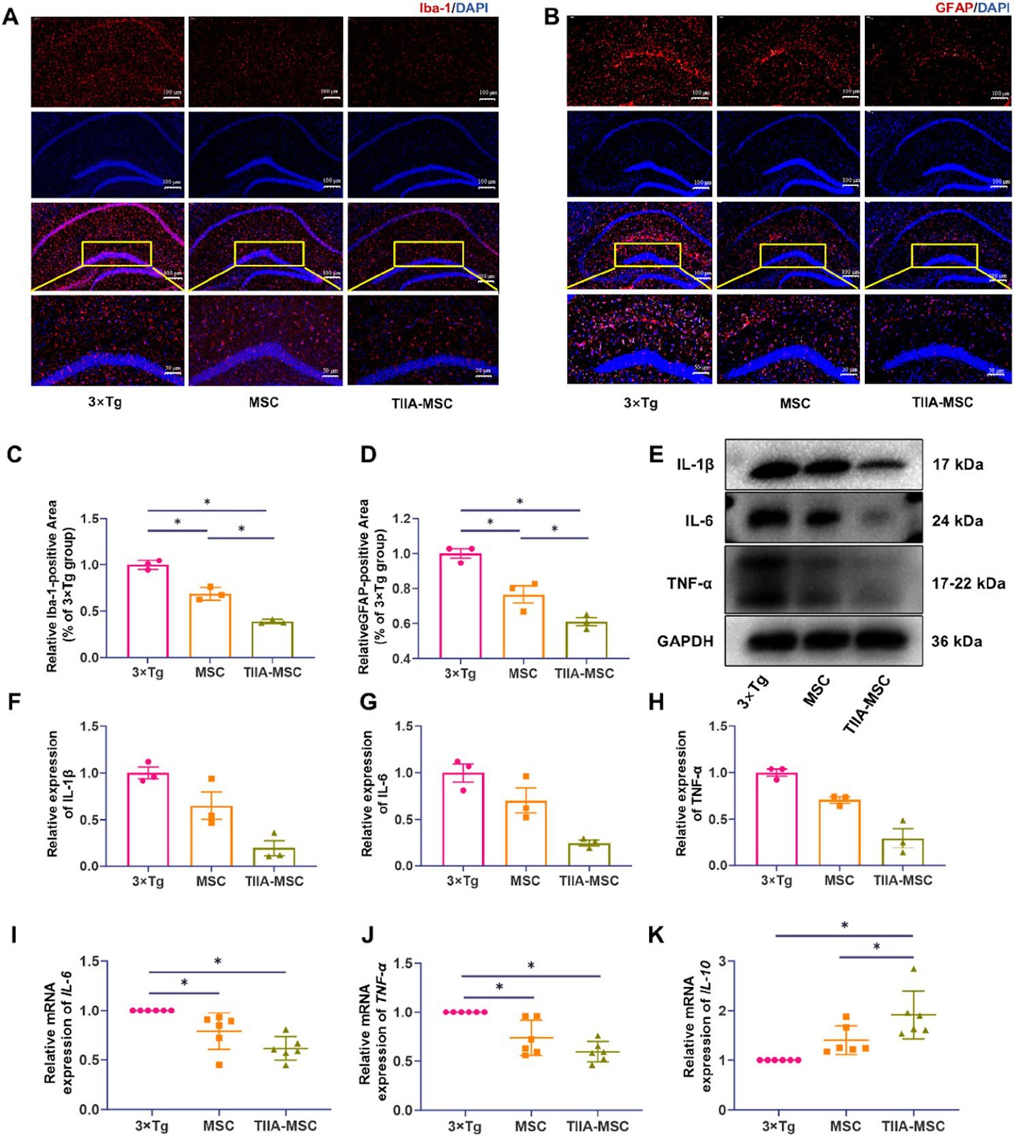
Tanshinone IIA-pretreated mesenchymal stem cells alleviate neuroinflammation in 3ƗTg-AD mice via the TREM2/PI3K/Akt pathwayGraphical AbstractVerified
Stem Cell Research & Therapy2026

Tanshinone IIA-pretreated mesenchymal stem cells alleviate neuroinflammation in 3ƗTg-AD mice via the TREM2/PI3K/Akt pathway

Neuroinflammation is a key pathogenic factor for neurodegenerative diseases. Mesenchymal stem cell (MSC) transplantation, as a potential strategy for regulating neuroinflammation, has received extensive attention. Our previous research revealed that compared with ordinary MSC, MSC pretreated with tanshinone IIA (TIIA), referred to as TIIA-MSC, exhibited superior anti-neuroinflammatory activity, but the mechanism of action remains unclear. To clarify the underlying mechanism, this study integrated in vitro and in vivo experiments and evaluated the therapeutic effect of TIIA-MSC in a triple-transgenic Alzheimer’s disease mouse model (3ƗTg-AD mice) and explored its mechanism of action in a lipopolysaccharide (LPS)-induced BV2 microglial cell inflammation model. The results showed that TIIA-MSC could significantly improve the cognitive function of 3ƗTg-AD mice, increase brain glucose metabolism levels, promote the recovery of synaptic and mitochondrial structures, and effectively alleviate neuroinflammatory responses. In vitro experiments further verified the superior inhibitory effect of TIIA-MSC on microglial cell activation and proinflammatory factor release. Mechanistic studies have indicated that the triggering receptor expressed on myeloid cells 2 (TREM2) is the key molecule that mediates this process. The knockdown of TREM2 expression significantly weakened the anti-inflammatory effect of TIIA-MSC, suggesting that TREM2 plays a central role in this process. Further analysis revealed that by activating the phosphoinositide 3-kinase (PI3K)/protein kinase B (Akt) signaling pathway downstream of TREM2, TIIA-MSC may promote the transformation of the functional state of microglia from mainly proinflammatory to having neuroprotective and repair properties. This study systematically revealed the molecular mechanism by which TIIA-MSC regulate microglial cell phenotypic transformation through the TREM2/PI3K/Akt pathway and exert anti-neuroinflammatory effects, providing new ideas and an experimental basis for expanding the application of MSC in the treatment of neurodegenerative diseases.

Read Full Abstract10.1186/s13287-026-04954-1
Deficiency of extracellular vesicles miR-32 from bone marrow mesenchymal stem cells alleviates vascular calcification in type 2 diabetes by inhibiting endothelial ferroptosisGraphical AbstractVerified
Stem Cell Research & Therapy2026

Deficiency of extracellular vesicles miR-32 from bone marrow mesenchymal stem cells alleviates vascular calcification in type 2 diabetes by inhibiting endothelial ferroptosis

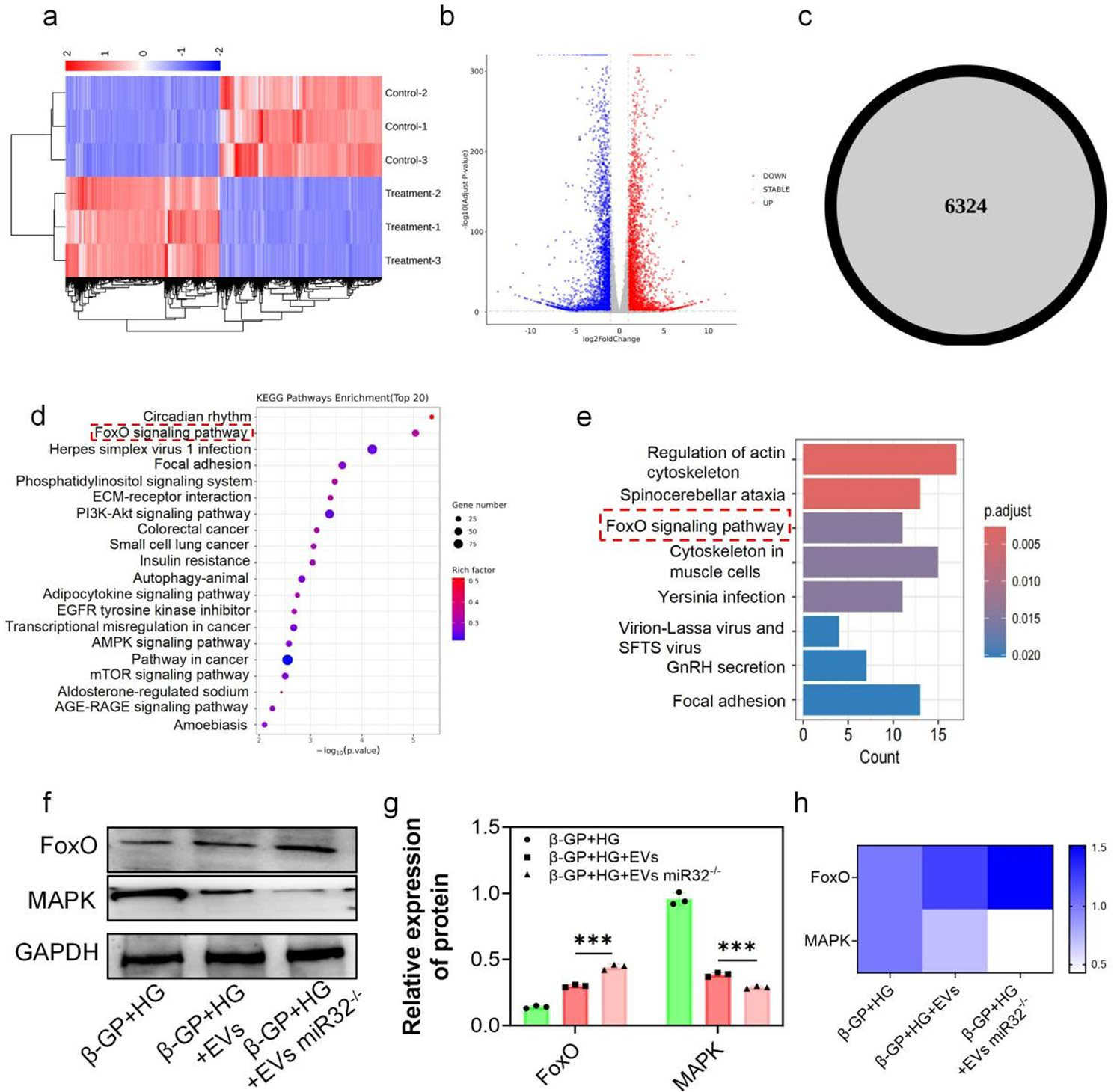
Background: The development of vascular calcification (VC) in diabetes is closely related to the endothelial-to-mesenchymal transition (EndMT). We found that microRNA-32-5p (miR-32) was elevated in the plasma of calcification patients. However, it is unclear whether miR-32 mediates the function of bone marrow mesenchymal stem cell-derived extracellular vesicles (BMSC-EVs) in type 2 diabetes (T2D) VC. Methods: BMSC-EVs were characterized by TEM, NTA, Western blotting, and confocal microscopy. Alizarin Red and ALP staining assessed the severity of VC. qRT-PCR and Western blotting evaluated the expression of BMP2, RUNX2, GPX4, SLC7A11, VE-cadherin, and N-cadherin, while immunofluorescence was used for detecting VE-cadherin and N-cadherin. In vivo validation was performed using miR-32–/– and ApoE–/– mice. RNA sequencing (RNA-seq) and bioinformatics analysis was conducted to explore underlying mechanisms. Results: We demonstrated that BMSC-EVs attenuate VC in endothelial cells (ECs) and inhibit EndMT. In vivo, histological analysis showed that treatment with BMSC-EVs significantly reduced the severity of VC associated with T2D. Notably, knockout of miR-32 further enhanced the inhibitory effect of BMSC-EVs on VC. Mechanistically, transcriptomic and functional analyses suggest that the protective effect of BMSC-EVs on VC is associated with regulation of the MAPK/FoxO signaling pathway, potentially mediated by modulation of ferroptosis. Conclusion: These findings demonstrate that BMSC-EVs attenuate T2D-associated VC, partially through miR-32-mediated suppression of EC ferroptosis.

Read Full Abstract10.1186/s13287-026-04896-8
Inhalation of Mesenchymal Stromal Cell-Derived Extracellular Vesicles Activates Macrophage Polarization through the miR-22-3p/NLRP3/IL-1β Pathway, Ameliorating Lung Ischemia-Reperfusion InjuryGraphical AbstractVerified
Stem Cell Research & Therapy2026

Inhalation of Mesenchymal Stromal Cell-Derived Extracellular Vesicles Activates Macrophage Polarization through the miR-22-3p/NLRP3/IL-1β Pathway, Ameliorating Lung Ischemia-Reperfusion Injury

Background: Lung ischemia–reperfusion injury (IRI) is a major contributor to primary graft dysfunction (PGD) after lung transplantation. Mesenchymal stem cell-derived extracellular vesicles (MSC-EVs) have emerged as promising therapeutic agents in inflammatory diseases by ameliorating tissue damage and promoting repair. However, the anti-inflammatory efficacy of these approaches and the underlying mechanisms in lung ischemia–reperfusion injury remain incompletely understood. Methods: The protective effects of mesenchymal stromal cell-derived extracellular vesicles (MSC-EVs) against lung ischemia–reperfusion injury were evaluated using two delivery approaches, inhalation and intravenous injection. Both in vivo and in vitro models were employed to assess the biological activity of MSC-EVs and to elucidate the underlying molecular mechanisms. In addition, a rat orthotopic lung transplantation (OLT) model was established to further examine the translational relevance of MSC-EVs. Results: MSC–EVs treatment significantly ameliorated lung IRI, with inhalation showing superior efficacy over intravenous delivery. Mechanistically, miR-22-3p within MSC-EVs targeted macrophage NLRP3, suppressing activation of the NLRP3/Caspase-1/IL-1β pathway and promoting M2 polarization. The protective efficacy was confirmed in a clinically relevant rat OLT model, underscoring their translational potential. Conclusions: Our findings indicate that inhaled MSC-derived extracellular vesicles attenuate lung ischemia–reperfusion injury by promoting macrophage polarization via the miR-22-3p/NLRP3/IL-1β pathway, supporting their potential as a cell-free therapeutic approach to mitigate primary graft dysfunction after lung transplantation.

Read Full Abstract10.1186/s13287-026-04921-w
Hypoxia-conditioned BMSC exosomes improve short-term spinal cord injury outcomes via the miR-615-3p/PDE4C-mediated cAMP/PKA pathwayGraphical AbstractVerified
Stem Cell Research & Therapy2026

Hypoxia-conditioned BMSC exosomes improve short-term spinal cord injury outcomes via the miR-615-3p/PDE4C-mediated cAMP/PKA pathway

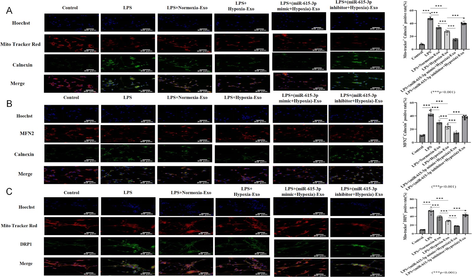
Spinal cord injury (SCI) remains a significant global health challenge with limited effective therapeutic options. Exosomes derived from mesenchymal stem cells (MSCs) have emerged as promising neuroprotective agents due to their biocompatibility and immunomodulatory properties. This study investigated the therapeutic potential of hypoxia-conditioned bone marrow MSC (BMSC)-derived exosomes in both in vitro and in vivo SCI models. Hypoxic preconditioning significantly enriched miR-615-3p in BMSC-derived exosomes. In spinal neuron injury models, hypoxic exosomes enhanced cell viability, reduced apoptosis, and ameliorated dysfunction of the mitochondria-associated endoplasmic reticulum membranes (MAMs). Mechanistically, miR-615-3p directly targeted and suppressed phosphodiesterase 4C (PDE4C), activating the cyclic adenosine monophosphate (cAMP)/protein kinase A (PKA) pathway. This in turn modulated calcium signaling, attenuated mitochondrial calcium overload, and reduced endoplasmic reticulum stress (ERS). In a mouse model of SCI, short-term treatment with hypoxic exosomes promoted functional recovery within a 14-day post-injury period, as evidenced by improved locomotor performance, reduced lesion volume, attenuated tissue edema, and decreased inflammatory cell infiltration. Furthermore, in vivo administration of hypoxic exosomes upregulated miR-615-3p and downregulated PDE4C expression in injured spinal cord tissues. These results demonstrate that hypoxia-conditioned BMSC-derived exosomes exert neuroprotective effects via the miR-615-3p/PDE4C axis, highlighting their potential as a novel therapeutic strategy for SCI by targeting calcium homeostasis and mitochondrial-ER dysfunction. These findings demonstrate the short-term therapeutic potential of hypoxia-conditioned exosomes in SCI. However, further preclinical studies, including long-term follow-up to assess the durability of recovery and potential late-onset effects, alongside clinical validation, are warranted before clinical translation.

Read Full Abstract10.1186/s13287-026-04895-9
Therapeutic potential of mesenchymal stromal cells in COVID-19: a meta-analysis of clinical trials conducted since the pandemic onsetGraphical AbstractVerified
Stem Cell Research & Therapy2026

Therapeutic potential of mesenchymal stromal cells in COVID-19: a meta-analysis of clinical trials conducted since the pandemic onset

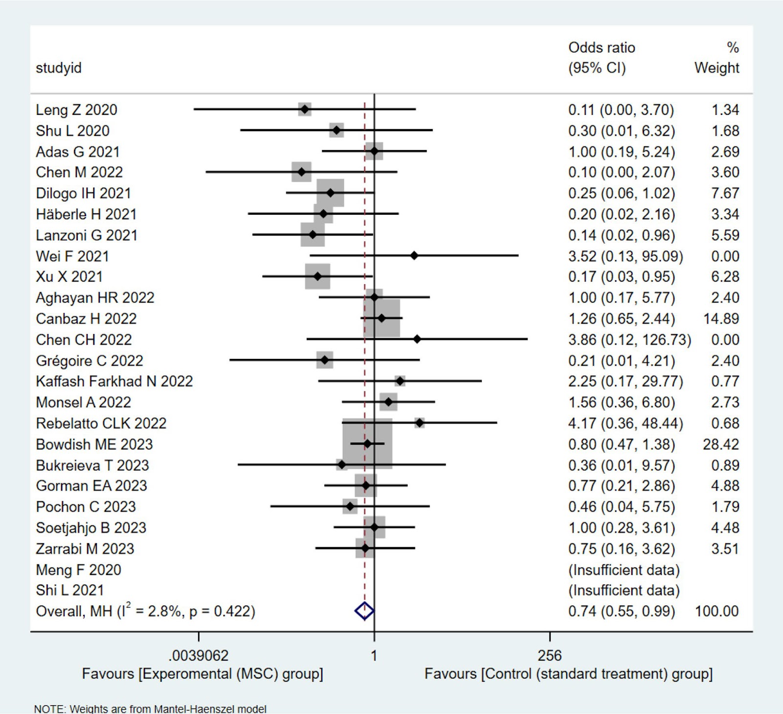
Background: Severe acute respiratory syndrome coronavirus 2 (SARS-CoV-2) infection can induce immune dysregulation and multi-organ injury; mesenchymal stromal cell (MSC) therapy has shown promise in clinical trials for COVID-19 and may have broader applicability to pneumonia induced by respiratory viruses (e.g., the influenza virus). This meta-analysis synthesized the available comparative clinical evidence on the safety and efficacy of MSCs in patients with moderate to critical COVID-19 and examined the reported outcomes relevant to Long-COVID. Methods: We searched the PubMed, Embase, and CNKI databases for original, comparative studies in moderate, severe, or critical COVID-19 published up to September 2, 2024. Twenty-four eligible studies (13 RCTs and 11 non-randomized controlled trials; n=1080) were included in the mortality meta-analysis. Patients were assigned to either the intervention group (MSC therapy plus standard care) or the control group (standard care with or without placebo). The primary efficacy outcome was all-cause mortality, while the primary safety outcomes were adverse events (AEs) and serious adverse events (SAEs). Secondary outcomes included clinical recovery, hospitalization metrics, chest imaging, and inflammatory biomarkers. We performed a pooled meta-analysis on mortality with subgroup analyses (by disease severity, administration route, dosing frequency, and study design), assessment of publication bias (using funnel plots and Egger's test), and evaluation of the quality of evidence via the GRADE approach. AEs/SAEs were analyzed using meta-analysis and descriptive statistics, while other secondary outcomes were summarized descriptively. Results: MSC therapy significantly reduced all-cause mortality (MSC: 26.4% vs control: 31.9%; fixed-effect OR=0.74, 95% CI 0.55–0.99), with low heterogeneity (I2=2.8%, P=0.422[Q-test]) and no publication bias. The quality of evidence was moderate (according to the GRADE assessment). The subgroup analysis revealed a significant survival benefit in severe/critical patients (OR=0.73, 95% CI 0.54–0.98) but not in studies that included moderate cases (OR=0.91, 95% CI 0.23–3.65). No significant heterogeneity was found across study designs, administration routes, or dosing frequencies, which confirmed the robustness of the primary findings while indicating insufficient evidence to determine the optimal regimen. The secondary outcomes suggested improvements in clinical recovery, pulmonary function, and pro-/anti-inflammatory cytokine balance in patients that received MSC therapy. Limited studies with long-term follow-up indicated potential benefits for Long-COVID outcomes (e.g., fatigue, quality of life, residual CT abnormalities, and exercise tolerance). No significant differences were observed in AEs or SAEs post-MSC infusion, which suggested that MSC therapy was well tolerated. Conclusion: This meta-analysis indicated that MSC therapy may reduce mortality in patients with severe or critical COVID-19, demonstrating a favorable safety profile and potential benefits for Long-COVID and other viral pneumonias. Further large-scale, rigorous RCTs and mechanistic studies are warranted to strengthen the evidence base and standardize MSC administration regimens (source, dosing, frequency).

Read Full Abstract10.1186/s13287-026-05020-6
Bone Marrow-Derived Mesenchymal Stem Cells Alleviate Hepatic Lipid Metabolism Disorders After Scald Injury: Integrating Liver Transcriptome and MetabolomeGraphical AbstractVerified
Stem Cell Research & Therapy2026

Bone Marrow-Derived Mesenchymal Stem Cells Alleviate Hepatic Lipid Metabolism Disorders After Scald Injury: Integrating Liver Transcriptome and Metabolome

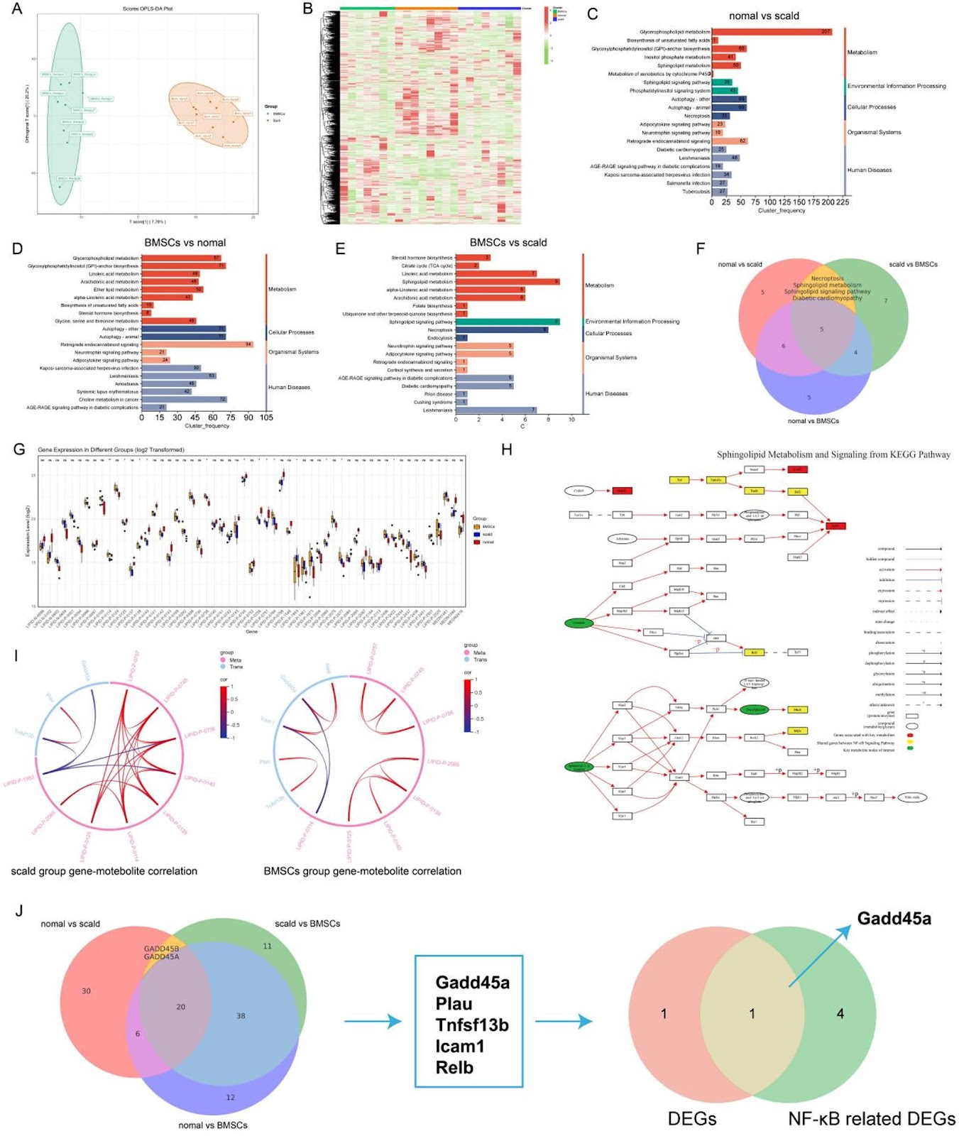
Previous studies have confirmed that scald injuries can lead to disturbances in hepatic lipid metabolism, and bone marrow-derived mesenchymal stem cells (BMSCs) have emerged as a promising therapeutic strategy for alleviating such disorders. However, research focusing on the regulation and restoration of liver lipid metabolic processes remains limited. In this study, we investigated the effects of BMSCs on hepatic lipid metabolism disorders induced by scald injury in rats through integrated transcriptomic and metabolomic analyses. The results demonstrated that portal vein infusion of BMSCs markedly improved body weight recovery, reduced hepatic lipid accumulation, normalized serum lipid profiles, and attenuated liver injury following scalding. Combined transcriptomic and metabolomic data further suggested that the therapeutic mechanism may involve inhibition of NF-ĪŗB/Gadd45a signaling in hepatocytes, restoration of sphingolipid metabolism, enhancement of hepatic lipid conversion, and suppression of adipocyte lipolysis. Overall, this study provides a theoretical basis for the potential clinical application of BMSCs in treating hepatic lipid metabolism disorders secondary to severe burn injury.

Read Full Abstract10.1186/s13287-025-04774-9
Fndc5 modification optimizes the therapeutic effect of rat MSCs on sepsis-induced ALI/ARDS via activating the PI3K/AKT signaling pathwayGraphical AbstractVerified
Stem Cell Research & Therapy2026

Fndc5 modification optimizes the therapeutic effect of rat MSCs on sepsis-induced ALI/ARDS via activating the PI3K/AKT signaling pathway

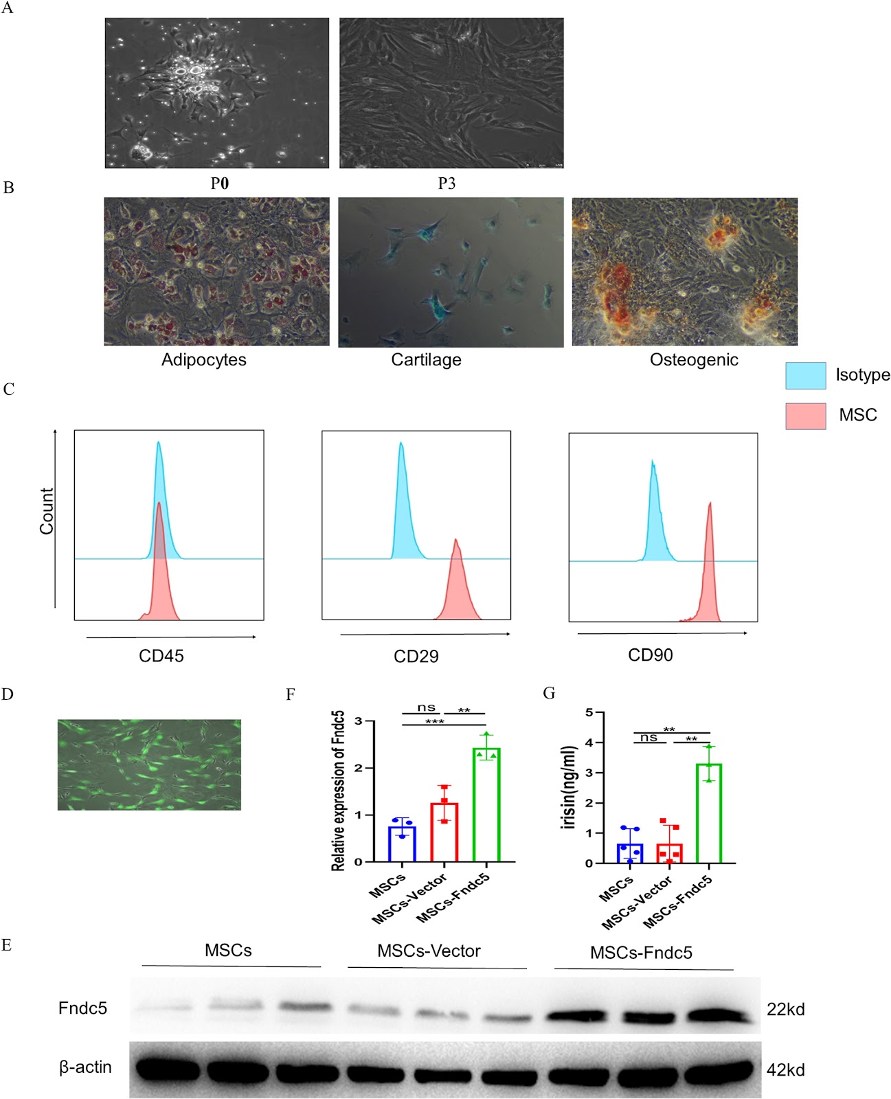
Background: Acute lung injury/Acute respiratory distress syndrome (ALI/ARDS) is a life-threatening inflammatory lung disorder characterized by high mortality rates and a lack of effective treatment options. Although mesenchymal stem cell (MSC)-based therapies have emerged as a promising approach for ARDS management, optimizing their therapeutic efficacy remains a significant challenge. Recent advances in gene modification techniques have opened new avenues for enhancing MSC functionality. Among these, Fibronectin type III domain-containing protein 5 (Fndc5)/irisin has attracted considerable attention due to its ability to improve endothelial function. This study aims to evaluate the therapeutic potential of Fndc5-modified MSCs in sepsis-induced ALI/ARDS and to elucidate the underlying molecular mechanisms driving their protective effects. Methods: To comprehensively evaluate the therapeutic potential of Fndc5-modified MSCs (MSCs-Fndc5) in ARDS, we employed both in vivo and in vitro experimental models. In vivo, a mouse model of sepsis-induced ALI was established through intraperitoneal injection of lipopolysaccharide (LPS), and the protective effects of MSCs-Fndc5 were systematically assessed by analyzing lung histopathology, inflammatory cytokine levels, vascular endothelial integrity, lung wet-to-dry weight ratio, and MSC retention in lung tissue. In parallel, in vitro studies were conducted to investigate the role of MSCs-Fndc5 in mitigating LPS-induced endothelial cell (EC) injury, with a focus on EC proliferation, angiogenesis, barrier permeability, apoptosis, and the regulation of key signaling pathways. Results: Fndc5 modification significantly increased the retention rate of MSCs in sepsis-induced ALI murine model while augmenting their in vitro proliferation and migration potential. In vivo, treatment with Fndc5-modified MSCs markedly attenuated lung inflammation, as evidenced by reduced levels of pro-inflammatory cytokines, decreased neutrophil infiltration, and improved lung histopathology. Additionally, MSCs-Fndc5 alleviated pulmonary edema, reduced fibrosis, lowered the lung wet-to-dry weight ratio, and preserved vascular endothelial integrity. In vitro, MSCs-Fndc5 significantly enhanced cell proliferation, migration, angiogenesis, endothelial barrier function, apoptosis inhibition, likely via PI3K/AKT pathway activation. Conclusions: Fndc5 overexpression in MSCs augments their therapeutic efficacy in sepsis-induced ALI/ARDS, which may be achieved by activating the endothelial PI3K/AKT pathway and improving MSCs retention in vivo. These findings propose MSCs-Fndc5 as a promising therapeutic strategy for sepsis-induced ALI/ARDS by enhancing endothelial repair, curbing inflammation, and modulating pivotal signaling pathways.

Read Full Abstract10.1186/s13287-026-04903-y
Mesenchymal stem cell-derived extracellular vesicles in the treatment of type 2 diabetes and its complications: current progress and future directionsGraphical AbstractVerified
Stem Cell Research & Therapy2026

Mesenchymal stem cell-derived extracellular vesicles in the treatment of type 2 diabetes and its complications: current progress and future directions

Type 2 diabetes (T2D) and its complications represent a complex disorder involving multiple pathophysiological processes. Although conventional therapeutic approaches partially regulate blood glucose, they fail to fundamentally reverse disease progression or effectively prevent complications. This review summarizes the current research advance and challenges of using different forms of mesenchymal stem cell-derived extracellular vesicles (MSC-EVs) in treating T2D and complications. It begins with an introduction to the characteristics of MSC-EVs. Subsequently, the mechanisms and therapeutic prospects of natural MSC-EVs are analyzed, with a focus on their roles in inflammatory modulation, tissue regeneration, and improving insulin resistance. Engineering MSC-EVs, covering strategies including optimizing MSC culture conditions, modifying EV contents, and establishing MSC-EV delivery systems based on bioactive materials are then discussed, which boost EV yield and quality while enhancing therapeutic efficacy. Current challenges, including the limited yield and high heterogeneity of natural MSC-EVs, as well as issues related to long-term safety, immunocompatibility, and large-scale production of engineered MSC-EVs are finally overviewed, with emphasizing artificial intelligence in guiding future research directions. These summaries are crucial for clinical translation of MSC-EVs and will ultimately provide T2D patients with an effective and safe treatment option.

Read Full Abstract10.1186/s13287-026-04991-w
Extracorporeal cardiac shock wave stimulation enhances the therapeutic efficacy of intravenously delivered endothelial colony-forming cells via PI3K/AKT signaling in a rat myocardial infarction modelGraphical AbstractVerified
Stem Cell Research & Therapy2026

Extracorporeal cardiac shock wave stimulation enhances the therapeutic efficacy of intravenously delivered endothelial colony-forming cells via PI3K/AKT signaling in a rat myocardial infarction model

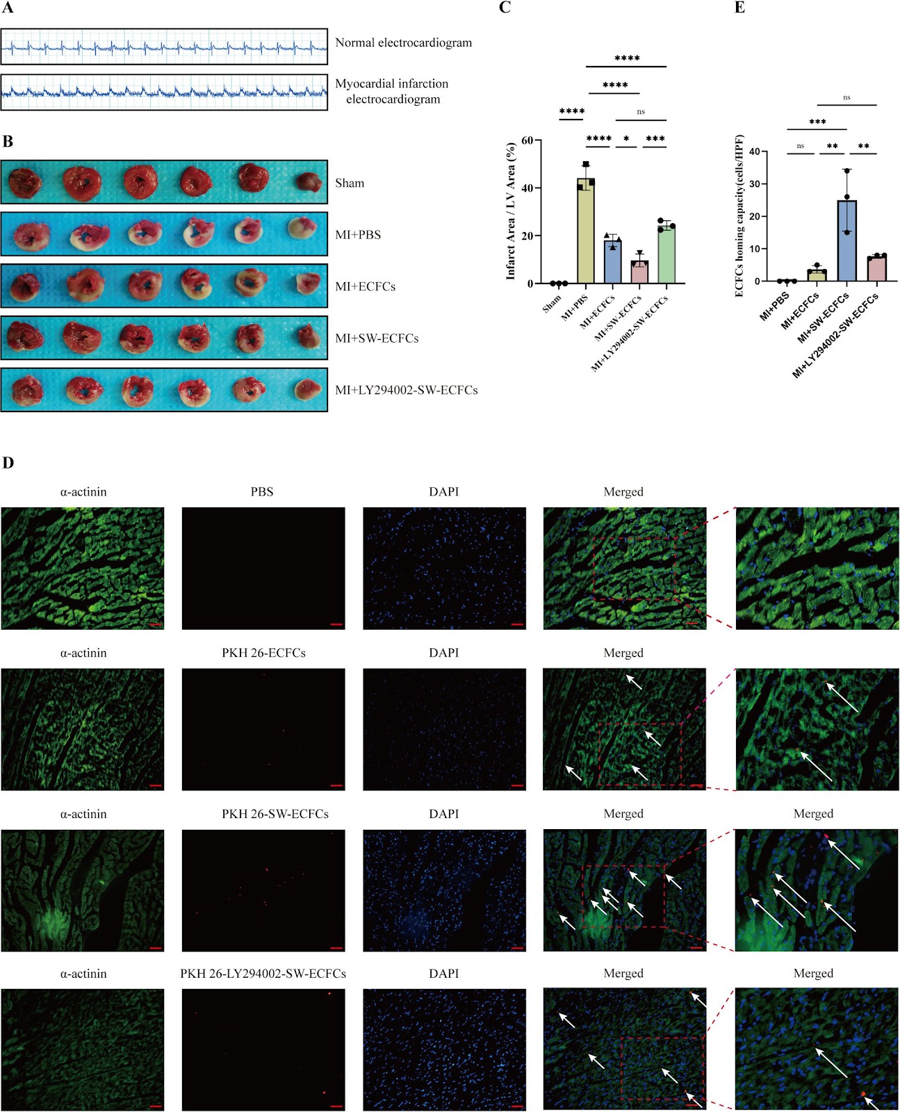
Background: Extracorporeal cardiac shock wave (ECSW) therapy enhances the function of endothelial colony-forming cells (ECFCs), but whether it can serve as a preconditioning strategy to enhance myocardial infarction (MI) therapy remains unclear. This study investigated the efficacy and mechanism of intravenously delivered ECSW-preconditioned ECFCs (SW-ECFCs) in a rat MI model. Methods: ECFCs were isolated from the bone marrow of ApoE-/- rats and fully characterized. RNA sequencing of control ECFCs versus SW-ECFCs revealed significant enrichment of the PI3K/AKT pathway. We therefore performed a series of in vitro functional assays on these cells, including Transwell migration, Matrigel tube formation, CCK-8 proliferation, flow cytometric apoptosis analysis, and VEGF-A ELISA. The role of the PI3K/AKT pathway was interrogated using the inhibitor LY294002. Subsequently, an acute MI model was established in ApoE-/- rats via left anterior descending coronary artery ligation. Rats were randomized into four groups: MI+PBS, MI+ECFCs, MI+SW-ECFCs, and MI+LY294002-pretreated SW-ECFCs (LY-SW-ECFCs), with sham-operated rats as controls. Comprehensive evaluations included echocardiography, serum injury biomarkers, TTC, and histopathological (H&E, Masson) staining, immunohistochemical detection of cardiomyocyte apoptosis and p-eNOS, immunofluorescence assessment of ECFC homing and vascular markers (CD31, α-SMA, VEGF-A), tissue/plasma nitric oxide measurement, and Western blot analysis of PI3K/AKT signaling proteins. Results: Transcriptomic analysis revealed significant enrichment of the PI3K/AKT pathway in SW-ECFCs. Functionally, ECSW enhanced ECFCs migration, tube formation, proliferation, and VEGF-A secretion, while reducing apoptosis; these effects were largely abolished by PI3K inhibition. In vivo, serum levels of CK, CK-MB, and LDH were significantly elevated in all MI groups compared to the Sham group (P<0.01), indicating comparable initial injury. However, no significant differences were observed among treatment groups (P>0.05). SW-ECFCs transplantation significantly improved cardiac function, reduced infarct size, fibrosis, and apoptosis, and enhanced angiogenesis (P<0.05). These benefits were associated with increased levels of p-AKT, p-eNOS, and BCL-2 protein as well as nitric oxide content, while suppressing the expression of cleaved caspase-3 (P<0.05). Crucially, all these therapeutic benefits were largely abolished by PI3K inhibition. Conclusion: In conclusion, this study demonstrates that preconditioning ECFCs with ECSW significantly enhances their therapeutic efficacy for myocardial infarction, improving both cardiac function and structural repair. These benefits are mediated primarily through activation of the PI3K/AKT signaling pathway, which augments cell homing, paracrine activity, and survival, thereby providing a novel and promising strategy for cardiac regeneration.

Read Full Abstract10.1186/s13287-026-04913-w
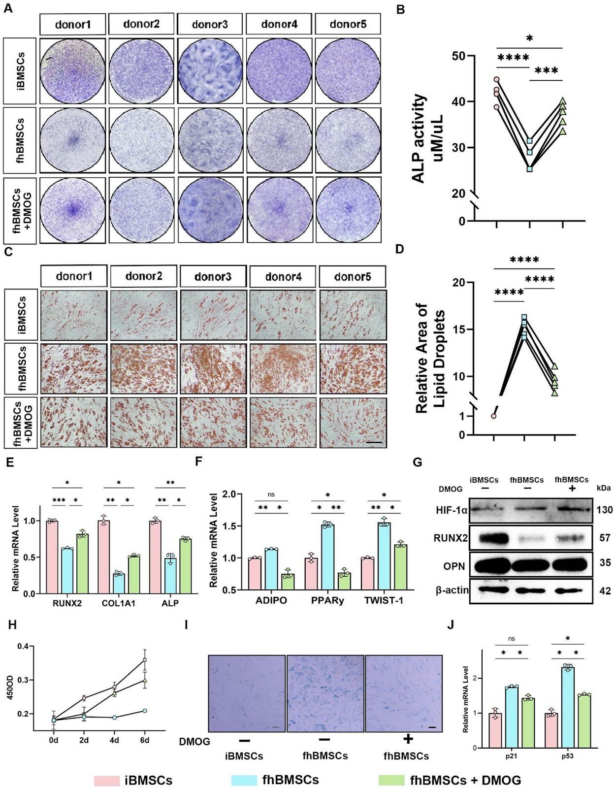
DMOG Pretreatment Restores Osteogenic–Adipogenic Balance and Mitochondrial Function in ONFH BMSCs through the HIF-1α/Homer3 PathwayGraphical AbstractVerified
Stem Cell Research & Therapy2026

DMOG Pretreatment Restores Osteogenic–Adipogenic Balance and Mitochondrial Function in ONFH BMSCs through the HIF-1α/Homer3 Pathway

Background: Osteonecrosis of the femoral head (ONFH) is a progressive orthopedic disorder that often culminates in femoral head collapse and joint failure. Dysfunction of bone marrow mesenchymal stem cells (BMSCs), including impaired osteogenesis, enhanced adipogenesis, and mitochondrial dysfunction, has been increasingly recognized as a central driver of ONFH pathogenesis. However, the molecular mechanisms linking metabolic stress to lineage imbalance remain poorly defined. Methods: Paired BMSCs were isolated from necrotic femoral head regions (fhBMSCs) and the iliac crest (iBMSCs) of ONFH patients. Functional assays, RNA sequencing, and molecular analyses were performed to evaluate the effects of the hypoxia mimetic dimethyloxalylglycine (DMOG) on osteogenic–adipogenic balance, mitochondrial function, and senescence. Loss-of-function experiments targeting hypoxia-inducible factor-1α (HIF-1α) and Homer3 were conducted to elucidate mechanistic pathways. Results: Compared with iBMSCs, fhBMSCs exhibited impaired osteogenesis, enhanced adipogenesis, mitochondrial dysfunction, and increased senescence. DMOG pretreatment restored osteogenic differentiation, suppressed adipogenesis, improved mitochondrial dynamics, reduced oxidative stress, and enhanced bioenergetic metabolism. These protective effects were dependent on HIF-1α stabilization. Transcriptomic profiling identified Homer3 as a downstream negative regulator of HIF-1α. Homer3 was aberrantly upregulated in fhBMSCs but suppressed by DMOG, and its knockdown mimicked the effects of DMOG by promoting osteogenesis, inhibiting adipogenesis, enhancing mitophagy, and restoring mitochondrial function. Conversely, silencing HIF-1α abolished DMOG-mediated benefits and reinstated Homer3 expression. Conclusions: This study identifies the HIF-1α/Homer3 axis as a central regulator of lineage balance and mitochondrial homeostasis in ONFH-derived BMSCs. Pharmacological targeting of this pathway with DMOG or related prolyl hydroxylase inhibitors may provide a promising joint-preserving therapeutic strategy for ONFH.

Read Full Abstract10.1186/s13287-026-05026-0
Generation of biologically responsive colon-like intestinal tissue patches from human induced pluripotent stem cells using a rapid co-differentiation platformGraphical AbstractVerified
Stem Cell Research & Therapy2026

Generation of biologically responsive colon-like intestinal tissue patches from human induced pluripotent stem cells using a rapid co-differentiation platform

The intestinal mucosa is a complex functional layer formed from diverse cell types including epithelial cells within crypts and villi and an array of mesenchymal cells. Many intestinal diseases involve loss of the surface mucosa, which is difficult to restore and delays healing. We hypothesized that a transplantable intestinal mucosal tissue graft could aid healing. To create such a graft, we developed a novel early-stage human induced pluripotent stem cell (hiPSC) co-differentiation platform capable of generating multiple intestinal cell lineages (epithelial, mesenchymal, and endothelial) in 8 days. This protocol is simple, serum-free, and reduces animal product use. We confirmed cell identity via RNA and protein expression profiles typical of intestinal lineages. Using bulk and single-cell RNA sequencing, we characterized global transcriptional profiles, showing intestinal identity with early colonic polarization. Results were replicated across multiple hiPSC lines and an independent center. Culturing derived cells on collagen hydrogels formed colon-like intestinal patches (CL-IPs). Upon transplantation into mouse subcutis, CL-IPs developed into colon-like tissue structures including crypts, stromal and muscle layers, and human-origin vasculature that anastomosed with murine vasculature. Teratoma assays and molecular analyses showed no residual pluripotency. This platform shows potential for intestinal mucosal regeneration therapy and as a physiologically relevant in vitro model of intestinal pathobiology.

Read Full Abstract10.1186/s13287-026-05006-4
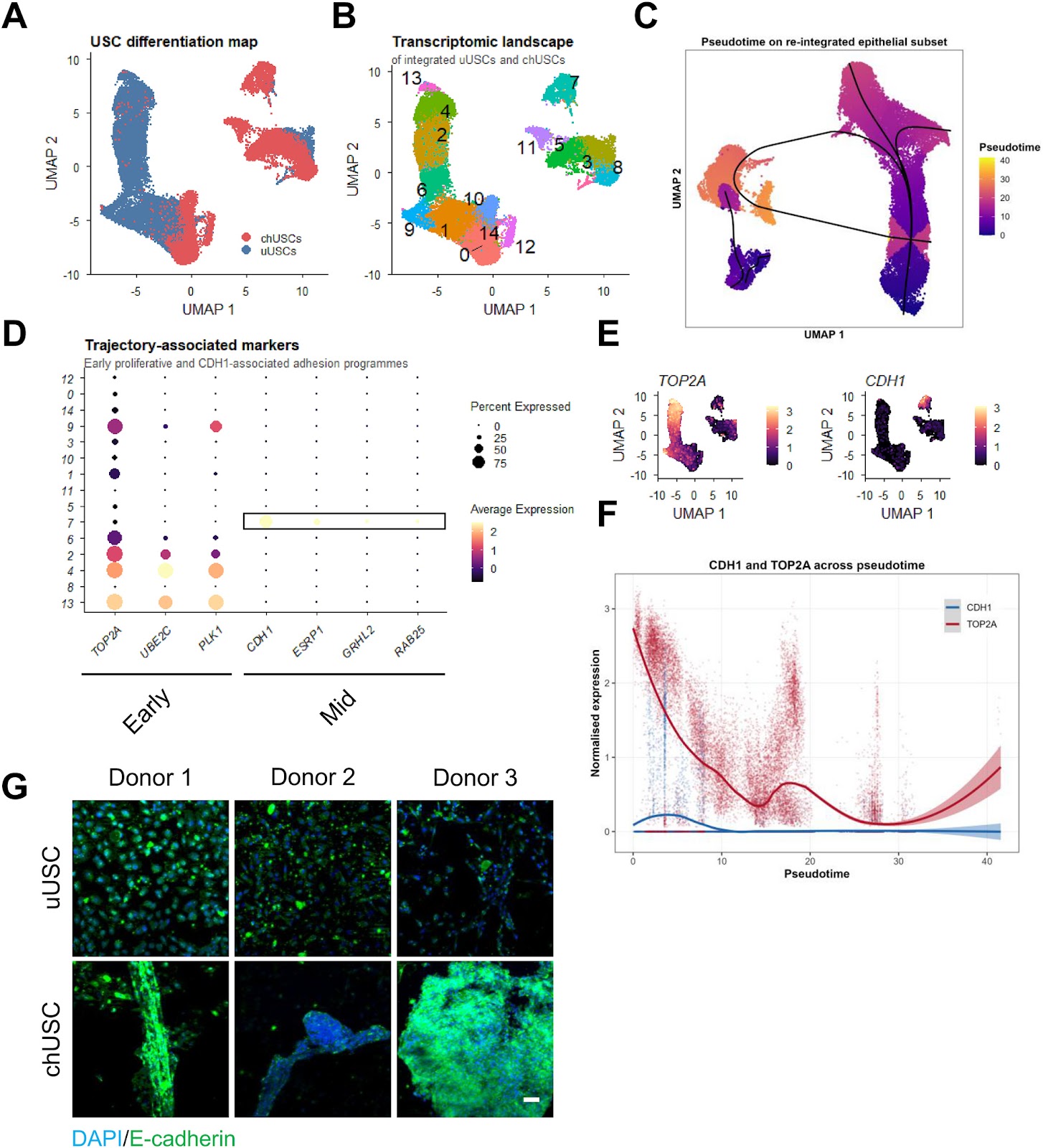
A single-donor proof-of-concept single-cell analysis maps heterogeneous differentiation trajectories toward cartilage-like states in human urine-derived stem cellsGraphical AbstractVerified
Stem Cell Research & Therapy2026

A single-donor proof-of-concept single-cell analysis maps heterogeneous differentiation trajectories toward cartilage-like states in human urine-derived stem cells

Background: Urine-derived stem cells (USCs) represent an accessible and non-invasive cell source with reported chondrogenic differentiation potential. However, the cellular heterogeneity and transcriptional dynamics underlying USC differentiation remain incompletely understood, limiting their translational interpretation. Methods: We combined functional differentiation assays with single-cell RNA sequencing to characterize USC differentiation at both phenotypic and transcriptional levels. Chondrogenic and osteogenic differentiation were assessed using histological staining, quantitative PCR, and three-dimensional spheroid cultures. Single-cell transcriptomic analysis was performed on integrated datasets of undifferentiated and differentiated USCs, followed by pseudotime trajectory inference and mapping to a human cartilage reference atlas. Results: Chondrogenic induction resulted in reproducible acquisition of cartilage-associated features, including glycosaminoglycan-rich extracellular matrix deposition, increased expression of SOX9, and formation of aggrecan-positive spheroids. In this donor, single-cell analysis mapped an inferred differentiation trajectory from proliferative states towards differentiated populations, although the fine-grained pseudotemporal ordering was sensitive to analytical choices and is therefore interpreted qualitatively. Along this inferred trajectory, we identified a candidate transient transcriptional state associated with elevated CDH1 expression and epithelial-like aggregation features. Probabilistic mapping to a human cartilage reference atlas indicated that overall mapping confidence was low (median prediction score 0.34) and that only a minority of cells showed confident transcriptional similarity (prediction score ≄0.5) to mature/articular cartilage-associated reference states (7.8% of all cells and 17.6% of chondrogenically induced cells). This confident similarity was concentrated in a few clusters at the differentiated end of the trajectory rather than representing the bulk of the culture, and label-transfer confidence was not equated with chondrocyte identity. Despite this enrichment, differentiated populations exhibited transcriptional heterogeneity, including subsets of cells associated with hypertrophic, fibrocartilage-like, and contractile gene programmes, indicating the presence of multiple differentiation trajectories. Conclusions: This single-donor proof-of-concept study suggests that USC differentiation may involve a candidate transient, aggregation-associated transcriptional state accompanied by CDH1 expression and gives rise to heterogeneous lineage-associated outcomes, with only a minority of cells acquiring confident transcriptional similarity to mature cartilage. Because these observations derive from one donor, they should be interpreted as hypothesis-generating and require validation across independent donors before donor-independent or translational conclusions for cartilage regeneration can be drawn. These findings nonetheless provide a single-cell resolution framework for future multi-donor validation of USC differentiation and its inherent transcriptional heterogeneity.

Read Full Abstract10.1186/s13287-026-05223-x
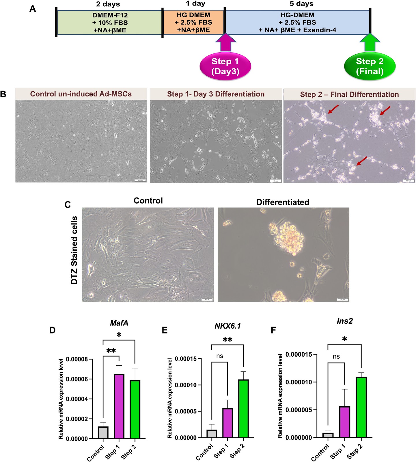
Emerging roles of the long non-coding RNAs MALAT1 and TUG1 during differentiation of adipose tissue-derived mesenchymal stem cells towards insulin-producing cellsGraphical AbstractVerified
Stem Cell Research & Therapy2026

Emerging roles of the long non-coding RNAs MALAT1 and TUG1 during differentiation of adipose tissue-derived mesenchymal stem cells towards insulin-producing cells

Background: Generation of insulin-producing cells (IPCs) from stem cells provides great hope for patients with diabetes mellitus (DM). Long non-coding RNAs (lncRNAs) ignited much interest regarding their role in determining the fate of stem cells. The lncRNAs MALAT1 and TUG1 have been reported to be interrelated with β-cell dysfunction and/or DM. However, their role during generation of IPCs from stem cells has not been adequately studied. Thus, the current study aimed to investigate the role of MALAT1 and TUG1 during differentiation of adipose tissue-derived mesenchymal stem cells (Ad-MSCs) towards IPCs. Methods: Ad-MSCs were isolated from rat epididymal fat pads, characterized and induced to differentiate towards IPCs. Assessment of differentiation was done by measuring expression levels of various β-cell-related markers using RT-qPCR, as well as morphological changes, and dithizone staining. Expression levels of MALAT1 and TUG1 were also measured by RT-qPCR. Several in-silico analyses were done using RNA–protein Association and Interaction Networks (RAIN) database. Results: MALAT1 and TUG1 expression levels were significantly increased during differentiation of Ad-MSCs into IPCs as compared to control uninduced cells. Furthermore, generated networks from RAIN database revealed an interplay between MALAT1 and TUG1, and between each of them with several common targets like GAS5, HOTAIR and TP53COR1. Conclusions: The current study portrays MALAT1 and TUG1 as novel interrelated molecular mediators and important regulatory nodes enhancing differentiation of Ad-MSCs towards IPCs. Their upregulation during differentiation can be interrelated with competitive endogenous RNA (ceRNA) networks, mediating various epigenetic modifications, orchestrating signaling pathways and overcoming cellular stress during reprogramming/differentiation.

Read Full Abstract10.1186/s13287-026-05125-y
Cadherin 19 deficiency inhibits osteogenic differentiation and bone formation by regulating PI3K/AKT signaling pathwayGraphical AbstractVerified
Stem Cell Research & Therapy2026

Cadherin 19 deficiency inhibits osteogenic differentiation and bone formation by regulating PI3K/AKT signaling pathway

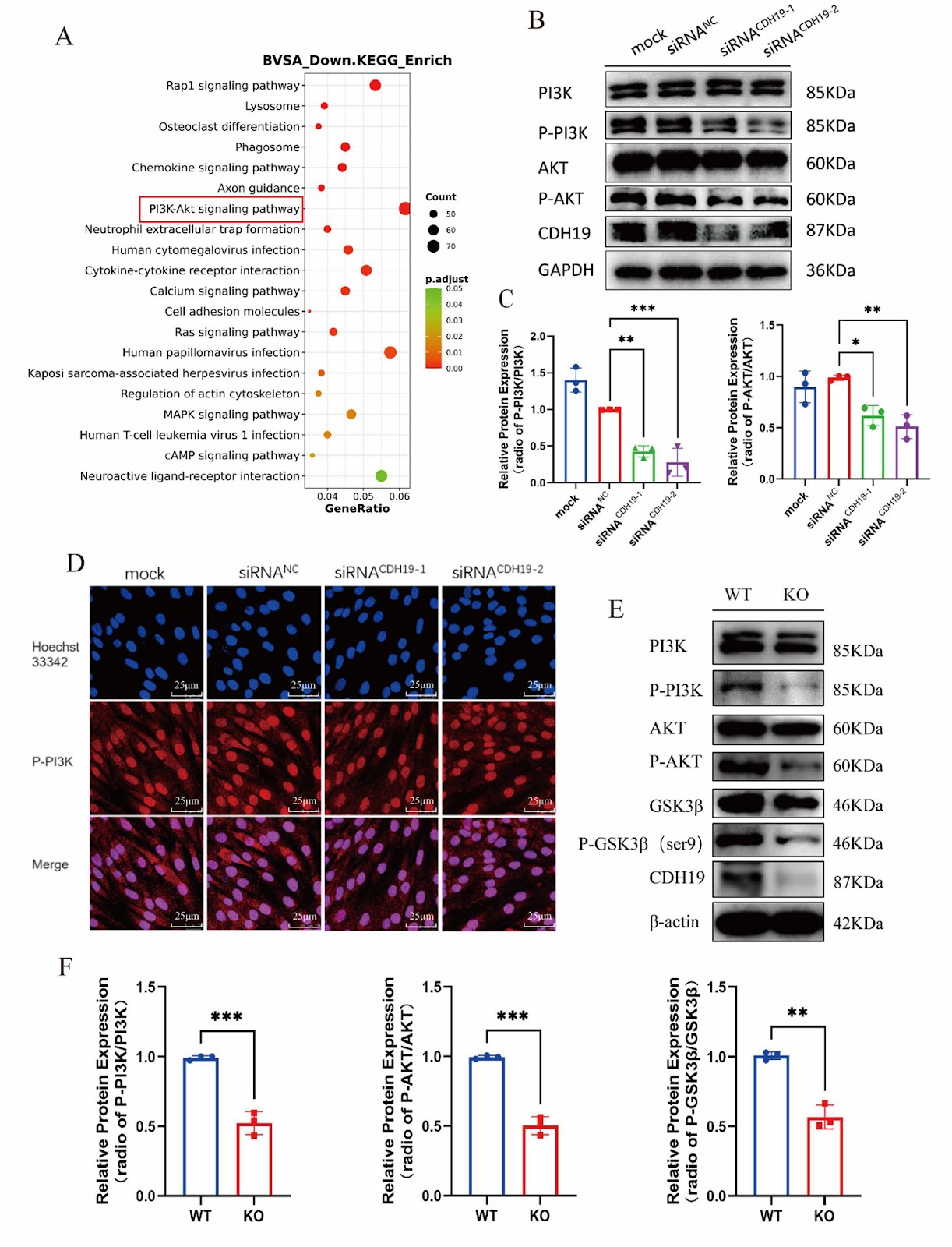
Background: Osteoporosis (OP) is a systemic bone disease characterized by damage to bone strength, leading to increased bone fragility and fracture risk. Cadherin 19 (CDH19) is located on chromosomes 18q22-q23, and 18q deletion is associated with terminal deletion diseases, including foot/hand deformities. However, the role of CDH19 in bone remains undefined. Methods: A conditional knockout mouse model of the CDH19 gene was constructed using the Cre-loxP system, and the bone mass and bone morphology in mice were investigated using microCT and histological staining. Osteoblasts were isolated and cultured from wild-type and CDH19 knockout mice. Cell proliferation and differentiation were explored through EdU labeling, qPCR, alkaline phosphatase (ALP)/ alizarin red S (ARS) staining, and Western blot assays. The expression of genes altered in CDH19 gene knockout osteoblast was checked by RNA sequencing (RNA-seq), and subsequently confirmed by immunofluorescence and Western blot. Results: We found that CDH19 could maintain the normal proliferation and differentiation in osteoblasts. After knocking out the CDH19 gene, the abilities of proliferation and osteogenesis were significantly inhibited in osteoblasts. Moreover, the bone mass of CDH19 knockout mice was significantly reduced, characterized by decreases in bone density, trabecular number, and bone volume fraction. The RNAseq analysis and western blot showed the PI3K/AKT signaling pathway was significantly inhibited in osteoblasts with CDH19 deletion. Furthermore, we demonstrated that administration of PI3K/AKT signaling pathway agonist 740Y-P partially alleviated the inhibition of osteogenic differentiation caused by CDH19 deletion in vitro and in vivo. Conclusion: This study demonstrated that CDH19 regulated osteogenic differentiation by modulating the PI3K/AKT signaling pathway in osteoblasts. CDH19 may become a novel target for the treatment of bone diseases.

Read Full Abstract10.1186/s13287-026-05061-x