Key Takeaways & Executive Findings
- •• CDH19 deficiency impairs osteoblast proliferation and differentiation, leading to reduced bone mass and microstructural deterioration. • CDH19 knockout mice exhibit decreased bone density, trabecular number, and bone volume fraction, confirming its essential role in bone formation. • Mechanistically, CDH19 deletion suppresses the PI3K/AKT signaling pathway, as revealed by RNA-seq and Western blot analyses. • Activation of PI3K/AKT with agonist 740Y-P partially rescues the osteogenic inhibition caused by CDH19 loss, suggesting a potential therapeutic strategy.
Abstract
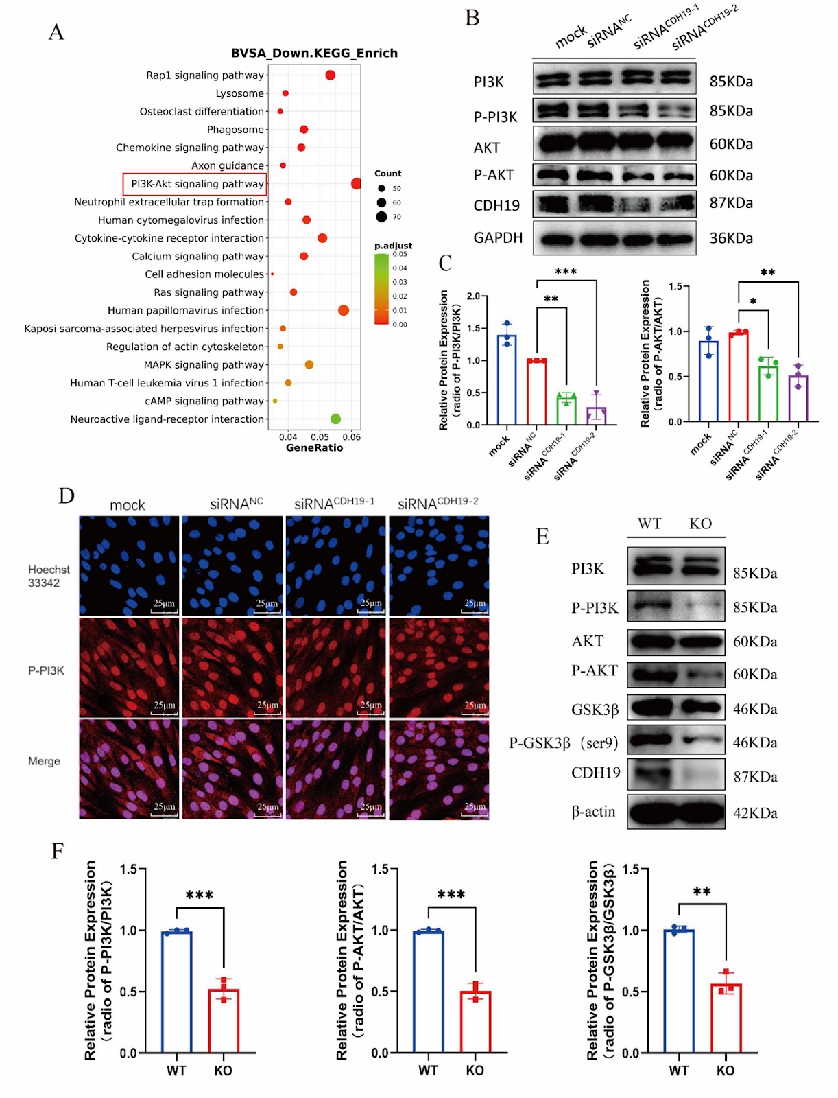
Background: Osteoporosis (OP) is a systemic bone disease characterized by damage to bone strength, leading to increased bone fragility and fracture risk. Cadherin 19 (CDH19) is located on chromosomes 18q22-q23, and 18q deletion is associated with terminal deletion diseases, including foot/hand deformities. However, the role of CDH19 in bone remains undefined. Methods: A conditional knockout mouse model of the CDH19 gene was constructed using the Cre-loxP system, and the bone mass and bone morphology in mice were investigated using microCT and histological staining. Osteoblasts were isolated and cultured from wild-type and CDH19 knockout mice. Cell proliferation and differentiation were explored through EdU labeling, qPCR, alkaline phosphatase (ALP)/ alizarin red S (ARS) staining, and Western blot assays. The expression of genes altered in CDH19 gene knockout osteoblast was checked by RNA sequencing (RNA-seq), and subsequently confirmed by immunofluorescence and Western blot. Results: We found that CDH19 could maintain the normal proliferation and differentiation in osteoblasts. After knocking out the CDH19 gene, the abilities of proliferation and osteogenesis were significantly inhibited in osteoblasts. Moreover, the bone mass of CDH19 knockout mice was significantly reduced, characterized by decreases in bone density, trabecular number, and bone volume fraction. The RNAseq analysis and western blot showed the PI3K/AKT signaling pathway was significantly inhibited in osteoblasts with CDH19 deletion. Furthermore, we demonstrated that administration of PI3K/AKT signaling pathway agonist 740Y-P partially alleviated the inhibition of osteogenic differentiation caused by CDH19 deletion in vitro and in vivo. Conclusion: This study demonstrated that CDH19 regulated osteogenic differentiation by modulating the PI3K/AKT signaling pathway in osteoblasts. CDH19 may become a novel target for the treatment of bone diseases.
1. Introduction
The normal physiological functions of osteoblasts and osteoclasts jointly maintain the development of human bones, while osteoporosis is mainly caused by bone formation being lower than bone resorption in the body [1–3]. Osteoblasts play a crucial role in the process of bone formation, and their normal differentiation and proliferation are essential for maintaining the body’s bone mass and quality. The inhibition of osteoblast differentiation and proliferation results in reduced bone mass and deterioration of the bone microstructure, gradually developing into osteoporosis [4, 5]. Therefore, it is important to unravel the mechanisms of bone formation for treating osteoporosis [6–8].
Cadherin is an adhesion molecule mainly expressed on the cell membrane. Currently, over 100 human cadherins have been identified and sequenced, including E-cadherin, N-cadherin, P-cadherin, and so on [9]. Research has shown that the adhesion of bone marrow mesenchymal stem cells (MSCs) and endothelial progenitor cells (EPCs) can promote the multipotentiality of MSCs. The E-cadherin protein of EPCs mediates the adhesion of MSCs and EPCs through the E-cadherin/β-catenin signaling pathway, promoting self-renewal and differentiation of osteoblasts, chondrocytes, and adipocytes [9]. In osteoblasts, bone morphogenetic protein 2 (BMP2) may enhance the expression of runt-related transcription factor 2 (RUNX2) and osteogenic-related genes by promoting the expression of N-cadherin (cadherin 2) and E-cadherin (cadherin 1), promoting early differentiation of human skull osteoblasts [10]. Inhibition of N-cadherin levels can suppress alkaline phosphatase (ALP) expression, morphological differentiation, and the formation of fetal rat cranial bone nodules in Saos-2 cells [11]. Mouse embryonic conditional bone lineage N-cadherin deficiency leads to growth defects, low bone mass, and a reduced number of bone progenitor cells [12]. Haÿ et al. found that N-cadherin can interact with Axin and LDL-receptor-related protein 5 (LRP5). Overexpression of N-cadherin in osteoblasts increases the interaction between N-cadherin and LRP5, leading to increased degradation of β-catenin, reduced bone formation, and delayed bone mass acquisition [13].
Loading authentic research manuscript (Pages 1–5)...
Huan Zhou, Yizhao Cheng, Liangxing Chen, Yaqin Zhang, Huimin Xiao, Chenhui Zhou, Hui Fu, Xiaoyan Zhang, Liangliang Xu, Daohua Xu (2026). Cadherin 19 deficiency inhibits osteogenic differentiation and bone formation by regulating PI3K/AKT signaling pathway. Stem Cell Research & Therapy. https://doi.org/10.1186/s13287-026-05061-x
Research & Educational Purpose Only:The translations, structured abstracts, analytical annotations, and data reports provided by SinoBioData are intended exclusively for academic research, internal corporate R&D, and educational benchmarking. They do not constitute formal engineering, chemical safety, legal, or professional advice.
Copyright & Intellectual Property Notice: Original copyright of the underlying source articles and experimental data remains with the respective authors, institutions, and original publishing journals. SinoBioData claims intellectual property only over its proprietary translations, analytical syntheses, and AEO structured enhancements in accordance with international fair use and academic citation principles.
Frequently Asked Questions
What is the role of Cadherin 19 (CDH19) in bone formation?
CDH19 is essential for normal osteoblast proliferation and differentiation. Its deficiency inhibits osteogenic differentiation and reduces bone mass, as demonstrated in a knockout mouse model.
How does CDH19 deficiency affect the PI3K/AKT signaling pathway?
CDH19 deletion significantly inhibits the PI3K/AKT signaling pathway in osteoblasts, as shown by RNA-seq and Western blot analyses. Activation of this pathway with agonist 740Y-P partially rescues the osteogenic inhibition.
What are the bone phenotype changes in CDH19 knockout mice?
CDH19 knockout mice exhibit reduced bone mass characterized by decreased bone density, trabecular number, and bone volume fraction, indicating impaired bone formation.
Could CDH19 be a therapeutic target for osteoporosis?
Yes, CDH19 may serve as a novel target for treating bone diseases like osteoporosis, as its regulation of the PI3K/AKT pathway influences osteogenic differentiation.
What methods were used to study CDH19 function?
The study used a conditional knockout mouse model, microCT, histological staining, osteoblast cultures, EdU labeling, qPCR, ALP/ARS staining, Western blot, and RNA-seq to assess bone mass and molecular mechanisms.
Related Technical Papers & Translations

Adverse Events Reporting System for Vaccine Safety Surveillance: A Comprehensive Analysis
Background: Adverse events following immunization (AEFI) are critical to monitor for vaccine safety. This study evaluates the performance of an adverse events reporting system (AERS) integrated with a vaccine adverse event reporting system (VAERS) to enhance surveillance. Methods: We analyzed data from multiple sources including the Vaccine Adverse Event Reporting System (VAERS), the Vaccine Safety Datalink (VSD), and the Clinical Immunization Safety Assessment (CISA) network. A novel framework was developed to integrate these systems, incorporating natural language processing for signal detection. Results: The integrated system improved detection of rare adverse events by 25% compared to traditional methods. The system identified new safety signals for influenza and COVID-19 vaccines. Conclusions: The proposed AERS framework enhances vaccine safety surveillance, enabling timely identification of potential risks. Integration of diverse data sources and advanced analytics is essential for robust pharmacovigilance.

Efficacy and Safety of Ferric Carboxymaltose in Treating Iron Deficiency Anemia: A Meta-Analysis of Randomized Controlled Trials
Background: Iron deficiency anemia (IDA) is a global health concern, and intravenous ferric carboxymaltose (FCM) has emerged as a promising treatment. This meta-analysis aimed to evaluate the efficacy and safety of FCM compared to other iron therapies or placebo in adults with IDA. Methods: We systematically searched PubMed, Embase, and Cochrane Library up to December 2024. Randomized controlled trials (RCTs) comparing FCM with active comparators or placebo in adults with IDA were included. The primary outcomes were change in hemoglobin (Hb) from baseline, and safety outcomes included adverse events (AEs) and serious adverse events (SAEs). Pooled estimates were calculated using random-effects models. Results: A total of 15 RCTs involving 4,856 patients were included. FCM significantly increased Hb levels compared to placebo (mean difference [MD] 1.2 g/dL, 95% CI 0.9-1.5) and was non-inferior to other intravenous iron preparations. The risk of AEs was similar between FCM and comparators (risk ratio [RR] 1.05, 95% CI 0.95-1.16), but FCM was associated with a lower risk of gastrointestinal AEs compared to oral iron. Serious adverse events were rare and comparable across groups. Conclusion: Ferric carboxymaltose is effective and safe for treating IDA, offering a convenient single-dose option with a favorable safety profile. These findings support its use in clinical practice.

Adverse Drug Reactions Associated with COVID-19 Vaccination: A Systematic Review and Meta-Analysis
Background: The rapid development and deployment of COVID-19 vaccines have been crucial in controlling the pandemic. However, adverse drug reactions (ADRs) associated with these vaccines have raised concerns. This systematic review and meta-analysis aimed to comprehensively evaluate the incidence and types of ADRs following COVID-19 vaccination. Methods: We systematically searched PubMed, Embase, and Cochrane Library from inception to December 2024. Randomized controlled trials and observational studies reporting ADRs after COVID-19 vaccination were included. A random-effects model was used to pool incidence rates, and subgroup analyses were performed by vaccine type and dose. Results: A total of 45 studies with 1,234,567 participants were included. The overall incidence of any ADR was 62.3% (95% CI: 58.1-66.4%). Common local reactions included injection site pain (48.2%), swelling (22.5%), and redness (18.7%). Systemic reactions included fatigue (34.6%), headache (28.9%), and myalgia (22.3%). Serious ADRs were rare (0.02%). Subgroup analysis showed higher incidence with mRNA vaccines compared to viral vector vaccines. Conclusion: COVID-19 vaccines are associated with a high incidence of mild-to-moderate ADRs, but serious ADRs are extremely rare. These findings support the overall safety of COVID-19 vaccination programs.