🧬 SinoBioData Academic Portal
📚 Peer-Reviewed Translated Literature

All Biomedical & Clinical Articles (Page 13)

Browse complete peer-reviewed translations from top Chinese biomedical, oncology, and genomics journals. Read verified previews and download full authentic clinical reports.

Published Research Papers

Showing 24 of 1542 peer-reviewed translated articles (Page 13 of 65)

Overexpression of collagen triple helix repeat-containing protein 1 promotes proliferation and osteogenic differentiation of human periodontal ligament stem cellsGraphical AbstractVerified
Chinese Journal of Tissue Engineering Research2026

Overexpression of collagen triple helix repeat-containing protein 1 promotes proliferation and osteogenic differentiation of human periodontal ligament stem cells

BACKGROUND: Collagen triple helix repeat-containing protein 1 (CTHRC1) is a positive regulator of bone formation. However, its role and underlying mechanisms in periodontal ligament stem cells (PDLSCs) remain unclear. OBJECTIVE: To investigate the effects of CTHRC1 on the proliferation and osteogenic differentiation of PDLSCs and its mechanism of action. METHODS: Human PDLSCs were isolated and cultured in vitro, and cells were transfected with a lentiviral vector overexpressing CTHRC1. The effect of CTHRC1 overexpression on the proliferation activity of PDLSCs was determined by CCK-8 assay and flow cytometry. The effect of CTHRC1 overexpression on the osteogenic differentiation of PDLSCs was determined by alkaline phosphatase (ALP) activity and Alizarin Red staining. Western blot was used to detect the expression of extracellular signal-regulated kinase 1/2 (ERK1/2) and phosphorylated ERK1/2 (p-ERK1/2) after CTHRC1 overexpression. After blocking the ERK1/2 signaling pathway, the expression of osteogenic differentiation-related factors Runt-related transcription factor 2 (Runx2), osteocalcin (OCN), and Osterix was detected by Western blot and qRT-PCR. RESULTS AND CONCLUSION: CCK-8 and flow cytometry results showed that CTHRC1 overexpression promoted the proliferation of PDLSCs. ALP activity and Alizarin Red staining showed that CTHRC1 overexpression promoted the osteogenic differentiation of PDLSCs. Western blot results showed that CTHRC1 overexpression activated the ERK1/2 signaling pathway. Western blot and qRT-PCR results showed that CTHRC1 overexpression promoted the expression of Runx2, OCN, and Osterix at both protein and mRNA levels. When the ERK signaling pathway was inhibited by the specific inhibitor PD98059, the upregulation of osteogenic-related factors was partially suppressed. These results suggest that overexpression of CTHRC1 can promote the proliferation and osteogenic differentiation of PDLSCs, and its osteogenic differentiation effect may be related to the activation of the ERK1/2 signaling pathway.

Read Full Abstract10.12307/2026.21341
Rutin promotes osteogenic differentiation of MC3T3-E1 cells: regulating the formation of neutrophil extracellular trapsGraphical AbstractVerified
Chinese Journal of Tissue Engineering Research2026

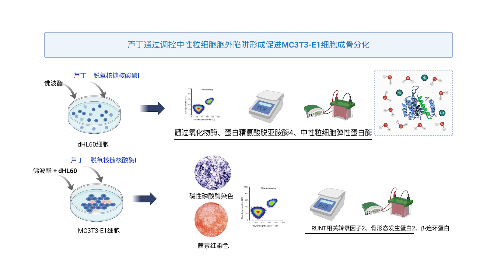
Rutin promotes osteogenic differentiation of MC3T3-E1 cells: regulating the formation of neutrophil extracellular traps

BACKGROUND: Rutin can effectively prevent osteoporosis, but its mechanism of action remains unclear. OBJECTIVE: To investigate the effect of rutin on osteogenesis of MC3T3-E1 cells under the action of neutrophil extracellular traps. METHODS: (1) Human myeloid leukemia dHL60 cells were stimulated with phorbol 12-myristate 13-acetate to induce neutrophil extracellular trap formation. dHL60 cells were divided into 4 groups: control group received Hank's balanced salt solution; the other three groups received 50 nmol/L phorbol 12-myristate 13-acetate; the latter two groups additionally received 250 μmol/L rutin or 250 μmol/L rutin plus 5 U/mL DNase I. Apoptosis of dHL60 cells was detected by flow cytometry; mRNA and protein expression of neutrophil extracellular trap marker genes were detected by RT-qPCR and western blot. (2) MC3T3-E1 cells were divided into 6 groups: control group received Hank's balanced salt solution; the other five groups received 50 nmol/L phorbol 12-myristate 13-acetate; dHL60 cells and 50 nmol/L phorbol 12-myristate 13-acetate; 100 μmol/L rutin; dHL60 cells, 50 nmol/L phorbol 12-myristate 13-acetate, and 250 μmol/L rutin; dHL60 cells, 50 nmol/L phorbol 12-myristate 13-acetate, 250 μmol/L rutin, and 5 U/mL DNase I. Apoptosis of MC3T3-E1 cells under neutrophil extracellular traps was detected by flow cytometry; alkaline phosphatase staining and alizarin red staining were used to determine osteogenic and mineralization abilities; RT-qPCR and western blot were used to detect osteogenic-related gene and protein expression. RESULTS AND CONCLUSION: (1) Compared with the blank control group, rutin significantly inhibited the mRNA and protein expression of protein arginine deiminase 4, myeloperoxidase, and neutrophil elastase in dHL60 cells (P < 0.000 1); compared with the rutin group, the combination of rutin and DNase I had a more significant downregulation effect (P < 0.05), indicating that rutin can significantly inhibit neutrophil extracellular trap formation. (2) After inducing neutrophil extracellular traps from dHL60 and co-culturing with MC3T3-E1, the mRNA and protein expression of Runt-related transcription factor 2, β-catenin, and bone morphogenetic protein 2 in MC3T3-E1 cells were significantly downregulated (P < 0.000 1), and the apoptosis rate significantly increased (P < 0.000 1), indicating that neutrophil extracellular traps can significantly inhibit osteogenic ability and promote apoptosis of MC3T3-E1 cells in vitro. After intervention with rutin alone or rutin combined with DNase I, the apoptosis and osteogenic ability of MC3T3-E1 cells under neutrophil extracellular traps were significantly improved, and the effect of rutin combined with DNase I was more significant than rutin alone, indicating that rutin may inhibit neutrophil extracellular trap formation, thereby improving the osteogenic ability of MC3T3-E1 cells. (3) Molecular docking and molecular dynamics simulations showed that rutin binds well to protein arginine deiminase 4, myeloperoxidase, and neutrophil elastase target proteins, indicating that rutin can target and inhibit neutrophil extracellular trap formation.

Read Full Abstract10.12307/2026.21347
Cerebrospinal fluid-contacting neurons differentiating into motor neurons promote functional recovery in spinal cord-injured miceGraphical AbstractVerified
Chinese Journal of Tissue Engineering Research2026

Cerebrospinal fluid-contacting neurons differentiating into motor neurons promote functional recovery in spinal cord-injured mice

BACKGROUND: Cell transplantation is one of the effective approaches for repairing spinal cord injury. Our research team previously found that transplanted cerebrospinal fluid-contacting neurons can survive and promote motor function recovery in mice with spinal cord injury. However, whether these transplanted cerebrospinal fluid-contacting neurons differentiate into functional neurons and thereby facilitate motor function recovery remains unclear. OBJECTIVE: To investigate whether transplanted cerebrospinal fluid-contacting neurons differentiate into functional neurons in vivo and contribute to motor function recovery after spinal cord injury. METHODS: Primary cells containing cerebrospinal fluid-contacting neurons were isolated from the cervical spinal cord of C57BL/6 neonatal mice within 24 hours of birth and cultured adherently. Cells were transduced with a lentivirus carrying a multimodal imaging gene, and cerebrospinal fluid-contacting neurons were selected and purified using puromycin. Differentiation was induced with serum-containing differentiation medium, and expression of neuronal marker NeuN and motor neuron marker ChAT was detected by immunofluorescence. Thirty C57BL/6 mice were randomly divided into three groups: transplantation group and PBS group underwent T10 spinal cord injury by clip compression, while sham group only had laminectomy. One week after injury, cerebrospinal fluid-contacting neurons were transplanted in situ in the transplantation group, and an equal volume of PBS was injected in the PBS group. At 1, 4, and 8 weeks after transplantation, immunofluorescence was used to detect expression of motor neuron marker ChAT in spinal cord tissue. At 8 weeks, immunofluorescence was used to detect synaptic marker SYN, inhibitory transmitter marker GAD65/67, and excitatory transmitter marker vGLUT1; hematoxylin-eosin staining was used to observe spinal cord morphology; BMS motor function score and footprint analysis were used to assess motor function recovery. RESULTS AND CONCLUSION: (1) Cerebrospinal fluid-contacting neurons expressed neural stem cell characteristics in vitro and could differentiate into motor neurons. (2) Transplanted cerebrospinal fluid-contacting neurons could survive long-term in vivo and differentiate into motor neurons. (3) The proportion of cerebrospinal fluid-contacting neurons differentiating into motor neurons was highest at 8 weeks (P < 0.0001). (4) At 8 weeks after transplantation, cerebrospinal fluid-contacting neurons co-expressed SYN, GAD65/67, and vGLUT1, indicating synaptic connections with host neurons. (5) BMS scores of PBS group were consistently lower than those of transplantation group (P < 0.001); footprint analysis showed more coordinated gait in transplantation group with only toe dragging, while PBS group showed obvious hindlimb dragging. (6) Hematoxylin-eosin staining showed large cavities in the injured area of PBS group, while cavities were reduced in transplantation group. These results indicate that transplanted cerebrospinal fluid-contacting neurons can differentiate into motor neurons both in vitro and in vivo, form synaptic connections, and thereby improve motor function in spinal cord-injured mice.

Read Full Abstract10.12307/2026.21346
Protective effect of optimization of the whole blood separation process to prepare therapeutic-grade platelet lysate on cardiomyocytes from hypoxic injuryGraphical AbstractVerified
Chinese Journal of Tissue Engineering Research2026

Protective effect of optimization of the whole blood separation process to prepare therapeutic-grade platelet lysate on cardiomyocytes from hypoxic injury

BACKGROUND: Platelets are important blood resources, yet in routine blood bank processes they are often filtered out along with white blood cells as medical waste. Optimizing whole blood separation processes to prepare platelet lysate products and exploring their applications in tissue engineering and regenerative medicine is of great value. OBJECTIVE: To optimize whole blood separation to prepare therapeutic-grade platelet lysate and to investigate the protective effect of platelet lysate on hypoxic injury of cardiomyocytes. METHODS: Platelets were isolated from 21 qualified whole blood units under closed blood bag and tubing conditions, and 21 platelet lysates were prepared by freeze-thawing. The mass concentration ranges of platelet-derived growth factor AA, platelet-derived growth factor BB, platelet-derived growth factor AB, vascular endothelial growth factor, epidermal growth factor, insulin-like growth factor 1, fibroblast growth factor, and transforming growth factor beta 1 in platelet lysates were measured using enzyme-linked immunosorbent assay kits. Bacterial contamination was assessed by colony culture method and mycoplasma contamination by PCR detection kit. A cardiomyocyte hypoxia model was established to evaluate the protective effect of platelet lysate on hypoxic injury. RESULTS AND CONCLUSION: (1) The mass concentration ranges of major growth factors and cytokines in platelet lysates were: platelet-derived growth factor AA 12.86-24.17 μg/L, platelet-derived growth factor BB 0.25-0.32 μg/L, platelet-derived growth factor AB 85.09-114.91 μg/L, vascular endothelial growth factor 10.57-58.37 μg/L, epidermal growth factor 0.43-0.69 μg/L, insulin-like growth factor 1 106-204.9 μg/L, fibroblast growth factor 0.03-0.06 μg/L, and transforming growth factor beta 1 124.17-192.38 μg/L. (2) Colony culture and mycoplasma detection results were negative. (3) Low volume fraction (1%) platelet lysate yielded the highest proliferation efficiency of cardiomyocytes; low volume fraction (1%) platelet lysate stimulated cardiomyocytes to produce high levels of superoxide dismutase and glutathione peroxidase to protect cardiomyocytes. This study established a method for preparing therapeutic-grade platelet lysate by optimizing the whole blood separation process, which can improve the utilization rate of blood resources. Platelet lysate has high levels of major growth factors and can significantly promote the repair of hypoxic injured cardiomyocytes.

Read Full Abstract10.12307/2026.21348
miR-9 regulates the differentiation of neural stem cells in mouse cerebral cortexGraphical AbstractVerified
Chinese Journal of Tissue Engineering Research2026

miR-9 regulates the differentiation of neural stem cells in mouse cerebral cortex

BACKGROUND: Neural stem cells located in the ventricular zone and subventricular zone are crucial for cortical neurodevelopment and the treatment of neurodegenerative diseases. However, their precise regulatory mechanisms remain incompletely understood. miRNA-9 is one of the most abundantly expressed miRNAs in the vertebrate embryonic and adult brain, playing diverse roles during development. Nevertheless, the role of miR-9 in neural stem cell differentiation remains unclear. OBJECTIVE: To investigate the role of miR-9 in regulating the differentiation of neural stem cells in the ventricular zone and subventricular zone. METHODS: Neural stem cells were isolated from the ventricular zone and subventricular zone of embryonic day 14.5 ICR mice and cultured in proliferation medium for 3-4 days to form neurospheres. Stemness was identified by Pax6/Nestin immunofluorescence double staining. The expression profile of miR-9 was detected by qRT-PCR in telencephalon tissues at embryonic days 12.5, 14.5, 16.5, 18.5 and postnatal days 0, 7, as well as in embryonic day 14.5 neural stem cells cultured in vitro. Neural stem cells were transfected with miR-9 inhibitor or mimic using transfection reagents. After 24 hours, cells were differentiated for 3-4 days (neurons) and 6-8 days (glial cells). The differentiation of each lineage was quantified by immunofluorescence staining for Tuj1 (neuronal marker), myelin basic protein (oligodendrocyte marker), and glial fibrillary acidic protein (astrocyte marker). RESULTS AND CONCLUSION: qRT-PCR results showed that miR-9 was highly expressed in early embryonic telencephalon (E12.5-E14.5) and gradually decreased with development (E16.5 to P7). In E14.5 neural stem cells, miR-9 expression level was close to 90% of the internal reference RNU6B. Functional experiments showed that compared with the control group, the miR-9 inhibition group had decreased proportions of Tuj1-positive neurons and myelin basic protein-positive oligodendrocytes, while the proportion of glial fibrillary acidic protein-positive astrocytes increased. Conversely, the miR-9 overexpression group had increased proportions of Tuj1-positive neurons and myelin basic protein-positive oligodendrocytes, and decreased proportion of glial fibrillary acidic protein-positive astrocytes, with significant differences (P < 0.001). These results indicate that miR-9 plays a bidirectional regulatory role in neural stem cell differentiation: (1) It participates in the temporal regulation of neurogenesis through developmental stage-specific expression patterns (high early, downregulated later); (2) It maintains the balance of trilineage differentiation by promoting neuronal and oligodendrocyte differentiation while inhibiting astrocyte generation.

Read Full Abstract10.12307/2026.21344
Role and mechanism of emodin in slowing down the senescence of HT-22 cells induced by high glucoseGraphical AbstractVerified
Chinese Journal of Tissue Engineering Research2026

Role and mechanism of emodin in slowing down the senescence of HT-22 cells induced by high glucose

BACKGROUND: The occurrence of diabetic encephalopathy may be closely related to neuronal aging, but its underlying molecular mechanism is not fully understood. Therefore, exploring the role of neuronal senescence in diabetic encephalopathy is of great significance for further revealing the pathogenesis of diabetic encephalopathy. OBJECTIVE: To investigate the effect and mechanism of emodin on senescence of HT-22 cells under high glucose conditions. METHODS: HT-22 cells were divided into control group (glucose concentration 25 mmol/L), high glucose group (glucose concentration 55 mmol/L), and high glucose + emodin group (glucose concentration 55 mmol/L, emodin concentration 100 µmol/L) and cultured for 48 h. The growth state of cells in each group was observed under microscope; CCK-8 assay was used to detect cell viability; ELISA was used to detect telomerase reverse transcriptase activity; RT-qPCR and western blot were used to detect the expression of senescence-related proteins P53, P21, and P16; immunofluorescence, RT-qPCR and western blot were used to detect the expression of lamin A/C. RESULTS AND CONCLUSION: Compared with the control group, the high glucose group showed obvious growth inhibition under microscope, characterized by decreased cell number, increased cell volume, and flattened morphology; compared with the high glucose group, the high glucose + emodin group showed significantly increased cell number and more regular morphology. Compared with the control group, cell viability was significantly decreased in the high glucose group (P < 0.0001); compared with the high glucose group, cell viability was significantly increased in the high glucose + emodin group (P < 0.0001). Compared with the control group, telomerase reverse transcriptase activity was significantly decreased in the high glucose group (P < 0.001). Compared with the control group, the expression levels of P53, P21, and P16 were significantly increased in the high glucose group (P < 0.05); compared with the high glucose group, the expression levels of P53, P21, and P16 were significantly decreased in the high glucose + emodin group (P < 0.05). Compared with the control group, the expression level of lamin A/C was significantly decreased in the high glucose group (P < 0.0001); compared with the high glucose group, the expression level of lamin A/C was significantly increased in the high glucose + emodin group (P < 0.05). The results indicate that emodin may slow down the senescence of HT-22 cells induced by high glucose by upregulating the expression of lamin A/C.

Read Full Abstract10.12307/2026.21343
Non-coding RNA-activated by DNA damage promotes proliferation and inhibits apoptosis of induced pluripotent stem cell-derived cardiomyocytesGraphical AbstractVerified
Chinese Journal of Tissue Engineering Research2026

Non-coding RNA-activated by DNA damage promotes proliferation and inhibits apoptosis of induced pluripotent stem cell-derived cardiomyocytes

BACKGROUND: Although cell transplantation offers a promising approach for the treatment of myocardial infarction, the low transplantation rate limits its application. Therefore, promoting the proliferation of transplanted cells and reducing apoptosis are the key issues to be solved urgently to improve the therapeutic effect. OBJECTIVE: To investigate the effects of non-coding RNA-activated by DNA damage (NORAD) on the proliferation of human induced pluripotent stem cell-derived cardiomyocytes and their apoptosis induced by oxygen-glucose deprivation/reoxygenation, as well as the effects of transplanting NORAD-overexpressing human induced pluripotent stem cell-derived cardiomyocytes (hiPSC-NORADOECMs) on cardiac function in a murine model of myocardial infarction. METHODS: The expression of NORAD in the hearts of mice at different ages (3 days old and 8 weeks old) was measured by real-time fluorescence quantitative polymerase chain reaction (RT-qPCR). A cellular model of hiPSC-NORADOECMs was established by infecting human induced pluripotent stem cells with a lentiviral vector designed to specifically upregulate NORAD, followed by directed differentiation into cardiomyocytes. The overexpression efficiency was confirmed by RT-qPCR. Proliferation was assessed by immunofluorescence staining for Ki67. Apoptosis was induced by oxygen-glucose deprivation/reoxygenation (OGD/R). Intracellular reactive oxygen species (ROS) levels were measured by flow cytometry, and the expression of apoptosis-related proteins (Bax, Bcl-2, Cleaved Caspase-3) was detected by western blot. In vivo, hiPSC-NORADOECMs were transplanted into the infarcted myocardium of mice, and cardiac function was evaluated by echocardiography after 4 weeks. RESULTS AND CONCLUSION: NORAD expression was significantly higher in 3-day-old neonatal mouse hearts compared with 8-week-old adult hearts. hiPSC-NORADOECMs were successfully generated, showing increased Ki67 expression compared with control cells. Overexpression of NORAD inhibited OGD/R-induced ROS production, decreased Bax and Cleaved Caspase-3 protein levels, and increased Bcl-2 levels. Transplantation of hiPSC-NORADOECMs significantly improved cardiac function in myocardial infarction mice. These findings indicate that NORAD overexpression promotes hiPSC-CM proliferation and inhibits apoptosis by reducing ROS production, thereby enhancing the reparative capacity of hiPSC-CMs in myocardial infarction.

Read Full Abstract10.12307/2026.21342
Retrospective analysis of central nervous system diseases related to non-primary infiltration after allogeneic hematopoietic stem cell transplantationGraphical AbstractVerified
Chinese Journal of Tissue Engineering Research2026

Retrospective analysis of central nervous system diseases related to non-primary infiltration after allogeneic hematopoietic stem cell transplantation

BACKGROUND: Allogeneic hematopoietic stem cell transplantation may be complicated by central nervous system diseases not related to primary disease infiltration. There is no clear conclusion on the clinical symptoms, possible causes, and prognosis. OBJECTIVE: To explore the clinical characteristics, risk factors, and prognosis of non-primary infiltration-related central nervous system diseases after allogeneic hematopoietic stem cell transplantation, in order to provide evidence-based basis for early clinical diagnosis, etiological intervention, and prognosis improvement. METHODS: A retrospective analysis was conducted on clinical data, laboratory characteristics, and treatment processes of 298 patients with hematological diseases who developed non-primary infiltration-related central nervous system diseases after allogeneic hematopoietic stem cell transplantation from January 2015 to June 2024. They were divided into a non-primary infiltration-related central nervous system disease group (n=19) and a control group (without such diseases, n=279). Risk factors were analyzed statistically, and clinical symptoms, possible causes, and prognosis were evaluated. RESULTS AND CONCLUSION: (1) Among 298 patients, 19 developed non-primary infiltration-related central nervous system diseases, with an incidence of 6.4%. (2) The median onset time was 16 days (2-45 days) after transplantation. The main initial symptom was convulsions, accompanied by elevated blood pressure, headache, visual decline, consciousness disorders, and psychiatric behavioral abnormalities. (3) Univariate analysis showed significant associations between the occurrence of these diseases and granulocyte engraftment time, platelet engraftment time, history of central nervous system leukemia before transplantation, and grade III-IV graft-versus-host disease. Etiological analysis revealed: calcineurin inhibitor-related encephalopathy (2 cases), central nervous system injury (4 cases), central nervous system infection (4 cases), transplantation-associated thrombotic microangiopathy (4 cases), central nervous system graft-versus-host disease (1 case), intracranial hemorrhage (2 cases), endocrine metabolic encephalopathy (1 case), and unknown cause (1 case). (4) As of the follow-up date, the cumulative mortality in the disease group was 47% (9/19), significantly higher than 28.6% (80/279) in the control group. Further analysis showed that the estimated overall survival rates at 1 and 2 years after transplantation were significantly lower in the disease group than in the control group. In conclusion, non-primary infiltration-related central nervous system diseases after allogeneic hematopoietic stem cell transplantation are caused by multiple transplantation-related factors. Timely identification of pathogenic factors and precise diagnosis and treatment are crucial for improving the prognosis of patients with these complications.

Read Full Abstract10.12307/2026.21340
Culture and identification of adipose-derived stem cells from periprostatic adipose tissueGraphical AbstractVerified
Chinese Journal of Tissue Engineering Research2026

Culture and identification of adipose-derived stem cells from periprostatic adipose tissue

BACKGROUND: Periprostatic adipose tissue is the white visceral adipose tissue that is closest to the prostate, which is part of the prostate cancer tumor microenvironment and plays a key role in the occurrence and progression of prostate cancer. OBJECTIVE: To investigate the ability of adipose-derived stem cells derived from periprostatic adipose tissue to form three-dimensional cell sheets. METHODS: Periprostatic adipose tissue was harvested from patients undergoing radical prostatectomy. Adipose-derived stem cell suspensions were prepared using a combination of enzymatic digestion and mechanical dissection. Adipose-derived stem cell proliferation was assessed using a CCK-8 assay. Expression of stem cell-associated antigens CD34/CD44/CD45/CD90/CD105 was determined by flow cytometry. Multidirectional differentiation potential of the stem cells was assessed using osteogenic/adipogenic/chondrogenic differentiation assays. Adipose-derived stem cells were cultured for three weeks in low-glucose DMEM containing 100 μg/mL vitamin C and 10% fetal bovine serum to construct cell sheets, followed by histological analysis and scanning electron microscopy. RESULTS AND CONCLUSION: Adipose-derived stem cells from periprostatic adipose tissue exhibited a long spindle or fusiform shape, aligned growth, and consistent morphology. Primary culture reached 95% confluence at 9-10 days with good cell viability, and no obvious senescence was observed up to passage 15. Flow cytometry showed expression rates of CD44, CD90, and CD105 at 98.24%, 84.99%, and 89.14%, respectively, while CD34 and CD45 were expressed at 0.64% and 1.02%. After 3 weeks of osteogenic, adipogenic, and chondrogenic induction, the cells could differentiate into osteoblasts, adipocytes, and chondrocytes. After continuous culture for 3 weeks, the cells formed a three-dimensional cell sheet with a smooth surface and uniform texture, rich in extracellular matrix components such as fibronectin and type I collagen. Scanning electron microscopy revealed a flat surface with aligned long spindle-shaped cells and abundant extracellular matrix deposition between cells. This study successfully isolated adipose-derived stem cells from periprostatic adipose tissue of prostate cancer patients and constructed a three-dimensional cell sheet by stimulating extracellular matrix secretion with vitamin C over 3 weeks of continuous culture.

Read Full Abstract10.12307/2026.21333
Exosomes derived from human umbilical cord mesenchymal stem cells in treatment of diabetic foot ulcersGraphical AbstractVerified
Chinese Journal of Tissue Engineering Research2026

Exosomes derived from human umbilical cord mesenchymal stem cells in treatment of diabetic foot ulcers

BACKGROUND: Exosomes derived from mesenchymal stem cells play an important role in regulating apoptosis, promoting cell regeneration, and improving the wound microenvironment, making them a hot research topic in the treatment of diabetic foot ulcers. OBJECTIVE: To explore the clinical application value of exosomes derived from human umbilical cord mesenchymal stem cells in the repair of diabetic foot ulcers. METHODS: A retrospective analysis was conducted on the data from 72 patients with diabetic foot ulcers treated between May 2022 and April 2025. Thirty-six patients received treatment with exosomes derived from human umbilical cord mesenchymal stem cells (observation group), and 36 patients were managed with vacuum-assisted closure therapy (control group). Ulcer healing rate, incidence of adverse events, serum inflammatory markers, growth factor levels, total wound healing time, and ulcer recurrence rate during follow-up were compared between the two groups after 2 weeks of treatment. RESULTS AND CONCLUSION: (1) After 2 weeks of treatment, the ulcer healing rate in the observation group (48.03±6.12)% was significantly higher than that in the control group (30.13±6.38)%, with a significant difference (P < 0.05). (2) Ulcers healed in both groups, with healing time in the observation group (30.42±2.30) d significantly shorter than that in the control group (43.94±3.46) d (P < 0.05). (3) The incidence of adverse events during treatment was 13.89% in the control group and 19.44% in the observation group, with no significant difference (P > 0.05). (4) Serum levels of interleukin-6, C-reactive protein, and procalcitonin decreased significantly in both groups, with a greater decrease in the observation group (P < 0.01); serum levels of vascular endothelial growth factor, basic fibroblast growth factor, and platelet-derived growth factor increased significantly in both groups, with a greater increase in the observation group (P < 0.01). (5) After ulcer healing, the observation group was followed for an average of 9.4 months, with 5 cases of ulcer recurrence; the control group was followed for an average of 9.8 months, with 10 cases of recurrence. The recurrence rate in the observation group was significantly lower than that in the control group (13.9% vs. 27.8%, P < 0.05). These results indicate that compared with vacuum-assisted closure therapy, application of exosomes derived from human umbilical cord mesenchymal stem cells for repairing diabetic foot ulcers can effectively inhibit inflammation, promote ulcer healing, and reduce recurrence rate without increasing the risk of related adverse events, demonstrating both efficacy and safety.

Read Full Abstract10.12307/2026.21338
Quercetin promotes osteogenic differentiation of senescent jaw bone marrow mesenchymal stem cellsGraphical AbstractVerified
Chinese Journal of Tissue Engineering Research2026

Quercetin promotes osteogenic differentiation of senescent jaw bone marrow mesenchymal stem cells

BACKGROUND: Age-related degeneration is closely associated with bone metabolic imbalance. In the jaw, this manifests as alveolar bone resorption, tooth loosening, and even loss. Impaired osteogenic differentiation potential of senescent jaw bone marrow mesenchymal stem cells is a critical factor hindering jaw bone regeneration. Quercetin, a natural flavonoid compound, exhibits antioxidant, anti-inflammatory, and cell differentiation-regulating properties, yet effect and mechanism of quercetin in osteogenic differentiation of senescent jaw bone marrow mesenchymal stem cells remain unclear. OBJECTIVE: To investigate the effects of quercetin on the proliferation, migration, osteogenic differentiation, and senescence of aged jaw bone marrow mesenchymal stem cells. METHODS: Jaw bone marrow mesenchymal stem cells were isolated from the mandibles of 10 8-week-old SD rats and cultured using a combination of bone marrow flushing and bone slice digestion. Jaw bone marrow mesenchymal stem cells were subcultured to the third and seventh passages, serving as the young and senescent groups, respectively. The quercetin group was treated with quercetin based on the senescent group. CCK-8 assay was used to detect the effects of 0.01, 0.1, 1, 10, 100 μmol/L quercetin on proliferation of senescent jaw bone marrow mesenchymal stem cells, and the optimal concentration was selected. Cell scratch assay was used to observe cell migration ability. RT-qPCR and western blot were used to detect the expression of senescence markers. β-galactosidase staining was used to observe the proportion of positive cells. After 7 days of osteogenic induction, RT-qPCR and western blot were used to detect the expression of osteogenic markers. After 14 days of osteogenic induction, alkaline phosphatase staining was performed. After 21 days of osteogenic induction, alizarin red staining was performed. Western blot was used to detect the expression of phosphorylated protein kinase B, protein kinase B, phosphorylated mammalian target of rapamycin, and mammalian target of rapamycin. RESULTS AND CONCLUSION: Compared with the young group, the proliferation ability of the senescent group decreased. Compared with the senescent group, 1 μmol/L quercetin significantly promoted the proliferation of senescent jaw bone marrow mesenchymal stem cells (P < 0.01). Compared with the senescent group, the migration ability of senescent jaw bone marrow mesenchymal stem cells in the quercetin group was improved, the proportion of β-galactosidase positive cells was significantly reduced, and the mRNA and protein expression of senescence-related P16, P53, and P21 were decreased (P < 0.05). After osteogenic induction, compared with the senescent group, the quercetin group showed increased calcium nodule formation, alkaline phosphatase staining area, and mRNA and protein expression of alkaline phosphatase, osteopontin, and Runt-related transcription factor 2 (P < 0.05). Compared with the senescent group, the phosphorylation levels of protein kinase B and mammalian target of rapamycin in the quercetin group were significantly reduced (P < 0.05). These results indicate that quercetin can inhibit senescence of jaw bone marrow mesenchymal stem cells caused by multiple passages and promote osteogenic differentiation by regulating the protein kinase B/mammalian target of rapamycin signaling pathway.

Read Full Abstract10.12307/2026.21331
Effect of lactylated mixed lineage kinase domain-like protein on stemness expression of breast tumor stem cellsGraphical AbstractVerified
Chinese Journal of Tissue Engineering Research2026

Effect of lactylated mixed lineage kinase domain-like protein on stemness expression of breast tumor stem cells

BACKGROUND: Mixed lineage kinase domain-like protein is one of the key executor proteins in the necroptosis pathway and plays an important role in various diseases. However, the mechanism by which its lactylation affects the formation and differentiation of breast tumor stem cells remains unclear. OBJECTIVE: To investigate the effect of mixed lineage kinase domain-like protein K230 site lactylation on the stemness expression of breast tumor stem cells. METHODS: The differences in protein lactylation between breast tumor MCF-7 adherent cells and spheroidal stem cells were analyzed by mass spectrometry. The mixed lineage kinase domain-like protein and its lactylation sites related to tumor stem cells were screened. A mixed lineage kinase domain-like protein K230R mutant plasmid vector was constructed and transfected into MCF-7 breast tumor cells. The proliferation and migration abilities of the cells were detected by CCK-8 and scratch assays. The effect of mixed lineage kinase domain-like protein K230R mutation on the formation of breast tumor stem cells was verified by suspension spheroid formation assay. Western blot was used to detect the expression of stemness and epithelial-mesenchymal transition-related proteins. RESULTS AND CONCLUSION: The lactylation level of mixed lineage kinase domain-like protein at K230 was higher in breast tumor stem cells. Compared with the wild-type group, the K230R mutant group showed significantly reduced scratch healing rate and spheroid formation rate, significantly increased expression of epithelial-related proteins, and significantly decreased expression of mesenchymal-related and stemness-related proteins. The study indicates that lactylation of mixed lineage kinase domain-like protein at K230 promotes epithelial-mesenchymal transition and enhances stemness expression of breast tumor stem cells, thereby promoting the occurrence and development of breast tumors.

Read Full Abstract10.12307/2026.21339
Transplantation of human umbilical cord mesenchymal stem cells to repair myelination disorders in neonatal rats with white matter injuryGraphical AbstractVerified
Chinese Journal of Tissue Engineering Research2026

Transplantation of human umbilical cord mesenchymal stem cells to repair myelination disorders in neonatal rats with white matter injury

BACKGROUND: Myelination deficits are a core feature of white matter injury in preterm infants. In recent years, human umbilical cord mesenchymal stem cells have been applied in various animal models of brain injury, demonstrating the capacity to promote myelin repair. Elucidating the regulatory mechanisms by which human umbilical cord mesenchymal stem cells enhance neural myelination will contribute to optimizing therapeutic strategies and facilitating clinical translation. OBJECTIVE: To clarify the reparative effect of human umbilical cord mesenchymal stem cells on myelination disorders caused by maturation arrest of the oligodendrocyte lineage in neonatal rats with white matter injury. METHODS: Seventy-two 2-day-old Sprague-Dawley rats were randomly divided into sham operation group, white matter injury group, and human umbilical cord mesenchymal stem cell transplantation group (n=24 per group). A neonatal rat model of white matter injury was established by combining low-dose lipopolysaccharide with hypoxia-ischemia. On day 14 after modeling, pathological changes in white matter were observed by hematoxylin-eosin staining; the positive expression, protein and mRNA levels of oligodendrocyte lineage transcription factor 2, neural/glial antigen 2, and myelin basic protein were detected by immunohistochemistry, western blot, and real-time quantitative PCR. On day 28 after modeling, myelin formation was observed by Luxol fast blue staining, and spatial learning and memory ability were tested by Morris water maze. RESULTS AND CONCLUSION: On day 14 after modeling, hematoxylin-eosin staining showed that in the white matter injury group, a large number of cells degenerated and necrosed, and nerve fibers were arranged disorderly; in the human umbilical cord mesenchymal stem cell transplantation group, cell morphology was close to normal and nerve fibers were arranged relatively neatly. On day 14 after modeling, there was no statistically significant difference in the positive expression, protein and mRNA levels of oligodendrocyte lineage transcription factor 2 among groups (P > 0.05). Compared with the sham operation group, the positive expression, protein and mRNA levels of neural/glial antigen 2 were upregulated (P < 0.05), while those of myelin basic protein were downregulated (P < 0.05) in the white matter injury group. Compared with the white matter injury group, the positive expression, protein and mRNA levels of neural/glial antigen 2 were downregulated (P < 0.05), while those of myelin basic protein were upregulated (P < 0.05) in the human umbilical cord mesenchymal stem cell transplantation group. On day 28 after modeling, Luxol fast blue staining showed that compared with the sham operation group, myelin expression was decreased in the white matter injury group (P < 0.05); compared with the white matter injury group, myelin expression was increased in the human umbilical cord mesenchymal stem cell transplantation group (P < 0.05). On day 28 after modeling, Morris water maze results showed that compared with the sham operation group, the escape latency was prolonged and the number of platform crossings was decreased in the white matter injury group (P < 0.05); compared with the white matter injury group, the escape latency was shortened and the number of platform crossings was increased in the human umbilical cord mesenchymal stem cell transplantation group (P < 0.05); there was no statistically significant difference in average swimming distance among groups (P > 0.05). These findings indicate that human umbilical cord mesenchymal stem cells can promote the maturation of the oligodendrocyte lineage, repair myelination disorders, and improve cognitive function in neonatal rats with white matter injury.

Read Full Abstract10.12307/2026.21337
Human adipose multilineage-differentiating stress-enduring cells on treatment of ischemic stroke in ratsGraphical AbstractVerified
Chinese Journal of Tissue Engineering Research2026

Human adipose multilineage-differentiating stress-enduring cells on treatment of ischemic stroke in rats

BACKGROUND: Mesenchymal stem cells have shown good therapeutic effects in ischemic stroke, while the role of multilineage-differentiating stress-enduring (Muse) cells isolated from adipose-derived mesenchymal stem cells in ischemic stroke needs further study. OBJECTIVE: To explore the neurorestorative effect of intravenous administration of adipose Muse cells on ischemic stroke in rats. METHODS: The Muse cells expressing stage-specific embryonic antigen 3 were sorted by magnetic beads after long-term (4 hours) trypsin incubation of human adipose-derived mesenchymal stem cells. The middle cerebral artery occlusion model was established in rats. After successful modeling, the adipose-derived mesenchymal stem cell group and adipose-derived Muse group were injected with 200 μL adipose-derived mesenchymal stem cell suspension or adipose Muse cell suspension (containing 2×10^5 cells) via the tail vein, while the saline group received 200 μL saline. Behavioral scores were assessed at days 3 and 7 after transplantation. Hematoxylin-eosin staining was used to observe brain tissue damage at day 3. Immunofluorescence was used to detect microtubule-associated protein 2 and Ki67 expression in the damaged area. TUNEL staining was used to observe apoptosis. Western blot was used to detect growth-associated protein 43 and Bcl-2/Bax protein expression. RESULTS AND CONCLUSION: After magnetic bead sorting, flow cytometry showed that the expression rate of stage-specific embryonic antigen 3 in positively sorted cells was as high as 80%. At day 7 after transplantation, compared with the saline group, the neurological deficit score was reduced in both the adipose-derived mesenchymal stem cell group and the adipose-derived Muse group (P < 0.05). At day 3, compared with the adipose-derived mesenchymal stem cell group, the adipose-derived Muse group showed a lower neurological deficit score (P < 0.05). Hematoxylin-eosin staining showed reduced inflammatory response and vacuolation in the cerebral cortex of rats in both cell-treated groups, and immunofluorescence for microtubule-associated protein 2 showed that the adipose-derived Muse group had more obvious inhibition of neuronal loss. TUNEL and Ki67 staining showed reduced apoptosis and increased proliferation in the damaged area in both cell-treated groups. Western blot results showed that the Bcl-2/Bax ratio and growth-associated protein 43 expression were increased in both cell-treated groups, and the adipose-derived Muse group was superior to the adipose-derived mesenchymal stem cell group in inhibiting apoptosis. These results indicate that both adipose-derived mesenchymal stem cells and adipose-derived Muse cells promote neural repair in rats, with adipose-derived Muse cells playing a better role in inhibiting apoptosis.

Read Full Abstract10.12307/2026.21332
Overexpression of programmed death ligand 1 enhances immunosuppressive capacity of human umbilical cord mesenchymal stromal cells against T cellsGraphical AbstractVerified
Chinese Journal of Tissue Engineering Research2026

Overexpression of programmed death ligand 1 enhances immunosuppressive capacity of human umbilical cord mesenchymal stromal cells against T cells

BACKGROUND: The T cell immunosuppressive activity of mesenchymal stromal cells offers new hope for the treatment of autoimmune diseases. Amimatoside injection, a drug of human umbilical cord mesenchymal stromal cells, has been approved for the treatment of acute graft-versus-host disease (GVHD) primarily affecting the digestive tract, after steroid therapy failure, in patients aged 14 years and older. Therefore, further exploration of the T cell immunosuppressive potential of mesenchymal stromal cells can lay a foundation for the treatment of autoimmune diseases. OBJECTIVE: To investigate the effect of programmed death ligand 1 (PD-L1) gene overexpression on the inhibition of CD4+ T cell proliferation by human umbilical cord mesenchymal stromal cells (hUC-MSCs). METHODS: (1) hUC-MSCs were cultured in vitro to passages 0, 1, 2, and 3, and the percentage of PD-L1-positive cells was detected by flow cytometry. (2) hUC-MSCs were divided into experimental group and negative control group. The experimental group was transduced with lentivirus-mediated PD-L1 gene, while the negative control group was transduced with lentivirus carrying empty plasmid vector. Transfection efficiency was assessed by flow cytometry, real-time quantitative PCR, and western blot. (3) CD4+ T cells were enriched from healthy human peripheral blood using magnetic beads, labeled with carboxyfluorescein diacetate succinimidyl ester (CFSE), and co-cultured with hUC-MSCs from the experimental and negative control groups at a ratio of 5:1. The proportion of CFSE-dim CD4+ T cells was detected by flow cytometry. (4) RNA sequencing was performed on hUC-MSCs from both groups, and single-cell RNA sequencing was performed on the experimental group. Subgroups were identified based on function, and bioinformatics analysis was used to depict heatmaps of marker genes, enriched signaling pathways, and gene regulatory networks for each subgroup. RESULTS AND CONCLUSION: (1) The percentage of PD-L1-positive cells in hUC-MSCs gradually decreased with increasing passage number. (2) hUC-MSCs stably overexpressing PD-L1 were successfully constructed, with significantly increased PD-L1 expression in the experimental group. (3) Transcriptome sequencing data suggested that PD-L1-overexpressing hUC-MSCs promoted the upregulation of genes related to immune effector regulatory pathways. (4) Co-culture of CD4+ T cells with hUC-MSCs showed a significant decrease in the proportion of CD4+ T cells in the experimental group. (5) Based on single-cell RNA sequencing results, PD-L1-overexpressing hUC-MSCs could be divided into three functional subgroups with heterogeneity; PD-L1 gene expression was higher in subgroup 1, and the significantly high expression of histone methyltransferase SETDB1 might be closely related to enhanced T cell immunosuppressive function. These findings indicate that PD-L1 gene overexpression can significantly enhance the T cell immunosuppressive capacity of hUC-MSCs. Single-cell RNA sequencing can effectively classify PD-L1 gene-modified hUC-MSCs based on their functional characteristics, providing theoretical support for improving the clinical efficacy of hUC-MSCs.

Read Full Abstract10.12307/2026.21335
Inhibitory effect of complement 1q/tumor necrosis factor-related protein 4 on 3T3-L1 preadipocyte differentiationGraphical AbstractVerified
Chinese Journal of Tissue Engineering Research2026

Inhibitory effect of complement 1q/tumor necrosis factor-related protein 4 on 3T3-L1 preadipocyte differentiation

BACKGROUND: Adipocyte differentiation is a complex biological process involving the transformation of preadipocytes into mature adipocytes. This process plays a key role in the development and progression of obesity and related metabolic diseases. In recent years, the complement C1q/tumor necrosis factor-related protein family, as a new member of the adipokine family, has become a research hotspot in the field of metabolic regulation. OBJECTIVE: To investigate the effect of complement 1q/tumor necrosis factor-related protein 4 on the differentiation of 3T3-L1 preadipocytes and its underlying mechanism. METHODS: The CCK-8 assay was used to determine the effects of different concentrations of complement 1q/tumor necrosis factor-related protein 4 on the viability of 3T3-L1 preadipocytes, and a safe concentration was selected for intervention. 3T3-L1 preadipocytes were divided into a control group, an inducer group, and a complement 1q/tumor necrosis factor-related protein 4 group. 3T3-L1 preadipocytes were first contact-inhibited for 2 days, followed by adipogenic inducer and complement 1q/tumor necrosis factor-related protein 4 treatment. On day 10 of differentiation, oil red O staining was used to observe lipid droplet formation; RT-qPCR and western blot were used to detect the mRNA and protein expression levels of CCAAT/enhancer binding protein alpha, peroxisome proliferator-activated receptor gamma, and fatty acid binding protein 4; and triglyceride and total cholesterol assay kits were used to measure intracellular triglyceride and total cholesterol levels. RESULTS AND CONCLUSION: The highest safe concentration of complement 1q/tumor necrosis factor-related protein 4 with no toxic effect on 3T3-L1 preadipocytes was 1,000 ng/mL. Compared with the control group, the inducer group showed increased lipid droplet formation, elevated triglyceride and total cholesterol levels (P < 0.05), and significantly upregulated mRNA and protein expression of CCAAT/enhancer binding protein alpha, peroxisome proliferator-activated receptor gamma, and fatty acid binding protein 4 (P < 0.05). Compared with the inducer group, the complement 1q/tumor necrosis factor-related protein 4 group showed reduced lipid droplet formation, decreased triglyceride and total cholesterol levels (P < 0.05), and significantly downregulated mRNA and protein expression of CCAAT/enhancer binding protein alpha, peroxisome proliferator-activated receptor gamma, and fatty acid binding protein 4 (P < 0.05). These results indicate that complement 1q/tumor necrosis factor-related protein 4 can inhibit the differentiation of preadipocytes into mature adipocytes, and the mechanism may be related to the downregulation of CCAAT/enhancer binding protein alpha, peroxisome proliferator-activated receptor gamma, and fatty acid binding protein 4 expression.

Read Full Abstract10.12307/2026.21334
Osteoarthritis characteristic genes and prediction of targeted food-medicine homology traditional Chinese medicine: bioinformatics analysis and kinetic simulationGraphical AbstractVerified
Chinese Journal of Tissue Engineering Research2026

Osteoarthritis characteristic genes and prediction of targeted food-medicine homology traditional Chinese medicine: bioinformatics analysis and kinetic simulation

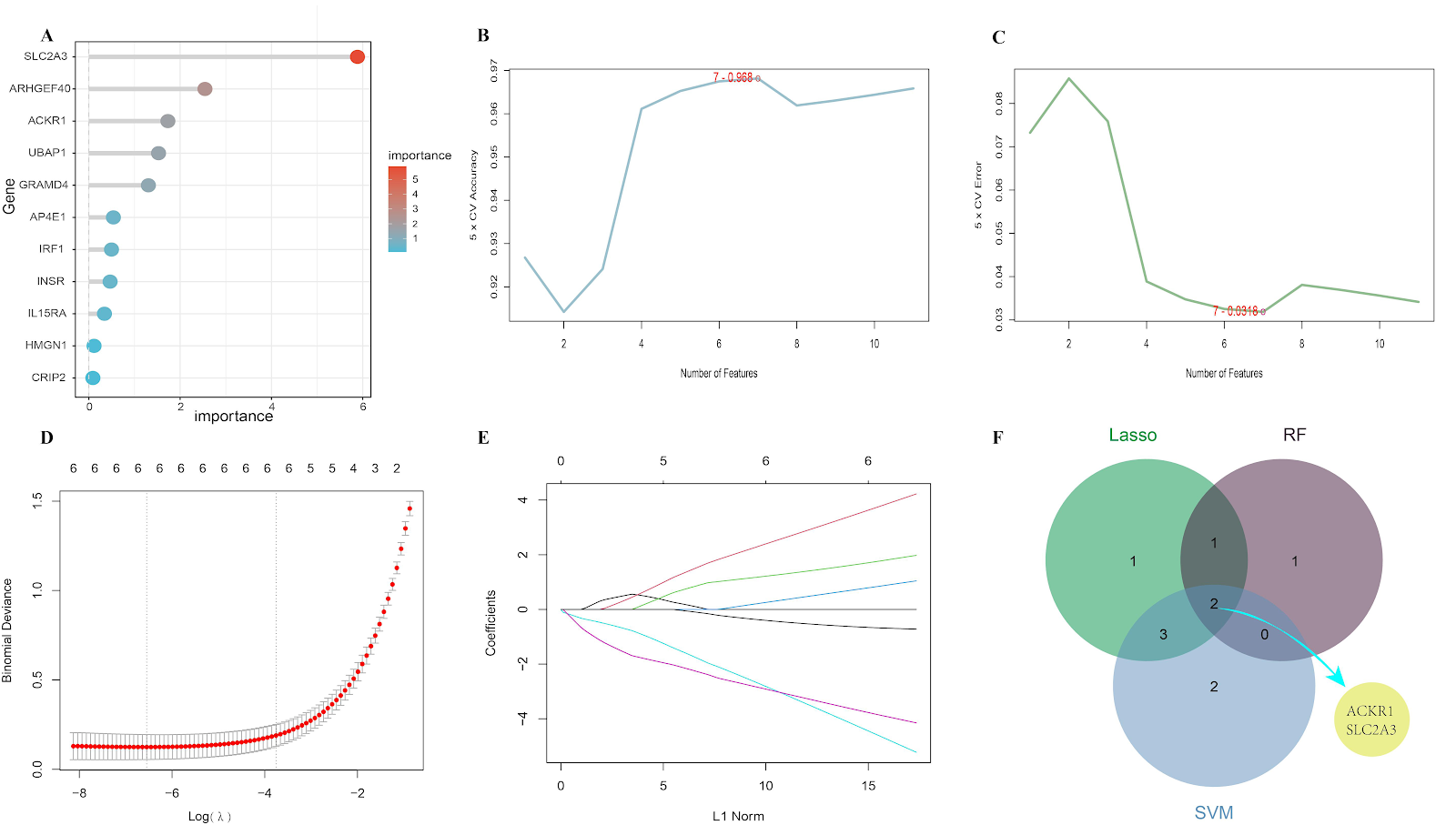
BACKGROUND: Early diagnosis and treatment of osteoarthritis remain a significant challenge due to the lack of highly specific biomarkers. OBJECTIVE: To screen characteristic genes of osteoarthritis, predict potential food-medicine homology traditional Chinese medicine and their core components, and validate their therapeutic potential through molecular docking and molecular dynamics simulations. METHODS: This study is based on three datasets (GSE55235, GSE169077, and GSE55457) from the GEO database, including a total of 25 normal samples and 26 osteoarthritis samples. It combines genes extracted from the eQTL database as exposure factors, and osteoarthritis data from the IEU openGWAS database (407,746 samples) as outcome factors. Core biomarkers were identified using least absolute shrinkage and selection operator regression, random forest, and support vector machine algorithms. CIBERSORT was used to evaluate immune infiltration characteristics and single-gene gene set enrichment analysis was performed. Potential traditional Chinese medicines were predicted using Coremine Medical and HERB databases, and food-medicine homology traditional Chinese medicines and their core components were screened, followed by molecular docking and molecular dynamics simulations. RESULTS AND CONCLUSION: ① Two genes, glucose transporter 3 (GLUT3) and atypical chemokine receptor 1 (ACKR1), were identified as characteristic genes of osteoarthritis, showing good diagnostic efficacy (AUC > 0.8) and involvement in metabolic regulation, cell signal transduction, and inflammatory responses, closely related to glucose metabolism, immune regulation, and inflammatory signaling pathways. ② Seven food-medicine homology traditional Chinese medicines were screened, including Cornus officinalis, Perilla frutescens, Ganoderma lucidum, Gastrodia elata, bitter almond, clove, and Rehmannia glutinosa, with core components β-sitosterol and stigmasterol. Molecular docking and dynamics simulations showed that stigmasterol had the best affinity with GLUT3 and the complex exhibited high stability. ③ This study systematically reveals the key roles of GLUT3 and ACKR1 in the pathogenesis of osteoarthritis, preliminarily validates the possibility of food-medicine homology traditional Chinese medicines intervening in the pathological process of osteoarthritis through multi-target and multi-pathway mechanisms, providing new molecular evidence for early diagnosis and targeted therapy, as well as theoretical support for prevention and treatment strategies. This research, from the perspective of traditional Chinese medicine and modern molecular biology, can provide a reference for the clinical application of traditional Chinese medicine in osteoarthritis.

Read Full Abstract10.12307/2026.21438
Potential targets and drug prediction for gout: identification of druggable genesGraphical AbstractVerified
Chinese Journal of Tissue Engineering Research2026

Potential targets and drug prediction for gout: identification of druggable genes

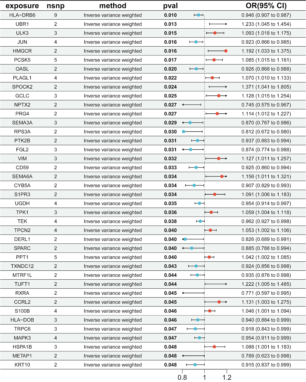
BACKGROUND: Existing pharmacological treatments for gout are frequently limited by substantial side effects, underscoring the urgent need to discover novel therapeutic targets and develop more targeted drugs. OBJECTIVE: To identify genetic targets for gout, and to predict promising therapeutic compounds as well as traditional Chinese medicines by integrating druggable gene datasets with Mendelian randomization and colocalization analysis approaches. This work will lay a foundation for in-depth exploration of the pathogenesis of gout in the Chinese population, and provide insights for the clinical management and development of new targeted drugs. METHODS: Gout-related datasets were obtained from the Finnish database FinnGen R11. Blood expression quantitative trait loci data were obtained from the GWAS catalog website developed by the MRC Integrative Epidemiology Unit at the University of Bristol. Mendelian randomization analysis was performed to identify potential targets; colocalization analysis was used to identify key susceptibility genes for gout. Gene Ontology and Kyoto Encyclopedia of Genes and Genomes enrichment analyses were used to explore gene functions, and protein-protein interaction networks were used to screen closely interacting targets. The Drug-Gene Interaction Database developed by Washington University School of Medicine in St. Louis was used to predict compounds with potential therapeutic effects. Molecular docking was used to predict the binding degree of compounds to core targets. The Coremine Medical database founded by PubGene was used to predict traditional Chinese medicines related to core genes. All databases used are public resources. A gout cell model was established using monosodium urate crystal-induced RAW264.7 cells to preliminarily verify the expression of key genes and the intervention effect of compounds. CCK-8 assay and cell invasion assay were used to screen safe doses and optimal administration concentrations. ELISA was used to measure inflammatory factor levels, and real-time fluorescence quantitative reverse transcription PCR was used to detect mRNA expression of key targets and pathways. RESULTS AND CONCLUSION: (1) Mendelian randomization analysis identified 40 potential gene targets significantly associated with gout; colocalization analysis identified Jun proto-oncogene as a key susceptibility gene for gout; protein-protein interaction network showed that Jun proto-oncogene, mitogen-activated protein kinase 3, and 3-hydroxy-3-methylglutaryl-CoA reductase had close interactions. (2) Gene Ontology and Kyoto Encyclopedia of Genes and Genomes enrichment results showed that potential targets mainly regulate mitogen-activated protein kinase, tumor necrosis factor, ErbB, interleukin-17, hypoxia-inducible factor 1, Toll-like receptor and other signaling pathways, and intervene in positive regulation of extracellular signal-regulated kinase 1/2 cascade, glutathione metabolism, ubiquitin protein regulation and other processes. (3) Based on potential targets, 372 compounds with potential intervention effects were predicted, including capsaicin, 5,6-benzoflavone, L-glutamic acid, quercetin, honokiol, kaempferol, cinnamaldehyde, and andrographolide. (4) Molecular docking showed that capsaicin and 5,6-benzoflavone had high binding affinity with core targets such as Jun proto-oncogene. (5) 79 potential targeted traditional Chinese medicines were predicted, including Atractylodes, Magnolia officinalis, Smilax glabra, Alisma orientale, and Salvia miltiorrhiza, with efficacy mainly concentrated in clearing heat and detoxifying, promoting blood circulation and removing blood stasis, and resolving phlegm and dampness. (6) In cell experiments, CCK-8 and cell invasion assay results showed that the optimal safe dose of capsaicin was 50 μmol/L. In the model group, the expression of key gene Jun proto-oncogene was significantly upregulated, and capsaicin could significantly downregulate the mRNA expression of Jun proto-oncogene and mitogen-activated protein kinase pathway-related genes such as c-Jun N-terminal kinase, extracellular signal-regulated kinase 1/2, and p38, and reduce the levels of interleukin-6, interleukin-1β, and tumor necrosis factor α in cell supernatant. (7) Data mining results suggest that compounds such as capsaicin and 5,6-benzoflavone and traditional Chinese medicines such as Atractylodes and Smilax glabra may exert therapeutic effects on gout by intervening in targets such as Jun proto-oncogene and mitogen-activated protein kinase 3, regulating tumor necrosis factor, Th-17, hypoxia-inducible factor 1 and other pathways, and mitogen-activated protein kinase cascade, protein ubiquitination, and glutathione metabolism. Among them, the key susceptibility gene JUN can serve as a potential diagnostic marker for gout. Treatment methods mainly focusing on clearing heat and detoxifying combined with promoting blood circulation and removing blood stasis can be key to gout treatment. (8) Cell experiments preliminarily verified the expression of JUN gene and mitogen-activated protein kinase pathway in gout cell model and the intervention effect of capsaicin, providing a basis and foundation for the next step of gout diagnosis and treatment targets and new drug development.
Read Full Abstract10.12307/2026.21434
Bibliometric analysis of exercise therapy interventions for adolescent idiopathic scoliosisGraphical AbstractVerified

Chinese Journal of Tissue Engineering Research2026

Bibliometric analysis of exercise therapy interventions for adolescent idiopathic scoliosis

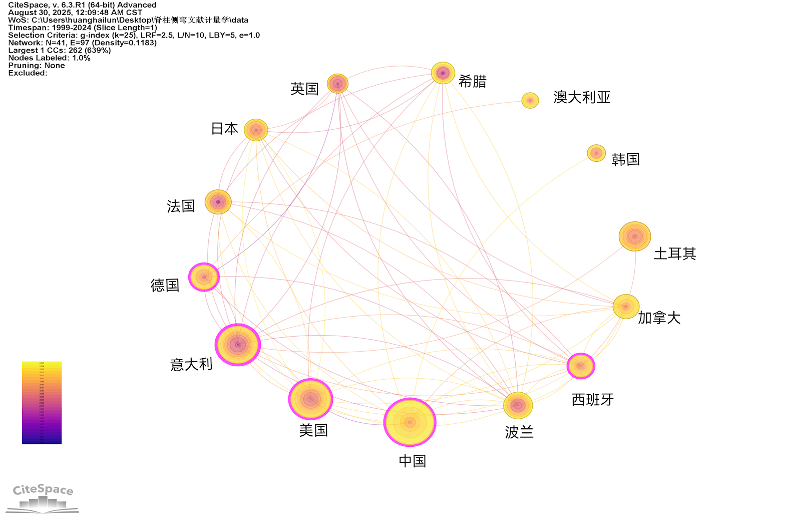
BACKGROUND: Adolescent idiopathic scoliosis is one of the most common spinal deformities in adolescents. As an important non-surgical treatment, exercise therapy for adolescent idiopathic scoliosis is widely used in clinical practice, yet there is a lack of systematic analysis of research trends and developments in this field. OBJECTIVE: To analyze the research hotspots, developmental dynamics, and international collaboration patterns in exercise therapy for improving adolescent idiopathic scoliosis from 1999 to 2024 using bibliometric methods, and to provide references for future research directions. METHODS: Based on the Web of Science Core Collection database, relevant literature published between January 1999 and September 2024 was retrieved, and 188 studies were ultimately included. Visualization analysis of countries/regions, institutions, authors, journals, keywords, and co-cited references was conducted using VOSviewer and CiteSpace software. RESULTS AND CONCLUSION: The number of publications in the field of exercise therapy for adolescent idiopathic scoliosis has undergone three stages, with sustained growth in recent years. China and the United States lead in publication output, while the United States has the highest average citations per paper, and Italy serves as a central hub for international collaboration. Schroth training, individualized treatment plans, and quality of life assessment are current research hotspots. Keyword evolution indicates a shift towards precise assessment and personalized intervention. Through visualization, this study identifies core author groups, high-impact journals, and key references in the field, revealing a clear path from general conservative treatment to specific, technology-integrated approaches, providing a systematic reference for scholars to quickly grasp the field's landscape and plan research directions.

Read Full Abstract10.12307/2026.21441
Acanthopanax exosome-like nanovesicles promote osteogenic differentiation of human bone marrow mesenchymal stem cellsGraphical AbstractVerified
Chinese Journal of Tissue Engineering Research2026

Acanthopanax exosome-like nanovesicles promote osteogenic differentiation of human bone marrow mesenchymal stem cells

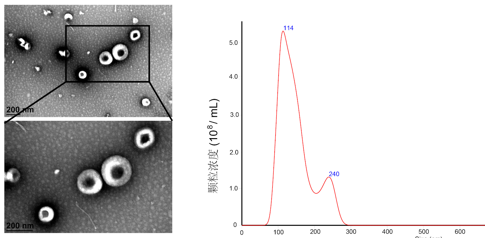
BACKGROUND: Acanthopanax and its extracts exhibit osteogenic effects, but the osteogenic potential and mechanisms of acanthopanax exosome-like nanovesicles remain unclear. OBJECTIVE: To investigate the molecular mechanism by which acanthopanax exosome-like nanovesicles promote osteogenic differentiation of human bone marrow mesenchymal stem cells and their preventive role in osteoporosis. METHODS: (1) Human bone marrow mesenchymal stem cells were extracted by gradient density centrifugation. Exosome-like nanovesicles derived from acanthopanax were isolated by differential centrifugation and sucrose gradient density centrifugation. (2) Human bone marrow mesenchymal stem cells were treated with 0, 2.5, and 5 μg/mL acanthopanax exosome-like nanovesicles. Osteogenic differentiation was assessed by alkaline phosphatase staining, Alizarin red staining, qRT-PCR, and western blot assay. (3) Key pathways were identified by transcriptome sequencing and verified using a transforming growth factor β1 receptor inhibitor. (4) An ovariectomized rat model of osteoporosis was established, and acanthopanax exosome-like nanovesicles were administered intraperitoneally for 12 weeks. Bone microarchitecture was analyzed by Micro-CT, and osteogenic protein expression was detected by histological staining. RESULTS AND CONCLUSION: (1) Acanthopanax exosome-like nanovesicles exhibited a typical cup-shaped or disc-shaped morphology. (2) In vitro experiments confirmed that acanthopanax exosome-like nanovesicles dose-dependently promoted osteogenic differentiation of bone marrow mesenchymal stem cells, as evidenced by increased alkaline phosphatase activity, enhanced mineralization nodule formation, and upregulated osteogenic gene expression. (3) Transcriptome analysis revealed that acanthopanax exosome-like nanovesicles activated the transforming growth factor β1/Smad2/3 signaling pathway, upregulating transforming growth factor β1 and phosphorylated Smad2/3 protein expression, and the transforming growth factor β1 receptor inhibitor partially suppressed their osteogenic effect. (4) Animal experiments showed that after intervention with 5 mg/kg acanthopanax exosome-like nanovesicles, bone mineral density, bone volume fraction, and trabecular thickness were significantly increased in ovariectomized rats (P < 0.05), collagen fiber formation was evident, and the expression of Runt-related transcription factor 2, osteocalcin, and transforming growth factor β1 proteins in bone tissue was upregulated. No obvious toxicity was observed in major organs by histological examination. These results indicate that acanthopanax exosome-like nanovesicles promote osteogenic differentiation of human bone marrow mesenchymal stem cells by activating the transforming growth factor β1/Smad2/3 pathway and effectively improve osteoporosis.

Read Full Abstract10.12307/2026.21329
Senescent bone marrow mesenchymal stem cells promote multiple myeloma cell proliferation through galectin-3Graphical AbstractVerified
Chinese Journal of Tissue Engineering Research2026

Senescent bone marrow mesenchymal stem cells promote multiple myeloma cell proliferation through galectin-3

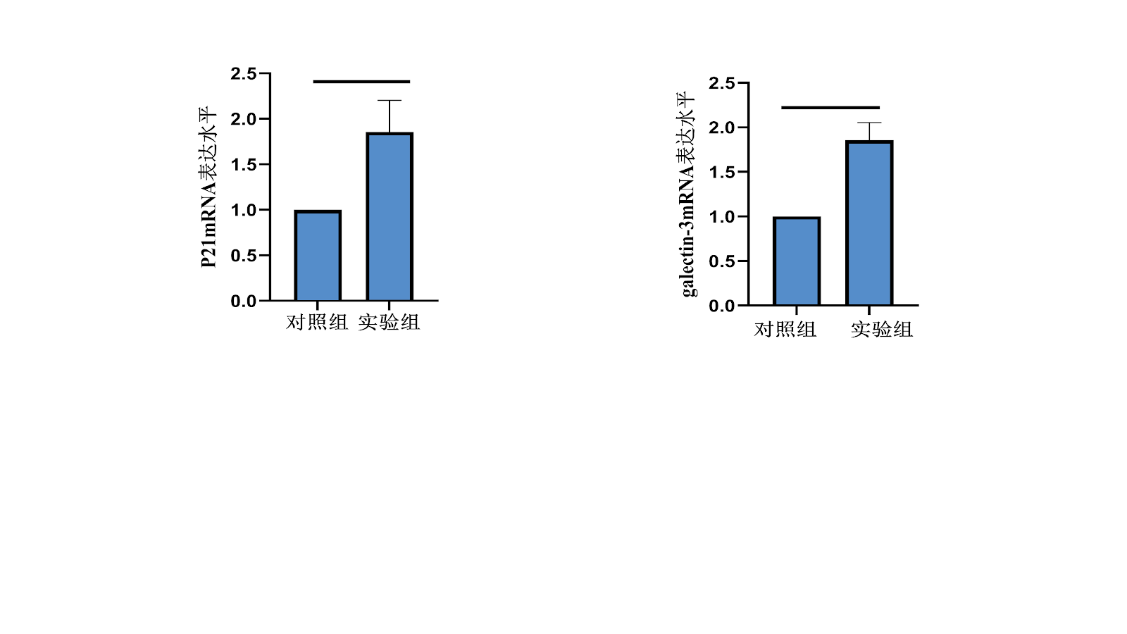
BACKGROUND: Studies showed that multiple myeloma microenvironment has the function of inducing mesenchymal stem cells to become senescent phenotype, while the effect of senescent bone marrow mesenchymal stem cells on multiple myeloma cells is rarely reported. OBJECTIVE: To investigate the effect of senescent bone marrow mesenchymal stem cells on the proliferation of multiple myeloma cells through paracrine galectin-3. METHODS: Bone marrow blood was collected from healthy donors, and bone marrow mesenchymal stem cells were extracted by Ficoll density gradient centrifugation and adherent purification. The third-generation bone marrow mesenchymal stem cells were taken and induced with 200 µmol/L hydrogen peroxide solution for 2 h, then cultured with L-DMEM complete medium for 24 h to construct a senescent bone marrow mesenchymal stem cell model. The model was identified by β-galactosidase staining and senescence gene P21 expression. RT-qPCR was used to detect the expression of galectin-3 in senescent bone marrow mesenchymal stem cells. The supernatant of senescent bone marrow mesenchymal stem cells was collected and concentrated by centrifugation to prepare conditioned medium, which was used to culture multiple myeloma cell line U266 for 24 h. CCK-8 was used to detect U266 cell proliferation, flow cytometry was used to detect U266 cell apoptosis, and RT-qPCR and western blot were used to detect BCL-2 protein and mRNA expression in U266 cells. Bone marrow from multiple myeloma patients and healthy individuals was collected, and galectin-3 levels were detected by ELISA. RESULTS AND CONCLUSION: After hydrogen peroxide induction, the number of β-galactosidase positive cells significantly increased, and the mRNA expression of P21 and galectin-3 was upregulated (P < 0.01). Compared with the control group, after culturing U266 cells with senescent bone marrow mesenchymal stem cell conditioned medium for 24 h, cell proliferation increased (P < 0.05), apoptosis rate decreased (P < 0.05), and BCL-2 protein and mRNA expression levels increased (P < 0.05). The level of galectin-3 in bone marrow of multiple myeloma patients was significantly higher than that of healthy individuals (P < 0.05). The results indicate that senescent bone marrow mesenchymal stem cells may promote the proliferation of multiple myeloma cells by upregulating BCL-2 expression through paracrine galectin-3.

Read Full Abstract10.12307/2026.21330
Causal relationship between plasma metabolites and osteonecrosis: a large sample analysis based on genome-wide association study database and FinnGen databaseGraphical AbstractVerified
Chinese Journal of Tissue Engineering Research2026

Causal relationship between plasma metabolites and osteonecrosis: a large sample analysis based on genome-wide association study database and FinnGen database

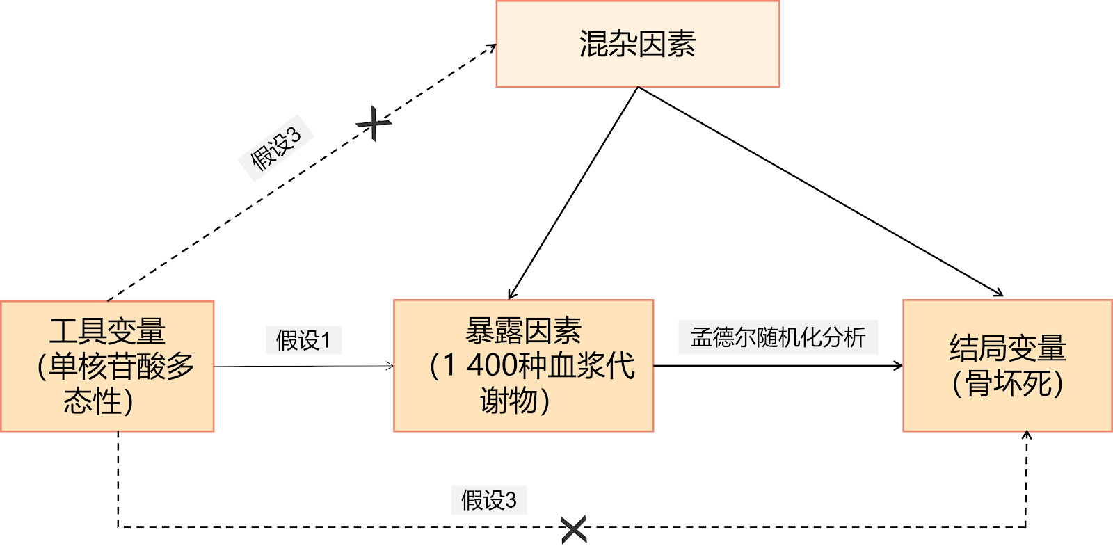
BACKGROUND: Osteonecrosis is a disabling and refractory disease with a high prevalence rate in China, necessitating the exploration of potential biomarkers for early prevention, diagnosis, and treatment. Metabolomic studies have demonstrated correlations between human metabolites and osteonecrosis; however, the causal relationship between plasma metabolites and osteonecrosis remains unclear. OBJECTIVE: To investigate the causal association between 1,400 plasma metabolites and osteonecrosis using Mendelian randomization and provide supporting evidence. METHODS: Public data on 1,400 plasma metabolites (exposure factors) and osteonecrosis (outcome factor) were collected. The plasma metabolite data were derived from a genome-wide association study (GWAS) on blood metabolites published in Nature Genetics in January 2023, which included 1,091 blood metabolites and 309 metabolite ratios from 8,299 individuals in the Canadian Longitudinal Study on Aging (CLSA) cohort. The single-nucleotide polymorphism data for osteonecrosis were obtained from the FinnGen public database R12 dataset, comprising 475,307 samples, including 2,043 osteonecrosis cases and 473,264 controls, all of European ancestry. Mendelian randomization analyses (inverse variance weighting, MR-Egger, weighted median, simple mode, and weighted mode) were performed using Rstudio software, followed by heterogeneity tests, pleiotropy tests, and Steiger directionality tests to ensure robustness and reliability. RESULTS AND CONCLUSION: Three plasma metabolites showed significant causal associations with osteonecrosis (P < 0.05): adenosine monophosphate to valine ratio (OR=1.303, 95%CI=1.110-1.531, P=0.001, PFDR=0.07), oxidized cysteinylglycine level (OR=0.888, 95%CI=0.791-0.998, P=0.046, PFDR=0.05), and 3β,17β-androstenediol disulfate level (OR=1.121, 95%CI=1.020-1.231, P=0.018, PFDR=0.06). The adenosine monophosphate to valine ratio and 3β,17β-androstenediol disulfate level were risk factors for osteonecrosis, while oxidized cysteinylglycine level was a protective factor. These findings suggest causal relationships between three plasma metabolites and osteonecrosis, potentially serving as biomarkers for early diagnosis and targets for intervention. Although based on European population data, this study provides valuable reference for osteonecrosis research in China, and future domestic researchers may achieve early diagnosis and precise treatment by detecting and regulating metabolite levels.

Read Full Abstract10.12307/2026.21437
Bibliometric analysis of research hotspots on mitochondria and spinal cord injury treatmentGraphical AbstractVerified
Chinese Journal of Tissue Engineering Research2026

Bibliometric analysis of research hotspots on mitochondria and spinal cord injury treatment

BACKGROUND: Mitochondria are vital for axonal regeneration and apoptosis following spinal cord injury, making them an essential area of therapeutic research for this condition. OBJECTIVE: To summarize literature on mitochondria and spinal cord injury treatment, using bibliometric methods to elucidate the current research landscape, global trends, and comprehensive academic understanding in this field. METHODS: We retrieved relevant literature published between 1990 and 2024 from the Web of Science Core Collection database on topics related to mitochondria and the treatment of spinal cord injury. Visual analysis was conducted using CiteSpace 5.8 and VOS Viewer 1.6.18 software to draw knowledge maps and examine metrics such as publications, centrality, and H-index. Keyword analysis, co-cited journal analysis, co-cited document analysis, highly co-cited document analysis, and publication burst analysis were conducted to identify research hotspots and directions within the field. RESULTS AND CONCLUSION: A total of 558 publications were included in this bibliometric analysis, involving 48 countries, 3,036 authors, 713 institutions, and 252 journals. The number of publications in the field of mitochondria and spinal cord injury treatment has shown an increasing trend year by year. The United States and China have performed prominently in this field, ranking among the top globally in terms of publication output, citation frequency, core authors, and research institutions. Currently, research hotspots in this field focus on apoptosis and oxidative stress. Future research directions may center on molecular mechanisms, inflammatory responses, and neural repair. The field of mitochondria and spinal cord injury treatment holds great promise, with steady improvement in overall research level in recent years, but still faces challenges such as limited high-quality literature and significant regional disparities in research progress, which require further efforts by researchers.

Read Full Abstract10.12307/2026.21442
Zinc finger DHHC-type containing 2 emerges as a novel therapeutic target in osteoarthritis pathogenesis: genome-wide data analysis in European populationsGraphical AbstractVerified
Chinese Journal of Tissue Engineering Research2026

Zinc finger DHHC-type containing 2 emerges as a novel therapeutic target in osteoarthritis pathogenesis: genome-wide data analysis in European populations

BACKGROUND: Studies have suggested that palmitoylation-mediated regulation offers distinct advantages in osteoarthritis. Therefore, it is essential to utilize whole-genome data to explore novel key drug-targetable tissue-constructing hubs of palmitoylation regulation in osteoarthritis from a genetic perspective. OBJECTIVE: To explore novel key drug targets involved in palmitoylation-mediated regulation of osteoarthritis pathogenesis through Mendelian randomization analysis, thereby providing valuable insights for developing targeted therapeutic strategies against osteoarthritis. METHODS: We identified 31 palmitylation-related genes from three independent studies and cross-referenced them with 15 695 druggable genes from the eQTLGen Consortium database (which is publicly available and aims to better understand diseases at the plasma proteome gene level, containing multiple druggable gene targets). This yielded 22 potential palmitoylation drug targets. Using drug-target Mendelian randomization, sensitivity analysis, and colocalization analysis, we identified novel drug targets for palmitoylation-regulated osteoarthritis (from GWAS Catalog database, established by the National Human Genome Research Institute, summarizing data from published genome-wide association studies). GeneMANIA and STRING interaction network analyses were performed to explore potential interacting proteins of the novel drug target. After further validation with osteoarthritis validation genes (also from GWAS Catalog), we determined the interacting proteins of the novel drug target. RESULTS AND CONCLUSION: (1) After matching 31 palmitoylation genes, 22 potential palmitoylation drug targets were obtained. Through Mendelian randomization, sensitivity analysis, and colocalization analysis, zinc finger DHHC-type containing 2 (ZDHHC2) was identified as a novel drug target for palmitoylation-regulated osteoarthritis. GeneMANIA and STRING analyses revealed 7 potential interacting proteins, and validation with osteoarthritis genes suggested a strong interaction between ZDHHC3 and ZDHHC2. These results indicate that ZDHHC2 is a novel key drug target in palmitoylation-regulated osteoarthritis pathogenesis, and ZDHHC3, as an interacting protein, may exert synergistic effects, facilitating future construction of efficient and safe drug prevention and treatment chains for osteoarthritis patients through palmitoylation regulation. (2) The use of international databases and European populations provides important reference for Chinese biomedical and clinical research, offering clues for osteoarthritis research in the Chinese population from a genetic perspective. This approach can also be used to screen drug gene targets in Chinese populations, target palmitoylation regulation, and promote personalized and precise medication for osteoarthritis prevention and treatment.

Read Full Abstract10.12307/2026.21435