🧬 SinoBioData Academic Portal
📚 Peer-Reviewed Translated Literature

All Biomedical & Clinical Articles (Page 12)

Browse complete peer-reviewed translations from top Chinese biomedical, oncology, and genomics journals. Read verified previews and download full authentic clinical reports.

Published Research Papers

Showing 24 of 1542 peer-reviewed translated articles (Page 12 of 65)

Synergistic imbalance in lumbar core muscles and novel targeted interventions for intervertebral disc degenerationGraphical AbstractVerified
Chinese Journal of Tissue Engineering Research2026

Synergistic imbalance in lumbar core muscles and novel targeted interventions for intervertebral disc degeneration

BACKGROUND: Research on the mechanical stability imbalance mechanism in intervertebral disc degeneration has long focused on the paraspinal muscles, with insufficient attention paid to the anterior/posterior abdominal wall and hip core muscle groups. There is a particular lack of systematic analysis of the synergistic actions of multiple muscle groups, and the link between molecular mechanisms and muscle function remains unclear. OBJECTIVE: To integrate evidence on the association between the anterior/posterior abdominal wall, paraspinal, and hip core muscle groups and intervertebral disc degeneration, to elucidate interaction of synergistic muscle imbalance with molecular pathways such as Piezo1–YAP, and to propose targeted prevention and treatment strategies. METHODS: A search was conducted in CNKI, WanFang, PubMed and Web of Science using a combination of MeSH terms (e.g., transversus abdominis[MeSH]) and free terms (e.g., TrA, IVDD) connected by Boolean operators (AND/OR) for muscle anatomy terms (transversus abdominis, gluteus maximus, etc.), disease terms (intervertebral disc degeneration, low back pain, etc.), and study types (RCT, cohort study, etc.). Finally, 61 articles were selected according to preset criteria for analysis. RESULTS AND CONCLUSION: There is a complex association between lumbar core muscles and intervertebral disc degeneration. The transversus abdominis maintains lumbar stability by regulating intra-abdominal pressure and thoracolumbar fascia tension; its decompensation (inhibition/atrophy) is an important pathological feature of intervertebral disc degeneration. Meanwhile, patients with intervertebral disc degeneration exhibit characteristic synergistic dysfunction of core muscles: (1) antagonistic compensation of abdominal wall muscles (overactivation of internal/external oblique to compensate for transversus abdominis dysfunction); (2) dual compensation in the quadratus lumborum region (intra-regional psoas-quadratus lumborum synergistic reorganization, inter-regional erector spinae-quadratus lumborum/psoas compensation); (3) gluteal muscle imbalance (gluteus maximus fatty infiltration/inhibition, gluteus medius protective compensation on the dominant side). These synergistic dysfunctions are core links in disrupting spinal stability and accelerating intervertebral disc degeneration. Multi-muscle synergistic imbalance (e.g., disruption of the gluteus-psoas-abdominal muscle kinetic chain) not only exacerbates local mechanical abnormalities but also affects overall spine-pelvic biomechanical balance through systemic compensation, and causes dysregulation of intra-abdominal pressure. Molecular mechanism studies indicate that abnormal mechanical loading activates the Piezo1-Ca²⁺-F-actin-YAP signaling axis, promoting extracellular matrix degradation and inflammatory responses; meanwhile, imbalance of the nuclear factor E2-related factor 2/nuclear factor κB pathway exacerbates oxidative stress and inflammatory microenvironment, forming a mechanical-biological vicious cycle. Intervention strategies targeting recovery of core muscle synergistic function (e.g., transversus abdominis targeted training, gluteal strengthening, correction of abnormal activation patterns) and their combination with molecular targeted drugs have important clinical potential. Future research should delve into the mechanisms of interaction among muscle groups and compensation patterns to optimize prevention and treatment strategies for intervertebral disc degeneration.

Read Full Abstract10.12307/2026.21322
Xanthohumol combined with swimming ameliorates hepatic injury in rats with metabolic associated fatty liver diseaseGraphical AbstractVerified
Chinese Journal of Tissue Engineering Research2026

Xanthohumol combined with swimming ameliorates hepatic injury in rats with metabolic associated fatty liver disease

BACKGROUND: Xanthohumol is a natural polyphenol that exhibits biological activities such as antioxidant and anti-inflammatory properties. Recently, it has been found to potentially improve lipid metabolism disorders. As an aerobic exercise, swimming can effectively regulate body energy metabolism and reduce hepatic fat accumulation. However, the intervention effect and mechanism of their combined application on metabolic associated fatty liver disease remain unclear. OBJECTIVE: To investigate the effect of xanthohumol combined with swimming on the ferroptosis pathway mediated by nuclear factor erythroid 2-related factor 2 in rats with metabolic associated fatty liver disease. METHODS: Rats were randomly divided into seven groups: control group, model group, exercise group, low-, medium-, and high-dose xanthohumol, and combination groups, with 12 rats in each group. The rats in control group were fed with normal feed, while the rats in other groups were used to prepare metabolic associated fatty liver disease models. Rats in the exercise and combination groups received swimming training once a day, 6 days per week, for a total of 8 weeks. Rats in other groups were raised quietly. Rats in the low-, medium-, and high-dose xanthohumol groups were intragastrically administered 2 mL of 25, 50, and 100 mg/(kg·d) xanthohumol, respectively; rats in the combination group were intragastrically administered 2 mL of 100 mg/(kg·d) xanthohumol while undergoing swimming training; other groups were intragastrically administered 2 mL of 0.3% sodium carboxymethyl cellulose, for a total of 8 weeks. After treatment, serum alanine aminotransferase, aspartate aminotransferase, and free fatty acid levels were measured; hepatic lipid accumulation was observed by hematoxylin-eosin staining; hepatic malondialdehyde and reduced glutathione levels were detected according to kit instructions; hepatic ferrous ion content was measured by microassay; Western blot was used to detect the protein expression of nuclear factor erythroid 2-related factor 2, Kelch-like ECH-associated protein 1, and glutathione peroxidase 4 in liver tissue; RT-qPCR was used to detect the mRNA levels of ferroptosis-related genes (glutathione peroxidase 4, solute carrier family 7 member 11, ferritin heavy chain 1, ferroportin 1, and cationic transport regulator-like protein 1). RESULTS AND CONCLUSION: Compared with the exercise group and high-dose xanthohumol group, the combination group showed lower serum alanine aminotransferase, aspartate aminotransferase, and free fatty acid levels, improved liver morphology, decreased hepatic malondialdehyde level, increased reduced glutathione level, decreased hepatic ferrous ion content, increased protein expression of nuclear factor erythroid 2-related factor 2 (nuclear) and glutathione peroxidase 4, decreased protein expression of Kelch-like ECH-associated protein 1, increased mRNA levels of glutathione peroxidase 4, solute carrier family 7 member 11, ferritin heavy chain 1, and ferroportin 1, and decreased mRNA level of cationic transport regulator-like protein 1 (P < 0.05). These findings suggest that xanthohumol combined with swimming may improve hepatic injury in rats with metabolic associated fatty liver disease by regulating the ferroptosis pathway mediated by nuclear factor erythroid 2-related factor 2.

Read Full Abstract10.12307/2026.21320
Hedgehog signaling pathway and diabetic osteoporosis: a potential target for specific drug therapyGraphical AbstractVerified
Chinese Journal of Tissue Engineering Research2026

Hedgehog signaling pathway and diabetic osteoporosis: a potential target for specific drug therapy

BACKGROUND: The Hedgehog signaling pathway has been demonstrated to play a crucial role in osteogenesis, promoting osteoblast differentiation and maturation, maintaining bone metabolic homeostasis, enhancing glucose metabolism, and improving insulin resistance, thereby offering therapeutic potential for both osteoporosis and diabetes. Currently, the primary treatment strategy for diabetic osteoporosis involves a combination of hypoglycemic agents and calcium supplements. However, studies indicate that some antidiabetic drugs may disrupt calcium and phosphate balance, accelerating bone loss. Therefore, identifying effective therapeutic targets for diabetic osteoporosis is imperative. OBJECTIVE: To explore the relationship between Hedgehog signaling pathway activation and diabetic osteoporosis pathogenesis, providing a reference and theoretical basis for the subsequent development of targeted drugs for diabetic osteoporosis. METHODS: Literature on the link between Hedgehog signaling pathway transduction and the pathological mechanism of diabetic osteoporosis published from the inception of PubMed and CNKI databases up to July 2025 was retrieved. Chinese search terms included "diabetic osteoporosis, Hedgehog signaling pathway, osteogenic differentiation, glucose metabolism, Runx2, advanced glycation end products"; English search terms included "diabetic osteoporosis, hedgehog signaling pathway, osteogenic differentiation, glucose metabolism, Runx2, AGEs". A total of 81 articles were included after screening for relevance and avoiding duplication. RESULTS AND CONCLUSION: (1) The prevention and treatment of diabetic osteoporosis requires simultaneous regulation of bone metabolism and glucose metabolism. Activation of the Hedgehog signaling pathway promotes osteoblast differentiation and increases bone mass by initiating transcription of the target gene Runt-related transcription factor 2 and synergistically regulating with the Wnt signaling pathway. (2) Additionally, Hedgehog signaling pathway expression promotes the activation of phosphatidylinositol 3-kinase-protein kinase B and AMP-activated protein kinase signaling pathways, and reduces advanced glycation end products. Activation of these pathways enhances glucose transport and utilization, improving glucose metabolism, while reduced AGEs alleviate stress responses, inhibit pancreatic β-cell apoptosis, and maintain glucose homeostasis.

Read Full Abstract10.12307/2026.21324
Molecular mechanisms of Toddalia asiatica against rheumatoid arthritis: bioinformatics and molecular dynamics simulationGraphical AbstractVerified
Chinese Journal of Tissue Engineering Research2026

Molecular mechanisms of Toddalia asiatica against rheumatoid arthritis: bioinformatics and molecular dynamics simulation

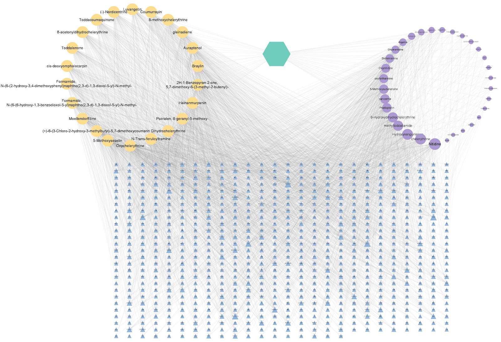
BACKGROUND: The therapeutic potential of Toddalia asiatica in rheumatoid arthritis has garnered increasing attention, yet its mechanisms remain incompletely elucidated. OBJECTIVE: To investigate the underlying mechanisms of Toddalia asiatica in treating rheumatoid arthritis using bioinformatics combined with molecular dynamics simulation. METHODS: Active ingredients of Toddalia asiatica and their targets were retrieved. Drug targets were intersected with rheumatoid arthritis-related targets, followed by enrichment analysis of the overlapping genes. Molecular docking and molecular dynamics simulation were performed to validate the binding mechanisms of core active ingredients with key targets. RESULTS AND CONCLUSION: Through literature retrieval, 22 core active ingredients of Toddalia asiatica and their key targets against rheumatoid arthritis were identified. Enrichment analysis indicated that Toddalia asiatica may exert therapeutic effects by modulating disease-related signaling pathways (including cancer, infectious diseases, metabolic diseases, and cardiovascular diseases) as well as biological pathways related to metabolism, immunity, and inflammation. Meanwhile, the main components Dihydrochelerythrine and 8-Methoxychelerythrine specifically target phospholipase C gamma 2 (PLCG2) and mitogen-activated protein kinase 8 (MAPK8), respectively, suggesting that Toddalia asiatica may exert anti-rheumatoid arthritis effects through synergistic multi-pathway regulation.

Read Full Abstract10.12307/2026.21316
Exosomes and neuropathic pain: visualization analysis on literatureGraphical AbstractVerified
Chinese Journal of Tissue Engineering Research2026

Exosomes and neuropathic pain: visualization analysis on literature

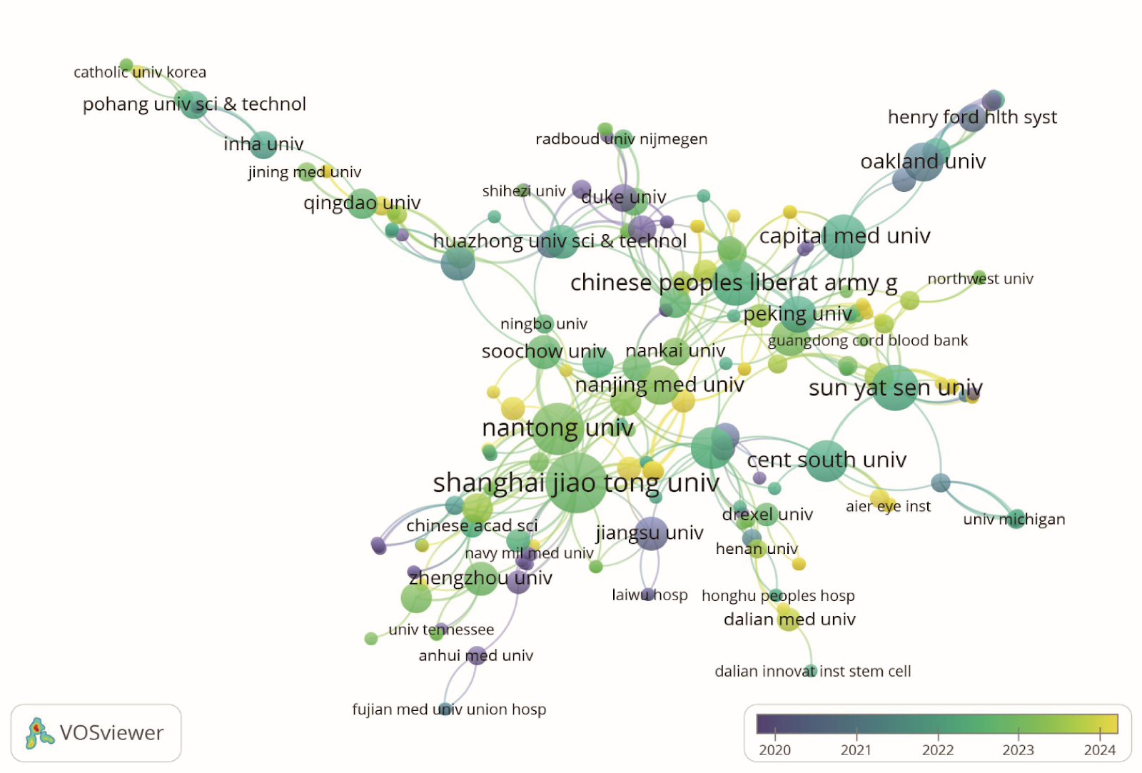
BACKGROUND: Neuropathic pain has a complex pathogenesis and limited clinical intervention outcomes. In recent years, exosomes have gradually emerged as a focal point in the study of neuropathic pain due to their unique intercellular communication functions and molecular delivery capabilities. OBJECTIVE: To systematically review research progress on exosomes in the field of neuropathic pain using bibliometric methods, summarize the knowledge framework and research hotspots, and provide theoretical foundations and translational strategies for advancing this field. METHODS: Based on the Web of Science Core Collection (WOSCC) database, literature on exosomes and neuropathic pain was retrieved from 2012-01-01 to 2025-03-31. VOSviewer and CiteSpace software were used for keyword co-occurrence, cluster analysis, burst detection, and collaboration network visualization. RESULTS AND CONCLUSION: A total of 313 articles were included. Among 42 countries or regions, the United States and China made significant contributions. Shanghai Jiao Tong University and Nantong University were the most prolific institutions. The most productive and co-cited journals were Neural Regeneration Research and International Journal of Molecular Sciences, respectively. A total of 382 authors were identified; Zhang Zhenggang had the most articles, and Zhang Yi had the most citations. Key high-frequency keywords included 'nerve regeneration', 'neuroinflammation', 'schwann cell', 'regenerative medicine', and 'spinal cord injury', which are key research areas for future development. Through bibliometric analysis, this study mapped the research trends of exosomes in neuropathic pain. Exosomes hold broad prospects in elucidating basic mechanisms and developing novel precision therapies for neuropathic pain. Future efforts should integrate multi-omics and engineered exosome platforms to advance this field.

Read Full Abstract10.12307/2026.21365
Differential proteomic analysis of exercise-induced and pathological cardiac hypertrophy models in miceGraphical AbstractVerified
Chinese Journal of Tissue Engineering Research2026

Differential proteomic analysis of exercise-induced and pathological cardiac hypertrophy models in mice

BACKGROUND: Improving outcomes for patients with pathological cardiac hypertrophy by leveraging the mechanisms of exercise-induced cardiac hypertrophy is currently a significant focus in cardiovascular research. However, the molecular mechanisms underlying the differences between exercise-induced and pathological cardiac hypertrophy remain incompletely understood. OBJECTIVE: To identify potential therapeutic targets for pathological cardiac hypertrophy based on cardiac proteomics using mouse models of exercise-induced and pathological cardiac hypertrophy. METHODS: Twenty-one 6-8-week-old male C57BL/6J mice were randomly divided into control, exercise, and isoproterenol groups (n=7 per group). Exercise-induced cardiac hypertrophy was established by 8 weeks of continuous training, while pathological cardiac hypertrophy was induced by subcutaneous injection of isoproterenol for 7 days. After confirming successful modeling via heart mass index, heart-to-tibia ratio, hematoxylin-eosin staining, wheat germ agglutinin staining, and Sirius red staining, tandem mass tag technology was used to reveal differential protein expression and functional characteristics between the two hypertrophy models. RESULTS AND CONCLUSION: Compared with the control group, heart mass index and heart-to-tibia ratio were significantly increased in both exercise and isoproterenol groups (P < 0.001, P < 0.05). Isoproterenol group showed disordered cardiomyocyte arrangement, extensive inflammatory cell infiltration, and obvious cardiomyocyte damage. Cardiomyocyte cross-sectional area was significantly increased in both exercise and isoproterenol groups (P < 0.05, P < 0.01), and myocardial fibrosis area was significantly higher in the isoproterenol group than in the control group (P < 0.01). Compared with the control group, 46 differentially expressed proteins were identified in the exercise group, 302 in the isoproterenol group, and 340 between exercise and isoproterenol groups. Among these, two overlapping proteins were peroxisomal acyl-coenzyme A oxidase 1 (Acox1) and galectin-3 (Gal-3). Gene Ontology and Kyoto Encyclopedia of Genes and Genomes enrichment analyses suggested that exercise may induce physiological cardiac hypertrophy by upregulating Acox1 to promote fatty acid metabolism, while isoproterenol may induce pathological cardiac hypertrophy by downregulating Acox1 leading to peroxisome dysfunction and lipotoxicity. These findings suggest that Acox1 and Gal-3 may serve as potential intervention targets for pathological cardiac hypertrophy.

Read Full Abstract10.12307/2026.21318
Transcriptomic analysis of potential targets of protocatechualdehyde in treatment of atherosclerosisGraphical AbstractVerified
Chinese Journal of Tissue Engineering Research2026

Transcriptomic analysis of potential targets of protocatechualdehyde in treatment of atherosclerosis

BACKGROUND: Protocatechualdehyde has the potential to delay the progression of atherosclerosis. Nevertheless, its specific mechanisms of action within multi-target regulatory networks remain unclear and require further investigation. OBJECTIVE: To investigate the potential targets of protocatechualdehyde in intervening atherosclerosis based on transcriptomics. METHODS: (1) Thirty ApoE-/- mice were randomly divided into a model group (n=10), a rosuvastatin group (n=10), and a protocatechualdehyde group (n=10). An atherosclerosis model was induced by feeding the mice with a high-fat diet for 12 weeks. Seven C57BL/6J mice were selected as a control group (without modeling). After successful modeling, the control group and model group were given physiological saline by gavage; the rosuvastatin group was given rosuvastatin by gavage, and the protocatechualdehyde group was given protocatechualdehyde by gavage, once a day for 12 consecutive weeks. After the last administration, samples were collected. Serum lipid levels were measured using an automatic biochemical analyzer. Aortic plaque pathology was assessed by gross oil red O staining, hematoxylin-eosin staining, and Masson staining of aortic root paraffin sections. (2) High-throughput sequencing was used to analyze the transcriptome expression profiles of aortic samples from the control, model, and protocatechualdehyde groups. Differential gene screening (FC > 2, q < 0.05), GO and KEGG enrichment analyses, weighted gene co-expression network analysis, and short time-series expression miner analysis were performed based on the Ouyi Cloud platform. A protein-protein interaction network was constructed using the STRING database, and core genes were screened using Cytoscape. (3) RT-PCR was used to detect the mRNA expression of Calm4 (calmodulin pseudogene 4), Kprp (keratinocyte proline-rich protein), Hrnr (filaggrin 2), and Lor (loricrin) in aortic samples from the control, model, and protocatechualdehyde groups to validate candidate targets. RESULTS AND CONCLUSION: (1) Protocatechualdehyde significantly reduced serum total cholesterol, triglycerides, high-density lipoprotein cholesterol, and low-density lipoprotein cholesterol levels in atherosclerotic mice. Gross oil red O staining and hematoxylin-eosin and Masson staining of aortic root paraffin sections showed that protocatechualdehyde reduced plaque formation, inhibited intimal thickening, increased collagen fiber content in plaques, and stabilized plaques. (2) Transcriptome analysis identified 191 differentially expressed genes, and Cytoscape analysis preliminarily identified Kprp, Calm4, Hrnr, and Lor as key candidate targets. (3) RT-PCR showed that the mRNA expression of Kprp, Calm4, Hrnr, and Lor in the model group was higher than that in the control group (P < 0.05), while the mRNA expression of Kprp, Calm4, and Lor in the protocatechualdehyde group was lower than that in the model group (P < 0.05). These results indicate that protocatechualdehyde intervention can significantly improve atherosclerotic plaques, and Kprp, Calm4, and Lor may be potential targets for protocatechualdehyde in the treatment of atherosclerosis.

Read Full Abstract10.12307/2026.21319
Exosomes promote diabetic wound healing: a visual analysis of research hotspots and evolutionary trendsGraphical AbstractVerified
Chinese Journal of Tissue Engineering Research2026

Exosomes promote diabetic wound healing: a visual analysis of research hotspots and evolutionary trends

BACKGROUND: Diabetes wound is one of the serious complications of diabetes patients, and its complex pathological mechanism and clinical treatment dilemma is still a major challenge. In recent years, exosomes have become a new focus in the field of diabetes wound research because they play a key role in intercellular communication, immune regulation, and tissue repair. OBJECTIVE: To investigate the research hotspots and evolutionary trends of exosomes in diabetic wound healing. METHODS: A systematic search was conducted in the Web of Science core collection to identify English literature focusing on exosomes in diabetic wound healing and published between the inception of the database and December 31, 2024. The annual publication volume was analyzed to track changes over time. Visual analyses using VOSviewer and CiteSpace software were performed on the retrieved literature to examine key aspects such as authors, countries, institutions, journals, and keywords, providing insights into the current research landscape and evolving hot topics in exosomes for diabetic wound healing. RESULTS AND CONCLUSION: From 2014 to 2024, a total of 424 publications on exosome-promoted diabetic wound healing were produced, contributed by 2,883 authors from 46 countries and featured in 199 journals. In the realm of exosome-promoted diabetic wound healing, China had the highest number of publications, followed by the United States. The journals 'Journal of Nanobiotechnology' and 'Advanced Healthcare Materials' published the most papers and had high influence. Author Chen Zhenbing and Huazhong University of Science and Technology were the most productive author and institution, respectively, but the researcher clusters have not yet reached a certain scale, and future collaboration needs to be strengthened. Global research focus mainly concentrated on 10 thematic clusters including adipose stem cells, diabetic wounds, diabetic wound healing, wound healing, endoplasmic reticulum stress, microvesicles, collagen, proteomics, and diabetic foot infection. The research hotspots in this field are undergoing a transition from molecular mechanisms to systematic interventions. Future research hotspots will focus on angiogenesis, macrophages, antibacterial, and hydrogels. On this basis, integrating multidisciplinary technologies to achieve more effective precision treatment and optimize management strategies for diabetic wounds.

Read Full Abstract10.12307/2026.21364
Function and molecular mechanism of physcion in regulating bone homeostasisGraphical AbstractVerified
Chinese Journal of Tissue Engineering Research2026

Function and molecular mechanism of physcion in regulating bone homeostasis

BACKGROUND: Although physcion has been shown to have protective effects against osteoporosis, the exact mechanism is not fully understood. OBJECTIVE: Through multidimensional analysis of the regulatory effect of physcion on the AKT signaling pathway, the molecular mechanism of its regulation on osteoclast induced differentiation and osteogenic function induced differentiation is revealed. METHODS: (1) RAW264.7 cells and C3H10T1/2 cells were cultured in vitro and subsequently exposed to 0, 10, 20, 30, 40, 50, and 60 µmol/L physcion, respectively. The cytotoxicity of physcion was detected by cell counting kit-8 assay. (2) RAW264.7 cells and C3H10T1/2 cells were treated with different concentrations (0, 20, 40 µmol/L) of physcion during osteoclast and osteoblast differentiation, respectively. Differentiation ability was assessed by qPCR, Western Blot, and alkaline phosphatase staining. (3) Network pharmacology was used to analyze the regulation of physcion on osteoclast differentiation and related signaling pathways, and molecular docking was performed for target proteins. (4) Western Blot was used to verify the phosphorylation level of AKT in the downstream target signaling pathway AKT axis regulated by physcion. RESULTS AND CONCLUSION: (1) At concentrations of 0-60 µmol/L, cell viability in all groups was greater than 90%, indicating no significant cytotoxicity. (2) Physcion significantly inhibited the expression of osteoclast differentiation-related genes, with Acp5, CTSK, DC-STAMP, and Nfatc1 showing downregulation, but had no significant effect on osteoblast differentiation-related genes COL1A1, Runx2, OSX expression or alkaline phosphatase staining intensity. (3) Network pharmacology and molecular docking suggested that physcion affects osteoclast differentiation and regulates the PI3K-AKT pathway, with a binding energy of -10.72 kJ/mol to AKT1, indicating strong binding activity. (4) During osteoclast differentiation, the p-AKT/AKT ratio in RAW264.7 cells increased (n=3, P=0.0063), while physcion decreased this ratio. These findings indicate that physcion inhibits osteoclast differentiation by regulating the AKT signaling pathway, thereby modulating bone homeostasis.

Read Full Abstract10.12307/2026.21312
Construction of an early knee osteoarthritis rat model: CatWalk-based gait analysis and evaluationGraphical AbstractVerified
Chinese Journal of Tissue Engineering Research2026

Construction of an early knee osteoarthritis rat model: CatWalk-based gait analysis and evaluation

BACKGROUND: Existing animal models of knee osteoarthritis predominantly focus on mechanical injury factors but fail to simulate and observe the "cold-dampness obstruction" syndrome characteristics in traditional Chinese medicine. OBJECTIVE: To construct a traditional Chinese medicine-Western medicine integrated knee osteoarthritis model for cold-dampness obstruction syndrome and validate its efficacy via a multidimensional assessment. METHODS: Twenty-four male Sprague-Dawley rats (SPF-grade) were randomly divided into sham-operated, model, and cold-dampness obstruction groups. The latter two groups underwent anterior cruciate ligament transection of the right hind knee. The cold-dampness obstruction group received artificial cold-damp environment intervention (temperature 10.5 °C, humidity 90%, 4 h/day, for 4 weeks) starting 14 days post-surgery. The sham-operated group had skin incision and immediate closure. Before modeling and at 1, 2 weeks post-modeling, and 4 weeks after cold-damp intervention, traditional Chinese medicine syndrome scores and CatWalk gait analysis were performed. Right hind knee joint tissues were harvested for histopathological observation and Mankin scoring. RESULTS AND CONCLUSION: (1) Traditional Chinese medicine syndrome scores: The cold-dampness obstruction group showed significant mental fatigue, reduced activity, loose stools, dark purple tongue, dull fur, decreased food intake, and slower weight gain (P < 0.01). (2) CatWalk gait parameters: At 1 week post-modeling, compared with the sham-operated group, the model and cold-dampness obstruction groups showed decreased maximum contact intensity, print length, maximum intensity, average intensity of 15 maximum pixels, and increased swing phase of the right hind paw (all P < 0.01). The cold-dampness obstruction group also showed significantly decreased swing speed (P < 0.05). After 4 weeks in the artificial climate chamber, compared with the sham-operated group, the cold-dampness obstruction group showed significantly decreased maximum contact intensity, maximum intensity, average intensity of 15 maximum pixels (P < 0.01), increased swing phase (P < 0.01), and decreased swing speed (P < 0.05). (3) Histopathology: Mankin scores in the model and cold-dampness obstruction groups were significantly higher than in the sham-operated group (P < 0.01), and the cold-dampness obstruction group had significantly higher scores than the model group (P < 0.01). These results indicate that anterior cruciate ligament transection combined with cold-damp environment can successfully construct a cold-dampness obstruction type early knee osteoarthritis rat model. CatWalk gait parameters and traditional Chinese medicine syndrome scores provide an objective evaluation system for studying the mechanisms of traditional Chinese medicine in knee osteoarthritis.

Read Full Abstract10.12307/2026.21317
Shaoyang Shenggu Fang inhibits oxidative stress and delays cartilage aging in rats with knee osteoarthritisGraphical AbstractVerified
Chinese Journal of Tissue Engineering Research2026

Shaoyang Shenggu Fang inhibits oxidative stress and delays cartilage aging in rats with knee osteoarthritis

BACKGROUND: Preliminary studies have demonstrated that Shaoyang Shenggu Fang can alleviate joint cartilage degeneration and promote cartilage repair, but its specific mechanism for alleviating knee osteoarthritis symptoms remains unclear. The Wnt/β-catenin pathway and oxidative stress play crucial roles in maintaining articular cartilage homeostasis. OBJECTIVE: To investigate the molecular mechanisms by which Shaoyang Shenggu Fang regulates the Wnt/β-catenin pathway to inhibit oxidative stress in cartilage and thereby delay cartilage aging in a rat model of knee osteoarthritis. METHODS: Thirty-two Sprague-Dawley rats were randomly divided into four groups: a blank control group, a model group, a Western medicine group, and a Chinese medicine group. Animal models of knee osteoarthritis were established in all groups except for the blank control group by transecting the anterior cruciate ligament and resecting the anterior horn of the medial meniscus. After 28 days of modeling, the Chinese medicine group was administered concentrated Shaoyang Shenggu Fang at a dose of 16 g/(kg·d) by gavage, the Western medicine group received glucosamine hydrochloride solution at 4 mL/d, and the blank and model groups received the same volume of normal saline. After 4 weeks, hematoxylin-eosin staining and Safranin O-fast green staining were used to observe the degree of cartilage damage and degeneration. ELISA was used to detect serum levels of inflammatory factors and oxidative stress indicators. Western blot was used to detect the expression of p21Cip1, p16INK4a, and Wnt signaling pathway-related proteins in knee cartilage. RESULTS AND CONCLUSION: Compared with the model group, the Western medicine and Chinese medicine groups showed significant improvement in cartilage defects, thinning of the cartilage layer, and decreased density, with significantly lower Mankin scores (P < 0.05). Compared with the model group, serum levels of interleukin-1β, tumor necrosis factor-α, and interleukin-6 were significantly decreased in the Western medicine and Chinese medicine groups (P < 0.05), while superoxide dismutase and glutathione peroxidase levels were increased and malondialdehyde concentration was decreased (all P < 0.05). In the Chinese medicine group, the expression levels of p21Cip1, p16INK4a, and Wnt5a proteins were significantly decreased (P < 0.05 and P < 0.01), β-catenin and C-Myc protein expression levels were decreased (P < 0.05), and glycogen synthase kinase-3β protein expression was significantly increased (P < 0.05). These results suggest that Shaoyang Shenggu Fang can significantly reduce inflammation and alleviate cartilage aging in rats with knee osteoarthritis, and the potential mechanism may be through regulation of the Wnt/β-catenin pathway to inhibit cartilage oxidative stress.

Read Full Abstract10.12307/2026.21314
Effects of platelet-rich fibrin on osteogenic genes and bone microstructure in rats with peri-implant bone defectGraphical AbstractVerified
Chinese Journal of Tissue Engineering Research2026

Effects of platelet-rich fibrin on osteogenic genes and bone microstructure in rats with peri-implant bone defect

BACKGROUND: Peri-implant bone defects may affect implant stability. Platelet-rich fibrin, a second-generation autologous platelet concentrate, contains abundant growth factors and fibrin scaffolds and can facilitate bone regeneration. Nevertheless, its mechanism of action in the context of peri-implant bone defects remains to be fully investigated. OBJECTIVE: To investigate the effects of platelet-rich fibrin on osteogenic genes, bone microstructure, and IκB kinase/inhibitor of nuclear factor-κB/nuclear factor-κB signaling pathway in rats with peri-implant bone defect using a rat tibia model to simulate peri-implant bone defects, combined with ligature-induced inflammation. METHODS: Thirty male Sprague-Dawley rats were selected, and 20 of them were selected to establish peri-implant bone defect model. After modeling, they were randomly divided into model group and platelet-rich fibrin group, with an average of 10 rats per group, and the remaining 10 rats were assigned to the control group. The control group and the model group were not treated with any intervention, and the platelet-rich fibrin group was treated with platelet-rich fibrin implantation at the bone defect site. After 8 weeks, Image-Pro-Plus software was used to detect implant-bone contact rate and new bone formation rate; Micro-CT was used to detect bone microstructure changes; hematoxylin-eosin staining was used to observe histopathological changes; western blot was used to detect the protein expression of nuclear factor-κB, inhibitor of nuclear factor-κB, and IκB kinase in tibial tissue; RT-PCR was used to detect the expression of osteogenic-related genes osteopontin, osteocalcin, and Runt-related transcription factor 2. RESULTS AND CONCLUSION: (1) At 4 and 8 weeks after surgery, the new bone formation rate and implant-bone contact rate in the model group and platelet-rich fibrin group were increased (P < 0.05); the new bone formation rate and implant-bone contact rate in the platelet-rich fibrin group were significantly higher than those in the model group (P < 0.05). (2) Compared with the control group, the model group showed decreased trabecular bone number, bone volume fraction, Lane-Sandhu histological score, and mRNA expression of Runt-related transcription factor 2, osteocalcin, and osteopontin (P < 0.05), while trabecular separation, and protein expression of IκB kinase, inhibitor of nuclear factor-κB, and nuclear factor-κB were increased (P < 0.05). Compared with the model group, the platelet-rich fibrin group showed increased trabecular bone number, bone volume fraction, Lane-Sandhu histological score, and mRNA expression of Runt-related transcription factor 2, osteocalcin, and osteopontin (P < 0.05), while trabecular separation, and protein expression of IκB kinase, inhibitor of nuclear factor-κB, and nuclear factor-κB were decreased (P < 0.05). (3) Micro-CT showed no new bone tissue formation in the model group, while a large amount of new bone formation and connection with bone ends were observed in the platelet-rich fibrin group. (4) Hematoxylin-eosin staining showed that the platelet-rich fibrin group had good bone repair status and a large number of new bone cells around the defect. These results suggest that platelet-rich fibrin can accelerate the process of bone cell repair, has a significant promoting effect on bone healing in rats with peri-implant bone defects, can increase the expression level of osteogenic-related genes, improve bone microstructure, and enhance the activity of the IκB kinase/inhibitor of nuclear factor-κB/nuclear factor-κB signaling pathway.

Read Full Abstract10.12307/2026.21313
Effects of human umbilical cord blood mesenchymal stem cells on pain and function in patients with knee osteoarthritis: a meta-analysisGraphical AbstractVerified
Chinese Journal of Tissue Engineering Research2026

Effects of human umbilical cord blood mesenchymal stem cells on pain and function in patients with knee osteoarthritis: a meta-analysis

OBJECTIVE: To conduct a meta-analysis concerning the effects of human umbilical cord blood mesenchymal stem cells on pain and function in patients with knee osteoarthritis. METHODS: Using the Chinese search terms “human umbilical cord blood, mesenchymal stem cells, knee joint-related diseases” and the English search terms “human cord blood, mesenchymal stem cell, MSC, knee osteoarthritis, knee joint disease, knee joint disorders, knee OA,” we conducted searches in the CNKI, WanFang, VIP, PubMed, Elsevier, and Web of Science databases. The search timeframe spanned from the establishment of each database until June 13, 2024. The quality of the included literature was assessed using the Cochrane Risk of Bias tool and the ROBINS-I tool. For meta-analysis, the Revman software was utilized, calculating mean differences for continuous variables and relative risks for dichotomous variables, along with 95% confidence intervals. RESULTS: Three randomized controlled trials and three case-control studies were included, totaling 248 subjects, with moderate quality. Meta-analysis showed: (1) The visual analog scale score in the experimental group was lower than that in the control group, with a significant difference (χ²=44.98, P < 0.001, I²=91%); (2) The Western Ontario and McMaster Universities Osteoarthritis Index in the experimental group was lower than that in the control group, with a significant difference (χ²=16.84, P < 0.001, I²=88%); (3) The Lysholm knee function score in the experimental group was higher than that in the control group, with a significant difference (χ²=0.12, P=0.73, I²=0%); (4) The incidence of adverse reactions in the experimental group was higher than that in the control group, with a significant difference (χ²=4.99, P < 0.001, I²=20%), with a combined risk difference of 0.21, translating to a number needed to treat of 5. CONCLUSION: Human umbilical cord blood mesenchymal stem cells can reduce pain and improve knee function in patients with knee osteoarthritis, achieving a good balance between safety and efficacy.

Read Full Abstract10.12307/2026.21363
In vitro simulation of cellular exercise environments: advancements in methodology and signal simulationGraphical AbstractVerified
Chinese Journal of Tissue Engineering Research2026

In vitro simulation of cellular exercise environments: advancements in methodology and signal simulation

BACKGROUND: With an increasing understanding of the health benefits of exercise, research on the mechanisms of exercise intervention has become a focal point. Traditional studies rely on in vivo animal models or multi-omics techniques to indirectly infer exercise intervention mechanisms, but the research is not in-depth enough, and many disease models cannot achieve the prescribed exercise intensity. Therefore, in vitro cell-based exercise environment simulation techniques are of particular significance. Existing technologies primarily focus on the replication of single signals, failing to comprehensively simulate the interaction of multi-dimensional signals during exercise, which limits the understanding of exercise adaptation mechanisms. OBJECTIVE: To explore the technological advancements in in vitro cell-based exercise environment simulation, analyze the advantages of existing signal simulation techniques, and propose a new framework integrating multi-dimensional signals to promote the precise replication of exercise mechanisms and application research in related fields. METHODS: This study conducted a search in the PubMed and Web of Science databases using keywords such as Exercise, Physiology, Molecular Signals, Myokines, Exerkines, etc. After initial screening and removal of duplicates, 5,046 relevant articles were identified, and 99 were finally included after further screening. RESULTS AND CONCLUSION: Existing in vitro cell exercise simulation techniques have made some progress in simulating specific attributes of exercise (e.g., mechanical stretching, electrical signals), but they still fail to fully replicate the multi-dimensional signal interactions during exercise. By integrating multiple signals such as mechanical forces, electrophysiological stimuli, and biological factors, future simulation technologies are expected to more realistically reproduce the effects of exercise on cellular metabolism, gene expression, and phenotypic remodeling, providing a more precise experimental platform for studying exercise mechanisms. Furthermore, innovations and optimizations in in vitro exercise simulation technologies will provide important support for sports medicine, drug development, and regenerative medicine.

Read Full Abstract10.12307/2026.21362
Human umbilical cord mesenchymal stem cell transplantation protects against reproductive damage induced by high-altitude hypoxia exposure in male miceGraphical AbstractVerified
Chinese Journal of Tissue Engineering Research2026

Human umbilical cord mesenchymal stem cell transplantation protects against reproductive damage induced by high-altitude hypoxia exposure in male mice

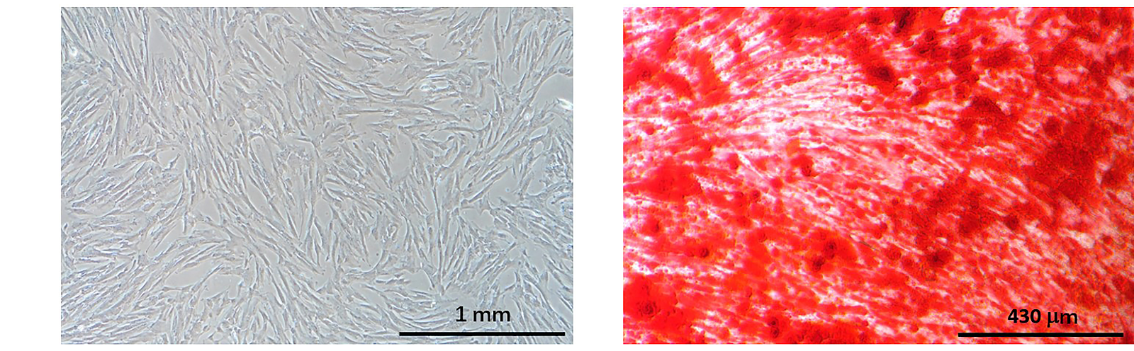
BACKGROUND: High-altitude hypoxia has been reported to damage the male reproductive system, but whether stem cells can protect against male reproductive damage caused by high-altitude hypoxia has not been reported. OBJECTIVE: To investigate the preventive effect of human umbilical cord mesenchymal stem cell transplantation on reproductive damage in hypoxia-exposed male mice. METHODS: Human umbilical cord mesenchymal stem cells were isolated and cultured, and three-lineage differentiation and flow cytometry identification were performed. Twenty-one C57BL/6 male mice were randomly divided into control, hypoxia, and stem cell groups (n=7). The hypoxia and stem cell groups were exposed to a chronic intermittent hypoxia model simulating an altitude of 5,000 m (11.1% oxygen). In the stem cell group, 1×10^6 human umbilical cord mesenchymal stem cells were injected via the tail vein once a week for 6 weeks, while the other groups received PBS. Body mass, food intake, and water intake were monitored. After hypoxia exposure, testicular tissue was analyzed for morphology, ultrastructure, reactive oxygen species levels, and mitochondrial membrane potential; epididymal tissue was analyzed by hematoxylin-eosin staining and sperm motility; and the homing ability of stem cells was observed by DiL fluorescence tracing. RESULTS AND CONCLUSION: Human umbilical cord mesenchymal stem cell transplantation significantly improved water and food intake in hypoxic mice but had no significant effect on body mass. Morphological analysis showed that hypoxia caused edema of the testis and epididymis and shedding of spermatogenic cells, while stem cell transplantation alleviated these structural damages and reversed mitochondrial swelling and atrophy in germ cells. Additionally, stem cell transplantation significantly inhibited hypoxia-induced increase in reactive oxygen species, restored mitochondrial membrane potential, and improved sperm motility. Tracing experiments showed that after entering the mice, stem cells mainly accumulated in lung tissue, with low homing to the testis. In conclusion, human umbilical cord mesenchymal stem cell transplantation can protect the structure and function of germ cell mitochondria, reduce hypoxia-induced testicular and epididymal edema, and thereby restore spermatogenesis and sperm motility.

Read Full Abstract10.12307/2026.21336
Mechanisms and clinical strategies of mesenchymal stem cell-derived extracellular vesicles intervening in cell regulatory networks to treat pulmonary fibrosisGraphical AbstractVerified
Chinese Journal of Tissue Engineering Research2026

Mechanisms and clinical strategies of mesenchymal stem cell-derived extracellular vesicles intervening in cell regulatory networks to treat pulmonary fibrosis

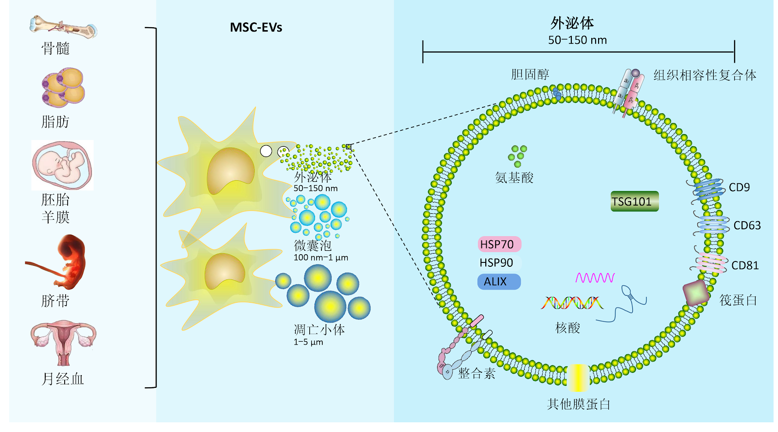
BACKGROUND: Pulmonary fibrosis is a chronic progressive lung disease characterized by abnormal deposition of extracellular matrix, with current therapeutic options remaining limited. Extracellular vesicles derived from mesenchymal stem cells, with their lipid membrane structure, can cross the internal barriers in the body and directly deliver various anti-fibrotic, immunomodulatory factors (such as growth factors, immunomodulatory cytokines, and chemokines), lipids and nucleic acids (mRNAs and miRNAs) and other bioactive substances to target cells in the lungs. OBJECTIVE: To systematically review the core mechanisms of mesenchymal stem cell extracellular vesicles in the treatment of pulmonary fibrosis, summarize and elaborate on how extracellular vesicles directly deliver the bioactive substances they carry to different target cells in the lungs, demonstrating their unique advantages in regulating the pulmonary fibrosis microenvironment, and provide a theoretical basis for future use of mesenchymal stem cell-derived extracellular vesicles in the treatment of pulmonary fibrosis. METHODS: A computer-based search was conducted in CNKI, PubMed, clinicaltrials.gov, and the Chinese Clinical Trial Registry. English search terms included "Mesenchymal stem cells, Extracellular vesicles, Pulmonary fibrosis, Alveolar epithelium, Microvascular endothelium, Macrophages, Neutrophils"; Chinese search terms included "间充质干细胞,细胞外囊泡,肺纤维化,上皮细胞,血管内皮细胞,巨噬细胞,中性粒细胞". A total of 56 articles were included for summary. RESULTS AND CONCLUSION: In alveolar epithelial cells, epithelial-mesenchymal transition is inhibited by regulating signaling pathways such as protein kinase B/glycogen synthase kinase 3β and transforming growth factor β/Smad, and specific miRNAs (e.g., miR-466f-3p, let-7) block pro-fibrotic pathway networks. In fibroblasts and endothelial cells, miR-21-5p and miR-218/miR-214-3p respectively interfere with fibroblast activation and endothelial-mesenchymal transition. Meanwhile, they reprogram monocytes, regulate macrophage polarization, inhibit dendritic cell maturation, and balance Th17/Treg responses, reshaping the immune microenvironment. Furthermore, engineered modifications (targeting peptide modification, drug co-loading) of mesenchymal stem cell-derived extracellular vesicles can enhance precise targeting of multiple cell subtypes in diseased areas, while vesicle heterogeneity, standardized production, and in vivo dynamic tracing remain key bottlenecks for clinical translation. Future research should combine single-cell sequencing and spatial multi-omics to deeply analyze the mechanisms of mesenchymal stem cell-derived extracellular vesicles in intervening in cell regulatory networks and develop novel cell-targeted clinical strategies for pulmonary fibrosis.

Read Full Abstract10.12307/2026.21358
Application and prospects of precision-medicine-driven breast cancer organoids in therapeutic drug discoveryGraphical AbstractVerified
Chinese Journal of Tissue Engineering Research2026

Application and prospects of precision-medicine-driven breast cancer organoids in therapeutic drug discovery

BACKGROUND: Breast cancer organoids, as a novel in vitro model, can not only simulate the biological characteristics of breast cancer but also to some extent reproduce the impact of the tumor microenvironment on the tumor, facilitating research on breast cancer and further promoting precision medicine. OBJECTIVE: To review the application status of breast cancer organoids in the field of therapeutic drugs, including chemotherapy, targeted therapy, and immunotherapy, over the past few years, and to discuss the existing limitations in order to further promote their application in breast cancer treatment. METHODS: The first author conducted a search in the China National Knowledge Infrastructure (CNKI) and PubMed databases in June 2025 for relevant literature published from January 2010 to June 2025. Chinese search terms included '类器官,乳腺类器官,乳腺癌类器官,乳腺癌模型实验验证,精准治疗,靶向治疗,化疗,免疫治疗,药物敏感性'; English search terms included 'organoid, breast organoid, breast cancer organoid, breast cancer experimental model, precision medicine, targeted therapy, chemotherapy, immunotherapy, drug sensitivity'. A total of 58 articles were included for review. RESULTS AND CONCLUSION: (1) Compared with traditional breast cancer cell experiments, which lack verification of tissue structure and cell-cell interactions as well as in vivo microenvironment, breast cancer organoids have diverse sources of primary tumor cells and continuously innovating culture systems. They can simulate cell-cell interactions and reproduce the biological characteristics of breast cancer and its tumor microenvironment, making breast cancer organoids one of the most promising tools in breast cancer research. This also provides greater potential for improving treatment resistance in clinical breast cancer patients through drug sensitivity screening. (2) The application of drug sensitivity test results from breast cancer organoids in clinical practice has yielded promising outcomes. By testing drug sensitivity in breast cancer organoids to common chemotherapeutic agents, targeted drugs, and immunotherapeutic drugs, the antitumor mechanisms and synergistic effects of multiple drugs can be verified, avoiding the use of drugs with primary resistance and high toxicity in patients, thereby enabling personalized treatment plans and evidence-based precision medicine strategies. (3) Breast cancer organoids still have limitations in practical applications such as drug screening and treatment, including low model construction success rates, difficulty in model growth, and lack of angiogenesis processes. To overcome these limitations, it is necessary to increase the source tissue volume, improve the culture system, and innovate culture techniques, which will facilitate comprehensive therapeutic drug selection through breast cancer organoids and aid in personalized precision medicine.

Read Full Abstract10.12307/2026.21359
Mechanism of acupuncture regulating proliferation and differentiation of stem cellsGraphical AbstractVerified
Chinese Journal of Tissue Engineering Research2026

Mechanism of acupuncture regulating proliferation and differentiation of stem cells

BACKGROUND: Stem cells have the potential for self-renewal and multi-directional differentiation, which can enhance tissue repair through direct differentiation or paracrine and immunomodulatory microenvironments. Acupuncture can promote the proliferation and differentiation of stem cells through multi-pathway synergy, which expands the application range of acupuncture. OBJECTIVE: To review the types of stem cells and their differentiation potential, and to explore the role and mechanism of acupuncture in promoting stem cell proliferation and differentiation. METHODS: Articles published before March 2025 were searched in PubMed and CNKI databases using English search terms 'Electroacupuncture, Acupuncture, Neural stem cells, Bone marrow mesenchymal stem cells, Adipose mesenchymal stem cells' and Chinese search terms '电针, 针刺, 神经干细胞, 骨髓间充质干细胞, 脂肪间充质干细胞'. Literature related to acupuncture promoting stem cell proliferation and differentiation was included, while irrelevant content was excluded. Finally, 76 articles were selected for analysis. RESULTS AND CONCLUSION: (1) Stem cells belong to the category of kidney essence, and stem cells and kidney essence play a synergistic role, which can be used to guide the treatment of diseases with kidney essence deficiency, explaining modern medicine with traditional Chinese medicine theory. (2) Acupuncture can activate multiple signaling pathways and regulate growth factor expression to promote the proliferation and differentiation of endogenous or exogenous stem cells, providing new ideas for clinical treatment. (3) Although acupuncture plays an important role in stem cell proliferation and differentiation, it also faces many challenges in application; the establishment of stem cell engineering standards and the innovative development of acupuncture therapy are the primary prerequisites for in-depth exploration of the combined mechanism of acupuncture and stem cell therapy, ensuring the stability and efficiency of treatment effects.

Read Full Abstract10.12307/2026.21361
Innovative application of kidney organoids in acute kidney injuryGraphical AbstractVerified
Chinese Journal of Tissue Engineering Research2026

Innovative application of kidney organoids in acute kidney injury

BACKGROUND: In recent years, the application of kidney organoid technology in acute kidney injury has gradually become a research hotspot. Traditional animal models have species differences from humans, and physiological and pathological processes of their kidneys cannot fully represent the human situation. Kidney organoid technology forms 3D kidney models through stem cell culture, which can simulate the complex structure and function of human kidneys. It has shown great potential in disease modeling and mechanism exploration of acute kidney injury, prediction of drug nephrotoxicity, and exploration of regeneration and repair mechanisms. OBJECTIVE: To summarize the application progress of kidney organoids in acute kidney injury research, providing new technical means and research strategies for the prevention and treatment of acute kidney injury. METHODS: Literature related to organoids and acute kidney injury was searched in CNKI and PubMed databases. Chinese search terms included "acute kidney injury, organoid, pluripotent stem cells, 3D bioprinting, kidney-on-a-chip, regenerative medicine, kidney transplantation"; English search terms included "acute kidney injury, organoid, pluripotent stem cells, 3D bioprinting, kidney-on-a-chip, regenerative medicine, kidney transplantation". All retrieved literature were original research articles and relevant reviews, with the search time limit from database inception to April 2025. Finally, 99 articles were screened for analysis and summary. RESULTS AND CONCLUSION: (1) The cell sources for inducing kidney organoid formation reported in the literature mainly include pluripotent stem cells, embryonic stem cells, and urine-derived stem cells. These induced kidney organoids play important roles in in vitro drug screening, kidney development, and disease modeling. (2) 3D bioprinting and kidney-on-a-chip technology are emerging techniques for constructing kidney organoids. 3D bioprinting can precisely and specifically construct complex multicellular structures, while kidney-on-a-chip technology has characteristics such as high gas permeability, sensitivity, and low cost, which can extend organoid lifespan, increase biocompatibility, and are suitable for preclinical drug development and toxicity screening. (3) The combination of gene editing technology with kidney organoid models brings new perspectives and tools for kidney disease research, drug development, and regenerative medicine. It can construct kidney organoids with specific reporter genes or sensitive indicators, and amplify and classify specific kidney cell types in kidney organoids. (4) Kidney organoids show unique advantages in disease simulation, drug evaluation, and exploration of regenerative therapeutic strategies for acute kidney injury. They can serve as in vitro models to study the toxicity mechanisms of drugs such as cisplatin, doxorubicin, and red yeast rice supplements that cause acute kidney injury, screen high-throughput drugs and therapeutic targets, and also play an important role in the field of renal transplantation regenerative medicine.

Read Full Abstract10.12307/2026.21360
Stem cell-derived exosomes modulate the inflammatory microenvironment and enhance regenerative capacity of oligodendrocytesGraphical AbstractVerified
Chinese Journal of Tissue Engineering Research2026

Stem cell-derived exosomes modulate the inflammatory microenvironment and enhance regenerative capacity of oligodendrocytes

BACKGROUND: The dynamic interplay between the inflammatory microenvironment and oligodendrocytes following neural injury constitutes a central pathological feature in neurodegenerative and demyelinating diseases. Stem cell-derived exosomes, leveraging their inherent low immunogenicity, efficient barrier-penetrating capacity, and targeted delivery of diverse pro-repair factors, play a pivotal role in modulating oligodendrocyte differentiation and the inflammatory microenvironment, thereby facilitating neural repair and regeneration. OBJECTIVE: To investigate the mechanisms by which stem cell-derived exosomes regulate the inflammatory microenvironment to enhance oligodendrocyte survival, differentiation, and myelin repair. It seeks to establish a novel "cell-free therapy" paradigm, utilizing exosome-mediated multi-component synergy (miRNAs, proteins, and metabolites) and microenvironmental adaptation for treating neurological disorders. METHODS: Literature searches were conducted in the China National Knowledge Infrastructure, PubMed, and WanFang databases, covering publications from 2010 to 2025. Chinese search terms included "exosomes, stem cells, engineered, diagnosis, inflammatory microenvironment, oligodendrocytes, signaling pathways," while English terms comprised "stem cell-derived exosomes, oligodendrocytes, inflammatory microenvironment, signaling pathway, regulatory mechanisms." Irrelevant studies were excluded, and 65 articles meeting inclusion criteria were systematically reviewed according to the inclusion and exclusion criteria. RESULTS AND CONCLUSION: (1) The biological characteristics of exosomes and their roles in the central nervous system were summarized, analyzing the impact of the inflammatory microenvironment on oligodendrocytes and the regulatory mechanisms of exosomes, including miRNA-mediated signaling pathway regulation, anti-inflammatory factor secretion, and immune cell function modulation. (2) The regulatory mechanisms of the inflammatory microenvironment on oligodendrocyte biological behavior and their roles in disease pathogenesis were elaborated. (3) An engineered exosome delivery system based on targeting peptide modification and functional molecule loading, combined with traditional Chinese medicine active ingredient regulation strategies, was proposed to construct a novel cell-free therapy paradigm. (4) The deep interaction between the exosome functional network and myelin homeostasis was explained at the molecular level, providing new therapeutic directions for the development of exosome-targeted delivery systems to intervene in central nervous system demyelinating diseases.

Read Full Abstract10.12307/2026.21354
Exosomal miRNA as an early diagnostic biomarker and potential therapeutic target for cerebral small vessel diseaseGraphical AbstractVerified
Chinese Journal of Tissue Engineering Research2026

Exosomal miRNA as an early diagnostic biomarker and potential therapeutic target for cerebral small vessel disease

BACKGROUND: In recent years, microRNA (miRNA) has received extensive attention in the pathogenesis and diagnosis and treatment of cerebral small vessel disease, and is involved in the regulation of various pathological processes of cerebral small vessel disease. OBJECTIVE: To review the role of miRNA in the pathogenesis, diagnosis, and treatment of cerebral small vessel disease, and to provide effective therapeutic targets and new potential biomarkers for the early diagnosis of cerebral small vessel disease. METHODS: “microRNA, cerebral small vessel disease, blood-brain barrier, chronic cerebral hypoperfusion, inflammation, apoptosis, diagnosis, biomarkers” were used as English search terms for PubMed search. “Exosomal miRNA, cerebral small vessel disease” were used as Chinese search terms for CNKI search. The search time limit was from inception to January 2025. Through the preliminary screening of reading titles and abstracts, the literature with poor relevance and duplicate content was excluded, and finally 72 articles were included for inductive discussion. RESULTS AND CONCLUSION: (1) Through the excavation and discussion of the biological functions and characteristics of exosomal miRNAs, it was confirmed that exosomal miRNAs are important related components in the occurrence and progression of cerebral small vessel disease diseases. (2) Exosomal miRNA participates in the regulation of various pathological processes of cerebral small vessel disease, playing an important role in the pathological mechanism of cerebral small vessel disease by protecting the blood-brain barrier, improving chronic cerebral hypoperfusion, reducing inflammatory responses, and inhibiting apoptosis. (3) Exosomal miRNA can intervene at different stages of pathological development by targeting multiple signaling pathways, effectively targeting different pathological links of cerebral small vessel disease. (4) The combined use of multiple exosomal miRNAs can effectively improve the progression of cerebral small vessel disease. Different miRNAs play different roles at various stages of the pathological mechanism, and constructing a miRNA interaction network is of great significance for regulating the development of cerebral small vessel disease. (5) Exosomal miRNAs are widely and stably present in various body fluids, and their specific and significant expression in urine, serum, blood and other body fluids of patients with cerebral small vessel disease can serve as an effective basis for diagnosis. (6) Currently, the main clinical treatment is to inject miRNA mimics or antagonists to regulate downstream target gene expression. How to achieve the most effective therapeutic effect of miRNA still requires further research. (7) miRNA as exosomal content has great application prospects in the treatment of cerebral small vessel disease. Future research should further explore its mechanism of action to provide effective ideas and further optimize clinical treatment plans for cerebral small vessel disease.

Read Full Abstract10.12307/2026.21353
Advantages and potential of cell-derived exosomes in oral tissue regenerationGraphical AbstractVerified
Chinese Journal of Tissue Engineering Research2026

Advantages and potential of cell-derived exosomes in oral tissue regeneration

BACKGROUND: Stem cells show a great potential in oral tissue regeneration but face challenges such as immune rejection and tumor formation. Exosomes are nanoscale extracellular vesicles secreted by cells, reducing immunogenicity and tumor risks while maintaining stem cell functions, such as promoting angiogenesis and tissue repair. OBJECTIVE: To summarize the mechanisms and roles of exosomes in oral tissue regeneration, explore exosome engineering strategies and the challenges and future directions in the application of exosomes in oral regenerative medicine. METHODS: The relevant literature published from the WanFang and PubMed databases from their inception to 2025 was searched using Chinese search terms “stem cells, exosomes, dental pulp regeneration, periodontal regeneration” and English search terms “exosomes, stem cells, dentistry, regenerate.” Finally, 94 articles were included for review and analysis. RESULTS AND CONCLUSION: (1) Exosomes have lower immunogenicity and no tumorigenic risk compared with stem cells. They are more stable and easier to store and transport. Additionally, exosomes can penetrate dense tissues for targeted delivery, avoiding ethical and immune rejection issues associated with stem cell therapy, making them a safer and more effective treatment option. (2) Exosomes have shown significant efficacy in regenerating dental pulp, periodontal tissues, craniofacial bone, salivary glands, nerves, and skin, promoting tissue repair and regeneration through multiple mechanisms, demonstrating broad application prospects. (3) Engineering strategies such as preconditioning, isolation and purification, and targeted modification can enhance exosome function, improving therapeutic potential and clinical feasibility. However, current technologies still have limitations, and further optimization is needed to promote widespread application of exosomes.

Read Full Abstract10.12307/2026.21356
Single-cell sequencing data identifies differentially expressed genes and immune cell subtypes in periodontitis patientsGraphical AbstractVerified
Chinese Journal of Tissue Engineering Research2026

Single-cell sequencing data identifies differentially expressed genes and immune cell subtypes in periodontitis patients

BACKGROUND: Periodontitis is a chronic inflammatory disease. Previous research has predominantly focused on specific immune cells or cytokines. Therefore, systematically elucidating its immune mechanisms and discovering novel therapeutic targets hold significant implications. OBJECTIVE: To analyze the expression profiles of periodontitis-associated immune cell subpopulations and identify key differentially expressed genes with a causal relationship to the disease, thereby exploring potential molecular mechanisms and key genes involved in periodontitis and immune cell dynamics. METHODS: Single-cell RNA sequencing data from the GEO database were used to analyze immune cell subset heterogeneity and identify differentially expressed genes. Mendelian randomization analysis was performed using expression quantitative trait loci data to infer causal relationships between immune cell gene expression and periodontitis risk. Pathway enrichment and immune infiltration analyses were performed on the identified causal genes to reveal the associations between differentially expressed genes and immune cells with the development and progression of periodontitis. CellChat trajectory analysis was used to explore intercellular communication. To validate key findings, gingival tissue samples were collected from 20 patients with periodontitis diagnosed by the Department of Stomatology at The First Affiliated Hospital of Shihezi University (periodontitis group) and 20 healthy gingival tissue samples from patients undergoing orthodontic or impacted tooth extraction (control group). RT-qPCR and immunohistochemistry were used to detect the expression of key genes. RESULTS AND CONCLUSION: Comprehensive analysis identified 23 immune cell clusters in periodontitis and three key genes with significant causal relationships to periodontitis risk: annexin A1 (ANXA1), solute carrier family 11 member 1 (SLC11A1), and vimentin (VIM). Pathway enrichment analysis revealed their involvement in key immune regulatory mechanisms. Further analyses of immune subtype receptor-ligand interactions and key cell subtype trajectories characterized the distinct roles of ANXA1, SLC11A1, and VIM in disease progression. Compared with healthy controls, the mRNA expression levels of ANXA1, SLC11A1, and VIM were upregulated in periodontitis tissues (P < 0.05). This study reveals the key roles of immune cell subpopulations in periodontitis and validates causal genes (ANXA1, SLC11A1, VIM) associated with the disease.

Read Full Abstract10.12307/2026.21345
Action mechanism of mesenchymal stem cells and their derivatives in the treatment of liver fibrosisGraphical AbstractVerified
Chinese Journal of Tissue Engineering Research2026

Action mechanism of mesenchymal stem cells and their derivatives in the treatment of liver fibrosis

BACKGROUND: Multiple chronic liver diseases that fail to heal will progress to the stage of liver fibrosis. If not treated in a timely manner, they will eventually develop into liver cancer, severely threatening the safety of patients' lives. However, there is currently no specific drug for the treatment of liver fibrosis. Recent studies have demonstrated that mesenchymal stem cell therapy has significant advantages over traditional treatment protocols, providing a new direction for the treatment of liver fibrosis. OBJECTIVE: To review the mechanisms of action of mesenchymal stem cells and their derivatives in the treatment of liver fibrosis. METHODS: The Chinese and English keywords "mesenchymal stem cells, mesenchymal stromal cells, MSCs, liver fibrosis, hepatic fibrosis, hepatocyte death, liver cell death, hepatocyte-like cells, immunomodulation, macrophage, hepatic stellate cells, clinical trials, clinical studies" were used and searched in CNKI and PubMed databases, a total of 81 eligible articles were selected for this review. RESULTS AND CONCLUSION: Through summarizing existing studies, the mechanisms by which mesenchymal stem cells and their derivatives exert anti-fibrotic effects and delay disease progression have been identified. These specific mechanisms include reducing hepatocyte death, differentiating into hepatocyte-like cells, regulating immune responses, and inhibiting hepatic stellate cell activation, confirming that mesenchymal stem cells and their derivatives can serve as a new direction for the treatment of liver fibrosis-related diseases.

Read Full Abstract10.12307/2026.21351