🧬 SinoBioData Academic Portal
šŸ“š Peer-Reviewed Translated Literature

All Biomedical & Clinical Articles (Page 14)

Browse complete peer-reviewed translations from top Chinese biomedical, oncology, and genomics journals. Read verified previews and download full authentic clinical reports.

Published Research Papers

Showing 24 of 1542 peer-reviewed translated articles (Page 14 of 65)

Bibliometric and visualization analysis of the mechanism of osteogenic factors and neurotransmitters in the bone-brain axisGraphical AbstractVerified
Chinese Journal of Tissue Engineering Research2026

Bibliometric and visualization analysis of the mechanism of osteogenic factors and neurotransmitters in the bone-brain axis

BACKGROUND: In recent years, numerous studies have confirmed a close relationship between the skeletal system and the central nervous system, making the bone-brain axis a research hotspot in interdisciplinary fields; however, no studies have yet conducted a bibliometric and visualization analysis of this field. OBJECTIVE: To comprehensively analyze the research trends, hotspots, and future development directions in the bone-brain axis field utilizing bibliometric methods, providing data support and reference for subsequent studies. METHODS: A systematic literature search was conducted in the Web of Science Core Collection database to collect studies related to bone-brain axis published between 2015 and 2024. Visualization tools such as VOSviewer and CiteSpace were employed to analyze publication trends, collaboration networks, institutional contributions, and keyword co-occurrence patterns. RESULTS AND CONCLUSION: ā‘ A total of 7,461 publications were included, showing a significant upward trend in publication volume over the past decade (2015-2024), indicating that bone-brain axis research has become an academic hotspot with increasing attention. ā‘”The United States and China dominated the field, with the USA publishing 2,397 papers (32.1%) and China 2,307 papers (30.9%). Harvard Medical School and Zhejiang University were the most productive and central institutions. ā‘¢Professor Wang Wei was the most prolific author, focusing on the interaction between bone marrow and neuroinflammation. ā‘£The journal Bone published the most papers (over 800), while PLOS ONE had the highest average citations per paper (45), indicating its influence. ⑤Core keywords included 'Bone Marrow', 'Stem Cells', 'Osteoporosis', and 'Neuroinflammation', reflecting fundamental research directions. Emerging frontiers included 'Extracellular Vesicles', 'Alzheimer's Disease', 'Inflammation', and 'Oxidative Stress', highlighting the importance of inflammation and neurodegenerative diseases. ā‘„Future research directions include exploring the specific mechanisms of osteogenic factors and neurotransmitters in the bone-brain axis, elucidating the molecular mechanisms of inflammation, oxidative stress, and extracellular vesicles in neurodegenerative diseases and bone metabolic disorders, and promoting the translation of basic research to clinical applications.


Read Full Abstract10.12307/2026.21436
Mendelian randomization analysis identifies potential drug targets for spinal osteoarthritisGraphical AbstractVerified
Chinese Journal of Tissue Engineering Research2026

Mendelian randomization analysis identifies potential drug targets for spinal osteoarthritis

BACKGROUND: Spinal osteoarthritis is a common degenerative spinal disease that severely affects quality of life of patients, but its exact molecular mechanism remains unclear. OBJECTIVE: To identify plasma proteins related to spinal osteoarthritis through Mendelian randomization analysis and provide a reference for finding new potential therapeutic targets in this disease field. METHODS: Protein data were obtained from the deCODE Genetics database (A total of 35 559 Icelandic individuals were included, and genetic association information of 4 907 plasma proteins was detected, https://www.decode.com/summarydata/). Spinal osteoarthritis data were obtained from the Osteoarthritis Genetics Consortium (A total of 826 690 samples are available for free download via https://msk.hugeamp.org/downloads.html). All data are open source and comply with ethical requirements. Wald ratio or inverse variance weighting was used to assess the causal relationship between 4 907 plasma proteins and spinal osteoarthritis, with Bonferroni correction applied to the P-values. In addition, Steiger directional test was performed to exclude reverse causality; colocalization analysis to exclude linkage disequilibrium; phenotype scanning to exclude horizontal pleiotropy; and external validation to exclude chance findings. Finally, the online analysis tool Enrichr was used to screen small-molecule compounds targeting causal proteins, and molecular docking was performed for the top-ranked compounds to predict their binding modes and energies, thereby identifying the most stable and possible binding modes. RESULTS AND CONCLUSION: Among 4 907 proteins, 1 878 significant protein quantitative trait loci for 1 553 proteins were screened. After Mendelian randomization analysis, four proteins were identified to have strong causal relationships with spinal osteoarthritis: monocyte CD14, interleukin-12 subunit beta (IL12B), hepatocyte growth factor-like protein (MST1), and Semaphorin-4A (SEMA4A). IL12B was negatively correlated with spinal osteoarthritis, while the others were positively correlated. Drug prediction results showed that tesmilifene targeting CD14, montelukast targeting IL12B, naphthylamine targeting MST1, and colchicine targeting SEMA4A all exhibited good binding abilities in molecular docking, with the lowest binding energies all below -6.0 kJ/mol. Comprehensive analysis indicates that these four plasma proteins may serve as potential biomarkers or drug targets for clinical screening, prevention, and intervention of spinal osteoarthritis, and also provide theoretical basis and reference value for related research in the Chinese population.

Read Full Abstract10.12307/2026.21440
Signaling pathways related to active ingredients of ginseng in the treatment of musculoskeletal degenerative diseasesGraphical AbstractVerified
Chinese Journal of Tissue Engineering Research2026

Signaling pathways related to active ingredients of ginseng in the treatment of musculoskeletal degenerative diseases

BACKGROUND: Current therapies for musculoskeletal degenerative diseases merely alleviate symptoms with significant adverse effects. As a traditional Chinese medicine, active ingredients of ginseng exhibit protective effects on bone and muscle through multi-target regulation of signaling pathways, and breakthroughs have been made in mechanism research in recent years. OBJECTIVE: To provide a systematical review of the latest molecular mechanisms of active ingredients of ginseng (ginsenosides, polysaccharides, and peptides) in preventing and treating musculoskeletal degenerative diseases via key signaling pathways, providing a basis for targeted drug development. METHODS: A systematic search was performed in multiple databases, including PubMed, Web of Science, Embase, CNKI, Wanfang, and VIP, with the search period from inception to April 2025. Chinese search terms included "musculoskeletal diseases, osteoporosis, osteoarthritis, intervertebral disc herniation, sarcopenia, ginsenosides, ginseng polysaccharides, ginseng peptides, signaling pathways"; English search terms included "musculoskeletal degenerative diseases, osteoporosis, osteoarthritis, intervertebral disc degeneration, sarcopenia, ginsenosides, ginseng polysaccharides, ginseng peptides, signaling pathways". Finally, 75 eligible articles were included. RESULTS AND CONCLUSION: (1) Ginsenosides (e.g., Rg3, Rh4, Rc): target the karyopherin α2-nuclear factor-κB axis to inhibit osteoclast differentiation and reduce bone resorption; activate the sirtuin 1 pathway to enhance mitochondrial biogenesis and delay sarcopenia; regulate Yes-associated protein 1/transcriptional coactivator and p38 mitogen-activated protein kinase to alleviate intervertebral disc degeneration. (2) Polysaccharides: processing techniques affect immunomodulatory activity, inhibiting inflammation via the mitogen-activated protein kinase/nuclear factor-κB pathway. (3) Peptides: activate the NAD+/sirtuin 1/peroxisome proliferator-activated receptor γ coactivator 1α axis to improve mitochondrial function. These findings suggest that ginseng exerts synergistic regulation of bone metabolism balance, inhibition of cartilage degradation, and delay of muscle aging through a "multi-component, multi-target" mechanism, but challenges remain regarding low bioavailability and lack of large-scale clinical trials for translation.

Read Full Abstract10.12307/2026.21423
Metabolic dysregulation in osteoarthritis: mechanisms and targeted therapeutic strategiesGraphical AbstractVerified
Chinese Journal of Tissue Engineering Research2026

Metabolic dysregulation in osteoarthritis: mechanisms and targeted therapeutic strategies

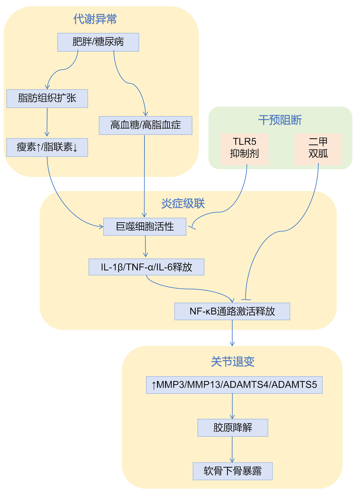
BACKGROUND: Osteoarthritis has traditionally been considered as a degenerative joint disorder, with the central role of metabolic dysregulation in its pathogenesis long overlooked. Recent studies reveal a strong association between metabolic syndromes, such as obesity and diabetes, and osteoarthritis progression, yet the molecular mechanisms by which metabolic dysregulation results in joint degeneration remain poorly elucidated. OBJECTIVE: To systematically review advances in the pathological mechanisms of metabolic dysregulation in osteoarthritis, integrating multi-omics evidence to decode the regulatory network of the "metabolism-joint axis," and to provide novel perspectives for the clinical prevention and treatment of metabolic dysregulation in osteoarthritis. METHODS: An online search of the China National Knowledge Infrastructure and Wanfang databases was conducted using the terms "osteoarthritis, metabolic dysregulation, adipokine, glycolysis, mitochondrial dysfunction, metabolomics, MRI" in Chinese to retrieve relevant literature. Additionally, PubMed and Web of Science were searched using the English terms "osteoarthritis, metabolic dysregulation, adipokine, glycolysis, mitochondrial dysfunction, metabolomics, MRI." After screening titles and abstracts, as well as evaluating full texts, 108 articles were finally included for review. RESULTS AND CONCLUSION: Epidemiological studies confirm that metabolic syndrome (obesity, diabetes, dyslipidemia) is positively correlated with osteoarthritis risk; each 1 kg/m2 increase in body mass index raises the risk of knee osteoarthritis by 15%, and involvement of non-weight-bearing joints supports the independent pathogenic role of metabolic factors. Imbalance of the adipokine network drives osteoarthritis progression: obesity leads to abnormal leptin/adiponectin ratio (leptin increased 3.2-fold, adiponectin decreased 40%), which accelerates cartilage degradation by activating matrix metalloproteinase 13 (increased 2.1-fold) and inhibiting tissue inhibitor of metalloproteinase 2. Intracellular metabolic reprogramming is prominent: osteoarthritic chondrocytes exhibit a triad of enhanced glycolysis, mitochondrial dysfunction, and lipid droplet deposition (free fatty acids increased 1.8-fold). A metabolic-inflammatory positive feedback loop: adipose tissue releases interleukin-1β/tumor necrosis factor-α to activate the nuclear factor-κB pathway in joints, which in turn inhibits insulin receptor signaling and exacerbates metabolic disturbance. Clinical translation: serum adiponectin combined with synovial fluid lactate can predict osteoarthritis progression; early use of AMP-activated protein kinase agonists combined with muscle training and nutritional intervention is recommended for patients with metabolic syndrome.

Read Full Abstract10.12307/2026.21426
Meta-analysis of blood flow restriction training to improve knee function and muscle strength in patients after anterior cruciate ligament reconstructionGraphical AbstractVerified
Chinese Journal of Tissue Engineering Research2026

Meta-analysis of blood flow restriction training to improve knee function and muscle strength in patients after anterior cruciate ligament reconstruction

OBJECTIVE: Blood flow restriction training is a low-load training method designed to enhance muscle strength and promote muscle hypertrophy. Its characteristics align well with the rehabilitation needs following anterior cruciate ligament reconstruction. However, its actual efficacy remains unclear. Therefore, this study systematically evaluated the effectiveness of blood flow restriction training on knee joint rehabilitation in patients after anterior cruciate ligament reconstruction and compared the differences in knee rehabilitation outcomes between blood flow restriction training and traditional resistance training. METHODS: By searching EBSCO, Embase, PubMed, The Cochrane Library, Web of Science English databases, CBM, CNKI, VIP, WanFang Data Chinese databases and clinical trial center database (ClinicalTrials.gov), the randomized controlled trials of knee rehabilitation after anterior cruciate ligament reconstruction were collected. The search time was from the establishment of the database to October 31, 2024. Two reviewers independently screened literature, extracted data, and assessed the risk of bias of included studies. The meta-analysis was performed using RevMan 5.4 software and Stata 14.0 software. RESULTS: A total of 11 studies involving 317 patients were included. Meta-analysis results showed that blood flow restriction training was superior to traditional resistance training in improving knee muscle strength [SMD=0.75, 95%CI (0.46, 1.04), P < 0.000 01], knee muscle mass [SMD=0.48, 95%CI (0.30, 0.66), P < 0.000 01], and knee function [SMD=2.69, 95%CI (1.32, 4.07), P=0.000 1]. Subgroup analysis showed that when training frequency was ≄3 times per week, knee muscle mass [SMD=0.43, 95%CI (0.23, 0.63), P < 0.000 1] was more improved than traditional resistance training. CONCLUSION: Blood flow restriction training can better improve knee muscle strength, knee muscle mass, and knee function in patients after anterior cruciate ligament reconstruction than traditional resistance training. Moreover, during the rehabilitation period, blood flow restriction training at least three times per week can better increase knee muscle mass.

Read Full Abstract10.12307/2026.21431
A new strategy for preventing and treating orthopedic diseases by regulating ferroptosis through signaling pathwaysGraphical AbstractVerified
Chinese Journal of Tissue Engineering Research2026

A new strategy for preventing and treating orthopedic diseases by regulating ferroptosis through signaling pathways

BACKGROUND: Experiments have confirmed that ferroptosis is closely associated with a variety of orthopedic diseases. However, the specific mechanisms by which the regulation of ferroptosis leads to orthopedic diseases remain unclear. Current evidence suggests that signaling pathways may be an important approach for regulating the occurrence of ferroptosis. OBJECTIVE: To summarize the relevant signaling pathways involved in the regulation of ferroptosis in orthopedic diseases (osteoarthritis, spinal cord injury, osteoporosis, intervertebral disc degeneration, rheumatoid arthritis, osteosarcoma, steroid-induced osteonecrosis of the femoral head), to describe the key regulators of the ferroptosis pathway in orthopedic diseases through the modulation of the conduction of signaling pathways, and to conduct an in-depth study on the regulatory mechanisms of ferroptosis in orthopedic diseases and provide a theoretical basis for the prevention and treatment of such diseases. METHODS: Databases including PubMed, Elsevier, Web of Science, and CNKI were searched for relevant literatures on ferroptosis and related orthopedic diseases from the establishment of these databases up to February 2025. The search terms were "ferroptosis, osteoarthritis, osteoporosis, spinal cord injury, intervertebral disc degeneration, osteosarcomas, rheumatoid arthritis, steroid-induced osteonecrosis of the femoral head". A total of 138 articles were included for review. RESULTS AND CONCLUSION: (1) Under the regulation of multiple signaling pathways, the accumulation of intracellular iron ions, reactive oxygen species, and other substances can be induced, causing ferroptosis in osteoblasts, chondrocytes, osteosarcoma cells, etc., leading to changes in the microenvironment, thereby promoting or inhibiting the occurrence of related orthopedic diseases. (2) Studies have confirmed that signaling pathway-regulated ferroptosis is of great significance in the pathogenesis of orthopedic diseases. (3) However, the interaction mechanisms among signaling pathways, ferroptosis, and orthopedic diseases are still in the preliminary stage, and further research is needed to provide more strategies for the treatment of orthopedic diseases.

Read Full Abstract10.12307/2026.21419
A new perspective on exercise for the prevention and treatment of type 2 diabetes mellitus: pyroptosisGraphical AbstractVerified
Chinese Journal of Tissue Engineering Research2026

A new perspective on exercise for the prevention and treatment of type 2 diabetes mellitus: pyroptosis

BACKGROUND: Pyroptosis, a pro-inflammatory form of programmed cell death, plays a key role in innate immunity. However, its excessive activation can induce the body's inflammatory response and the development of disease, e.g., pancreatic β-cell pyroptosis is closely associated with the pathogenesis of type 2 diabetes mellitus. Recent studies suggest that exercise may suppress β-cell pyroptosis, thereby offering potential benefits for the prevention and management of type 2 diabetes mellitus and its complications. Nevertheless, the precise mechanisms underlying the interplay among β-cell pyroptosis, type 2 diabetes mellitus, and exercise remain unclear. OBJECTIVE: To review a series of pyroptosis pathways and the molecular mechanisms of pancreatic β-cell pyroptosis and nucleotide-binding oligomerization domain-like receptor protein 3 (NLRP3) inflammasome in the pathophysiological process of type 2 diabetes mellitus, and to summarize the effects and potential mechanisms of different exercise modalities in regulating the pyroptosis of pancreatic β-cells and preventing and controlling type 2 diabetes mellitus, thereby providing theoretical and practical guidance for the prevention and control of type 2 diabetes mellitus and for the intervention of exercise. METHODS: A systematic literature search was conducted in CNKI, VIP, WanFang, Google Scholar, Web of Science, and PubMed for relevant literature published from January 2000 to April 2025. High-quality studies on exercise regulation of pyroptosis in the prevention and treatment of type 2 diabetes mellitus were screened. Based on explicit inclusion and exclusion criteria, the quality of the literature was assessed and integrated, and finally 57 articles were analyzed and summarized. RESULTS AND CONCLUSION: ā‘  NLRP3 inflammasome-mediated pyroptosis plays an important role in the development and progression of type 2 diabetes mellitus; ā‘” Various exercise interventions, especially aerobic exercise, can effectively improve pancreatic β-cell damage and enhance insulin sensitivity by inhibiting NLRP3-related pathways and reducing inflammatory responses; ā‘¢ Tai Chi, Yijinjing, resistance exercise, and hypoxic exercise also show certain regulatory potential. In summary, exercise intervenes in pyroptosis through multiple pathways, providing theoretical support and practical directions for new targets in the prevention and treatment of type 2 diabetes mellitus.

Read Full Abstract10.12307/2026.21430
Outcome measures in randomized controlled trials of vestibular peripheral vertigo: a systematic evaluation of traditional Chinese medicine treatmentGraphical AbstractVerified
Chinese Journal of Tissue Engineering Research2026

Outcome measures in randomized controlled trials of vestibular peripheral vertigo: a systematic evaluation of traditional Chinese medicine treatment

Background: Vestibular peripheral vertigo is characterized by a wide range of diseases, profound impact, and difficulty in prevention and treatment, posing a significant public health issue globally. Currently, randomized controlled trials (RCTs) of traditional Chinese medicine (TCM) for vestibular peripheral vertigo are complex and diverse, lacking standardized and systematic categorization, which constrains the quality of evidence-based evidence and clinical translation value. Objective: To systematically review RCTs of TCM for vestibular peripheral vertigo, analyze clinical research characteristics and outcome measures, to optimize the development of clinical guideline indicator sets and provide reference for future clinical trial design. Methods: PubMed, Web of Science, The Cochrane Library, EMbase, CNKI, VIP, Wanfang, and China Biology Medicine disc were searched from inception to April 18, 2025, to collect RCTs of TCM for vestibular peripheral vertigo. Two researchers independently conducted literature screening, data extraction, and quality assessment. Qualitative analysis was used to summarize clinical outcome measures and related trial design elements. Results and Conclusion: A total of 166 RCTs involving 14,718 patients were included, with 119 types of outcome measures. Outcome measures were categorized into 7 domains based on functional attributes: symptoms/signs, TCM syndromes, physical and chemical examinations, safety, long-term prognosis, quality of life, and others. Currently, the design of RCTs of TCM for vestibular peripheral vertigo has not formed a unified standard, with lack of standardization in TCM syndrome types and measurement tools, and numerous methodological quality issues. Clinical outcome measures exhibit prominent heterogeneity, imbalance between endpoint and surrogate indicators, unreasonable selection, neglect of economic indicators, and incomplete safety event and long-term prognosis evaluation systems. It is recommended that future researchers improve methodological quality, rationally design outcome measures, and align with the characteristics of TCM clinical trial efficacy evaluation, to promote the standardization of clinical guidelines and core indicator sets for vestibular peripheral vertigo, and provide more scientific and effective evidence-based medicine evidence for precise prevention and treatment.

Read Full Abstract10.12307/2026.21433
Role of non-coding RNAs in Alzheimer’s disease and treatment with traditional Chinese medicineGraphical AbstractVerified
Chinese Journal of Tissue Engineering Research2026

Role of non-coding RNAs in Alzheimer’s disease and treatment with traditional Chinese medicine

BACKGROUND: The etiology of Alzheimer’s disease is diverse and its pathogenesis remains complex and incompletely understood. In recent years, non-coding RNAs have been demonstrated to play a key role in the regulation of amyloid-β abnormal deposition, tau hyperphosphorylation, neuroinflammation activation, mitochondrial dysfunction, and synaptic damage, offering new perspectives for elucidating mechanisms underlying diseases and development of drugs. Additionally, by regulating non-coding RNA networks, traditional Chinese medicine exhibits the advantages of multi-pathway intervention. OBJECTIVE: To review recent studies on the role of regulatory non-coding RNAs and transfer RNAs in the pathological mechanisms of Alzheimer’s disease, summarize the current status of traditional Chinese medicine monomers, compound formulas, and acupuncture in regulating different non-coding RNAs to exert anti-Alzheimer’s disease effects, and provide theoretical basis and direction reference for future optimization of clinical treatment strategies and development of novel drugs. METHODS: Using Chinese and English search terms including 'non-coding RNA, microRNA, long non-coding RNA, circular RNA, transfer RNA, Alzheimer’s disease, traditional Chinese medicine', relevant literature published from January 2015 to July 2025 was retrieved from CNKI and PubMed databases. According to inclusion and exclusion criteria, 101 articles were finally included for review. RESULTS AND CONCLUSION: (1) The occurrence and development of Alzheimer’s disease originate from a multifactorial interrelated pathological network, mainly including amyloid-β deposition, tau hyperphosphorylation, neuroinflammation activation, mitochondrial dysfunction, oxidative stress, synaptic structural and functional abnormalities, and calcium homeostasis imbalance, which can also interweave and synergistically promote disease progression. (2) Various regulatory non-coding RNAs such as microRNAs, long non-coding RNAs, circular RNAs, and transfer RNAs among housekeeping non-coding RNAs can regulate the above pathological processes at different levels, thereby affecting disease progression. (3) Many active components of traditional Chinese medicine monomers such as berberine, catalpol, Panax notoginseng saponins, ginsenoside Rg1, β-asarone, triptolide, and tanshinone IIA; traditional Chinese medicine compound formulas such as Anshen Dingzhi Formula, Tiaoxin Formula, Bushen Tiansui Formula; and acupuncture and moxibustion can upregulate or downregulate specific non-coding RNAs to intervene in multiple pathological links of Alzheimer’s disease, exert neuroprotective effects, and delay the occurrence and development of the disease.

Read Full Abstract10.12307/2026.21429
Optimal exercise prescription for chronic low back pain in adults: a network meta-analysisGraphical AbstractVerified
Chinese Journal of Tissue Engineering Research2026

Optimal exercise prescription for chronic low back pain in adults: a network meta-analysis

OBJECTIVE: Traditional meta-analyses have confirmed that exercise intervention can effectively improve chronic low back pain, but the evidence for which specific exercise method is more effective is still insufficient. Therefore, this article used a network meta-analysis to explore the differences in the effects of different exercise elements on improving chronic low back pain in adults. METHODS: Randomized controlled trials of exercise intervention for nonspecific chronic low back pain were searched in PubMed, Web of Science, Embase, Cochrane, CNKI, WanFang, and VIP databases. The trial group received any type of exercise intervention, while the control group received non-exercise interventions such as drug therapy and physical therapy. The search period was from the database inception to March 1, 2025. Boolean operators (AND/OR) were used to combine keywords for the search. Two reviewers independently completed literature screening, data extraction, and risk of bias assessment. Stata 17.0 software was used for network meta-analysis, and the surface under the cumulative ranking curve (SUCRA) was used to rank the effects of exercise dose variables. RESULTS: A total of 40 randomized controlled trials were included. Network meta-analysis showed that core stability training, mat exercise, traditional Chinese exercises, combined exercise, and other exercises were superior to the control group in improving chronic low back pain [SMD=-0.76, 95%CI(-1.39, -0.13), P < 0.05; SMD=-1.67, 95%CI(-2.48, -0.86), P < 0.05; SMD=-2.09, 95%CI(-3.37, -0.80), P < 0.05; SMD=-1.60, 95%CI(-2.71, -0.49), P < 0.05; SMD=-1.40, 95%CI(-2.40, -0.40), P < 0.05]. Suspension training was less effective than traditional Chinese exercises and mat exercise [SMD=1.50, 95%CI(0.05, 2.95), P < 0.05; SMD=1.09, 95%CI(0.11, 2.06), P < 0.05]. Traditional Chinese exercises were superior to core stability training [SMD=-1.32, 95%CI(-2.64, -0.01), P < 0.05]. Regarding exercise duration, sessions of 15-20 min and 30-40 min were superior to control [SMD=-1.96, 95%CI(-3.55, -0.36), P < 0.05; SMD=-1.44, 95%CI(-2.12, -0.76), P < 0.05]. Regarding frequency, 3 times/week and 6-7 times/week were superior to control [SMD=-1.03, 95%CI(-1.69, -0.37), P < 0.05; SMD=-1.83, 95%CI(-2.75, -0.91), P < 0.05], and 6-7 times/week was superior to 1-2 times/week [SMD=-1.30, 95%CI(-2.61, -0.06), P < 0.05]. Regarding duration of program, 4 weeks, 12-13 weeks, and ≄16 weeks were significantly superior to control [SMD=-0.81, 95%CI(-1.50, -0.12), P < 0.05; SMD=-1.63, 95%CI(-2.82, -0.43), P < 0.05; SMD=-2.14, 95%CI(-3.36, -0.92), P < 0.05], and ≄16 weeks was superior to 6 weeks [SMD=-1.55, 95%CI(-3.03, -0.07), P < 0.05]. SUCRA results showed that traditional Chinese exercises, 15-20 min per session, 6-7 times per week, and ≄16 weeks ranked highest in their respective categories. CONCLUSION: Traditional Chinese exercises (Tai Chi, Qigong, Wuqinxi, Baduanjin), 15-20 min per session, 6-7 times per week, and ≄16 weeks may be the most effective for relieving chronic low back pain in adults. However, due to the limited number of included studies, further research is needed to provide stronger evidence.

Read Full Abstract10.12307/2026.21432
Blood cells and the occurrence and progression of osteoporosis: biomarkers and emerging therapeutic strategiesGraphical AbstractVerified
Chinese Journal of Tissue Engineering Research2026

Blood cells and the occurrence and progression of osteoporosis: biomarkers and emerging therapeutic strategies

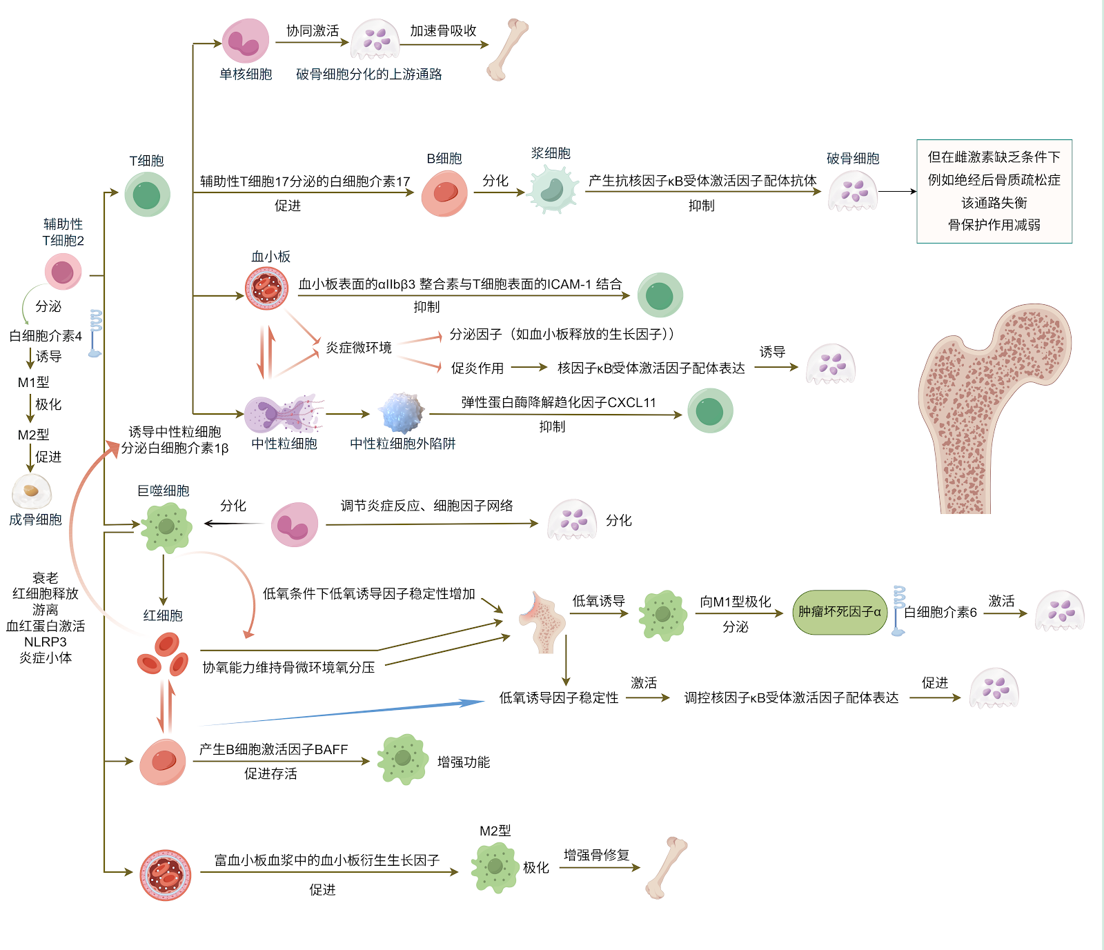
BACKGROUND: Osteoporosis is a systemic bone metabolic disease characterized by deterioration of bone microstructure and increased bone fragility. Blood is not only a medium of bone metabolism, but also a major factor in the regulation of bone metabolism. The traditional theory of "osteogenesis-osteoclast" is difficult to fully explain its complex pathogenesis, but bone immunology reveals the core role of the interaction between the immune system and bone, and the effects of blood cells in osteoporosis have become a research hotspot. OBJECTIVE: To summarize the mechanism of action of blood cells in osteoporosis and evaluate its potential for related biomarkers and emerging therapeutic approaches. METHODS: A systematic literature search was conducted in CNKI, Wanfang, PubMed, and Web of Science databases using Chinese and English search terms including "blood cells, osteoporosis, biomarkers, bone metabolism, neutrophils, macrophages, bone marrow mesenchymal stem cells". A total of 65 articles were included for review. RESULTS AND CONCLUSION: Blood cells influence the occurrence and development of osteoporosis through mechanisms such as inflammation, immune response, and metabolic regulation. Blood cell-based biomarkers can serve as indicators for early screening of osteoporosis and provide effective targets for diagnosis and treatment. With advances in CRISPR-Cas9 gene editing, single-cell technology, and novel monoclonal antibody development, blood cells are expected to play an important role in the treatment of osteoporosis.

Read Full Abstract10.12307/2026.21422
Systematic review of the effect of 3D-printed exoskeleton on hand function rehabilitation in stroke patientsGraphical AbstractVerified
Chinese Journal of Tissue Engineering Research2026

Systematic review of the effect of 3D-printed exoskeleton on hand function rehabilitation in stroke patients

OBJECTIVE: To systematically evaluate the clinical efficacy of different types of 3D-printed exoskeletons in hand function rehabilitation for stroke patients based on the International Classification of Functioning, Disability and Health framework. METHODS: A comprehensive search was conducted in PubMed, Cochrane Library, Web of Science, Embase, CNKI, and CBM databases for literature published between January 2015 and December 2024 on 3D-printed exoskeleton interventions for post-stroke hand function rehabilitation. Two researchers independently screened studies, extracted data, and assessed methodological quality following the PRISMA guidelines for systematic reviews. RESULTS: A total of 13 studies involving 62 stroke patients from the United States, China, the Netherlands, Italy, Singapore, and Turkey were finally included. Based on the Brunnstrom stages, exoskeletons were categorized into four types: assistive (Stage I), corrective (Stages II–III), training-oriented (Stages IV–VI), and compensatory (Stages IV–VI). Interventions were delivered via institution-based, home-based, or combined institution-home rehabilitation, with session durations ranging from 15 to 90 minutes, frequencies of 3 to 7 sessions per week, and durations of 5 to 8 weeks. Results showed that assistive exoskeletons improved fine hand use and self-care; corrective exoskeletons improved joint mobility, muscle tone, grip strength, and fine hand use; training-oriented exoskeletons improved joint mobility, upper limb muscle strength, and self-care; compensatory exoskeletons enhanced fine hand use and hand and arm use. CONCLUSION: Based on the ICF framework, evidence indicates that 3D-printed exoskeletons improve hand function, activity, and participation in stroke patients. It is recommended to select matching exoskeleton types according to Brunnstrom stages and develop individualized intervention plans to achieve optimal rehabilitation outcomes.

Read Full Abstract10.12307/2026.21425
Interaction between vascular-lymphatic system imbalance and immune microenvironment in intervertebral disc degenerationGraphical AbstractVerified
Chinese Journal of Tissue Engineering Research2026

Interaction between vascular-lymphatic system imbalance and immune microenvironment in intervertebral disc degeneration

BACKGROUND: The pathological process of intervertebral disc degeneration is accompanied by angiogenesis-lymphatic imbalance and changes in the immune microenvironment, both of which play important roles in intervertebral disc degeneration. OBJECTIVE: To systematically summarize the roles of angiogenesis-lymphatic imbalance-related cytokines in intervertebral disc degeneration. METHODS: The first author searched relevant literature published between January 2000 and April 2025 in the PubMed, Web of Science, and China National Knowledge Infrastructure (CNKI) databases. The Chinese search terms included "intervertebral disc degeneration, vascular-lymphatic imbalance, angiogenesis, vascular endothelial growth factor (VEGF), lymphatic vessel, Prox-1, immune microenvironment." A total of 65 articles were ultimately included for review. RESULTS AND CONCLUSION: During intervertebral disc degeneration, the vascular-lymphatic system and immune microenvironment play crucial roles in maintaining disc homeostasis. When disc degeneration occurs, angiogenesis and lymphatic disruption increase inflammatory factors within the disc, leading to enhanced degradation of the extracellular matrix. Concurrently, changes in the immune microenvironment, characterized by increased immune cell infiltration, elevated levels of pro-inflammatory cytokines and chemokines, and activation of local immune responses, modulate vascular and lymphatic vessels, ultimately reducing disc repair capacity and inducing chronic pain, thereby exacerbating the degree of disc degeneration.

Read Full Abstract10.12307/2026.21407
Hand tendon suturing: optimization of traditional suture techniques and application of novel repair materialsGraphical AbstractVerified
Chinese Journal of Tissue Engineering Research2026

Hand tendon suturing: optimization of traditional suture techniques and application of novel repair materials

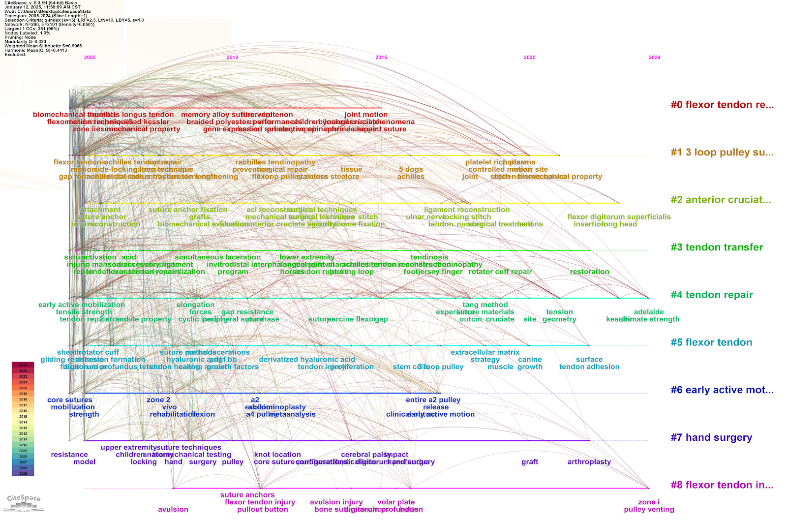
BACKGROUND: The tendon suturing technique for the hand has been continuously innovated with the development of biomechanics, minimally invasive techniques, and regenerative medicine. Over the past two decades, research has focused on optimizing traditional suturing techniques and the application of new repair materials, improving the effectiveness of tendon repair and the level of functional recovery in the hand. OBJECTIVE: To assess the global research status and development trends of hand tendon repair techniques over the past two decades through bibliometric analysis, identify research hotspots and their evolution. METHODS: Relevant literature was selected from the Web of Science database from 2005 to 2024, and bibliometric methods were employed for analysis. Data were organized using Microsoft Excel and analyzed for publication trends using the R language Bibliometrix package. VOSviewer was used to visualize keyword co-occurrence and collaboration networks, while CiteSpace was utilized to identify research hotspots and their temporal evolution. RESULTS AND CONCLUSION: Over the past two decades, research in the field of tendon suturing has shown a fluctuating growth trend. The United States, China, and Europe are the main contributing countries, with the United States occupying a central position in the global research network. Research on flexor tendon repair mainly focuses on biomechanics and the development of new repair materials, while extensor tendon research emphasizes postoperative functional recovery and complex injury repair. In recent years, biomaterials and regenerative medicine have gradually become research hotspots, promoting the application of precision medicine in tendon repair. In the future, interdisciplinary collaboration and the combination of advanced materials will further optimize hand tendon repair techniques.

Read Full Abstract10.12307/2026.21474
Biomechanical analysis during non-anticipated stop-jump cutting before and after exercise fatigue in functional ankle instability and healthy populationsGraphical AbstractVerified
Chinese Journal of Tissue Engineering Research2026

Biomechanical analysis during non-anticipated stop-jump cutting before and after exercise fatigue in functional ankle instability and healthy populations

BACKGROUND: Systemic fatigue increases injury risk in individuals with functional ankle instability, while stop-jump cutting is a high risk for ankle injuries. The biomechanical mechanisms underlying non-anticipated stop-jump cutting during systemic exercise fatigue in this population remain unclear. OBJECTIVE: To quantify differences in kinematic and kinetic characteristics during non-anticipated stop-jump cutting before and after exercise fatigue between individuals with functional ankle instability and healthy controls, revealing the impact of exercise fatigue on stop-jump cutting in individuals with functional ankle instability. METHODS: Fifteen male participants with unilateral functional ankle instability and 15 healthy male controls were recruited. Kinematic (peak angles of ankle dorsiflexion, plantarflexion, inversion, knee flexion, knee varus, knee valgus, hip flexion, and hip abduction) and kinetic (joint stiffness of hip, knee, and ankle) parameters were collected during non-anticipated stop-jump cutting before and after exercise fatigue. Two-way repeated measures ANOVA was used to analyze peak joint angles and joint stiffness. Statistical parametric mapping (SPM) was further used to analyze the effects of fatigue on time-series data of ankle angle and ground reaction forces. RESULTS AND CONCLUSION: Kinematic results: Two-way ANOVA revealed significant main effects of fatigue and group-by-fatigue interactions for peak ankle inversion, knee flexion, knee valgus, and hip flexion angles (P < 0.05). Compared with pre-fatigue, peak ankle inversion increased in the functional ankle instability group after fatigue (P < 0.05), peak knee flexion increased in both groups (P < 0.05), and peak hip flexion increased in the healthy control group (P < 0.05). After fatigue, the functional ankle instability group showed smaller peak ankle inversion and hip flexion angles but larger peak knee valgus and knee flexion angles than the healthy control group (P < 0.05). SPM analysis revealed that ankle inversion/eversion angle was greater during 4%-18% of the cutting movement after fatigue in the functional ankle instability group (P < 0.05). Kinetic results: Two-way ANOVA revealed significant main effects of fatigue and group-by-fatigue interactions for hip, knee, and ankle joint stiffness (P < 0.05). Compared with pre-fatigue, hip and ankle stiffness decreased in the healthy control group (P < 0.05), while knee and ankle stiffness decreased in the functional ankle instability group (P < 0.05). SPM analysis showed that vertical ground reaction force was greater during 5%-16% of the cutting movement, and mediolateral ground reaction force was greater during 35%-49% of the movement after fatigue in the functional ankle instability group (P < 0.05). CONCLUSION: Exercise fatigue alters kinematic and kinetic characteristics during non-anticipated stop-jump cutting in individuals with functional ankle instability, particularly affecting knee and ankle stability and shock absorption. Fatigue reduces joint stiffness and control, increasing injury risk, especially during the initial and transitional phases of the cutting movement.

Read Full Abstract10.12307/2026.21394
Lycium barbarum polysaccharide-mediated intestinal flora remodeling improves glycolipid abnormalities in type 2 diabetic ratsGraphical AbstractVerified
Chinese Journal of Tissue Engineering Research2026

Lycium barbarum polysaccharide-mediated intestinal flora remodeling improves glycolipid abnormalities in type 2 diabetic rats

BACKGROUND: Lycium barbarum polysaccharide is a natural active ingredient with potential to lower blood glucose and improve diabetes-related symptoms. However, from the perspective of gut microbiota, the underlying factors for the effects of Lycium barbarum polysaccharide on glycolipid abnormalities have not been fully elucidated. OBJECTIVE: To investigate the effect of Lycium barbarum polysaccharide on type 2 diabetes mellitus and its related mechanism. METHODS: Six male Sprague-Dawley rats aged 8 weeks were randomly selected from 18 rats to form a blank control group. The remaining 12 rats were fed a high-sugar, high-fat diet for 8 weeks and then received a single tail vein injection of 1% streptozotocin to establish a type 2 diabetes model. After successful modeling, the rat models were then randomly divided into a model control group (n=6) and a Lycium barbarum polysaccharide group (n=6). Rats in the Lycium barbarum polysaccharide group were administered Lycium barbarum polysaccharide solution via gavage at a dose of 200 mg/(kgĀ·d), 2 mL per dose, once daily, for 12 consecutive weeks. After the intervention, rat serum and feces were collected. 16S rDNA sequencing was used to analyze gut microbiota, and alpha diversity, beta diversity, and principal component analysis were used to characterize microbial abundance and structure. Liquid chromatography-mass spectrometry was used to detect short-chain fatty acid levels. ELISA was used to detect glycolipid metabolism, insulin resistance, and inflammatory response indicators. RESULTS AND CONCLUSION: Compared with the model control group, the Lycium barbarum polysaccharide group had increased high-density lipoprotein cholesterol levels and decreased total cholesterol, triglyceride, and low-density lipoprotein cholesterol levels, indicating that Lycium barbarum polysaccharide improved lipid accumulation and inhibited weight loss in type 2 diabetic rats. Compared with the model control group, the Lycium barbarum polysaccharide group had decreased levels of interleukin-6, tumor necrosis factor-alpha, and blood glucose, and increased levels of insulin and glucagon-like peptide-1, indicating that Lycium barbarum polysaccharide effectively inhibited inflammatory response and insulin resistance. Lycium barbarum polysaccharide significantly improved the composition of gut microbiota, increasing the abundance of Lachnospiraceae_NK4A136_group, Clostridia_UCG-014_unclassified, Monoglobus, Phascolarctobacterium, Candidatus_Saccharimonas, Desulfovibrionaceae unclassified, and Desulfovibrio, and decreasing the abundance of Muribaculaceae_unclassified and Enterorhabdus. Lycium barbarum polysaccharide also significantly increased short-chain fatty acid levels, and Clostridia_UCG-014_unclassified, Candidatus_Saccharimonas, and Muribaculaceae_unclassified may participate in regulating butyric acid production to improve glycolipid metabolism in type 2 diabetic rats. Lachnospiraceae_NK4A136_group, Monoglobus, and Desulfovibrionaceae unclassified may participate in regulating isobutyric acid production to inhibit insulin resistance and improve lipid metabolism. Lycium barbarum polysaccharide can improve the inflammatory response in type 2 diabetic rats by increasing the abundance of Firmicutes_unclassified, Clostridia_UCG-014_unclassified, Intestinimona, and Colidextribacter, and inhibiting the abundance of Kineothrix.

Read Full Abstract10.12307/2026.21403
Effects of different lunge exercises on lower limb mechanics and muscle activation in patellofemoral pain syndrome patientsGraphical AbstractVerified
Chinese Journal of Tissue Engineering Research2026

Effects of different lunge exercises on lower limb mechanics and muscle activation in patellofemoral pain syndrome patients

BACKGROUND: Lunge exercises are often used in strengthening programs for patellofemoral pain syndrome and have been shown to help strengthen the quadriceps. However, limited data exist to evaluate the effects of lunge exercises with medial or lateral resistance on patellofemoral joint stress and lower limb muscle activation. OBJECTIVE: To compare the effect of three lunge exercises (traditional lunge, lunge with hip adduction resistance, and lunge with hip abduction resistance) on lower limb muscle activation and patellofemoral joint stress in subjects with patellofemoral pain syndrome and health control group. METHODS: Totally 29 subjects with patellofemoral pain syndrome and 29 healthy subjects completed three different lunge exercises. Lower limb dynamics and electromyography data were simultaneously acquired using an infrared motion capture system, a three-dimensional force table, and a surface electromyography analyzer. A mixed-design analysis of variance was used to determine the effects of group and exercise on patellofemoral joint stress and lower limb muscle activation characteristics during different lunge exercises. RESULTS AND CONCLUSION: (1) During the squatting phase, the vastus lateralis muscle activation was significantly lower in both groups during the lunge exercises with hip adduction (P < 0.05), while the vastus medialis/vastus lateralis activation ratio was significantly increased in the healthy group (P < 0.05). In the patellofemoral pain syndrome group, the gluteus medius activation was significantly increased (P < 0.05) and the peak patellofemoral joint stress was significantly reduced (P < 0.05) during the lunge with hip abduction. (2) During the pushing phase, the vastus lateralis activation was significantly lower in both groups during the lunge with hip abduction (P < 0.05), and the patellofemoral pain syndrome group showed significantly reduced peak patellofemoral joint stress (P < 0.05) and enhanced gluteus medius activation (P < 0.05). In the healthy group, the vastus medialis/vastus lateralis activation ratio was significantly increased during the lunge with hip adduction (P < 0.05), and the patellofemoral pain syndrome group had significantly lower peak patellofemoral joint stress than the healthy group (P < 0.05). (3) These findings suggest that traditional lunge can be one of the preferred training methods for strengthening the quadriceps in the early rehabilitation of patellofemoral pain syndrome. Lunge exercises with lateral adduction/abduction resistance have advantages in improving vastus medialis/vastus lateralis activation imbalance, strengthening gluteus medius activation, and reducing patellofemoral joint stress during squatting, which may help rebuild neuromuscular control of the lower limb kinetic chain in the later rehabilitation stage. For patients with patellofemoral pain syndrome, lunge with lateral abduction resistance is more recommended to alleviate pain and improve function.

Read Full Abstract10.12307/2026.21390
Proteomic analysis of the mechanism of moxibustion intervention in a rat model of atopic dermatitisGraphical AbstractVerified
Chinese Journal of Tissue Engineering Research2026

Proteomic analysis of the mechanism of moxibustion intervention in a rat model of atopic dermatitis

BACKGROUND: Moxibustion has been demonstrated as an effective therapeutic approach for atopic dermatitis, yet its underlying mechanisms remain to be further elucidated. OBJECTIVE: To investigate the mechanisms of moxibustion intervention in atopic dermatitis using proteomics technology. METHODS: Thirty-four Sprague-Dawley rats were randomly divided into three groups: a model group (n=12), a moxibustion group (n=12), and a blank group (n=10). The first two groups were induced to develop an atopic dermatitis model using 2,4-dinitrochlorobenzene. Following successful modeling, the moxibustion group received moxibustion therapy with moxa sticks applied to the Ashi point for 30 minutes per session, maintaining a local temperature of (43±1) °C, administered every other day over a 14-day intervention period. The model group and blank control group underwent restraint and fixation procedures of the same duration and intensity. Skin lesion severity after modeling was evaluated using the Eczema Area and Severity Index. Proteomic analysis of rat skin tissue was performed using a data-independent acquisition approach on a high-performance liquid chromatography-tandem mass spectrometry platform. Mass spectrometry data processing, protein identification, differential protein expression analysis, functional annotation, and bioinformatics analyses were conducted using MaxQuant, Perseus software, DAVID, STRING, and Cytoscape. RESULTS AND CONCLUSION: After moxibustion treatment, the skin lesion score of atopic dermatitis model rats was significantly lower than that of the model group (P < 0.05), indicating successful modeling. Moxibustion reversed the upregulation of 28 differentially expressed proteins and the downregulation of 40 differentially expressed proteins in the model group. Bioinformatics analysis indicated that the main signaling pathways involved in atopic dermatitis pathogenesis include neuroactive ligand-receptor interaction, viral protein interaction with cytokine and cytokine receptor, inflammatory mediator regulation of TRP channels, and neutrophil extracellular trap formation. Among these, the expression regulation of integrin β3 and β-1,4-galactosyltransferase proteins may be most relevant to the pathogenesis of atopic dermatitis. The mechanisms of moxibustion in treating atopic dermatitis mainly involve herpes simplex virus 1 infection, olfactory transduction, influenza A, steroid hormone biosynthesis, and other infection- or immune-related signaling pathways. The most relevant proteins include nuclear factor κB subunit 1, major histocompatibility complex protein, RT1-Bb, Jak1, and Cdk6. These findings suggest that moxibustion exerts a multi-target, multi-pathway, and multi-channel intervention effect on atopic dermatitis, reversing the inflammatory response caused by atopic dermatitis and exerting anti-inflammatory and antioxidant effects.

Read Full Abstract10.12307/2026.21398
Effect of magnetic field mitochondrial regulation technology combined with low-load blood flow restriction on the strength of lower limb muscle groupsGraphical AbstractVerified
Chinese Journal of Tissue Engineering Research2026

Effect of magnetic field mitochondrial regulation technology combined with low-load blood flow restriction on the strength of lower limb muscle groups

BACKGROUND: The magnetic field mitochondrial regulation technology has been proven to enhance skeletal muscle function. Low-load blood flow restriction training can effectively induce adaptive growth of muscle strength through metabolic emergency mechanisms. Currently, both technologies have become hotspots in the application and research of skeletal muscle function improvement and treatment. However, the differences in their effects on muscle strength enhancement and whether their combined application can produce a synergistic effect remain unclear. OBJECTIVE: To observe the differences in the effects of low-frequency pulsed magnetic stimulation (1.5 mT, 3 300 Hz) and low-load blood flow restriction training on muscle strength enhancement and the impact of their combined intervention on lower limb muscle strength. METHODS: Fifty-six healthy subjects were recruited and randomly divided into magnetic stimulation group (high-load squat training + magnetic stimulation), blood flow restriction group (low-load blood flow restriction squat training), combined group (low-load blood flow restriction squat training + magnetic stimulation), and control group (high-load squat training). The trial lasted 4 weeks, with training three times per week, and low-frequency pulsed magnetic stimulation (1.5 mT, 3 300 Hz) was administered every 48 hours. After the trial, changes in maximal strength, explosive power, and strength endurance of the lower limb muscles were observed among groups. RESULTS AND CONCLUSION: Fifty subjects completed the trial and were included in the analysis. ā‘  After 4 weeks of intervention, the maximal strength, explosive power, and strength endurance of the lower limbs in the magnetic stimulation, blood flow restriction, and combined groups significantly increased. ā‘” In terms of maximal strength increase, blood flow restriction was superior to magnetic field mitochondrial regulation technology; low-load blood flow restriction also enhanced distal muscle strength, while magnetic field mitochondrial regulation technology had the advantage of improving maximal strength without fatigue accumulation. ā‘¢ In terms of explosive power increase, both technologies had similar effects; magnetic stimulation was more advantageous for explosive power in single-joint movements, while low-load blood flow restriction training was more advantageous for explosive power in multi-joint coordinated movements. ā‘£ In terms of strength endurance increase, magnetic stimulation technology, due to its mitochondrial function regulation, effectively improved muscle fatigue resistance. The results suggest that the combined application of magnetic stimulation and low-load blood flow restriction can produce synergistic effects on maximal strength, explosive power, and strength endurance of the lower limbs. This technical approach may provide a novel and efficient auxiliary training protocol for lower limb muscle strength enhancement in postoperative rehabilitation and sports injury patients who cannot undergo high-intensity resistance training.

Read Full Abstract10.12307/2026.21387
Correlation of bone mineral density assessed using quantitative CT with obesity indicators, blood pressure, platelet count, and blood lipids in perimenopausal womenGraphical AbstractVerified
Chinese Journal of Tissue Engineering Research2026

Correlation of bone mineral density assessed using quantitative CT with obesity indicators, blood pressure, platelet count, and blood lipids in perimenopausal women

BACKGROUND: Due to the decline in ovarian function, reduced estrogen secretion, and endocrine disorders, perimenopausal women are more prone to osteoporosis. There are few studies on the correlation of bone mineral density with obesity indicators, blood pressure, blood routine, and glycolipid metabolism in perimenopause women, and the research results are controversial. OBJECTIVE: To investigate the correlation of bone mineral density with quantitative CT with obesity indicators, blood pressure, platelet count, and glycolipid metabolism in perimenopause women. METHODS: This is a single-center retrospective study. 490 perimenopause women who were admitted to Guizhou Hospital of Beijing Jishuitan Hospital from January 1, 2022 to December 31, 2024 were selected. Their age, height, body mass, waist circumference, blood pressure, fasting blood glucose, blood routine, and blood biochemical indicators were collected to calculate body mass index, waist height ratio, body shape index, body roundness index, taper index, visceral fat index, and lipid accumulation index. Quantitative CT was used to measure lumbar spine bone mineral density. According to bone mineral density, the participants were divided into normal bone mass group, low bone mass group, and osteoporosis group. Kruskal-Wallis rank sum test was used to compare the differences in various indicators among different bone mineral density groups. Spearman correlation analysis and partial correlation analysis were used to analyze the correlation between bone mineral density and various indicators. RESULTS AND CONCLUSION: (1) There were significant differences in age, waist circumference, waist height ratio, body shape index, body roundness index, taper index, visceral fat index, lipid accumulation index, bone mineral density, systolic blood pressure, diastolic blood pressure, platelet count, total cholesterol, triglycerides, and postmenopausal status among different bone mineral density groups (P < 0.05). (2) Spearman correlation analysis showed that L1 bone mineral density, L2 bone mineral density, and average bone mineral density of L1 and L2 were negatively correlated with waist circumference, waist height ratio, body shape index, body roundness index, taper index, visceral fat index, lipid accumulation index, systolic blood pressure, and diastolic blood pressure (P < 0.05), and positively correlated with platelet count (P < 0.05). L1 bone mineral density and average bone mineral density were negatively correlated with total cholesterol (P < 0.05); L1 bone mineral density was negatively correlated with triglycerides (P < 0.05). (3) After adjusting for confounding factors, L1 bone mineral density, L2 bone mineral density, and average bone mineral density were still negatively correlated with waist circumference, body shape index, and taper index (P < 0.05); L1 bone mineral density and average bone mineral density were still negatively correlated with waist height ratio and body roundness index (P < 0.05); L2 bone mineral density was positively correlated with platelet count (P < 0.05). Among abdominal obesity indicators, body shape index and taper index had the largest correlation coefficients, especially body shape index. The results indicate that for perimenopausal women, after adjusting for confounding factors, quantitative CT bone mineral density has a certain correlation with abdominal obesity indicators and platelet count, but has no correlation with systolic blood pressure, diastolic blood pressure, blood lipids, and fasting blood glucose. The research results provide reference for clinical early prevention of osteoporosis or delaying the development of osteoporosis, and have certain clinical value.

Read Full Abstract10.12307/2026.21385
A customizable vascular network biomimetic design for nutrient supply in large-scale engineering tissuesGraphical AbstractVerified
Chinese Journal of Tissue Engineering Research2026

A customizable vascular network biomimetic design for nutrient supply in large-scale engineering tissues

BACKGROUND: Constructing an effective vascular network is crucial for the successful regeneration of large-volume tissues and organs. Currently, vascular network design methods predominantly rely on predefined geometric patterns, making them inadequate to meet the metabolic demands of engineered tissues with diverse material properties and complex morphologies. Existing hierarchical vascular networks suffer from insufficient diffusion coverage and low nutrient supply rates due to their hierarchical rules. OBJECTIVE: To propose a vascular network model design method based on developmental biomimetic principles, aiming to automatically generate customized voxel vascular network structures tailored to the metabolically active distance of target tissues. METHODS: The method integrated voxelization techniques to simulate biological behaviors of vascular endothelial cells, such as migration and aggregation. Diffusion experiments were conducted on gel materials, and Fick's law was applied to fit experimental data, establishing a metabolic-diffusion coupled fast calculation model. Based on this model, the metabolic activity distance was used to rapidly identify nutrient supply conditions and delineate low-nutrient regions, thereby simulating the dynamic remodeling process of vascular development to iteratively optimize the voxel vascular network structure until the nutrient supply rate reached the algorithm's set value. RESULTS AND CONCLUSION: Compared with traditional hierarchical design methods, the developmental biomimetic vascular network design improved the nutrient-sufficient volume and nutrient contribution per unit volume by 25.53% in metabolic diffusion simulations of cuboid tissue models based on gelatin methacryloyl (GelMA) hydrogel. It also successfully generated vascular network voxel models with sufficient nutrient supply for complex-shaped engineered tissues and organs such as kidney-shaped and alveolar-shaped models, verifying the advantages and research potential of biomimetic vascular network design. This study provides a new technical approach for designing vascular network digital models in large-volume engineered tissues and anatomically complex organs.

Read Full Abstract10.12307/2026.21383
Mechanism by which kaempferol inhibits intervertebral disc degeneration in rats by regulating mitophagy levelsGraphical AbstractVerified
Chinese Journal of Tissue Engineering Research2026

Mechanism by which kaempferol inhibits intervertebral disc degeneration in rats by regulating mitophagy levels

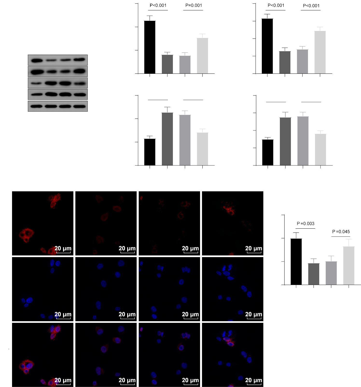
BACKGROUND: Oxidative stress is a primary factor accelerating the progression of intervertebral disc degeneration. Mitophagy plays a crucial role in mitigating oxidative stress and preventing mitochondrial dysfunction and associated diseases. Prior investigations have demonstrated that kaempferol enhances the proliferative capacity of degenerating nucleus pulposus cells, diminishes inflammation, and decelerates the progression of intervertebral disc degeneration in rat models. OBJECTIVE: To investigate the effects of kaempferol on mitophagy in degenerated intervertebral disc tissue. METHODS: Fifteen Sprague-Dawley rats were randomly allocated into five intervention groups: a blank group (n=3), a model group (n=3), a low-dose kaempferol group (n=3), a medium-dose kaempferol group (n=3), and a high-dose kaempferol group (n=3). A coccygeal disc degeneration model was established in all groups except the blank group via full-thickness annulus fibrosus puncture. Commencing 4 weeks post-surgery, the blank and model groups received normal saline via gavage, while the low-dose, medium-dose, and high-dose kaempferol groups were administered 25, 50, and 100 mg/kg kaempferol via gavage, respectively, once daily for 8 consecutive weeks. Following the final administration, samples were collected for serum inflammatory cytokine level measurement, MRI imaging, and hematoxylin-eosin staining. In cell experiments, second-passage rat nucleus pulposus cells were divided into five groups: blank group (no treatment), model group (H2O2-induced oxidative stress), kaempferol group (H2O2 for 24 h then 10 μmol/L kaempferol), autophagy inhibitor group (H2O2 for 24 h then 3-methyladenine), and kaempferol + autophagy inhibitor group (H2O2 for 24 h then 10 μmol/L kaempferol plus 3-methyladenine). After 24 h of further culture, cell viability was assessed by CCK-8 assay, protein expression of inflammatory cytokines and autophagy markers by western blot, gene expression by RT-qPCR, type II collagen expression by immunofluorescence staining, and reactive oxygen species levels by DCFH-DA fluorescent probe. RESULTS AND CONCLUSION: In animal experiments, compared with the blank group, serum levels of interleukin-1β, interleukin-6, and tumor necrosis factor-α were elevated in the model group (P < 0.05), while kaempferol treatment at all doses reduced these levels (P < 0.05), with the medium dose showing the most pronounced effect. MRI and hematoxylin-eosin staining revealed that kaempferol treatment significantly ameliorated the pathological features of disc degeneration, with clearer and more continuous nucleus pulposus contours and increased cell numbers, particularly in the medium-dose group. In cell experiments, compared with the model group, kaempferol increased cell viability, type II collagen expression, and the gene and protein expression of PTEN-induced putative kinase 1 and Parkin (P < 0.05), while decreasing reactive oxygen species levels and the expression of inflammatory cytokines (P < 0.05). Kaempferol also increased the LC3-II/LC3-I ratio and LC3B mRNA expression (P < 0.05). 3-Methyladenine inhibited these effects of kaempferol. These findings indicate that kaempferol may alleviate oxidative stress injury and delay intervertebral disc degeneration by regulating mitophagy.

Read Full Abstract10.12307/2026.21380
Regulating mitochondrial dynamics balance in nucleus pulposus cells inhibits cell apoptosisGraphical AbstractVerified
Chinese Journal of Tissue Engineering Research2026

Regulating mitochondrial dynamics balance in nucleus pulposus cells inhibits cell apoptosis

BACKGROUND: Mitochondrial dysfunction is increasingly recognized as a key factor during intervertebral disc degeneration. Sirt3, a major mitochondrial deacetylase, mediates AMPK pathway activation by directly phosphorylating and inhibiting Drp1 activity while indirectly regulating mitochondrial function through downstream signaling. However, the specific mechanisms of Sirt3 and the AMPK/Drp1 pathway in nucleus pulposus cells during intervertebral disc degeneration remain unclear. OBJECTIVE: To investigate whether Sirt3 regulates mitochondrial dynamics balance in nucleus pulposus cells induced by tert-butyl hydroperoxide by mediating the AMPK/Drp1 pathway, thereby inhibiting cell apoptosis. METHODS: Human nucleus pulposus cells were cultured in vitro, and a degeneration model was established by oxidative damage with tert-butyl hydroperoxide. Cells were divided into the following groups: control, model, model + oe-NC, model + oe-Sirt3, model + oe-Sirt3 + Compound C (AMPK inhibitor), and Compound C alone. After 24 h of treatment, cell viability was assessed by CCK-8, apoptosis by flow cytometry, and expression of apoptosis-related proteins (Bax, Bcl2), disc degeneration-related proteins (aggrecan, collagen type II), Sirt3, mitochondrial fission proteins (Fis1, Mff), fusion proteins (Mfn1, Mfn2), and AMPK/Drp1 pathway proteins by western blot. ATP and reactive oxygen species levels were measured using kits, and mitochondrial DNA copy number was determined by RT-qPCR. RESULTS AND CONCLUSION: Compared with the control group, the model group showed significantly decreased cell viability and expression of Bcl2, aggrecan, collagen type II, and Sirt3, while apoptosis rate and Bax level were significantly increased (all P < 0.05), indicating successful establishment of the degeneration model. Additionally, ATP levels, mitochondrial membrane potential, mtDNA copy number, and Mfn1/Mfn2 expression were significantly reduced, while reactive oxygen species, Fis1, and Mff levels were elevated (all P < 0.05), indicating mitochondrial dynamics imbalance. Overexpression of Sirt3 in the model+oe-Sirt3 group inhibited TBHP-induced apoptosis, improved cell viability, restored mitochondrial dynamics balance, activated the AMPK/Drp1 pathway, and suppressed mitochondrial fission. However, the protective effects of Sirt3 overexpression were partially reversed by the AMPK inhibitor Compound C. These findings suggest that Sirt3 is a potential target for inhibiting nucleus pulposus cell apoptosis and may serve as a novel therapeutic direction for intervertebral disc degeneration.

Read Full Abstract10.12307/2026.21379
Mechanism of Gugutou Huaisiyu Capsule in alleviating peripheral pain sensitization in rats with steroid-induced osteonecrosis of the femoral headGraphical AbstractVerified
Chinese Journal of Tissue Engineering Research2026

Mechanism of Gugutou Huaisiyu Capsule in alleviating peripheral pain sensitization in rats with steroid-induced osteonecrosis of the femoral head

BACKGROUND: Previous studies have demonstrated that Gugutou Huaisiyu Capsule effectively alleviate pain in patients with osteonecrosis of femoral head; however, the mechanism of action remains unclear. OBJECTIVE: To investigate the mechanism by which Gugutou Huaisiyu Capsule mitigate peripheral pain sensitization in rats with steroid-induced osteonecrosis of the femoral head. METHODS: Thirty-two Sprague-Dawley rats were randomly assigned to four groups: blank control, model, low-dose Gugutou Huaisiyu Capsule, and high-dose Gugutou Huaisiyu Capsule groups with eight rats in each group. Steroid-induced femoral head necrosis rat models were established via intraperitoneal injection of lipopolysaccharide combined with gluteal injection of methylprednisolone in the later three groups. After 7 days of modeling, the low- and high-dose Chinese medicine groups were administered 0.33 and 0.67 g/kg of Gugutou Huaisiyu Capsule decoction by gavage, respectively, while the blank control and model groups received normal saline, once daily for 8 weeks. After the last administration, Micro-CT was used to analyze the microstructure of the femoral head cancellous bone; Von-Frey test was used to detect mechanical pain threshold; ELISA was used to detect serum levels of pain mediators calcitonin gene-related peptide (CGRP) and substance P; multiplex immunofluorescence staining and real-time quantitative PCR were used to detect protein and gene expression of tyrosine kinase receptor A (TrkA), CGRP, and neuronal marker β3-tubulin in the femoral head and dorsal root ganglion. RESULTS AND CONCLUSION: (1) Micro-CT and pain behavior tests showed that compared with the blank control group, the model group had significant destruction of the femoral head cancellous bone microstructure, decreased bone mineral density and bone volume fraction, increased trabecular separation, and decreased mechanical pain threshold. Compared with the model group, the low- and high-dose Chinese medicine groups had significantly improved trabecular structure, increased bone mineral density and bone volume fraction, decreased trabecular separation, and increased mechanical pain threshold. (2) ELISA showed that the levels of CGRP and substance P in the model group were significantly higher than those in the blank control group, while the levels in the low- and high-dose Chinese medicine groups were lower than those in the model group. (3) Multiplex immunofluorescence staining and real-time quantitative PCR showed that compared with the blank control group, the protein and gene expression of CGRP, TrkA, and β3-tubulin in the femoral head and dorsal root ganglion were increased in the model group; compared with the model group, the expression of these proteins and genes was decreased in the low- and high-dose Chinese medicine groups. (4) These results indicate that Gugutou Huaisiyu Capsule can increase the mechanical pain threshold, inhibit abnormal neuronal activation, and alleviate pain sensitization in rats with steroid-induced osteonecrosis of the femoral head, and the mechanism may be related to the inhibition of TrkA.

Read Full Abstract10.12307/2026.21378