🧬 SinoBioData Academic Portal
šŸ“š Peer-Reviewed Translated Literature

All Biomedical & Clinical Articles (Page 15)

Browse complete peer-reviewed translations from top Chinese biomedical, oncology, and genomics journals. Read verified previews and download full authentic clinical reports.

Published Research Papers

Showing 24 of 1542 peer-reviewed translated articles (Page 15 of 65)

Clinical trial of a robotic system for puncture navigation and positioningGraphical AbstractVerified
Chinese Journal of Tissue Engineering Research2026

Clinical trial of a robotic system for puncture navigation and positioning

BACKGROUND: Conventional percutaneous CT-guided interventional puncture cannot be monitored in real time, and the operation takes a long time. In some high-risk puncture sites, multiple CT scans and adjustments to the position of the puncture needle are required, causing greater radiation damage to the patient. A self-developed surgical navigation and positioning system provides an effective way to solve the clinical problems of percutaneous puncture information perception and accurate and safe target puncture in complex intraoperative environments, achieving precise puncture positioning of the chest and abdomen. OBJECTIVE: To evaluate the safety, effectiveness and usability of the self-developed percutaneous puncture navigation robotic system in clinical application. METHODS: A retrospective analysis was conducted on clinical trial data from percutaneous lung nodule biopsy and tumor ablation procedures guided by a puncture navigation robotic system at the First Affiliated Hospital of Guangzhou Medical University and the Second Affiliated Hospital of Soochow University between November 1, 2021 and June 28, 2022. A multicenter, open-label, parallel controlled clinical study was conducted, and 120 subjects were randomly divided into an experimental and a control group, with 60 subjects in each group. The experimental group underwent puncture guided by a puncture navigation robotic system, while the control group underwent conventional CT-guided percutaneous puncture. The primary effectiveness endpoint was puncture accuracy rate, and secondary endpoints included number of needle adjustments, one-time success rate of puncture, number of CT scans, and system usability. RESULTS AND CONCLUSION: No unsafe events occurred during the entire clinical trial. The one-time success rate of puncture was 98.31% in the experimental group and 15.00% in the control group; complication rates were 6.78% and 13.33%, respectively. The system usability satisfaction rate was 100%. The system achieved interactive modeling of puncture needle and soft tissue, dynamic reconstruction of complex operative environment and real-time perception of puncture information, and dynamic navigation and tracking compensation under physiological motion and puncture interaction, providing an effective solution to the clinical challenges of information perception and accurate and safe target puncture in complex intraoperative environments.


Read Full Abstract10.12307/2026.21377
Research status and trends of nanotechnology in improving photodynamic therapy for hypoxic tumorsGraphical AbstractVerified
Chinese Journal of Tissue Engineering Research2026

Research status and trends of nanotechnology in improving photodynamic therapy for hypoxic tumors

BACKGROUND: Photodynamic therapy, a novel tumor treatment, is limited by the hypoxic tumor microenvironment. Nanotechnology-based oxygen regulation strategies offer a novel approach to overcoming this bottleneck. OBJECTIVE: To systematically analyze the research status of nanotechnology in improving photodynamic therapy for hypoxic solid tumors using bibliometric methods, identify hotspots, and predict future directions. METHODS: Publications and reviews from 2016 to 2025 on nanotechnology for regulating tumor hypoxia and enhancing photodynamic therapy were retrieved from the Web of Science Core Collection. Excel, CiteSpace, VOSviewer, and Bibliometrix were used for visual analysis of categories, publication trends, countries, institutions, authors, co-cited references, and keywords. RESULTS AND CONCLUSION: A total of 1,879 articles were included, with 'nanoscience & nanotechnology' as the core category. From 2016 to 2022, publications increased steadily, with a slight decline in 2023 and a subsequent rise. China was the leading country, with the Chinese Academy of Sciences having the highest output, and Liu Zhuang from Soochow University being the most prolific author. The most cited paper was by Zhou ZJ et al. (2016) in Chemical Society Reviews. The field focuses on cancer treatment, particularly microenvironment-responsive optical therapeutic strategies using nanomaterials. Keywords 'Photodynamic therapy' and 'Nanoparticles' appeared most frequently. Bibliometric analysis indicates that nanotechnology offers advantages in enhancing photodynamic therapy for hypoxic tumors, with promising efficacy and safety. Future hotspots may focus on combination with immunotherapy.

Read Full Abstract10.12307/2026.21473
A network meta-analysis of therapeutic effects of different bone repair materials on apical bone defectsGraphical AbstractVerified
Chinese Journal of Tissue Engineering Research2026

A network meta-analysis of therapeutic effects of different bone repair materials on apical bone defects

OBJECTIVE: To promote the regeneration of apical bone defects, autologous bone, xenograft bone, synthetic bone, and bioactive materials are commonly used clinically. However, a systematic comparison of the efficacy of each repair material is lacking. Therefore, this study used a network meta-analysis to comprehensively compare and evaluate the differences in efficacy of different bone repair materials in treating apical bone defects. METHODS: Randomized controlled trials on the treatment of apical bone defects caused by chronic periapical periodontitis or apical cysts using bone repair materials were searched in databases including CNKI, WanFang Data, VIP, SinoMed, PubMed, Embase, Web of Science, and Cochrane Library. The search period was from database inception to July 20, 2025. According to the Cochrane Handbook of Evaluation, RevMan 5.4 software was used to assess the risk of bias in the included literature. Stata 17MP was used for statistical analysis. RESULTS: A total of 21 studies involving 1,286 patients were included, evaluating 10 interventions: hydroxyapatite, deproteinized bovine bone mineral, collagen membrane, hydroxyapatite + collagen membrane, deproteinized bovine bone mineral + collagen membrane, platelet concentrate growth factor, platelet-rich fibrin, gelatin sponge, blank control (blood clot healing), and platelet-rich plasma. Network meta-analysis results showed that: (1) The top three treatments in terms of effective rate were hydroxyapatite + collagen membrane > deproteinized bovine bone mineral + collagen membrane > platelet concentrate growth factor; (2) The adverse reaction rate from high to low was: gelatin sponge > blank control > hydroxyapatite > hydroxyapatite + collagen membrane. CONCLUSION: For the treatment of apical bone defects, hydroxyapatite combined with collagen membrane has the most significant efficacy and the lowest postoperative adverse reaction rate. Limited by the quantity and quality of literature, more clinical trials are still needed for verification in the future.

Read Full Abstract10.12307/2026.21472
Development and application of natural oral hydrogels in drug delivery systemsGraphical AbstractVerified
Chinese Journal of Tissue Engineering Research2026

Development and application of natural oral hydrogels in drug delivery systems

BACKGROUND: Oral drug delivery has consistently been the most preferred route of administration due to its high patient compliance, significantly enhancing the overall treatment experience compared to injectables. However, the gastrointestinal environment severely limits drug bioavailability. As the demand for biocompatibility and biodegradability in the medical field continues to grow, natural hydrogels have emerged as ideal drug delivery carriers, attracting widespread attention. OBJECTIVE: To explore the development and application of oral hydrogels made from various natural materials, from material selection to synthesis methods. METHODS: A comprehensive literature search was conducted in the PubMed and Web of Science databases using the English search terms ā€œoral hydrogels, physical crosslinking, chemical crosslinking, natural material, therapy, drug delivery, application of disease researchā€ to identify the most recent relevant articles published from 2009 to 2024. A total of 83 articles were selected for review. RESULTS AND CONCLUSION: Hydrogels, as a promising new drug delivery system, exhibit significant advantages in achieving precise drug delivery and controlled release. Oral natural hydrogels stand out in the field of drug delivery due to their excellent biocompatibility, good degradability, and extremely low potential toxicity. They not only enable precise drug delivery but also effectively avoid irritation caused by direct contact between drugs and the gastrointestinal tract, providing a safer and more effective route for drug administration. With the continuous exploration of researchers, novel intelligent hydrogel delivery systems based on natural materials are emerging, such as pectin-based pH-responsive hydrogels and hyaluronic acid-based reactive oxygen species-responsive hydrogels. These new materials open new avenues for intelligent and precise drug delivery. However, natural material hydrogels also expose some issues to be solved during application: on one hand, natural materials generally suffer from insufficient mechanical properties and tensile strength, making it difficult to meet complex drug delivery needs; on the other hand, although natural materials originate from nature, they may still trigger immune responses in the human body.

Read Full Abstract10.12307/2026.21470
Main preparation methods of new fluorescent nanomaterial carbon quantum dots and their applications in tumor diagnosis and treatmentGraphical AbstractVerified
Chinese Journal of Tissue Engineering Research2026

Main preparation methods of new fluorescent nanomaterial carbon quantum dots and their applications in tumor diagnosis and treatment

BACKGROUND: Carbon quantum dots are a novel fluorescent nanomaterial that, thanks to their excellent optical properties, good biocompatibility, and low toxicity, show significant potential in the field of tumor diagnosis and treatment. OBJECTIVE: To systematically review the methods used to prepare carbon quantum dots and their applications in tumor diagnosis and treatment. METHODS: Relevant literature was retrieved from the China National Knowledge Infrastructure (CNKI) and PubMed databases using computer searches. Chinese search terms were ā€œtumor therapy, carbon quantum dots, arc discharge, electrochemical discharge, drug delivery, fluorescence imaging.ā€ English search terms were ā€œCQD tumor therapy, carbon quantum dot preparation, carbon quantum dots, tumor diagnosis and treatment.ā€ According to the inclusion and exclusion criteria, 102 articles were finally included in the review. RESULTS AND CONCLUSION: Common preparation methods for carbon quantum dots are top-down method and bottom-up method. In terms of preparation, the top-down method (arc discharge, electrochemical, laser ablation) uses graphite and other carbon materials as precursors, which is simple to operate but produces many by-products and has low quantum yield; after purification, the yield can be improved. The bottom-up method (hydrothermal, microwave, and template) utilizes biomass or small-molecule carbon sources, offering environmental friendliness and excellent water solubility of the products. In tumor diagnosis and therapy, carbon quantum dots exert their effects through multiple synergistic mechanisms. Fluorescence imaging enables early diagnosis and real-time monitoring by labeling tumor cells; photothermal therapy converts light energy into heat to kill tumor cells; photodynamic therapy generates reactive oxygen species to damage tumor cell biomolecules; as drug carriers, carbon quantum dots can target delivery of antitumor drugs to tumor sites, reducing adverse effects; in immunotherapy, they trigger immunogenic cell death, reverse the immunosuppressive microenvironment, and activate the stimulator of interferon genes pathway, converting ā€œcoldā€ tumors to ā€œhotā€ tumors; in multimodal therapy, carbon quantum dots integrate chemotherapy, phototherapy, and immunotherapy to achieve high tumor inhibition rates.

Read Full Abstract10.12307/2026.21471
Three-dimensional bioprinting and tendon repair: application advances and future directionsGraphical AbstractVerified
Chinese Journal of Tissue Engineering Research2026

Three-dimensional bioprinting and tendon repair: application advances and future directions

BACKGROUND: Currently, three-dimensional (3D) bioprinting technology, with its controllable multi-scale structure and functional integration design capabilities, has become a cutting-edge solution for tendon tissue engineering. OBJECTIVE: To systematically summarize the latest research progress of 3D bioprinting technology in tendon repair. METHODS: Using the keywords ā€œ3D printing, bioink, myotendinous junction, tendon repair, tendon-bone junction, bionic scaffold,ā€ literature searches were conducted in the PubMed and Web of Science databases, as well as in the China National Knowledge Infrastructure (CNKI) with the same keywords. Articles with weak relevance to the topic were excluded, and 109 articles were ultimately included for review. RESULTS AND CONCLUSION: 3D bioprinting technology, through multi-material integration and controllable biomimetic structural design, effectively reproduces the multi-level structure of tendons. Mainstream technologies (such as melt electrowriting, extrusion-based printing, etc.) play differentiated advantages in fiber alignment, interface simulation, and dynamic regulation, constructing mechanical transition layers at the muscle-tendon interface and four-zone gradient structures at the tendon-bone interface. Functionalized bioink innovations (immunomodulatory materials, cross-species oxygen-supplying scaffolds, etc.) and multi-technology synergy (aligned fiber deposition + photocuring reinforcement) enhance scaffold bioactivity and mechanical-biological coupling. In the full healing cycle (support in the inflammatory phase, guidance in the proliferative phase, regulation in the remodeling phase), precise intervention from molecular to macroscopic levels is achieved, optimizing collagen alignment and repair mechanical properties. Differentiated repair strategies (multi-material gradients, aligned fibers, gradient scaffolds) for the muscle-tendon interface, tendon body, and tendon-bone interface have made progress. Despite challenges such as resolution-efficiency contradictions and insufficient material matching, 3D printing technology still provides new strategies for tendon repair from structural biomimicry to functional regeneration. In the future, the integration of intelligent materials (photothermal/piezoelectric) and multimodal technologies (4D printing, organoids) is expected to promote dynamic functional regeneration and provide technical references for interface repair.

Read Full Abstract10.12307/2026.21469
Construction and performance evaluation of pre-vascularized three-dimensional porous bioprinted hydrogelGraphical AbstractVerified
Chinese Journal of Tissue Engineering Research2026

Construction and performance evaluation of pre-vascularized three-dimensional porous bioprinted hydrogel

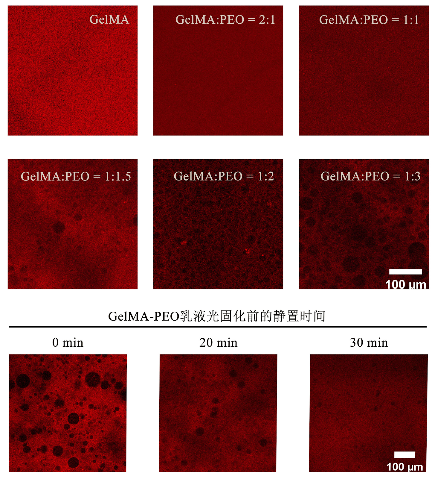
BACKGROUND: Three-dimensional bioprinted hydrogels have become an important research direction for the repair of oral tissue defects. Pre-vascularization of hydrogels can be achieved by loading endothelial cells and stromal cells. However, the dense hydrogel fibers often limit cell viability and extension. Whether increasing the internal porosity of the hydrogel can improve pre-vascularization remains unclear. OBJECTIVE: To construct porous three-dimensional bioprinted hydrogels loaded with human umbilical vein endothelial cells and human dental pulp stem cells, and to explore the relationship between hydrogel pore size and pre-vascularization. METHODS: (1) Methacrylate gelatin solution and poly (ethylene oxide) solution were mixed at volume ratios of 2:1, 1:1, 1:1.5, 1:2, and 1:3, with pure methacrylate gelatin solution as a control. Three-dimensional bioprinting was performed, and after curing and crosslinking, poly (ethylene oxide) was leached out to form pores. Based on porosity measurements, the mixed solutions with methacrylate gelatin solution and poly (ethylene oxide) solution volume ratios of 1:1, 1:2, and 1:3, and pure methacrylate gelatin solution were selected for subsequent experiments. (2) The above four solutions were used as bioinks to encapsulate human umbilical vein endothelial cells or human dental pulp stem cells for three-dimensional bioprinting. After curing and crosslinking, poly (ethylene oxide) was leached out to form pores. Live/dead staining was used to detect cell viability. Both cells were co-encapsulated for three-dimensional bioprinting, and after curing and crosslinking, poly (ethylene oxide) was leached out to form pores. Tube formation assay was used to detect vascular network formation. (3) The four groups of three-dimensional bioprinted hydrogels with or without encapsulated cells were implanted subcutaneously into CB17-SCID mice. After 14 days, samples were harvested, and hematoxylin-eosin and CD31 immunohistochemical staining were used to observe vascular formation within the hydrogels. RESULTS AND CONCLUSION: (1) The pure methacrylate gelatin group had the smallest pores. As the proportion of poly (ethylene oxide) solution in the bioink increased, the pore size of the hydrogels increased. The 2:1 group had too small pores, and the 1:2 and 1:1.5 groups had similar pore sizes; therefore, the 2:1 and 1:1.5 groups were excluded from subsequent experiments. (2) Live/dead staining showed that human umbilical vein endothelial cells in the four groups did not spread significantly, while human dental pulp stem cells in the 1:2 and 1:3 groups spread significantly. There was no significant difference in cell viability of human umbilical vein endothelial cells or human dental pulp stem cells cultured for 3 days among the groups. The pure methacrylate gelatin group had the least vascular formation, and as the proportion of poly (ethylene oxide) solution increased, vascular formation in the three-dimensional bioprinted hydrogels increased, with denser network structures. (3) Hematoxylin-eosin and CD31 immunohistochemical staining showed no vascular formation in hydrogels without cells, and no vascular formation in the pure methacrylate gelatin group and the 1:1 group with cells, while obvious vascular formation was observed in the other two groups. (4) These results indicate that the internal pores of three-dimensional bioprinted methacrylate gelatin hydrogels can promote the formation of vascular-like structures in vitro by human umbilical vein endothelial cells and human dental pulp stem cells, and promote in vivo vascularization of the hydrogels.

Read Full Abstract10.12307/2026.21453
Selenium effect on human bone health and its application in bone materialsGraphical AbstractVerified
Chinese Journal of Tissue Engineering Research2026

Selenium effect on human bone health and its application in bone materials

BACKGROUND: In recent years, selenium modified bone repair materials have shown great potential in the treatment of bone diseases and regenerative repair. OBJECTIVE: To summarize the construction strategies of different types of selenium modified bone repair materials, as well as the effects, mechanisms, and repair promoting effects of regulating the immune microenvironment of bone regeneration. METHODS: CNKI, WanFang, PubMed, and ScienceDirect databases were searched for literature published from database inception to 2025, using the Chinese search terms ā€œselenium, bone defect, osteoporosis, Kashin Beck disease, osteosarcomaā€ and English search terms ā€œselenium, bone defect, osteoporosis, osteoarthritis, Kashin Beck disease, osteosarcoma.ā€ By reading literature for initial screening, duplicate and irrelevant articles were excluded, and ultimately 64 articles were included for review. RESULTS AND CONCLUSION: Selenium has anti-inflammatory, antioxidant, and bone promoting properties, which can effectively improve the immune microenvironment for bone regeneration. Selenium has high toxicity, but introducing selenium into bone materials can effectively diminish the toxicity of selenium. Selenium is often incorporated into bone repair materials using methods like mesoporous particles, nanoparticles, hydrogen bonds, and covalent bonds (diselenides). In composite materials, selenium uses its antioxidant and anti-inflammatory abilities to regulate the immune microenvironment and promote osteogenic gene expression, creating an ideal microenvironment for bone defect repair and regeneration. By anti-apoptosis, promoting chondrocyte self-renewal, and regulating the cellular microenvironment, it provides a potential direction for the treatment of Kashin-Beck disease. It can improve the surrounding environment of chondrocytes through anti-apoptosis, antioxidant, anti-aging, and enhancing cartilage anabolism, offering a new perspective for the treatment of osteoarthritis. However, most current research remains at a relatively basic level, and there are still many challenges before clinical application.

Read Full Abstract10.12307/2026.21468
Frontiers and hot topics of nanobiomedicine in delaying the progression of osteoarthritisGraphical AbstractVerified
Chinese Journal of Tissue Engineering Research2026

Frontiers and hot topics of nanobiomedicine in delaying the progression of osteoarthritis

BACKGROUND: The application of nanobiomedicine can effectively alleviate oxidative stress in osteoarthritis, reduce inflammatory responses of osteoarthritis, and promote joint surface repair, thus delaying the occurrence and development of osteoarthritis. OBJECTIVE: To review the research status, future development, and challenges of nanobiomedicine in delaying the progression of osteoarthritis. METHODS: The first author searched for articles indexed in the CNKI, PubMed, Scopus, and Web of Science databases. The literature search time limit was from the establishment of each database to April 2025. The Chinese and the English search terms were "nanobiomedicine, nanocomposited hydrogel, engineered organisms, osteoarthritis." Finally, 90 articles that met the criteria were selected for review. RESULTS AND CONCLUSION: Nanobiomedical research is constantly evolving, with nanobiomaterials becoming a mainstream research direction in the treatment of osteoarthritis. Nanobiomedical materials possess anti-inflammatory and antioxidant properties, cartilage repair, precise drug delivery, cell differentiation promotion, and targeted therapy, offering a new avenue for the treatment of osteoarthritis that transcends the limitations of traditional therapies (such as short-term drug effects, significant adverse reactions, and surgical trauma). The design of nanobiomedicine is based on multi-responsive mechanisms such as pH, enzyme, and temperature, exhibiting integrated characteristics of intelligent response and functional enhancement, thereby achieving precise and controlled drug release of nano-drug delivery systems.

Read Full Abstract10.12307/2026.21467
Degradation characteristics and biotoxicity of new domestic polyglycolic acid neural catheterGraphical AbstractVerified
Chinese Journal of Tissue Engineering Research2026

Degradation characteristics and biotoxicity of new domestic polyglycolic acid neural catheter

BACKGROUND: Neural catheterization repair of peripheral nerve defects is a research hotspot in the field of biomedical engineering, but the autologous nerve graft repair method as the gold standard has limitations, so there is an urgent need for a method that can replace autologous nerve grafting to repair peripheral nerve defects. OBJECTIVE: To observe the degradation characteristics and biological toxicity of the new domestic polyglycolic acid neural catheters. METHODS: (1) Degradation performance: PBS was added to the test tubes of the blank control group. PBS and new domestic polyglycolic acid neural catheter were added to the test tubes of the fluid exchange group, with PBS changed every 3 days. PBS and the new domestic polyglycolic acid neural catheter were added to the test tubes of the non-fluid exchange group, without changing the fluid. All three groups of test tubes were placed in a 37ā„ƒ incubator, and the pH value of the liquid in each test tube was measured weekly. (2) Cell experiment: Human fibroblasts were divided into two groups: the control group was added with pure medium, and the experimental group was added with medium containing the extract of the new domestic polyglycolic acid neural catheter. The cytocompatibility of the neural catheter was evaluated by cell morphology, CCK-8 assay, scratch test, and Transwell assay. (3) In vivo histocompatibility: The new domestic polyglycolic acid neural catheter and an imported neural catheter were implanted between the biceps femoris and gluteus maximus muscles of SD rats to evaluate the degradation characteristics and biotoxicity of the neural catheters. RESULTS AND CONCLUSION: (1) In vitro degradation experiments showed that under fluid exchange conditions, the degradation of the new domestic polyglycolic acid neural catheter had little effect on the pH value of the surrounding fluid; under non-fluid exchange conditions, the degradation of the new domestic polyglycolic acid neural catheter could reduce the pH value of the surrounding fluid. (2) The growth state of cells in both groups was good, and the cell morphology and volume were normal. CCK-8 assay showed that the new domestic polyglycolic acid neural catheter did not affect the proliferation of human fibroblasts. Scratch test and Transwell assay showed that the new domestic polyglycolic acid neural catheter did not affect the migration of human fibroblasts. (3) The degradation of the new domestic polyglycolic acid neural catheter was similar to that of the imported neural catheter. Hematoxylin-eosin staining showed that the new domestic polyglycolic acid neural catheter had no obvious effect on the main organs of rats. Masson staining showed that the tissue around the neural catheter in both groups was normal, and no inflammatory cell infiltration was observed. (4) The results indicate that the new domestic polyglycolic acid neural catheter has good degradability and no biotoxicity.

Read Full Abstract10.12307/2026.21465
Properties of boron nitride nanosheet-reinforced resin-matrix ceramicsGraphical AbstractVerified
Chinese Journal of Tissue Engineering Research2026

Properties of boron nitride nanosheet-reinforced resin-matrix ceramics

BACKGROUND: Boron nitride nanosheets have demonstrated remarkable advantages in enhancing the properties of dental materials. However, the current research on boron nitride nanosheets-reinforced resin-matrix ceramic materials is still in the preliminary exploration stage. OBJECTIVE: To investigate the effect of boron nitride nanosheet addition on the properties of resin-matrix ceramics. METHODS: 60% bisphenol A glycidyl methacrylate and 40% triethylene glycol dimethacrylate were used as the resin matrix, and barium glass powder was used as the inorganic filler. Resin-matrix ceramics were prepared by mixing 83% (mass fraction) of the resin matrix with 17% (mass fraction) of the barium glass powder. Meanwhile, boron nitride nanosheets were added to replace the barium glass powder at mass fractions of 0.3%, 0.5%, 0.7%, and 0.9% to prepare 0.3%, 0.5%, 0.7%, and 0.9% (mass fractions) of boron nitride nanosheet/resin-matrix ceramics, respectively. Resin-matrix ceramics were co-cultured with extracts of 0.3%, 0.5%, 0.7%, and 0.9% boron nitride nanosheet/resin-matrix ceramics and mouse fibroblast L929 cells (or human umbilical vein endothelial cells). CCK-8 assay was used to detect the survival rate of the two cells, and live/dead staining was used to detect L929 cell activity. The wettability, mechanical properties, and wear properties of resin-matrix ceramics and 0.3%, 0.5%, 0.7%, and 0.9% boron nitride nanosheet/resin-matrix ceramics were tested. RESULTS AND CONCLUSION: (1) After co-culture with resin-matrix ceramics and extracts of 0.3%, 0.5%, 0.7%, and 0.9% boron nitride nanosheet/resin-matrix ceramics, the survival rates of L929 cells and human umbilical vein endothelial cells all exceeded 85%, with no obvious cytotoxicity; live/dead staining showed that the extracts of resin-matrix ceramics and 0.3%, 0.5%, 0.7%, and 0.9% boron nitride nanosheet/resin-matrix ceramics did not affect L929 cell activity. (2) With the increase of boron nitride nanosheet mass fraction in the material, the water contact angle of resin-matrix ceramics increased, and the flexural strength and microhardness first increased and then decreased, among which 0.5% boron nitride nanosheet/resin-matrix ceramics had the highest flexural strength and microhardness. After 60,000 cycles, with the increase of boron nitride nanosheet mass fraction, the wear depth and wear volume of resin-matrix ceramics gradually increased; after 120,000 cycles, with the increase of boron nitride nanosheet mass fraction, the wear depth and wear volume of resin-matrix ceramics first decreased and then increased. The results show that appropriate addition of boron nitride nanosheets can significantly improve the comprehensive properties of resin-matrix ceramics.

Read Full Abstract10.12307/2026.21466
Ready-to-use sodium alginate@paper material for three-dimensional cell cultureGraphical AbstractVerified
Chinese Journal of Tissue Engineering Research2026

Ready-to-use sodium alginate@paper material for three-dimensional cell culture

Background: CiGiP is a three-dimensional culture technique that encapsulates cells in paper fibers using hydrogels, providing a good idea for the development of three-dimensional cell culture. However, the hydrogel needs to be prepared in advance and then added to the paper material, which lacks convenience and hinders the widespread application of CiGiP. Objective: To prepare a ready-to-use sodium alginate@paper material and analyze its application in three-dimensional cell culture. Methods: ā‘  2% sodium alginate solution was dripped onto filter paper to uniformly permeate the paper material, obtaining a freshly prepared sodium alginate@paper material. A mixture of 1-ethyl-(3-dimethylaminopropyl) carbodiimide hydrochloride and N-hydroxysuccinimide was dripped onto the freshly prepared sodium alginate@paper material, followed by different concentrations (5%, 10%, 15%, 20%) of polyethylene glycol-diamine. After freeze-drying, ready-to-use sodium alginate@paper materials were obtained. The swelling rate was used to select 15% polyethylene glycol-diamine for subsequent preparation of ready-to-use sodium alginate@paper material. ā‘” Human embryonic kidney cells (HEK 293) were cultured in the ready-to-use sodium alginate@paper material, with two-dimensional cultured cells as control. Cytotoxicity of the ready-to-use sodium alginate@paper material was assessed by lactate dehydrogenase release assay. HEK 293 cells labeled with SYTOā„¢ 9 green fluorescent nucleic acid dye were cultured in ready-to-use sodium alginate@paper material and freshly prepared sodium alginate@paper material, respectively. Cell adhesion was observed under confocal laser scanning microscopy and scanning electron microscopy. ā‘¢ The ready-to-use sodium alginate@paper material was stored in a clean culture dish at room temperature for 0, 40, 80, and 120 days. The microstructure, chemical structure, and porosity of the material were detected. HEK 293 cells were cultured in ready-to-use sodium alginate@paper materials stored for 0, 40, 80, and 120 days, and cytotoxicity was assessed by lactate dehydrogenase release assay. HEK 293 cells labeled with SYTOā„¢ 9 green fluorescent nucleic acid dye were cultured in ready-to-use sodium alginate@paper materials stored for 0, 40, 80, and 120 days, and cell adhesion was observed under confocal laser scanning microscopy. Results and Conclusion: ā‘  Lactate dehydrogenase release assay showed that the ready-to-use sodium alginate@paper material had no cytotoxicity. Confocal laser scanning microscopy showed that HEK 293 cells adhered uniformly to both materials, with no significant difference in cell adhesion between the two groups. Scanning electron microscopy showed that HEK 293 cells maintained cell-cell interactions in both materials, with no significant difference between the two groups. ā‘” After storage for 120 days, the sodium alginate hydrogel in the ready-to-use sodium alginate@paper material did not detach from the paper fibers, and the porosity showed no significant change. Lactate dehydrogenase release assay showed that the ready-to-use sodium alginate@paper materials stored for 40, 80, and 120 days had no cytotoxicity. Confocal laser scanning microscopy showed that HEK 293 cells adhered uniformly to the ready-to-use sodium alginate@paper materials stored for 40, 80, and 120 days, with no significant difference compared to the non-stored material. These results indicate that the ready-to-use sodium alginate@paper material has good stability.

Read Full Abstract10.12307/2026.21463
Biocompatibility evaluation of polylactic acid/collagen electrospinning bilayer guided tissue regeneration membraneGraphical AbstractVerified
Chinese Journal of Tissue Engineering Research2026

Biocompatibility evaluation of polylactic acid/collagen electrospinning bilayer guided tissue regeneration membrane

BACKGROUND: Marine collagen can promote the proliferation and differentiation of periodontal ligament fibroblasts and the proliferation of vascular endothelial cells. However, simple collagen membranes have low mechanical strength and rapid degradation, necessitating composite materials. Polylactic acid, a biodegradable medical material approved by the US Food and Drug Administration for implantation, can be composited with collagen to improve the mechanical strength of simple collagen. OBJECTIVE: To prepare a polylactic acid/collagen electrospinning bilayer guided tissue regeneration membrane and investigate its biocompatibility. METHODS: A 7% polylactic acid solution was used as the spinning dope for the dense layer and a 14% polylactic acid-collagen solution was used as the spinning dope for the loose layer. The polylactic acid/collagen double-layer guided tissue regeneration membrane was prepared by electrospinning technology. The membranes were characterized for micromorphology, pore size, and porosity. The membranes were cross-linked using three methods: glutaraldehyde vapor, glutaraldehyde solution, and carbodiimide/hydroxysuccinimide. Tensile tests were performed to identify the membranes with the best mechanical properties for subsequent experiments. The hydrophilic and hydrophobic properties of the membrane were evaluated by water contact angle measurements. The biocompatibility of the membrane was evaluated by cytotoxicity test, pyrogen test, hemolysis test, acute systemic toxicity test, subchronic systemic toxicity test, sensitization test, and intradermal irritation test. RESULTS AND CONCLUSION: The dense layer of the polylactic acid/collagen electrospinning bilayer guided tissue regeneration membrane had a fiber diameter of (0.45±0.11) μm, pore size of (2.43±1.31) μm, and porosity of (29.86±2.89)%. The loose layer had a fiber diameter of (0.85±0.19) μm, pore size of (11.71±4.41) μm, and porosity of (48.54±1.33)%. Based on the tensile strength, elastic modulus, and elongation at break, glutaraldehyde vapor cross-linking was selected, with a cross-linking degree of (17.42±1.67)%. The loose layer exhibited hydrophilicity, while the dense layer exhibited hydrophobicity. The polylactic acid/collagen electrospinning bilayer guided tissue regeneration membrane showed no cytotoxicity, no hemolysis, no pyrogenicity, no potential toxicity, no irritation, and no sensitization, indicating good biocompatibility.

Read Full Abstract10.12307/2026.21464
Apical sealing and resistance strength of C-Root BP material in in vitro environmentGraphical AbstractVerified
Chinese Journal of Tissue Engineering Research2026

Apical sealing and resistance strength of C-Root BP material in in vitro environment

BACKGROUND: In recent years, bioceramic materials have become the preferred materials for retrograde apical filling due to their excellent biocompatibility and sealing properties. The bioceramic materials C-Root BP and iRoot BP Plus both exhibit excellent biocompatibility and sealing properties. OBJECTIVE: To compare the apical sealing performance and resistance strength of C-Root BP and iRoot BP Plus materials in vitro. METHODS: From June 2022 to June 2024, 56 freshly extracted single detached teeth at the Department of Stomatology, Shijiazhuang Second Hospital due to orthodontics or periodontal disease were collected and randomly divided into four groups. The iRoot BP Plus group (n=16) and C-Root BP group (n=16) were respectively treated with iRoot BP Plus and C-Root BP materials for root tip filling. The positive control group (n=16) was treated with distilled water for root tip filling, while the negative control group (n=8) was not treated with root tip filling and only underwent routine root canal preparation and disinfection. Dye penetration method was used to detect apical sealing; bacterial microleakage was evaluated using an in vitro model; push-out test was used to measure the bond strength between filling material and dentin, and fracture patterns were observed under microscope. RESULTS AND CONCLUSION: After 7 days of dye staining, the dye penetration length in the positive control group was greater than that in the iRoot BP Plus and C-Root BP groups (P < 0.05), with no significant difference between the iRoot BP Plus and C-Root BP groups (P > 0.05). After 90 days of culture, the incidence of bacterial microleakage in the positive control group was higher than that in the iRoot BP Plus and C-Root BP groups (P < 0.05), with no significant difference between the iRoot BP Plus and C-Root BP groups (P > 0.05). The bond strength between filling material and dentin in the C-Root BP group was greater than that in the iRoot BP Plus group (P < 0.05), and there was no significant difference in fracture patterns between the two groups (P > 0.05). These results indicate that C-Root BP and iRoot BP Plus materials can produce similar apical sealing effects and fracture patterns, but C-Root BP material has better bond strength.

Read Full Abstract10.12307/2026.21462
Biocompatibility and preclinical experiments of a Chinese-made 3D-printed minimally invasive tungsten alloy needle electrodesGraphical AbstractVerified
Chinese Journal of Tissue Engineering Research2026

Biocompatibility and preclinical experiments of a Chinese-made 3D-printed minimally invasive tungsten alloy needle electrodes

BACKGROUND: The increasing prevalence of minimally invasive surgery has placed higher demands on high-frequency electrosurgical equipment. Imported minimally invasive tungsten alloy electrodes offer high cutting precision, low tissue adhesion, and good biocompatibility, but their high cost limits their widespread application. Therefore, conducting biocompatibility and preclinical animal studies on Chinese-made 3D-printed minimally invasive tungsten alloy needle electrodes can provide a scientific basis for the research and development of Chinese-made minimally invasive electrodes. OBJECTIVE: To evaluate the biocompatibility and preclinical safety of Chinese-made 3D-printed tungsten alloy needle-shaped electrodes. METHODS: (1) Biocompatibility: L-929 cells were co-cultured with extracts from Chinese-made 3D-printed minimally invasive tungsten alloy needle electrodes at different concentrations (100%, 50%, 25%, and 12.5%), and the cytotoxicity of the materials was assessed using the MTT assay. Intradermal stimulation experiments were performed on New Zealand white rabbits to evaluate the skin irritation of the Chinese-made 3D-printed minimally invasive tungsten alloy needle electrodes. Skin sensitization was evaluated in albino guinea pigs. (2) Preclinical animal experiments: 36 SD rats were randomly divided into three groups (n=12 per group): 304 stainless steel electrode group, imported minimally invasive tungsten alloy needle electrode group, and Chinese-made 3D-printed minimally invasive tungsten alloy needle electrode group. The corresponding electrodes were used to cut subcutaneous tissue and abdominal wall muscle, and the incisions were sutured. The amount of adherent material on the electrode surface, intraoperative blood loss, and smoke formation were recorded. At 14 days postoperatively, wound healing, fat liquefaction, and histological morphology of the incision were observed. RESULTS AND CONCLUSION: (1) MTT assay showed that the cell survival rate in the extract group of Chinese-made 3D-printed minimally invasive tungsten alloy needle electrodes was higher than 80%, indicating no obvious cytotoxicity. Intradermal stimulation and sensitization tests showed no significant skin irritation or sensitization reaction. (2) Compared with the 304 stainless steel electrode group, the Chinese-made 3D-printed minimally invasive tungsten alloy needle electrode group had reduced electrode surface adhesion, increased intraoperative blood loss and smoke formation (P < 0.05). There were no significant differences between the Chinese-made and imported tungsten alloy needle electrode groups in terms of electrode surface adhesion, intraoperative blood loss, and smoke formation (P > 0.05). There were no significant differences among the three groups in wound healing, fat liquefaction, and incision adverse reactions (P > 0.05). Hematoxylin-eosin staining showed mild inflammatory cell infiltration in all three groups, consistent with normal wound repair pathology, with no abnormal immune reaction or delayed healing. (3) The results indicate that Chinese-made 3D-printed minimally invasive tungsten alloy needle electrodes have good biocompatibility and safety, with overall performance comparable to imported tungsten needle electrodes.

Read Full Abstract10.12307/2026.21461
Preparation and biocompatibility of odanacatib microspheres-gel composite sustained-release carrierGraphical AbstractVerified
Chinese Journal of Tissue Engineering Research2026

Preparation and biocompatibility of odanacatib microspheres-gel composite sustained-release carrier

BACKGROUND: Odanacatib effectively exerts anti-inflammatory effects and promotes alveolar bone repair in periodontitis-affected areas. However, multiple injections are required to ensure efficacy, which is cumbersome. OBJECTIVE: To prepare an odanacatib-loaded microsphere-gel composite sustained-release carrier and characterize its biocompatibility. METHODS: (1) Poly(lactic-co-glycolic acid) microspheres loaded with different masses of odanacatib (denoted as ODN-MS) were prepared by emulsion-solvent evaporation method. Based on drug loading and encapsulation efficiency, microspheres prepared with 5 mg odanacatib and 40 mg PLGA were selected for subsequent experiments. Different masses of ODN-MS were mixed with methacrylated gelatin (GelMA) solution to prepare gel composite sustained-release carriers (denoted as ODN-MS-Gel), with ODN-MS mass concentrations of 250 and 500 μg/mL. The microstructure and in vitro drug release properties of ODN-MS and 250 μg/mL ODN-MS-Gel were characterized. (2) Rabbit bone marrow mesenchymal stem cells (BMSCs) were cultured with extracts of GelMA gel, PLGA microsphere-loaded gel composite sustained-release carrier, and 250 and 500 μg/mL ODN-MS-Gel. CCK-8 assay was used to detect cell proliferation. Rabbit BMSCs were cultured with extracts of GelMA gel, PLGA microsphere-loaded gel composite sustained-release carrier, and 250 μg/mL ODN-MS-Gel. Live/dead staining was used to detect cell viability. Rabbit BMSCs were seeded on GelMA gel, PLGA microsphere-loaded gel composite sustained-release carrier, and 250 μg/mL ODN-MS-Gel. Phalloidin staining and scanning electron microscopy were used to observe cell adhesion. RESULTS AND CONCLUSION: (1) Under optical microscope, ODN-MS were spherical, uniformly distributed, and without agglomeration. Scanning electron microscopy showed that ODN-MS surface had fine porous structure; 250 μg/mL ODN-MS-Gel hydrogel had porous structure, and ODN-MS were distributed in the porous structure. Both ODN-MS and 250 μg/mL ODN-MS-Gel could achieve sustained drug release, and the 250 μg/mL ODN-MS-Gel system released drug more gently, achieving dual sustained-release effect. (2) CCK-8 assay showed that 250 and 500 μg/mL ODN-MS-Gel extracts could promote the proliferation of rabbit BMSCs. Live/dead staining showed that 250 μg/mL ODN-MS-Gel extract did not affect the viability of rabbit BMSCs. Phalloidin staining and scanning electron microscopy showed that compared with the other two materials, 250 μg/mL ODN-MS-Gel promoted the adhesion of rabbit BMSCs. These results indicate that ODN-MS-Gel can achieve sustained release of odanacatib and has good biocompatibility.

Read Full Abstract10.12307/2026.21459
Preparation of recombinant humanized type III collagen and its structural characterization and safety evaluationGraphical AbstractVerified
Chinese Journal of Tissue Engineering Research2026

Preparation of recombinant humanized type III collagen and its structural characterization and safety evaluation

BACKGROUND: Recombinant collagen can avoid the risk of viral transmission associated with animal-derived collagen and has good water solubility and excellent biological properties. It holds broad application prospects in medical, cosmetic, and food fields. However, there is a lack of systematic reports on strain construction, production process, structural characterization, quality research, and safety evaluation. OBJECTIVE: To construct a high-yield strain of recombinant humanized type III collagen, establish fermentation and purification processes, and characterize and evaluate the safety of the purified product. METHODS: A recombinant humanized type III collagen-expressing Escherichia coli strain was constructed. High-density fermentation was used to achieve high expression of the target protein. The target protein — recombinant humanized type III collagen — was extracted using immobilized metal affinity chromatography and ion exchange chromatography. The impurity residue and structure of the recombinant humanized type III collagen were analyzed by quantitative PCR, ELISA, ultra high performance liquid chromatography-mass spectrometry, and differential scanning calorimetry. The safety of the recombinant humanized type III collagen was evaluated through intradermal reaction test, skin sensitization test, acute systemic toxicity test, cell proliferation and cell migration experiments. RESULTS AND CONCLUSION: The constructed high-yield strain achieved a yield of 10 g/L in a 5 L fermenter. The peptide coverage and molecular mass of the purified product were consistent with the designed sequence. The melting temperature of the purified product was 79.72 °C, far above body temperature. Residual exogenous DNA, E. coli proteins, and bacterial endotoxins met standard requirements. Intradermal reaction, skin sensitization, acute systemic toxicity, cell proliferation and migration tests indicated that the recombinant humanized type III collagen product has good safety.

Read Full Abstract10.12307/2026.21460
Strontium/Birabresib-loaded bioactive glass modulating bone microenvironment for osteoporosis therapyGraphical AbstractVerified
Chinese Journal of Tissue Engineering Research2026

Strontium/Birabresib-loaded bioactive glass modulating bone microenvironment for osteoporosis therapy

BACKGROUND: Existing treatments can effectively reduce fracture risk in patients with osteoporosis, but their effectiveness is limited in patients with concurrent inflammatory diseases (such as rheumatoid arthritis) or severe postmenopausal osteoporosis. Therefore, the development of novel therapeutic strategies with both anti-inflammatory and anti-osteoclast properties is of great clinical significance. OBJECTIVE: To develop an innovative Sr²⁺ and bromodomain inhibitor Birabresib-loaded nanocomposite material (Bir@Sr-MBG) and characterize their cytocompatibility and in vitro immunomodulatory, anti-osteoclast differentiation, and osteoclast differentiation-promoting effects. METHODS: (1) Strontium-bioactive glass (Sr-MBG) was synthesized using a modified microemulsion-assisted sol-gel method. Birabresib was loaded into the mesoporous structure of Sr-MBG using an optimized solution adsorption method. The resulting material, designated Bir@Sr-MBG, was characterized for drug encapsulation efficiency, drug loading rate, and in vitro drug release. (2) Primary mouse bone marrow macrophages were cultured with different concentrations of Birabresib or Bir@Sr-MBG, and cytocompatibility was assessed by CCK-8 assay. (3) For immunomodulation, cells were divided into five groups: control, lipopolysaccharide (LPS), LPS+Sr-MBG, LPS+Birabresib, and LPS+Bir@Sr-MBG. After 24 h incubation, immunofluorescence staining for iNOS (M1 marker) and CD206 (M2 marker) was performed; qPCR and ELISA were used to measure expression of IL-1β, IL-6, TNF-α, and IL-4. (4) For osteoclast differentiation, bone marrow macrophages were induced with RANKL and divided into four groups: control, Sr-MBG, Birabresib, and Bir@Sr-MBG. After 5 days, TRAP staining, cytoskeletal staining, and scanning electron microscopy were performed; qPCR was used to measure osteoclast-related genes (CTSK, c-Fos, TRAP, NFATc1). (5) For osteogenic differentiation, rat bone marrow mesenchymal stem cells were cultured in osteogenic medium and divided into five groups: control, LPS, LPS+Sr-MBG, LPS+Birabresib, and LPS+Bir@Sr-MBG. After 7 days, alkaline phosphatase and alizarin red staining were performed; qPCR was used to measure osteogenic genes (ALP, Runx2, OCN, OPN). RESULTS AND CONCLUSION: (1) The drug encapsulation efficiency of Bir@Sr-MBG was 44.82%, drug loading rate was 7.47%, and sustained release of Birabresib was observed for over 168 h. (2) CCK-8 assay showed good cytocompatibility for Birabresib at 0.1-1 μg/mL and Bir@Sr-MBG at 20-200 μg/mL. (3) Immunofluorescence staining showed that Bir@Sr-MBG improved the inflammatory microenvironment by regulating macrophage polarization, with stronger anti-inflammatory effects than Sr-MBG or Birabresib alone. qPCR and ELISA confirmed that Bir@Sr-MBG downregulated pro-inflammatory cytokines (IL-1β, IL-6, TNF-α) and upregulated anti-inflammatory cytokine (IL-4) more effectively than Sr-MBG or Birabresib. (4) TRAP staining, cytoskeletal staining, SEM, and qPCR showed that Bir@Sr-MBG had stronger anti-osteoclast differentiation effects than Sr-MBG or Birabresib. (5) ALP staining, alizarin red staining, and qPCR showed that under inflammatory conditions, Bir@Sr-MBG promoted osteogenic differentiation of rat bone marrow mesenchymal stem cells more effectively than Sr-MBG or Birabresib. (6) These results indicate that Bir@Sr-MBG effectively regulates bone metabolism and improves the bone microenvironment through a dual mechanism, showing significant therapeutic potential for osteoporosis.

Read Full Abstract10.12307/2026.21456
Physicochemical properties and angiogenesis-promoting effects of copper-containing calcium sulfate bone cementGraphical AbstractVerified
Chinese Journal of Tissue Engineering Research2026

Physicochemical properties and angiogenesis-promoting effects of copper-containing calcium sulfate bone cement

BACKGROUND: Calcium sulfate has been widely used as a bone graft for the treatment of alveolar bone loss, endodontic lesions, and periodontal disease. Copper plays an important role in various biological processes, including angiogenesis and cell migration. OBJECTIVE: To prepare copper-containing calcium sulfate bone cement and characterize its physicochemical properties and angiogenesis. METHODS: (1) Calcium sulfate hemihydrate was used as the solid phase and copper sulfate pentahydrate solutions of varying concentrations were used as the liquid phase. The solid and liquid phases were mixed at a ratio of 1.7 g/1 mL. The mass ratios of copper sulfate pentahydrate to calcium sulfate hemihydrate were 0.1%, 0.5%, 1%, and 2.5%, respectively. The prepared copper-containing calcium sulfate bone cements were designated 0.1%Cu-CS, 0.5%Cu-CS, 1%Cu-CS, and 2.5%Cu-CS, respectively. Pure calcium sulfate bone cement was also prepared. The micromorphology, compressive strength, and copper and calcium ion release in the in vitro degradation solution of the five cements were characterized. (2) Thirty SD rats selected and a single cortical bone defect model with a diameter of 3 mm and a length of 5 mm was established on the left tibia. These models were randomly divided into three groups: a blank group (n=10) received no intervention; a control group (n=10) received calcium sulfate bone cement implantation; an experimental group (n=10) received 0.5%Cu-CS bone cement implantation. At 6 weeks postoperatively, vascular Microfil perfusion followed by Micro-CT scanning was performed to observe angiogenesis at the tibial defect site, and CD31 immunohistochemical staining was used to observe angiogenesis. RESULTS AND CONCLUSION: (1) Scanning electron microscopy showed rod-like calcium sulfate crystals on the surface of calcium sulfate bone cement, while plate-like gypsum crystals were present on the surface of copper-containing calcium sulfate bone cement. With increasing copper sulfate pentahydrate content, the number of plate-like gypsum crystals increased. The compressive strength of 0.5%Cu-CS and 1%Cu-CS cements was higher than that of calcium sulfate cement and 0.1%Cu-CS cement (P < 0.05), and the compressive strength of 2.5%Cu-CS cement was higher than that of 0.5%Cu-CS and 1%Cu-CS cements (P < 0.05). After immersion in simulated body fluid for 6 weeks, calcium sulfate cement showed the highest calcium ion release concentration, while 2.5%Cu-CS cement showed the lowest. No copper ion release was detected from calcium sulfate cement; among copper-containing cements, 2.5%Cu-CS had the highest copper ion release, and 0.1%Cu-CS had the lowest. (2) Micro-CT scanning showed that the blank group had the least new blood vessel formation at the bone defect, while the experimental group had the most. CD31 immunohistochemical staining showed that the blank and control groups only exhibited punctate or linear new vascular structures, whereas the experimental group showed abundant and dense vascular formation. (3) These results indicate that copper-containing calcium sulfate bone cement possesses good mechanical properties and sustained copper ion release, and can promote angiogenesis.

Read Full Abstract10.12307/2026.21458
Hydrogel loaded with fibroblast exosomes promotes endothelial cell function recovery and diabetic wound healingGraphical AbstractVerified
Chinese Journal of Tissue Engineering Research2026

Hydrogel loaded with fibroblast exosomes promotes endothelial cell function recovery and diabetic wound healing

BACKGROUND: Exosomes, as an important mediator of intercellular communication, have been widely used in tissue repair and regeneration. Exosome-loaded hydrogels can significantly improve the stability and bioavailability of exosomes, thereby enhancing therapeutic efficacy. OBJECTIVE: To investigate the effects of fibroblast-exosome-loaded hydrogels on endothelial cell function recovery and wound repair in diabetic rats. METHODS: Exosomes isolated from human skin fibroblasts were added to PF-127 hydrogels to prepare fibroblast-exosome-loaded PF-127 hydrogels. (1) Cellular experiment: A suspension of third-generation human umbilical vein endothelial cells was divided into four groups: control group (5 mmol/L glucose), high glucose group (50 mmol/L glucose), high glucose + hydrogel group (50 mmol/L glucose + PF-127 hydrogel), and high glucose + exosome-loaded hydrogel group (50 mmol/L glucose + exosome-loaded PF-127 hydrogel). Cell proliferation was detected by EdU staining, migration ability by scratch and Transwell assays, tube formation ability by tube formation assay, and ferroptosis by Western blot and transmission electron microscopy. (2) Animal experiment: Twenty-four SD rats were randomly divided into four groups: control group (n=6) received a full-thickness skin defect wound of 1 cm diameter on the back without treatment; diabetic group (n=6) received the same wound after establishing type 1 diabetes model without treatment; diabetic + hydrogel group (n=6) and diabetic + exosome-loaded hydrogel group (n=6) received the wound and were injected with PF-127 hydrogel or exosome-loaded PF-127 hydrogel, respectively, twice a week for 3 weeks. Wound healing was observed during treatment. After treatment, samples were collected for hematoxylin-eosin staining and CD31 immunohistochemical staining. RESULTS AND CONCLUSION: (1) Cellular experiment: High glucose treatment inhibited proliferation, migration, and tube formation of human umbilical vein endothelial cells and induced ferroptosis; exosome-loaded PF-127 hydrogel significantly improved these functions and inhibited ferroptosis under high glucose conditions. (2) Animal experiment: The wound closure rate in the diabetic + exosome-loaded hydrogel group was faster than that in the diabetic and diabetic + hydrogel groups. Hematoxylin-eosin staining showed poor wound healing quality in the diabetic and diabetic + hydrogel groups, while the diabetic + exosome-loaded hydrogel group had better healing quality but not as good as the control group. CD31 immunohistochemical staining showed less angiogenesis in the diabetic and diabetic + hydrogel groups compared with the control and diabetic + exosome-loaded hydrogel groups. (3) These results indicate that fibroblast-exosome-loaded PF-127 hydrogel accelerates diabetic wound healing by inhibiting ferroptosis and promoting endothelial cell function recovery.

Read Full Abstract10.12307/2026.21454
Mechanism of ferrostatin-1 hydrogel in treatment of lumbar disc herniationGraphical AbstractVerified
Chinese Journal of Tissue Engineering Research2026

Mechanism of ferrostatin-1 hydrogel in treatment of lumbar disc herniation

BACKGROUND: Targeting the molecular mechanisms of ferrostatin, intervening in iron metabolism or inhibiting lipid peroxidation is expected to be a new strategy for the treatment of lumbar disc herniation, providing a new research direction for disease prevention and treatment. OBJECTIVE: To investigate the mechanism of action of the ferroptosis inhibitor ferrostatin-1 on lumbar disc herniation through in vitro cell experiments and in vivo animal studies using poly(lactic-co-glycolic acid)-poly(ethylene glycol)-poly(lactic-co-glycolic acid) hydrogel as a carrier. METHODS: (1) Third-generation mouse nucleus pulposus cells were divided into three treatment groups: the control group received no treatment; the model group received 10 ng/mL interleukin-1β, and the ferrostatin-1 group received 10 ng/mL interleukin-1β plus 25 μmol/L ferrostatin-1. Intracellular malondialdehyde levels, glutathione levels, iron ion content, and the mRNA expression of extracellular matrix-related genes type II collagen, aggrecan, matrix metalloproteinase 3 were detected. (2) Ferrostatin-1-loaded poly(lactic-co-glycolic acid)-poly(ethylene glycol)-poly(lactic-co-glycolic acid) hydrogel (drug-loaded hydrogel) was prepared, and its microstructure and in vitro drug release were characterized. Eighty C57BL/6 mice were randomly divided into normal, model, free drug, and drug-loaded hydrogel groups (n=20 per group). Except for the normal group, the other three groups were established as L5/6 lumbar disc herniation models. The model, free drug, and drug-loaded hydrogel groups received perivertebral injections of PBS, ferrostatin-1 solution, and drug-loaded hydrogel, respectively. Mechanical and thermal pain thresholds were dynamically monitored. On day 7 after administration, nucleus pulposus tissues were harvested to detect inflammatory factors (tumor necrosis factor α and interleukin-1β), malondialdehyde, glutathione levels, and ferroptosis pathway-related genes glutathione peroxidase 4 and solute carrier family 7 member 11 mRNA expression. RESULTS AND CONCLUSION: (1) Compared with the control group, the model group showed increased intracellular malondialdehyde levels, iron ion accumulation, and matrix metalloproteinase 3 mRNA expression (P < 0.05), while glutathione levels and type II collagen and aggrecan mRNA expression decreased (P < 0.05). Compared with the model group, the ferrostatin-1 group showed decreased intracellular malondialdehyde levels, iron ion accumulation, and matrix metalloproteinase 3 mRNA expression (P < 0.05), while glutathione levels and type II collagen and aggrecan mRNA expression increased (P < 0.05). (2) Scanning electron microscopy showed that the drug-loaded hydrogel had a loose porous structure with vacuoles of varying sizes, and the hydrogel exhibited good sustained-release properties. Compared with the model group, both free drug and drug-loaded hydrogel groups showed pain relief, decreased inflammatory factors and malondialdehyde levels (P < 0.05), and increased glutathione levels and glutathione peroxidase 4 and solute carrier family 7 member 11 mRNA expression (P < 0.05), with the drug-loaded hydrogel showing stronger effects than the free drug. (3) These results indicate that ferrostatin-1 exerts a protective effect on nucleus pulposus cells by regulating oxidative stress and ferroptosis-related gene expression, thereby treating lumbar disc herniation in mice.

Read Full Abstract10.12307/2026.21455
Quantitative analysis of bone cement dispersion height and efficacy comparison in percutaneous vertebroplasty and percutaneous kyphoplastyGraphical AbstractVerified
Chinese Journal of Tissue Engineering Research2026

Quantitative analysis of bone cement dispersion height and efficacy comparison in percutaneous vertebroplasty and percutaneous kyphoplasty

BACKGROUND: Studies have shown that patients with osteoporotic vertebral compression fractures exhibit significant individual differences in prognostic outcomes. The underlying mechanisms are closely associated with the choice of surgical technique, the distribution characteristics of bone cement within the vertebral body, and the degree of height restoration of the injured vertebra. OBJECTIVE: To explore the difference in bone cement dispersion height between percutaneous vertebroplasty and percutaneous kyphoplasty in the treatment of osteoporotic vertebral compression fractures. METHODS: A total of 112 patients with single-segment osteoporotic vertebral compression fractures admitted to the Fifth Affiliated Hospital of Xinjiang Medical University from May 2019 to February 2025 were included. According to the surgical method, they were divided into percutaneous vertebroplasty group (n=57) and percutaneous kyphoplasty group (n=55). The visual analog scale score, Oswestry disability index, local kyphotic angle of the injured vertebra, and complication incidence were compared between the two groups at 6 months postoperatively. The bone cement dispersion height in the anterior and middle columns of the vertebral body and the maximum dispersion height were compared between the two groups. Pearson correlation coefficient was used to analyze the correlation between bone cement dispersion height and Oswestry disability index at 6 months postoperatively. RESULTS AND CONCLUSION: (1) At 6 months postoperatively, the visual analog scale score, Oswestry disability index, and local kyphotic angle of the injured vertebra in both groups were lower than those before surgery (P < 0.05). At 6 months postoperatively, the visual analog scale score, Oswestry disability index, and local kyphotic angle in the percutaneous kyphoplasty group were lower than those in the percutaneous vertebroplasty group (P < 0.05). There was no significant difference in bone cement leakage rate and adjacent vertebral fracture incidence between the two groups (P > 0.05). (2) The bone cement dispersion height in the anterior and middle columns of the vertebral body and the maximum dispersion height in the percutaneous kyphoplasty group were higher than those in the percutaneous vertebroplasty group (P < 0.05). (3) Pearson correlation coefficient analysis showed that in the percutaneous vertebroplasty group and percutaneous kyphoplasty group, the bone cement dispersion height in the anterior column and the maximum dispersion height were significantly negatively correlated with the Oswestry disability index at 6 months postoperatively (r=-0.730, P < 0.001; r=-0.700, P < 0.001; r=-0.581, P < 0.001; r=-0.468, P < 0.001). There was no significant correlation between the bone cement dispersion height in the middle column and the Oswestry disability index at 6 months postoperatively in the two groups (r=-0.089, P=0.520; r=-0.024, P=0.859). (4) The results indicate that the three-dimensional dispersion height of bone cement in the vertebral body can serve as a potential imaging predictor for evaluating the efficacy of percutaneous vertebroplasty and percutaneous kyphoplasty.

Read Full Abstract10.12307/2026.21457
Curcumin-loaded chitosan/sodium β-glycerophosphate thermosensitive hydrogel promotes tendon healing in ratsGraphical AbstractVerified
Chinese Journal of Tissue Engineering Research2026

Curcumin-loaded chitosan/sodium β-glycerophosphate thermosensitive hydrogel promotes tendon healing in rats

BACKGROUND: Tendon injury repair is often compromised by inflammatory cascades and disordered collagen metabolism, leading to scar formation and mechanical deterioration. Curcumin exhibits anti-inflammatory, antioxidant, and pro-repair potential, but its rapid metabolism and low bioavailability limit clinical application. OBJECTIVE: To construct a thermosensitive injectable curcumin-loaded chitosan/sodium β-glycerophosphate hydrogel and evaluate its efficacy in tendon repair. METHODS: (1) Rat tendon stem cells were cultured with different concentrations of curcumin for 24 hours. Cell viability was assessed using the CCK-8 assay, and the 20 µmol/L concentration was selected for subsequent experiments. Rat tendon stem cells were cultured with 0 (control) and 20 µmol/L curcumin, and cell migration was assessed using a wound healing assay. Rat tendon stem cells were cultured in three groups: a control group received no treatment; a model group received tert-butyl hydroperoxide to induce oxidative stress; a curcumin group received tert-butyl hydroperoxide plus 20 µmol/L curcumin. qRT-PCR and western blot were used to detect the expression of matrix metalloproteinase 3, matrix metalloproteinase 13, collagen type I alpha 1 chain, collagen type III alpha 1 chain, Bcl-2, and Bax. (2) Chitosan/sodium β-glycerophosphate thermosensitive injectable hydrogels with or without curcumin (final concentration 20 µmol/L) were prepared. The microstructure and drug release were characterized. Rat tendon stem cells were co-cultured with the hydrogels, and cell compatibility was evaluated by live/dead staining and cytoskeletal staining. (3) Sixty SD rats were randomly divided into five groups: sham surgery (n=12), model (n=12), hydrogel only (n=12), curcumin solution (n=12), and curcumin-loaded hydrogel (n=12). The Achilles tendon rupture model was established, and treatments were injected at the tendon stump, with a second injection after 4 days. At 8 weeks post-surgery, peritendinous adhesion, hematoxylin-eosin staining, Masson staining, immunohistochemistry for cyclooxygenase-2 and collagen type I alpha 1 chain, and biomechanical analysis were performed. RESULTS AND CONCLUSION: (1) Curcumin promoted the migration of rat tendon stem cells. Compared with the model group, the curcumin group showed decreased mRNA and protein expression of matrix metalloproteinase 3, matrix metalloproteinase 13, collagen type III alpha 1 chain, and Bax protein (P < 0.05), and increased expression of collagen type I alpha 1 chain and Bcl-2 protein (P < 0.05). (2) Scanning electron microscopy revealed a typical three-dimensional porous network structure of the hydrogel with uniform pore size and interconnected pores. The curcumin-loaded hydrogel exhibited good sustained release. Live/dead and cytoskeletal staining showed good cytocompatibility. (3) The curcumin-loaded hydrogel group had lower peritendinous adhesion than the model, hydrogel only, and curcumin solution groups. Hematoxylin-eosin and Masson staining showed reduced inflammatory cell infiltration and orderly collagen deposition in the curcumin-loaded hydrogel group. Immunohistochemistry showed lower cyclooxygenase-2 expression and higher collagen type I alpha 1 chain expression in the curcumin-loaded hydrogel group compared with the model and hydrogel only groups (P < 0.05). The maximum tensile stress and elastic modulus were higher in the curcumin-loaded hydrogel group than in the model, hydrogel only, and curcumin solution groups (P < 0.05). In conclusion, the curcumin-loaded chitosan/sodium β-glycerophosphate thermosensitive hydrogel synergistically exerts anti-inflammatory effects and promotes orderly collagen deposition, significantly improving the quality of tendon repair.

Read Full Abstract10.12307/2026.21452
Different physical factor therapies for knee osteoarthritis: a network meta-analysis of efficacy and safetyGraphical AbstractVerified
Chinese Journal of Tissue Engineering Research2026

Different physical factor therapies for knee osteoarthritis: a network meta-analysis of efficacy and safety

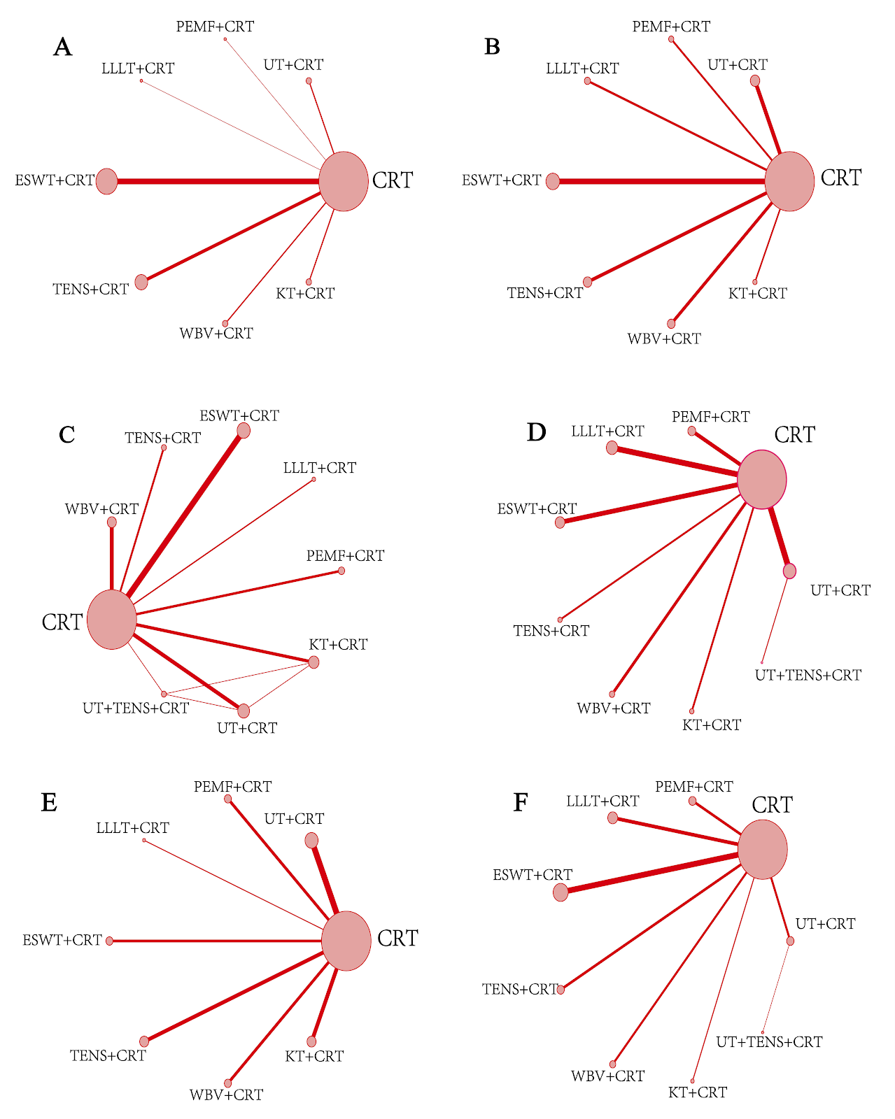
OBJECTIVE: The therapeutic modalities of physical factor interventions for knee osteoarthritis have been increasingly diversified; however, comprehensive comparative evaluations of their efficacy remain limited. This study aims to compare the efficacy and safety of various physical factor therapies for knee osteoarthritis through a network meta-analysis. METHODS: Randomized controlled trials on physical factor therapy for knee osteoarthritis were retrieved from PubMed, Web of Science, Cochrane Library, EMbase, CNKI, VIP, Wanfang, and CBM databases from inception to July 25, 2025. After literature screening and data extraction, the quality of included studies was assessed using the Cochrane risk-of-bias tool. Statistical analyses were performed using Stata 16.0 and RevMan 5.4.1. RESULTS: A total of 65 studies involving 3,418 patients (1,726 in treatment groups, 1,692 in control groups) were included, covering seven physical factor therapies. Network meta-analysis showed that for improving total effective rate, the top three interventions by surface under the cumulative ranking curve (SUCRA) were pulsed electromagnetic field + conventional rehabilitation, ultrasound + conventional rehabilitation, and transcutaneous electrical stimulation + conventional rehabilitation. For improving visual analogue scale (VAS) score, the top three were transcutaneous electrical stimulation + conventional rehabilitation, extracorporeal shock wave + conventional rehabilitation, and ultrasound + conventional rehabilitation. For improving WOMAC total score, the top three were ultrasound + conventional rehabilitation, pulsed electromagnetic field + conventional rehabilitation, and ultrasound + transcutaneous electrical stimulation + conventional rehabilitation. For reducing WOMAC stiffness score, the top three were transcutaneous electrical stimulation + conventional rehabilitation, ultrasound + conventional rehabilitation, and ultrasound + transcutaneous electrical stimulation + conventional rehabilitation. For improving SF-36 quality of life score, the top three were pulsed electromagnetic field + conventional rehabilitation, extracorporeal shock wave + conventional rehabilitation, and ultrasound + conventional rehabilitation. For improving Lysholm knee score, the top three were ultrasound + conventional rehabilitation, ultrasound + transcutaneous electrical stimulation + conventional rehabilitation, and extracorporeal shock wave + conventional rehabilitation. Regarding adverse events, no serious adverse events were reported; most studies reported only mild skin irritation or allergic reactions. CONCLUSION: Transcutaneous electrical stimulation combined with conventional rehabilitation showed superior advantages in improving VAS and WOMAC stiffness scores; ultrasound combined with conventional rehabilitation performed relatively better in improving Lysholm knee score and WOMAC total score; pulsed electromagnetic field combined with conventional rehabilitation had potential advantages in improving overall quality of life. Each physical factor has its unique advantages, but limited by the quality and quantity of included studies, these conclusions need to be verified by more high-quality, multi-center, large-sample randomized controlled trials.

Read Full Abstract10.12307/2026.21502