🧬 SinoBioData Academic Portal
🏛️ Indexed Academic JournalOriginal: 干细胞研究与转化

Stem Cell Research & Therapy

Premier Chinese Biomedical Journal indexed in SinoBioData: Stem Cell Research & Therapy (干细胞研究与转化).

Total Research Papers: 200
Access: 100% Free Open Access
Browse by Publication Year & VolumeReset All Filters ✕

Published Research PapersFiltered: Year 2025 • 16 • 1

Showing 13 of 200 peer-reviewed papers with full Graphical Abstracts.

Original ResearchVol. 16, Issue 1 • pp. 115DOI: 10.1186/s13287-025-04213-9

Purine metabolism in bone marrow microenvironment inhibits hematopoietic stem cell differentiation under microgravity

Authors: Xiru Liu, Hao Zhang, Jinxiao Yan, Penghui Ye, Yanran Wang, Nu Zhang, Zhenhao Tian, Bin Liu, Hui Yang

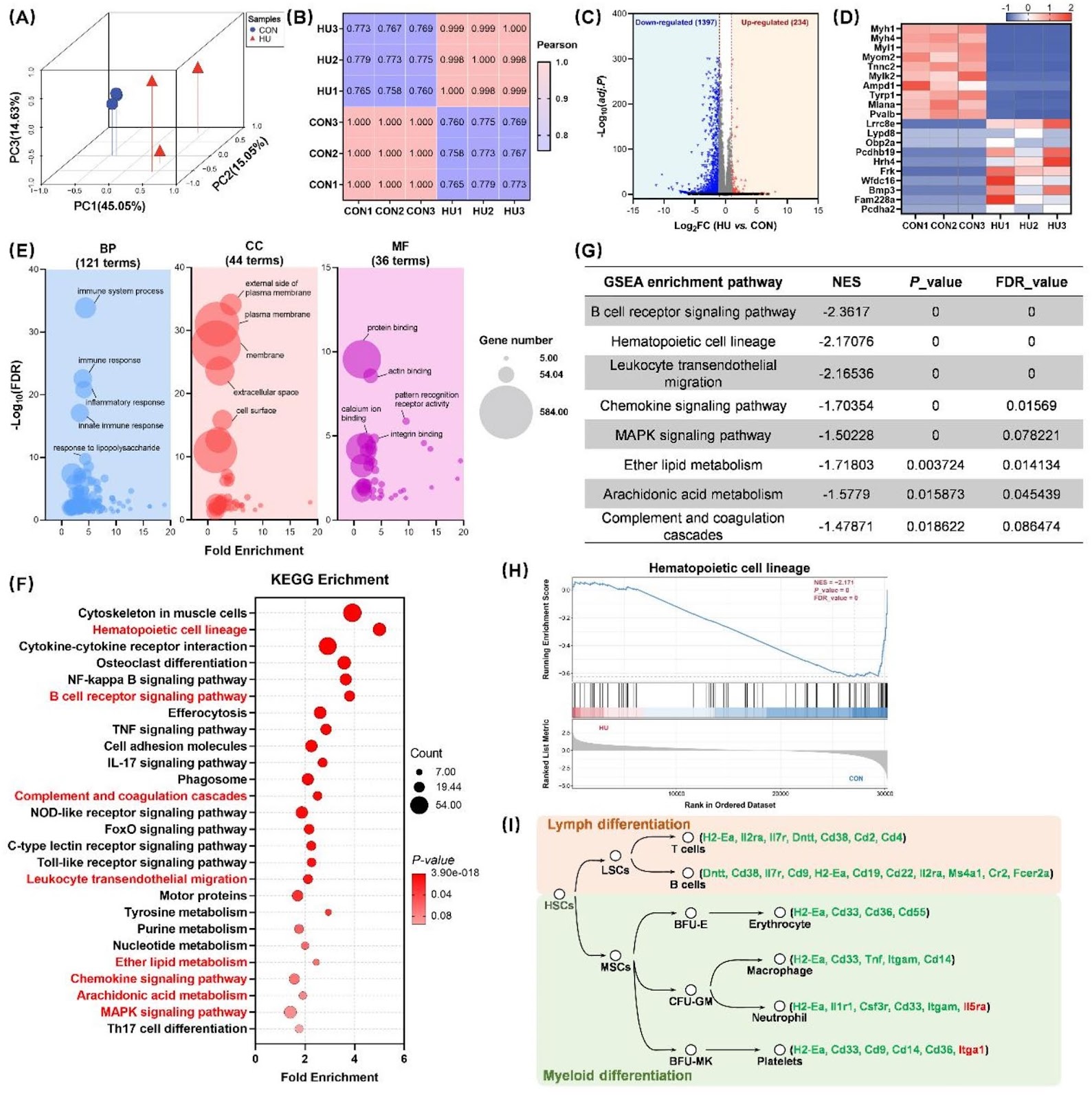
Background Spaceflight and microgravity environments have been shown to cause significant health impairments, including bone loss, immune dysfunction, and hematopoietic disorders. Hematopoietic stem cells (HSCs), as progenitors of the hematopoietic system, are critical for the continuous renewal and regulation of immune cells. Therefore, elucidating the regulatory mechanisms governing HSC fate and differentiation in microgravity environments is of paramount importance. Methods In this study, hindlimb unloading (HU) was employed in mice to simulate microgravity conditions. After 28 days of HU, cells were isolated for analysis. Flow cytometry and colony-forming assays were utilized to assess changes in HSC proliferation and differentiation. Additionally, transcriptomic and untargeted metabolomic sequencing were performed to elucidate alterations in the metabolic pathways of the bone marrow microenvironment and their molecular regulatory effects on HSCs fate. Results Our findings revealed that 28 days of HU impaired hematopoietic function, leading to multi-organ damage and hematological disorders. The simulated microgravity environment significantly increased the HSCs population in the bone marrow, particularly within the long-term and short-term subtypes, while severely compromising the differentiation capacity of hematopoietic stem/progenitor cells. Transcriptomic analysis of HSCs, combined with metabolomic profiling of bone marrow supernatants, identified 1,631 differentially expressed genes and 58 metabolites with altered abundance. Gene set enrichment analysis indicated that HU suppressed key pathways, including hematopoietic cell lineage and MAPK signaling. Furthermore, integrated analyses revealed that metabolites affected by HU, particularly hypoxanthine enriched in the purine metabolism pathway, were closely associated with hematopoietic cell lineage and MAPK signaling pathways. Molecular docking simulations and in vitro experiments confirmed that hypoxanthine interacts directly with core molecules within these pathways, influencing their expression. Conclusions These findings demonstrate that hypoxanthine in the bone marrow supernatant acts as a signaling mediator under microgravity, influencing HSCs fate by modulating hematopoietic cell lineage and MAPK signaling pathways. This study offers novel insights into the impact of microgravity on HSC fate and gene expression, underscoring the pivotal role of bone marrow microenvironmental metabolic changes in regulating key signaling pathways that determine hematopoietic destiny.
Purine metabolism in bone marrow microenvironment inhibits hematopoietic stem cell differentiation under microgravity
Graphical Abstract
Original ResearchVol. 16, Issue 1 • pp. 63DOI: 10.1186/s13287-025-04179-8

Induction of the p21/CDK6 pathway and alteration of the immune microenvironment by the stem cell marker CBX3 in melanoma

Authors: Wanxian Chen, Linsa Zhou, Jingjing Jiang, Jiasheng Chen, Deyi Geng, Yaokun Chen, Xiaosha Han, Qihu Xie, Genghong Guo, Xuefen Chen, Shijie Tang, Xiaoping Zhong

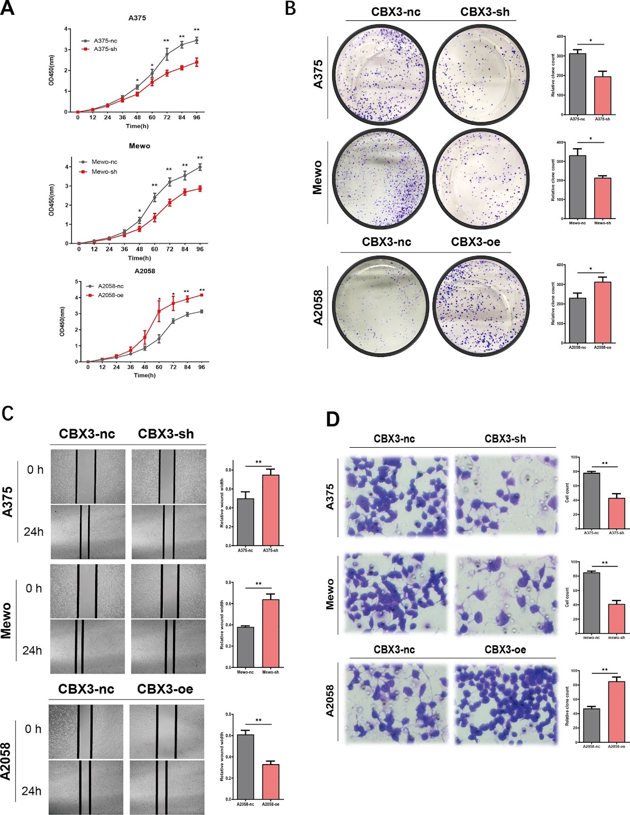
Background As one of the stem cell markers, chromobox protein homolog 3 (CBX3) participates in multiple signaling pathways that affect the progression of various tumors. However, the role of CBX3 in melanoma remains unclear, and the mechanisms by which CBX3 may regulate immunotherapy outcome remain largely unknown. Methods We used the Cancer Genome Atlas, Genotype-Tissue Expression portal, and Gene Expression Omnibus database to estimate CBX3 expression and its prognostic effect in melanoma. The role of CBX3 in proliferation and migration of melanoma cells were examined using the CCK8, cloning, wound healing, and transwell assays. The effect of CBX3 on melanoma tumorigenesis was assessed using an in vivo animal model. The role of CBX3 in cell cycle was examined using flow cytometry, and expression levels of cell cycle-related genes and proteins in cells with altered CBX3 levels were analyzed using qPCR and western blotting. The function of CBX3 in the immune microenvironment of melanoma was studied using single-cell RNA sequencing and public databases. Results We found that CBX3 was highly expressed in melanoma with poor prognosis. CBX3 promoted the proliferation and migration of melanoma cells in vivo and in vitro. Functional analysis revealed that CBX3 regulates cell cycle, as it accelerated the G1 to S transition, decreased p21 expression, and increased CDK6 expression. Finally, single-cell sequencing and immune-related assays showed that CBX3 is immunogenic and can change the immune microenvironment of melanoma. Conclusions We conclude that the stem cell marker, CBX3 activates the p21/CDK6 pathway and alters the immune microenvironment in melanoma.

Induction of the p21/CDK6 pathway and alteration of the immune microenvironment by the stem cell marker CBX3 in melanoma
Graphical Abstract
Original ResearchVol. 16, Issue 1 • pp. 609DOI: 10.1186/s13287-025-04755-y

Apremilast improves cardiomyocyte cohesion and arrhythmia in different models for arrhythmogenic cardiomyopathy

Authors: Konstanze Stangner, Orsela Dervishi, Janina Kuhnert, Carl Wendt, Soumyata Pathak, Maria Shoykhet, Silvana Olivares-Florez, Sina Moztarzadeh, Jens Opsteen, Ni Luh Cathrin Suniasih Wohlfarth, Ruth Biller, Elisabeth Graf, Dominik S. Westphal, Tatjana Williams, Brenda Gerull, Tomo Šarić, Sunil Yeruva, Jens Waschke

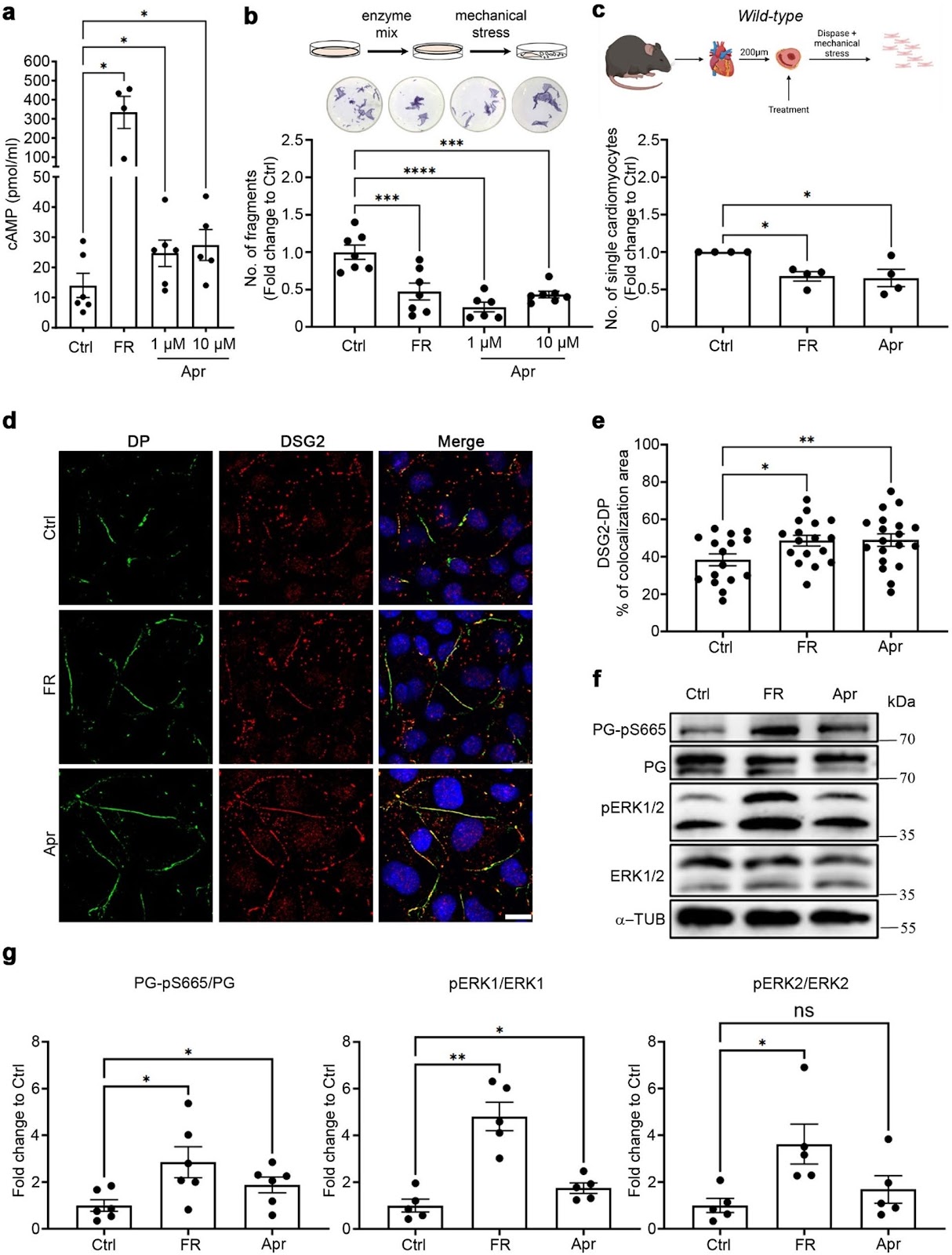
Background Arrhythmogenic cardiomyopathy (ACM) is a genetically inherited desmosome heart disease leading to life-threatening arrhythmias and sudden cardiac death. Currently, ACM treatment paradigms are merely symptom targeting. Recently, apremilast was shown to stabilize keratinocyte adhesion in the desmosomal disease pemphigus vulgaris. Therefore, this study investigated whether apremilast can be a therapeutic option for ACM. Methods Human induced pluripotent stem cells from a healthy control (hiPSC) and an ACM index patient (ACM-hiPSC) carrying a heterozygous desmoplakin (DSP) gene mutation (c.2854G > T, p.Glu952Ter), confirmed by whole exome sequencing (WES), were established. Cyclic-AMP ELISA, dissociation assay, immunostaining, and Western blotting analyses were performed in human iPSC-derived cardiomyocytes (hiPSC-CMs), murine HL-1 cardiomyocytes, and cardiac slices derived from wild-type (WT) mice, plakoglobin (PG, Jup) knockout (Jup−/−) (murine ACM model) or PG Serine 665 phosphodeficient (JUP-S665A) mice. Microelectrode array (MEA) analyses in ventricular cardiac slices and Langendorff heart perfusion were performed to analyze heart rate variability and arrhythmia. Results ACM-hiPSC derived cardiomyocytes (ACM-hiPSC-CMs) revealed a significant loss of cohesion, which was rescued by apremilast. Further, treatment with apremilast strengthened basal cardiomyocyte cohesion in HL-1 cells and WT murine cardiac slices, paralleled by phosphorylation of PG at Serine 665 in human and murine models. In HL-1 cells, apremilast in addition activated ERK1/2, inhibition of which abolished apremilast-enhanced cardiomyocyte cohesion. Further, dissociation assays in slice cultures from JUP-S665A and Jup−/− mice revealed that PG is crucial for apremilast's effects. Additionally, apremilast reduced arrhythmic events in ventricular cardiac slices and Langendorff-perfused hearts. Conclusion Apremilast improves cardiomyocyte cohesion and reduces arrhythmia in different models of ACM, suggesting a novel therapeutic strategy for this disease.

Apremilast improves cardiomyocyte cohesion and arrhythmia in different models for arrhythmogenic cardiomyopathy
Graphical Abstract
Original ResearchVol. 16, Issue 1 • pp. 84DOI: 10.1186/s13287-025-04229-1

OCT4 translationally promotes AKT signaling as an RNA-binding protein in stressed pluripotent stem cells

Authors: Wenjie Chen, Xinyu Chen, Cheng Chen, Shiqi She, Xia Li, Lina Shan, Xiaobing Zhang, Songsong Dan, Yisha Wang, Yan-Wen Zhou, Qingyi Cao, Wenxin Wang, Jianwen Hu, Yaxun Wei, Yaqiang Xue, Yi Zhang, Songying Zhang, Ying-Jie Wang, Bo Kang

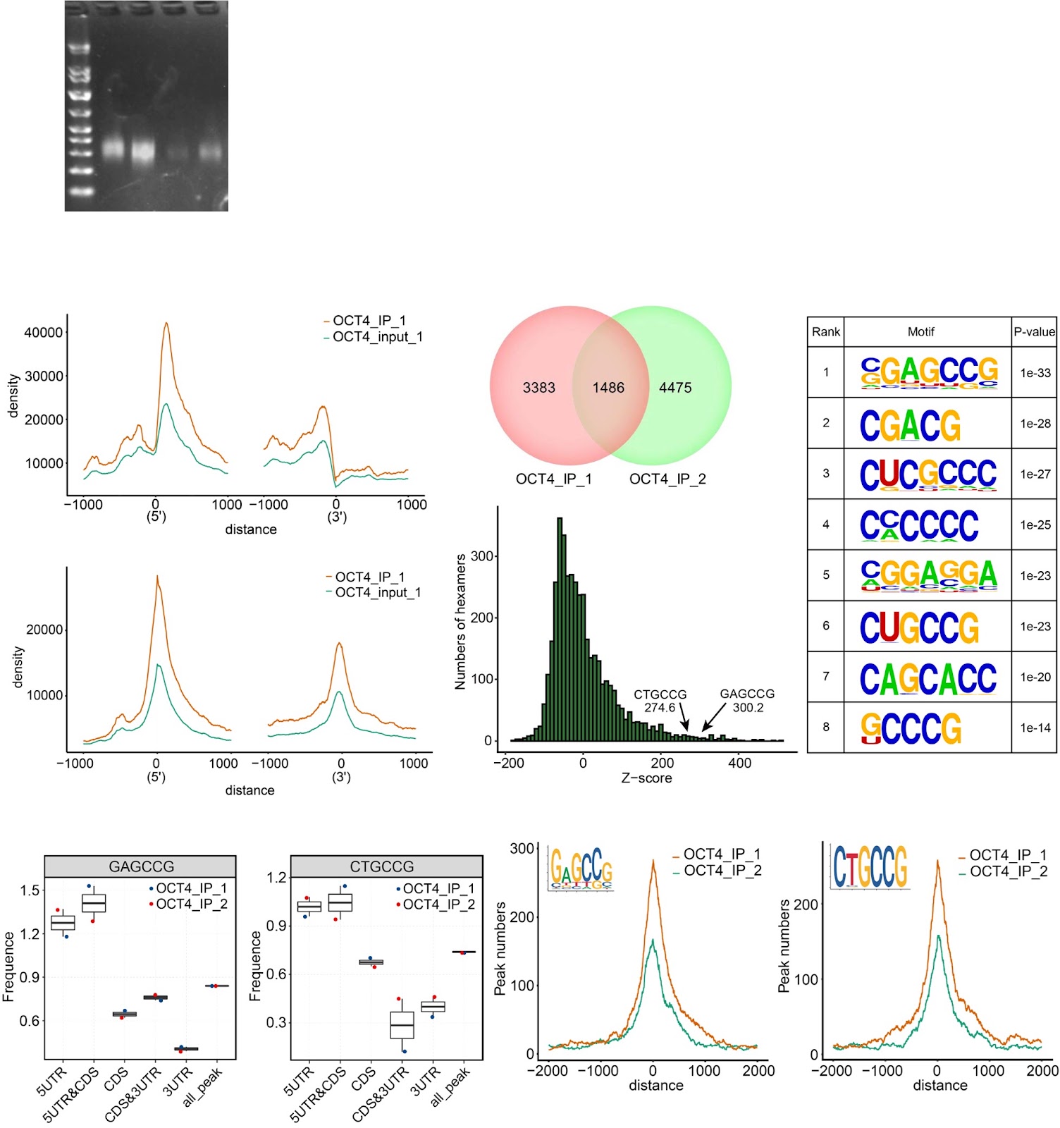
Background Despite numerous studies addressing the molecular mechanisms by which pluripotent stem cells (PSCs) maintain self-renewal and pluripotency under normal culture conditions, the fundamental question of how PSCs manage to survive stressful conditions remains largely unresolved. Post-transcriptional/translational regulation emerges to be vital for PSCs, but how PSCs coordinate and balance their survival and differentiation at translational level under extrinsic and intrinsic stress conditions is unclear. Methods The high-throughput sequencing of cross-linking immunoprecipitation cDNA library (HITS-CLIP) was employed to decipher the genome-wide OCT4-RNA interactome in human PSCs, a combined RNC-seq/RNA-seq analysis to assess the role of OCT4 in translational regulation of hypoxic PSCs, and an OCT4-protein interactome to search for OCT4 binding partners that regulate cap-independent translation initiation. By taking the Heterozygous Knocking In N-terminal Tags (HKINT) approach that specifically disrupts the 5'-UTR secondary structure and tagging its protein product of the mRNA from one allele while leaving that from the other allele intact, we examined the effect of disrupting the OCT4/5'-UTR interaction on translation of AKT1 mRNA. Results We revealed OCT4 as a bona fide RNA-binding protein (RBP) in human PSCs that bound to the 5'-UTR, 3'-UTR and CDS regions of mRNAs. Multiple known proteins participating in IRES-mediated translation initiation were detected in the OCT4-protein interactome, and a combined RNC-seq/RNA-seq analysis further confirmed a crucial role of OCT4 in translational regulation of PSCs in response to hypoxic stress. Remarkably, OCT4 bound to the GC-rich elements in the 5'-UTR of AKT1 and multiple PI3K/AKT-pathway-gene mRNAs, and promoted their translation initiation via IRES-mediated pathways under stress conditions. Specifically disrupting the AKT1 mRNA 5'-UTR structure and the OCT4/5'-UTR interaction by the HKINT approach significantly reduced the translation level of AKT1 that led to a higher susceptibility of PSCs to oxidative stress-induced apoptotic death and prioritized differentiation toward ectoderm and endoderm. Conclusions Our results reveal OCT4 as an anti-stress RBP for translational regulation that critically coordinates the survival and differentiation of PSCs in response to various stressors.

OCT4 translationally promotes AKT signaling as an RNA-binding protein in stressed pluripotent stem cells
Graphical Abstract
Original ResearchVol. 16, Issue 1 • pp. 81DOI: 10.1186/s13287-025-04202-y

Human spindle-shaped urine-derived stem cell exosomes alleviate severe fatty liver ischemia–reperfusion injury by inhibiting ferroptosis via GPX4

Authors: Shangheng Shi, Cunle Zhu, Shangxuan Shi, Xinqiang Li, Imran Muhammad, Qingguo Xu, Xinwei Li, Ziyin Zhao, Huan Liu, Guangming Fu, Meiying Song, Xijian Huang, Feng Wang, Jinzhen Cai

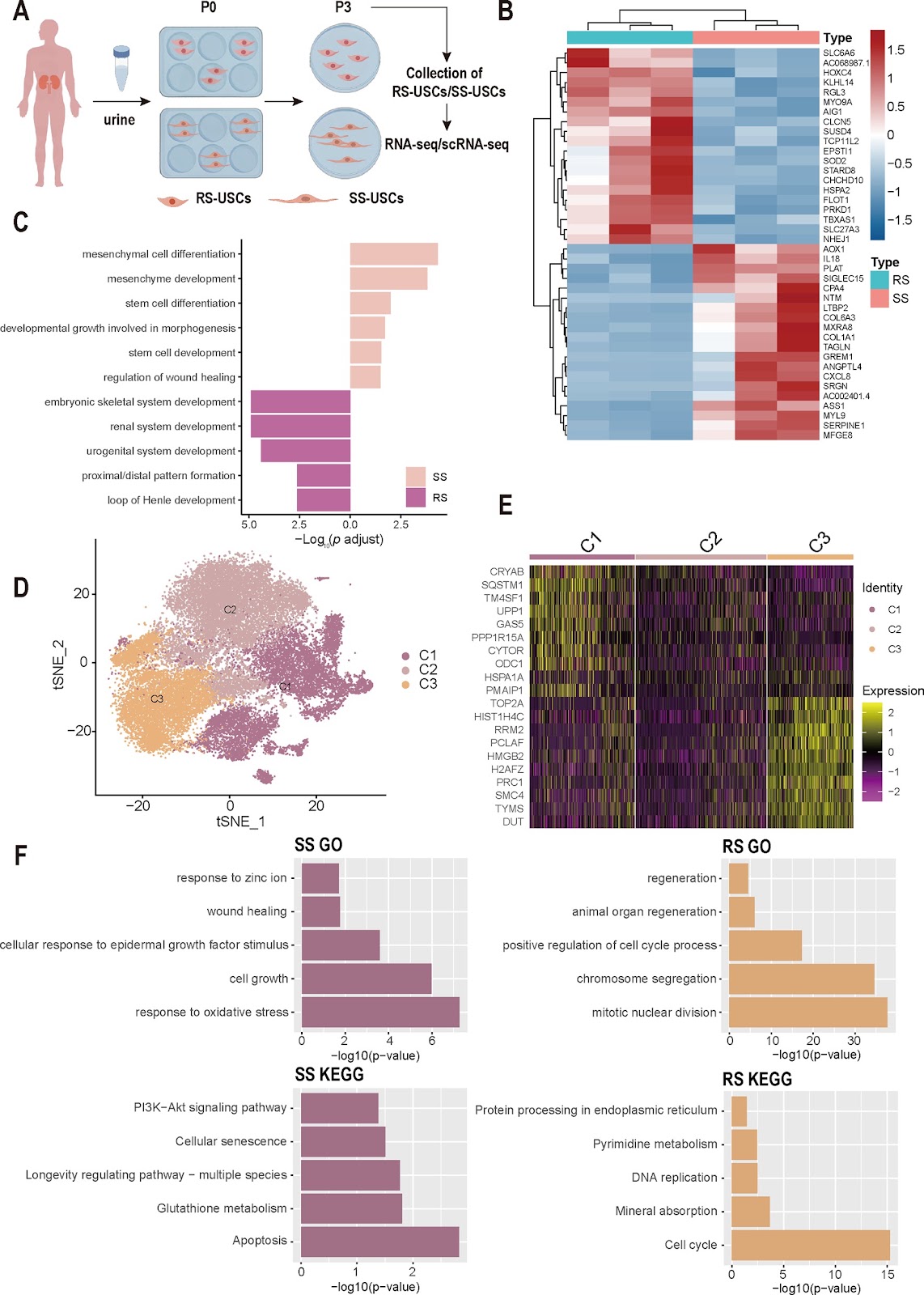
Background Severe hepatic steatosis can exacerbate Ischemia–reperfusion injury (IRI), potentially leading to early graft dysfunction and primary non-function. In this study, we investigated the heterogeneity of different subpopulations of Urine-derived stem cells (USCs) to explore the most suitable cell subtype for treating severe steatotic liver IRI. Methods This study utilized scRNA-seq and Bulk RNA-seq to investigate the transcriptional heterogeneity between Spindle-shaped USCs (SS-USCs) and Rice-shaped USCs (RS-USCs). Additionally, rat fatty Liver transplantation (LT) model, mouse fatty liver IRI model, and Steatotic Hepatocyte Hypoxia-Reoxygenation (SHP-HR) model were constructed. Extracellular vesicles derived from SS-USCs and RS-USCs were isolated and subjected to mass spectrometry analysis. The therapeutic effects of Spindle-shaped USCs Exosomes (SS-USCs-Exo) and Rice-shaped USCs Exosomes (RS-USCs-Exo) were explored, elucidating their potential mechanisms in inhibiting ferroptosis and alleviating IRI. Results Multiple omics analyses confirmed that SS-USCs possess strong tissue repair and antioxidant capabilities, while RS-USCs have the potential to differentiate towards specific directions such as the kidney, nervous system, and skeletal system, particularly showing great application potential in renal system reconstruction. Further experiments demonstrated in vivo and in vitro models confirming that SS-USCs and SS-USCs-Exo significantly inhibit ferroptosis and alleviate severe fatty liver IRI, whereas the effects of RS-USCs/RS-USCs-Exo are less pronounced. Analysis comparing the proteomic differences between SS-USCs-Exo and RS-USCs-Exo revealed that SS-USCs-Exo primarily inhibit ferroptosis and improve cellular viability by secreting exosomes containing Glutathione Peroxidase 4 (GPX4) protein. This highlights the most suitable cell subtype for treating severe fatty liver IRI. Conclusions SS-USCs possess strong tissue repair and antioxidant capabilities, primarily alleviating ferroptosis in the donor liver of fatty liver through the presence of GPX4 protein in their exosomes. This highlights SS-USCs as the most appropriate cell subtype for treating severe fatty liver IRI.

Human spindle-shaped urine-derived stem cell exosomes alleviate severe fatty liver ischemia–reperfusion injury by inhibiting ferroptosis via GPX4
Graphical Abstract
Original ResearchVol. 16, Issue 1 • pp. 493DOI: 10.1186/s13287-025-04577-y

Allogeneic bone marrow-derived mesenchymal stem cells in the aging kidney: secondary results of a Parkinson's disease clinical trial

Authors: Juan D. Martinez-Lemus, Donald A. Molony, Jessika Suescun, Emily Tharp, Tia S. Thomas, Charles Green, Chiamaka Onuigbo, Robert Ritter III, Mya C. Schiess

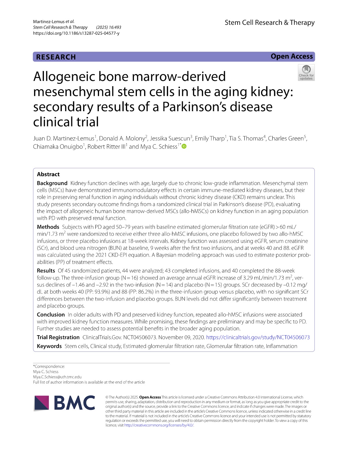
Background Kidney function declines with age, largely due to chronic low-grade inflammation. Mesenchymal stem cells (MSCs) have demonstrated immunomodulatory effects in certain immune-mediated kidney diseases, but their role in preserving renal function in aging individuals without chronic kidney disease (CKD) remains unclear. This study presents secondary outcome findings from a randomized clinical trial in Parkinson's disease (PD), evaluating the impact of allogeneic human bone marrow-derived MSCs (allo-hMSCs) on kidney function in an aging population with PD with preserved renal function. Methods Subjects with PD aged 50–79 years with baseline estimated glomerular filtration rate (eGFR) > 60 mL/min/1.73 m2 were randomized to receive either three allo-hMSC infusions, one placebo followed by two allo-hMSC infusions, or three placebo infusions at 18-week intervals. Kidney function was assessed using eGFR, serum creatinine (SCr), and blood urea nitrogen (BUN) at baseline, 9 weeks after the first two infusions, and at weeks 40 and 88. eGFR was calculated using the 2021 CKD-EPI equation. A Bayesian modeling approach was used to estimate posterior probabilities (PP) of treatment effects. Results Of 45 randomized patients, 44 were analyzed; 43 completed infusions, and 40 completed the 88-week follow-up. The three-infusion group (N=16) showed an average annual eGFR increase of 3.29 mL/min/1.73 m2, versus declines of –1.46 and –2.92 in the two-infusion (N=14) and placebo (N=15) groups. SCr decreased by –0.12 mg/dL at both weeks 40 (PP: 93.9%) and 88 (PP: 86.2%) in the three-infusion group versus placebo, with no significant SCr differences between the two-infusion and placebo groups. BUN levels did not differ significantly between treatment and placebo groups. Conclusion In older adults with PD and preserved kidney function, repeated allo-hMSC infusions were associated with improved kidney function measures. While promising, these findings are preliminary and may be specific to PD. Further studies are needed to assess potential benefits in the broader aging population. Trial Registration ClinicalTrials.Gov. NCT04506073. November 09, 2020. https://clinicaltrials.gov/study/NCT04506073

Allogeneic bone marrow-derived mesenchymal stem cells in the aging kidney: secondary results of a Parkinson's disease clinical trial
Graphical Abstract
Original ResearchVol. 16, Issue 1 • pp. 14DOI: 10.1186/s13287-025-04131-w

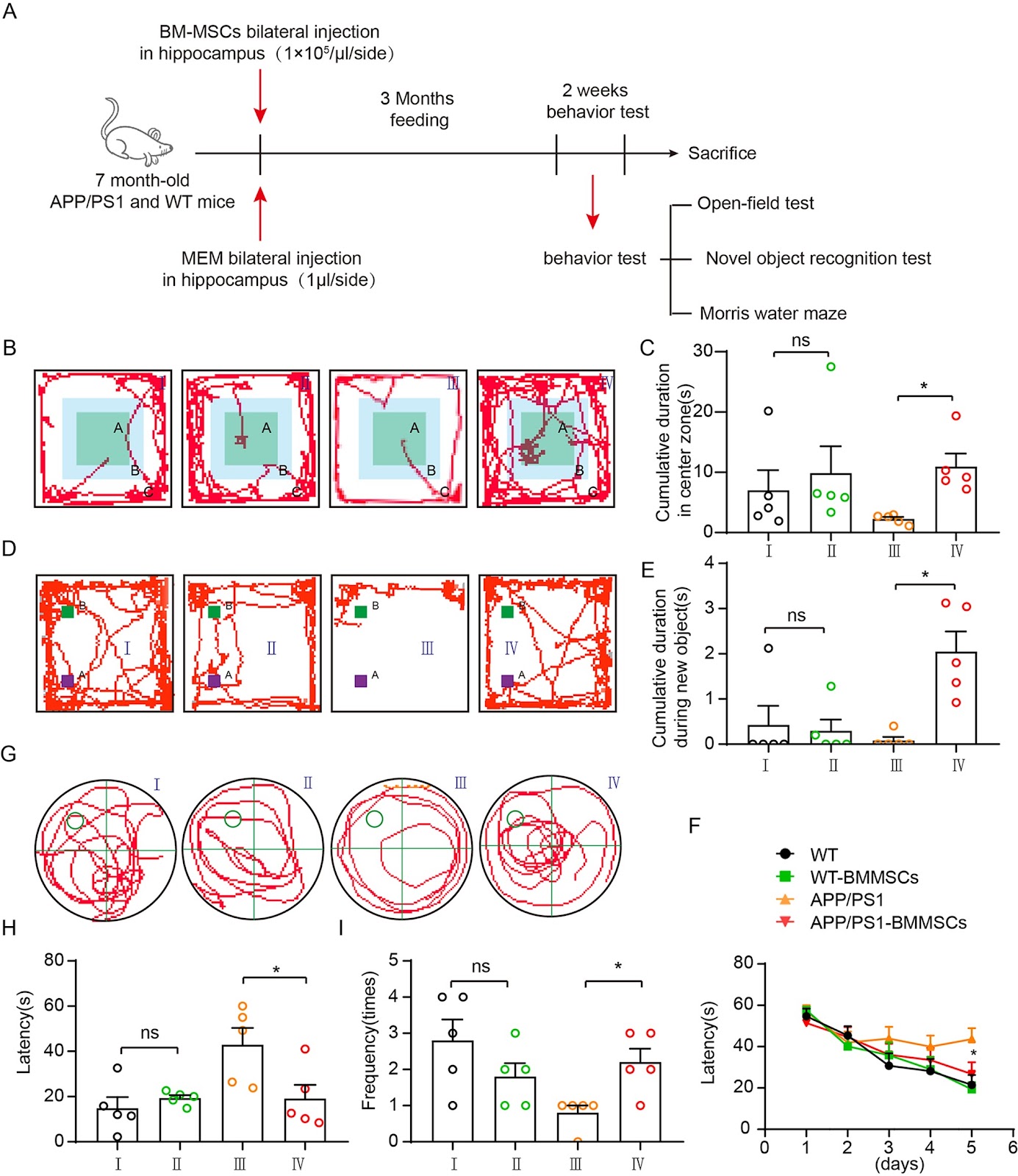
Bone marrow mesenchymal stem cells derived cytokines associated with AKT/IAPs signaling ameliorate Alzheimer’s disease development

Authors: Yalan Lu, Yanfeng Xu, Li Zhou, Siyuan Wang, Yunlin Han, Kewei Wang, Chuan Qin

Background Alzheimer’s disease (AD) is a progressive neurodegenerative condition affecting around 50 million people worldwide. Bone marrow-derived mesenchymal stem cells (BMMSCs) have emerged as a promising source for cellular therapy due to their ability to differentiate into multiple cell types and their paracrine effects. However, the direct injection of BMMSCs can lead to potential unpredictable impairments, prompting a renewed interest in their paracrine effects for AD treatment. The specific mechanism and central role of cytokines in this process have not been fully elucidated. Methods Mouse BMMSCs were isolated, validated, and then transplanted intracerebrally into APP/PS1 female mice. The behavioral tests, including open-field test, novel object recognition test, and Morris water maze were performed, followed by β-amyloidosis plaque and neuron apoptosis analyses. Then the tissue RNA sequencing and mBMMSC cytokine analysis were performed. A cytokine antibody array for BMMSCs and the brain slice models were performed with AD model tissues were used to elucidate the molecular mechanisms. Finally, APP/PS1 mice were administrated with cytokine mixture for cognitive recovery. Results Our results demonstrated that BMMSCs significantly improved cognitive function, reduced beta-amyloid plaque deposition, and decreased apoptotic neurons through the activation of the AKT signaling pathway. Using a cytokine antibody array, we identified three highly expressed AKT pathway regulated neuroprotective factors in BMMSCs: IGF1, VEGF, and Periostin2. These cytokines were found to upregulate inhibitors of apoptosis family proteins (IAPs) and suppress Caspase-3 activity in brain slices induced with beta amyloidosis (Aβ), okadaic acid (OA), and lipopolysaccharide (LPS). When injection of this cytokine mixture to APP/PS1 mice also resulted in a mitigation of cognitive impairment. Conclusions These findings suggest that the secretory factors IGF1, VEGF, and Periostin2 derived from BMMSCs play a crucial role in neuroprotection by modulating the AKT/IAPs pathway to restore neuronal function. These cytokine sets could be a potential therapeutic strategy for AD and lay the groundwork for promising clinical applications.

Bone marrow mesenchymal stem cells derived cytokines associated with AKT/IAPs signaling ameliorate Alzheimer’s disease development
Graphical Abstract
Original ResearchVol. 16, Issue 1 • pp. 22DOI: 10.1186/s13287-025-04146-3

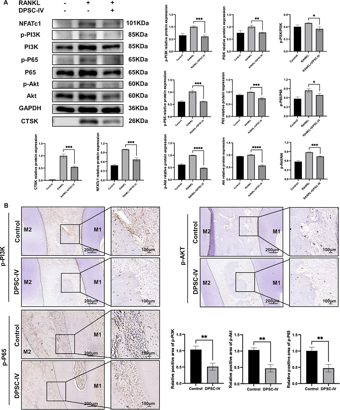
Dental pulp stem cell-derived intracellular vesicles prevent orthodontic relapse by inhibiting PI3K/Akt/NF-κB-mediated osteoclast activity

Authors: Boyuan Peng, Ziwei Li, Yong Cheng, Henghua Jiang, Qingsong Ye, Guangli Han

Background Orthodontic relapse, the undesired deviation of teeth from their corrected positions, remains a significant challenge in clinical orthodontics. Incomplete periodontal bone remodeling has been identified as a key factor in this process. Despite decades of research, currently there are no effective strategies to prevent relapse. Methods We isolated and identified dental pulp stem cell-derived intracellular vesicles (DPSC-IV) from human dental pulp tissue. To investigate its effect, DPSC-IV was added to osteoblast or osteoclast differentiation medium. During the orthodontic retention period, DPSC-IV was administrated to rats by subgingival injection. Relapse distance and relapse rate were calculated to evaluate DPSC-IV's ability to prevent relapse. Additionally, Western blot analysis were used to examine DPSC-IV's inhibitory effect on osteoclast differentiation. Results DPSC-IV significantly promoted osteoblast differentiation and inhibited osteoclast differentiation. Application of DPSC-IV during retention resulted in a significant reduction in both relapse distance and relapse rate, with improved periodontal structure and decreased osteoclast activity. This effect was mediated by the PI3K/Akt/NF-κB signaling pathway and could be reversed by the PI3K activator insulin-like growth factor-1 (IGF-1). Conclusion This study highlights the potential of DPSC-IV as a novel preventive approach against orthodontic relapse, offering a novel strategy for maintaining long-term orthodontic stability.

Dental pulp stem cell-derived intracellular vesicles prevent orthodontic relapse by inhibiting PI3K/Akt/NF-κB-mediated osteoclast activity
Graphical Abstract
Original ResearchVol. 16, Issue 1 • pp. 417DOI: 10.1186/s13287-025-04547-4

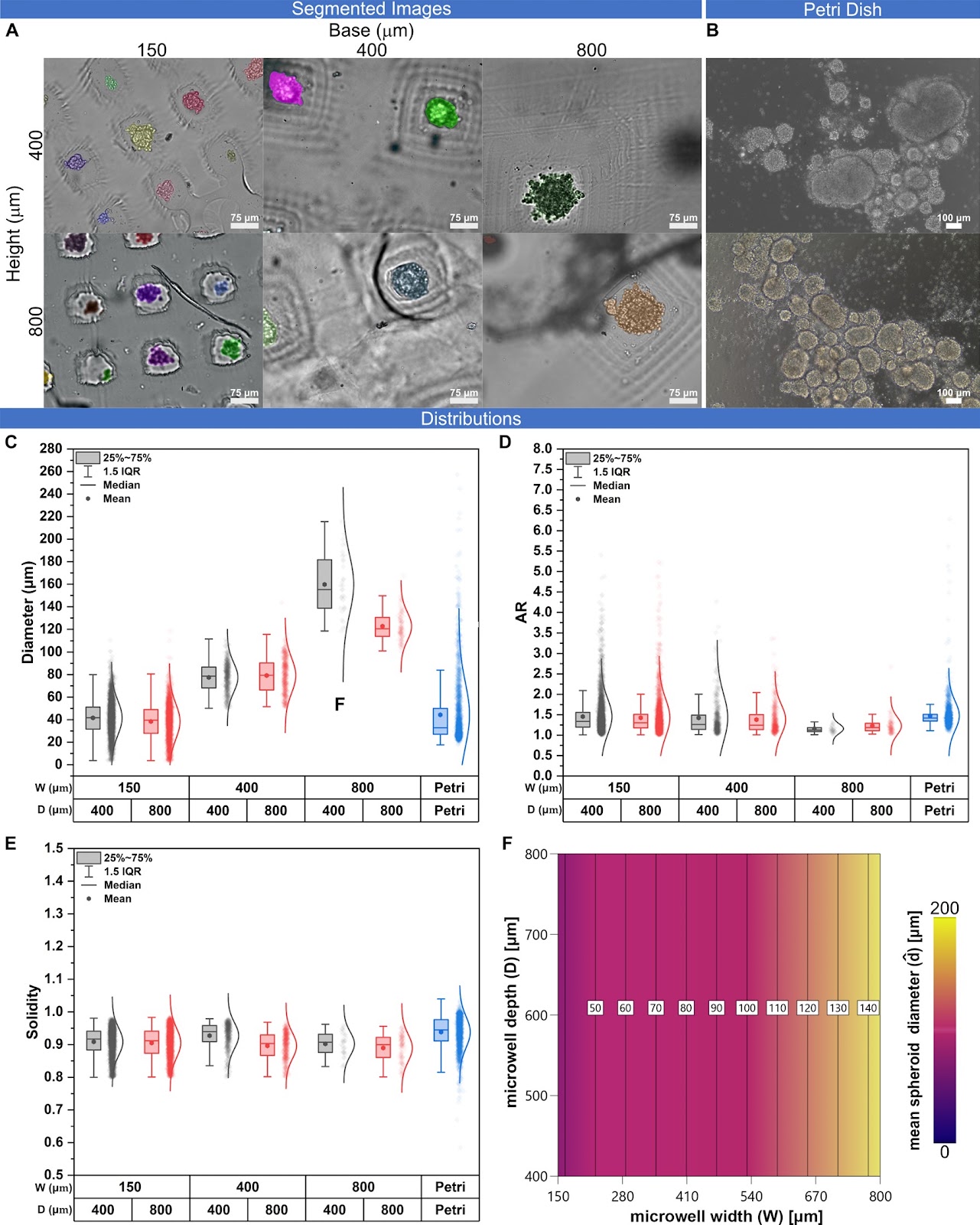
Geometrical constraints dictate assembly and phenotype of human iPSC-derived motoneuronal spheroids

Authors: Eleonora Mello, Stefano Sorrentino, Alessio Bucciarelli, Ermanno Cordelli, Elisa De Luca, Haakon Nygaard, Stefan Wendt, Alberto Rainer, Giuseppe Gigli, Lorenzo Moroni, Alessandro Polini, Pamela Mozetic

Background Neuronal spheroids represent an easy and versatile solution to model neuronal tissue in vitro. Conventional approaches to generate spheroids lack accurate size control, scalability, and customizability. This is even more exacerbated in case of pluripotent stem cell (PSC) derived spheroids, which remain challenging to standardize. Microwell devices address these limitations, providing an optimal balance between accessibility and scalability. With the aim of optimizing culture conditions, we parametrically investigated the role of microwell geometry on the formation and maturation of iPSC-derived motor neuron precursor (MNP) spheroids. Methods We developed a customizable mold device using Digital Light Processing (DLP) 3D printing to fabricate agarose microwell arrays with distinct aspect ratios for culturing hiPSC-derived MNP spheroids with high reproducibility. We generated nine different pyramidal microwell array geometries for culturing size-controlled spheroids in the 40–140 μm diameter range. We then evaluated the differential expression of genes related to cell proliferation and motor-neuron differentiation as function of microwell geometry and spheroid size. Results Our results indicate that spheroid size is significantly influenced by the microwell geometry, reliably due to cell partitioning at the seeding stage. Expression of proliferation and differentiation markers, such as motor neuron and pancreas homeobox 1 (MNX1) and Islet-1 (ISL1) transcription factors, is also dependent on microwell geometry and spheroid morphological descriptors. Conclusion Our approach enables the scalable production of size-controlled MNP spheroids and underscores the effect of geometrical confinement on regulating motor neuron differentiation.

Geometrical constraints dictate assembly and phenotype of human iPSC-derived motoneuronal spheroids
Graphical Abstract
Original ResearchVol. 16, Issue 1 • pp. 50DOI: 10.1186/s13287-025-04165-0

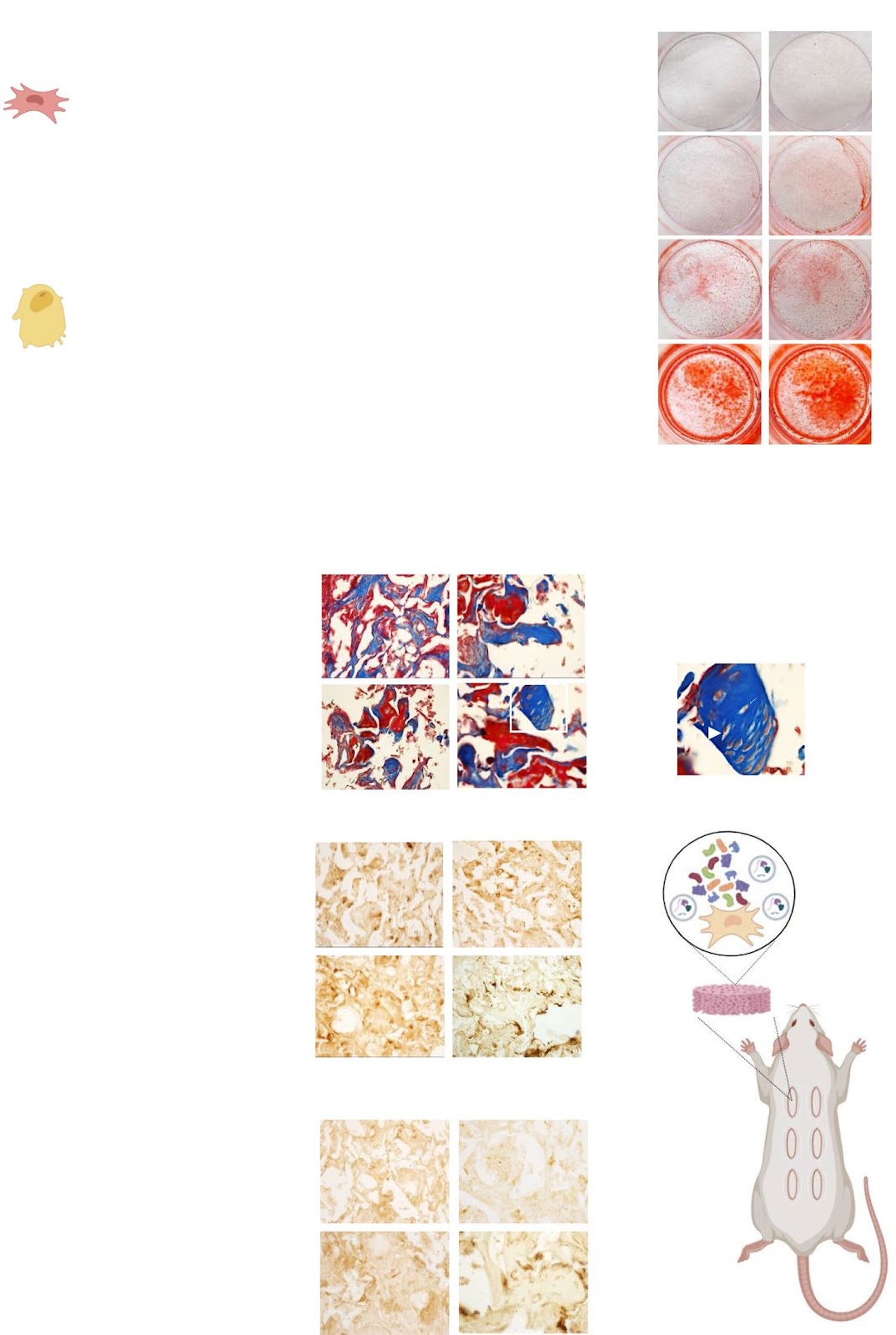
Paracrine activity of Smurf1-silenced mesenchymal stem cells enhances bone regeneration and reduces bone loss in postmenopausal osteoporosis

Authors: Alberto González-González, Itziar Álvarez-Iglesias, Daniel García-Sánchez, Monica Dotta, Ricardo Reyes, Ana Alfonso-Fernández, Alfonso Bolado-Carrancio, Patricia Díaz-Rodríguez, María Isabel Pérez-Núñez, José Carlos Rodríguez-Rey, Jesús Delgado-Calle, Flor M. Pérez-Campo

Background: Osteoporosis (OP), characterized by reduced bone mass and mineral density, is a global metabolic disorder that severely impacts the quality of life in affected individuals. Although current pharmacological treatments are effective, their long-term use is often associated with adverse effects, highlighting the need for safer, more sustainable therapeutic strategies. This study investigates the pro-osteogenic and anti-resorptive potential of the secretome from Smurf1-silenced mesenchymal stem cells (MSCs) as a promising cell-free therapy for bone regeneration. Methods: Conditioned media (CM) from Smurf1-silenced rat (rCM-Smur1) and human MSCs (hCM-Smurf1) was collected and analyzed. Pro-osteogenic potential was assessed by measuring in vitro mineralization in human and rat MSCs cultures. In vivo, studies were conducted using a rat ectopic bone formation model and a post-menopausal osteoporotic mouse model. Additionally, primary human osteoporotic MSCs were preconditioned with hCM-Smurf1, and their osteogenic capacity was compared to that induced by BMP2 treatment. Ex vivo, human bone explants were treated with hCM-Smurf1 to assess anti-resorptive effects. Proteomic analysis of the soluble and vesicular CM fractions identified key proteins involved in bone regeneration. Results: CM from Smurf1-silenced MSCs significantly enhanced mineralization in vitro and bone formation in vivo. Preconditioning human osteoporotic MSCs with hCM-Smurf1 significantly increases in vitro mineralization, with levels comparable to those achieved with BMP2 treatment. Additionally, in ex vivo human bone cultures, treatment with hCM-Smurf1 significantly reduced RANKL expression without affecting OPG levels, indicating an anti-resorptive effect. In vivo, CM from Smurf1-silenced MSCs significantly increased bone formation in a rat ectopic model, and its local administration reduced trabecular bone loss by 50% in a post-menopausal osteoporotic mouse model after a single administration within just four weeks. Proteomic analysis revealed both soluble and vesicular fractions of hCM-Smurf1 were enriched with proteins essential for ossification and extracellular matrix organization, enhancing osteogenic differentiation. Conclusions: The Smurf1-silenced MSCs’ secretome shows potent osteogenic and anti-resorptive effects, significantly enhancing bone formation and reducing bone loss. This study provides compelling evidence for the therapeutic potential of Smurf1-silenced MSC-derived secretome as a non-toxic and targeted treatment for osteoporosis. These findings warrant further in vivo studies and clinical trials to validate its therapeutic efficacy and safety.

Paracrine activity of Smurf1-silenced mesenchymal stem cells enhances bone regeneration and reduces bone loss in postmenopausal osteoporosis
Graphical Abstract
Original ResearchVol. 16, Issue 1 • pp. 26DOI: 10.1186/s13287-025-04145-4

Migrasomes derived from human umbilical cord mesenchymal stem cells: a new therapeutic agent for ovalbumin-induced asthma in mice

Authors: Weifeng Gu, Tingting Zheng, Wen Li, Xinkai Luo, Xiaowei Xu, Ying Wang, Chaoming Mao, Yongbin Ma, Liyang Dong

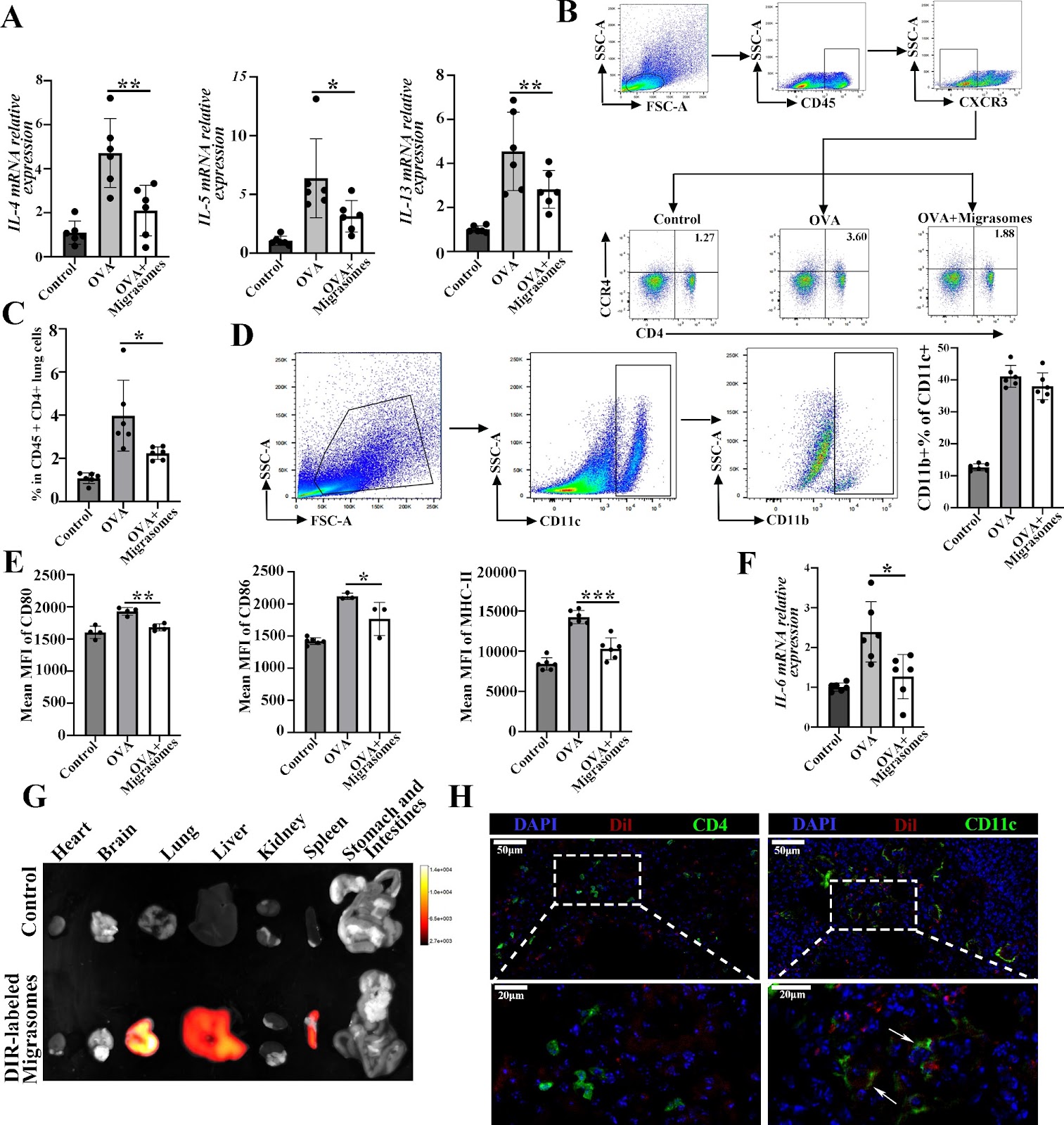
Background  Asthma is a prevalent respiratory disease, and its management remains largely unsatisfactory. Mesenchymal stem cells (MSCs) have been demonstrated to be efficacious in reducing airway inflammation in experimental allergic diseases, representing a potential alternative treatment for asthma. Migrasomes are recently identified extracellular vesicles (EVs) generated in migrating cells and facilitate intercellular communication. The objective of this study was to investigate the therapeutic effects of migrasomes obtained from MSC in a model of asthma. Methods  Migrasomes produced by human umbilical cord MSCs (hUCMSCs) were isolated by sequential centrifugation. Characterization of hUCMSC-derived migrasomes were carried out by transmission electron microscopy and western blot analysis. The therapeutic effects of migrasomes on airway inflammation in ovalbumin (OVA)-induced asthmatic mice were evaluated by hematoxylin-eosin (HE) and periodic-acid schiff (PAS) staining, and their mechanism were further testified by immunofluorescent staining, real-time PCR and flow cytometry. Results  Here, we showed that inhibition of migrasomes’ production dramatically impaired the anti-inflammatory effects of hUCMSCs in OVA animals, as evidenced by a notable increase in both the infiltration of inflammatory cells and the number of epithelial goblet cells. We successfully isolated hUCMSC-migrasomes, which were morphologically intact and positive for the specific migrasomes markers. The administration of hUCMSC-migrasomes was observed to significantly ameliorate the symptoms of airway inflammation and mucus production in asthmatic mice. Additionally, the expression of Th2 cytokines (IL-4, IL-5 and IL-13) were found to be reduced, while the activation of dendritic cells (DCs) was inhibited. HUCMSC-migrasomes could possibly be delivered to lung region after injection, and were able to be taken in by DCs both in vivo and in vitro. Notably, in vitro, migraosmes decreased the capacity of

Migrasomes derived from human umbilical cord mesenchymal stem cells: a new therapeutic agent for ovalbumin-induced asthma in mice
Graphical Abstract
Original ResearchVol. 16, Issue 1 • pp. 9DOI: 10.1186/s13287-024-04090-8

Combination of rapamycin and adipose-derived mesenchymal stromal cells enhances therapeutic potential for osteoarthritis

Authors: Damien Veret, Gautier Tejedor, Esther Perez, Alison Chomette, Maylis Farno, Rosanna Ferreira-Lopez, Louis Dagneaux, Yves-Marie Pers, Christian Jorgsensen, Claire Gondeau, Jean-Marc Brondello

Background The regenerative potential of mesenchymal stromal/stem cells (MSCs) has been extensively studied in clinical trials in the past decade. However, despite the promising regenerative properties documented in preclinical studies, for instance in osteoarthritis (OA), the therapeutic translation of these results in patients has not been fully conclusive. One factor contributing to this therapeutic barrier could be the presence of senescent cells in OA joints. Methods This study evaluated a novel approach to OA treatment by combining adipose tissue-derived MSCs (AD-MSCs) with rapamycin, a clinically approved immunosuppressive drug with anti-senescence properties. First, rapamycin effects on senescence and fibrosis markers were investigated in freshly isolated OA chondrocytes by immunostaining. Next, the in vitro differentiation capacities of AD-MSCs, their regulatory immune functions on activated immune cells and their regenerative effects on OA chondrocyte signature were assessed in the presence of rapamycin. Results In OA chondrocytes, rapamycin reduced the senescence marker p15INK4B and the fibrosis marker COL1A1 without affecting the expression of the master chondrogenic markers SOX9 and COL2. Rapamycin also enhanced AD-MSC differentiation into chondrocytes and reduced their differentiation into adipocytes. In addition, rapamycin improved AD-MSC immunoregulatory functions by promoting the expression of immunosuppressive factors, such as IDO1, PTGS2 and also CD274 (encoding PD-L1). Finally, RNA sequencing analysis showed that in the presence of rapamycin, AD-MSCs displayed improved chondroprotective regenerative effects on co-cultured OA chondrocytes. Conclusions Our findings suggest that the rapamycin and AD-MSC combination enhances the therapeutic efficacy of these cells in senescence-driven degenerative diseases such as OA, notably by improving their anti-fibrotic and anti-inflammatory properties.

Combination of rapamycin and adipose-derived mesenchymal stromal cells enhances therapeutic potential for osteoarthritis
Graphical Abstract
Original ResearchVol. 16, Issue 1 • pp. 141DOI: 10.1186/s13287-025-04267-9

Enhancing myelinogenesis through LIN28A rescues impaired cognition in PWMI mice

Authors: Xuan Wu, Zhechun Hu, Huimin Yue, Chao Wang, Jie Li, Yinxiang Yang, Zuo Luan, Liang Wang, Ying Shen, Yan Gu

Background: In premature newborn infants, preterm white matter injury (PWMI) causes motor and cognitive disabilities. Accumulating evidence suggests that PWMI may result from defected differentiation of oligodendrocyte precursor cells (OPCs) and impaired maturation of oligodendrocytes. However, the underlying mechanisms remain unclear. Methods: Using RNAscope, we analyzed the expression level of RNA-binding protein LIN28A in individual OPCs. Knockout of one or both alleles of Lin28a in OPCs was achieved by administrating tamoxifen to NG2CreER::Ai14::Lin28aflox/+ or NG2CreER::Ai14::Lin28aflox/flox mice. Lentivirus expressing FLEX-Lin28a was used in NG2CreER mice to overexpress LIN28A in OPCs. A series of behavioral tests were performed to assess the cognitive functions of mice. Two-tailed unpaired t-tests was carried out for statistical analysis between groups. Results: We found that the expression of Lin28a was decreased in OPCs in a PWMI mouse model. Knockout of one or both alleles of Lin28a in OPCs postnatally resulted in reduced OPC differentiation, decreased myelinogenesis and impaired cognitive functions. Supplementing LIN28A in OPCs postnatally was able to promote OPC differentiation and enhance myelinogenesis, thus rescuing the cognitive functions in PWMI mice. Conclusion: Our study reveals that LIN28A is critical in regulating postnatal myelinogenesis. Overexpression of LIN28A in OPCs rescues cognitive deficits in PWMI mice by promoting myelinogenesis, thus providing a potential strategy for the treatment of PWMI.

Enhancing myelinogenesis through LIN28A rescues impaired cognition in PWMI mice
Graphical Abstract