🧬 SinoBioData Academic Portal
🏛️ Indexed Academic JournalOriginal: 生物化学与生物物理学报

Acta Biochimica et Biophysica Sinica

Premier Chinese Biomedical Journal indexed in SinoBioData: Acta Biochimica et Biophysica Sinica (生物化学与生物物理学报).

Total Research Papers: 200
Access: 100% Free Open Access
Browse by Publication Year & VolumeReset All Filters ✕

Published Research PapersFiltered: Year 2026 • 58 • 3

Showing 11 of 200 peer-reviewed papers with full Graphical Abstracts.

Original ResearchVol. 58, Issue 3 • pp. 638-648DOI: 10.3724/abbs.2025148

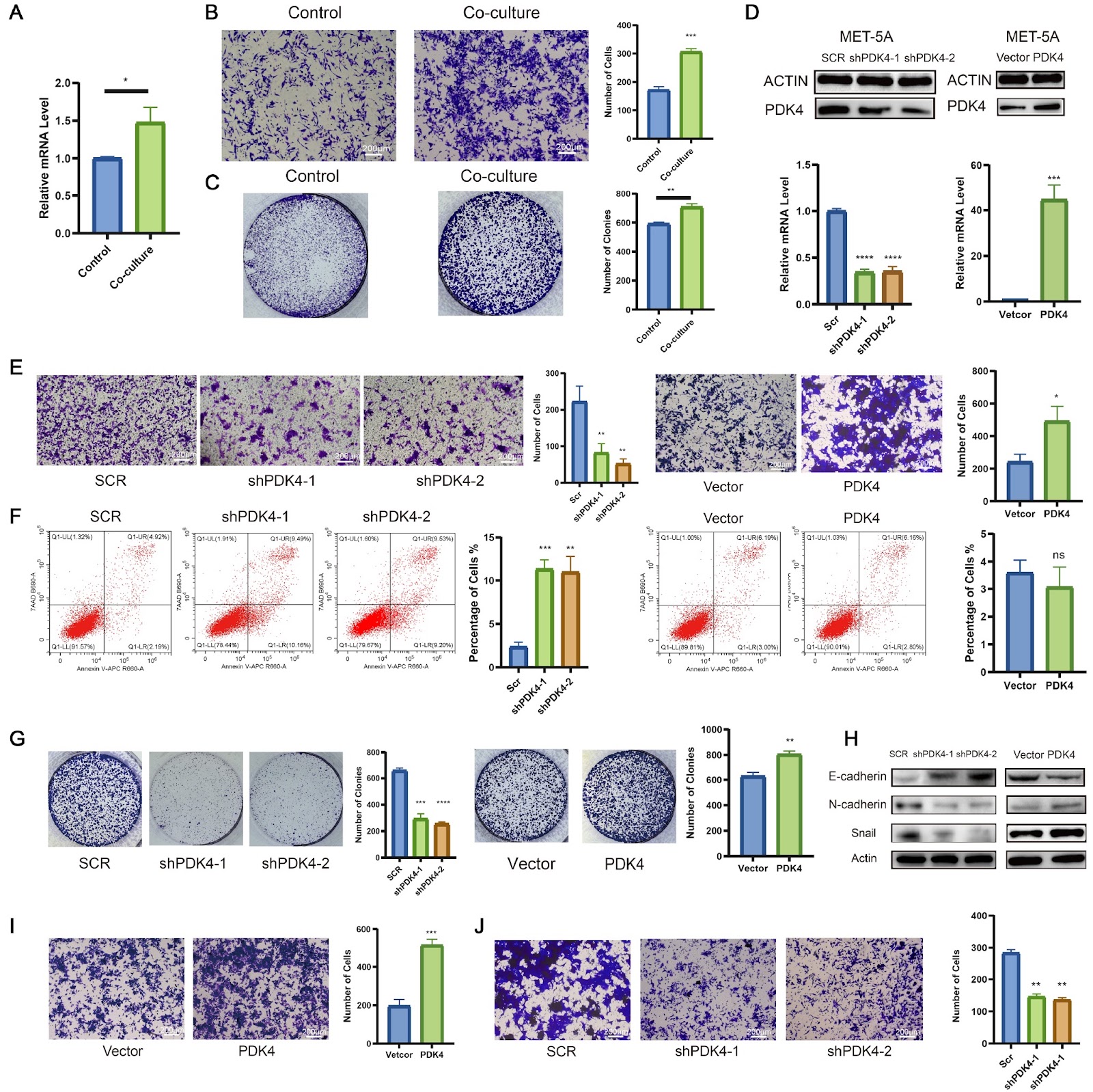
PDK4-driven metabolic reprogramming enhances mesothelial cell invasion in colorectal cancer peritoneal metastasis

Authors: Lingyu Han, Qinyuan Zhang, Yuchen Wu, Wenqin Luo, Shaobo Mo, Hongsheng Fang, Qingguo Li, Renjie Wang, Guoxiang Cai, Weixing Dai

Mesothelial cells play an important role in colorectal cancer peritoneal metastasis (CRC-PM), where they support tumor growth and invasion. In this study, we investigate the molecular mechanisms by which mesothelial cells contribute to CRC metastasis. Using single-cell RNA sequencing (scRNA-seq) on tissue samples from 12 CRC patients with peritoneal metastasis, we identify PDK4 as a key gene in mesothelial cells during metastasis. The expression of PDK4 is significantly greater in mesothelial cells undergoing mesothelial-to-mesenchymal transition (MMT) compared to normal peritoneal cells, suggesting its involvement in mesothelial cell reprogramming during peritoneal metastasis. In vitro experiments show that coculturing mesothelial cells with CRC cells leads to increased PDK4 expression, which in turn enhances mesothelial cell migration and invasion. Knockdown of PDK4 reduces mesothelial cell invasion, while overexpression of PDK4 increases invasive ability, highlighting its critical role in mesothelial cell invasion. Additionally, PDK4 promotes metabolic changes, specifically increasing fatty acid oxidation (FAO), which is necessary for mesothelial cell invasion. Blocking FAO reduces the invasive ability of PDK4-overexpressing mesothelial cells, while restoring FAO in PDK4-knockdown cells rescues their invasion potential. Further analysis shows that PDK4 enhances the acetylation of β-catenin, a protein involved in cell movement, and that this modification is crucial for mesothelial cell invasion. Our results suggest that PDK4 regulates mesothelial cell invasion through β-catenin acetylation following metabolic reprogramming, offering a potential target for therapies aimed at inhibiting CRC-PM.

PDK4-driven metabolic reprogramming enhances mesothelial cell invasion in colorectal cancer peritoneal metastasis
Graphical Abstract
Original ResearchVol. 58, Issue 3 • pp. 663-680DOI: 10.3724/abbs.2025114

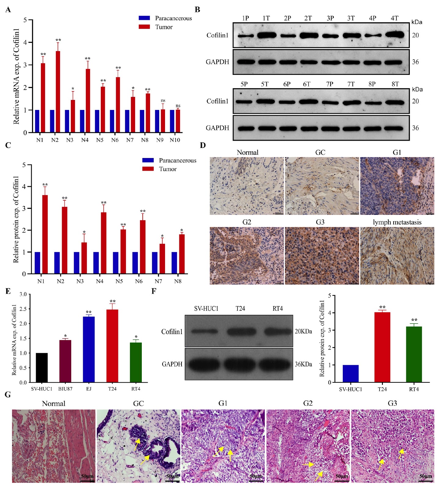
The TCF7L2/miR-206/Cofilin1 axis promotes the metastasis of bladder cancer cells by regulating the formation of invadopodia

Authors: Yuzhen Jie, Yinggui Yang, Chengyan Guo, Qinghui Wu, Zhewen Ou, Weifu Wang, Ning Xu, Wei Peng, Yingguang Wu, Jiangfan Peng, Shengchao Ma, Shufang Zhang, Fei Wang

Bladder cancer (BCa) is one of the most common malignant tumors of the urinary system, but its pathogenesis is still unclear. T1G3 BCa is particularly invasive and relapses readily after treatment, with progression to invasive cancer or distant metastasis. Therefore, identification of the molecular mechanism by which it invades and metastasizes to guide treatment and predict patient prognosis is needed. Cofilin1 plays an important role in regulating gene expression and the invasiveness of tumors. In this study, we show that Cofilin1 is highly expressed in BCa and lymph nodes with metastasis, which is positively related to the grade of BCa, and is significantly related to clinicopathological parameters and cancer-specific survival. Phenotypic analysis reveals that Cofilin1 knockout inhibits the proliferation and migration of BCa cells, whereas Cofilin1 overexpression promotes the opposite phenotype. Cofilin1 binds to cortactin, thereby reducing the expression of F-actin and promoting the formation of invadopodia in BCa cells. Further experiments reveal that TCF7L2 can bind to the promoter of Cofilin1 and transactivate it, promoting a malignant phenotype. TCF7L2 may also reverse the inhibitory effect of miR-206 on the binding of Cofilin1 and cortactin and promote the metastasis of BCa by inhibiting the transcription maturation of miR-206. This study confirms that Cofilin1 is an oncogene in T1G3 BCa, and the TCF7L2/miR-206/Cofilin1 signaling pathway plays an important role in the formation of invadopodia in BCa.

The TCF7L2/miR-206/Cofilin1 axis promotes the metastasis of bladder cancer cells by regulating the formation of invadopodia
Graphical Abstract
Original ResearchVol. 58, Issue 3 • pp. 516-529DOI: 10.3724/abbs.2025112

Scutellarin inhibits ferroptosis by promoting cellular antioxidant capacity through regulating Nrf2 signaling

Authors: Haiyan Yang, Onkei Chan, Xiaodi Huang, Liang Yan, Nuo Sun, Yaping Li, Zijian Shi, Qingbing Zha, Dongyun Ouyang, Jinhua Li, Xianhui He

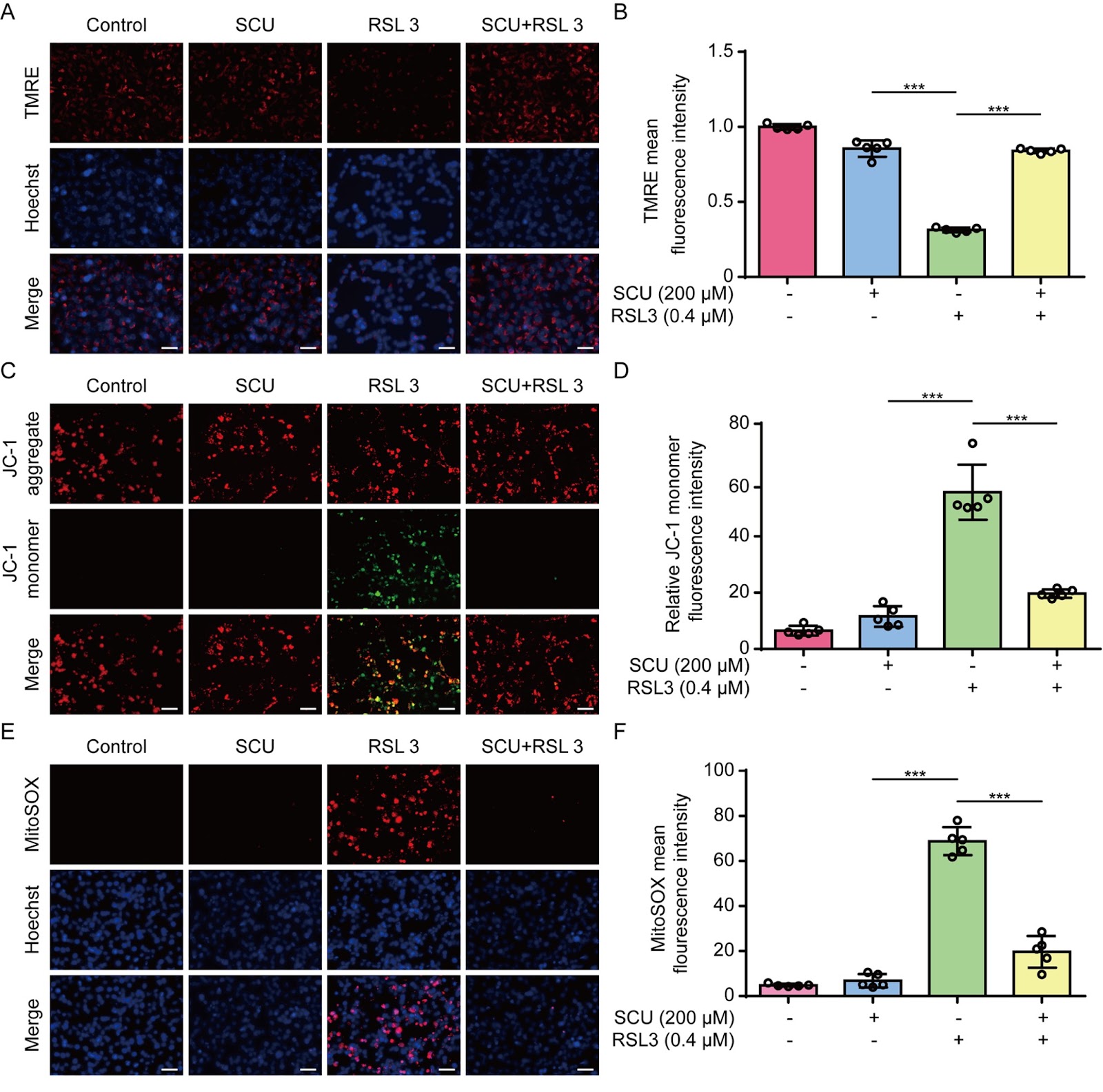
Ferroptosis is a lytic form of regulated cell death that is driven by iron-dependent lipid peroxidation and has been implicated in various diseases, including acute kidney injury (AKI). Scutellarin is a flavonoid isolated from Erigeron breviscapus (Vant.) Hand.-Mazz. and possesses various pharmacological activities, including anti-inflammatory and antioxidative properties. However, it is unclear whether scutellarin can inhibit ferroptosis and mitigate related diseases. In this study, we show that scutellarin can inhibit ferroptosis in both human HK-2 cells and mouse bone marrow-derived macrophages stimulated with RSL3 or erastin. Mitochondrial dysfunction and reactive oxygen species generation are counteracted by scutellarin treatment, suggesting the involvement of its antioxidative activity. Furthermore, scutellarin increases the nuclear levels of Nrf2 and the expressions of its target genes, including HO-1 and GPX4. Scutellarin-mediated inhibition of ferroptosis and increases in these proteins are abrogated by co-treatment with brusatol, an Nrf2 inhibitor, indicating an essential role for Nrf2 in this process. In a mouse model of folic acid-induced AKI, scutellarin mitigates acute renal damage, as revealed by histopathological analysis and serum blood urea nitrogen and creatinine assays. Folic acid-induced acute renal injury is associated with increased ferroptosis, as revealed by elevated level of 4-hydroxynonenal (4-HNE), a surrogate marker of ferroptosis, which is diminished by scutellarin co-treatment. Specifically, the elevated 4-HNE levels in macrophages (MAC-2 positive) and other renal cells are suppressed by scutellarin. Overall, scutellarin can inhibit ferroptosis both in cultured cells and in a mouse model of AKI by regulating Nrf2 signaling.

Scutellarin inhibits ferroptosis by promoting cellular antioxidant capacity through regulating Nrf2 signaling
Graphical Abstract
Original ResearchVol. 58, Issue 3 • pp. 530-540DOI: 10.3724/abbs.2025142

Combined ex vivo and in vivo evaluation of dolutegravir embryotoxicity: NTDs and yolk sac vascular abnormalities

Authors: Ruifang Ao, Ran Li, Zelin Li, Guicai Wu, Haoran Xu, Xuecong Wang, Jiayi Du, Xiaozheng Zhang, Jun Xie

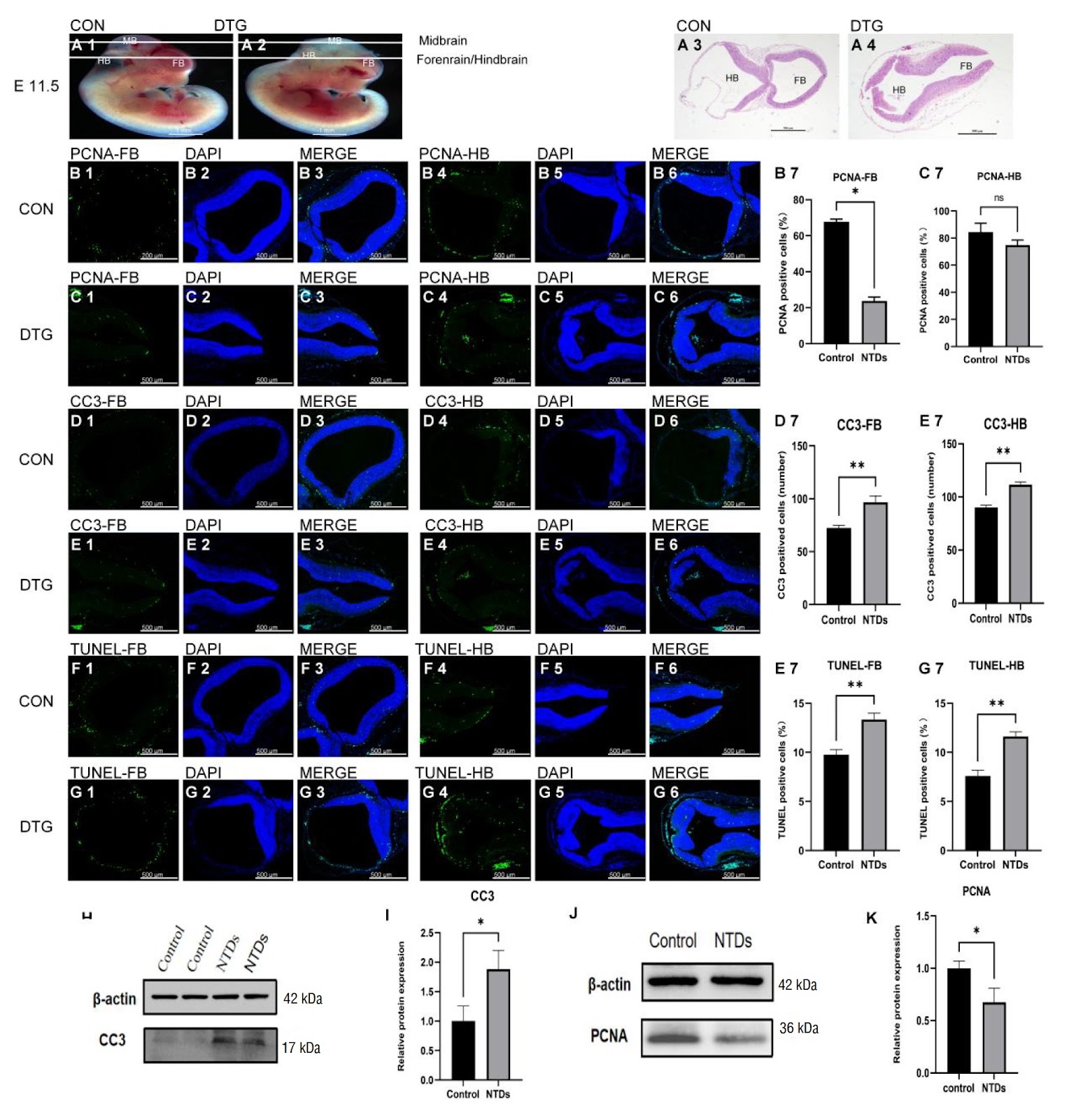
Dolutegravir (DTG) disrupts mouse embryonic development in a dose-dependent manner, culminating in neural-tube defects (NTDs). Using whole embryo culture (WEC), mouse embryos at embryonic day 8.5 (E8.5) are cultured for 24–48 h with 8, 10, or 12 μM DTG. The results reveal that higher DTG concentrations dose-dependently disrupt yolk sac development and markedly increase the frequency of NTDs. In vivo NTD models are generated by intraperitoneally injecting DTG at a dose of 7.5 mg/kg, and the resulting embryos exhibit disrupted yolk sac blood circulation, embryonic growth restriction, and malformations. Mechanistic studies suggest that DTG contributes to NTDs by inducing apoptosis: DTG exposure activates the Nrf2-SOD1/CAT antioxidant axis, yet it culminates in increased apoptosis and suppressed proliferation, ultimately impairing yolksac vasculogenesis and neuralepithelial closure, thereby producing NTDs. This study provides new evidence for assessing the potential risk of DTG in embryonic development and highlights the need to re-evaluate its clinical safety in future applications.

Combined ex vivo and in vivo evaluation of dolutegravir embryotoxicity: NTDs and yolk sac vascular abnormalities
Graphical Abstract
Original ResearchVol. 58, Issue 3 • pp. 649-662DOI: 10.3724/abbs.2025144

Exploring DNA topoisomerase II alpha in adrenocortical carcinoma through multi-omics analysis: a potential biomarker and therapeutic target

Authors: Jianming Lu, Pei Deng, Zhenjie Wu, Yuxiang Liang, Yangjia Zhuo, Yongding Wu, Yingke Liang, Jianheng Ye, Wenjie Xie, Zhouda Cai, Chao Cai, Jiahong Chen, Le Zhang, Junhong Deng, Weide Zhong, Jiaojiao Tang, Zhaodong Han

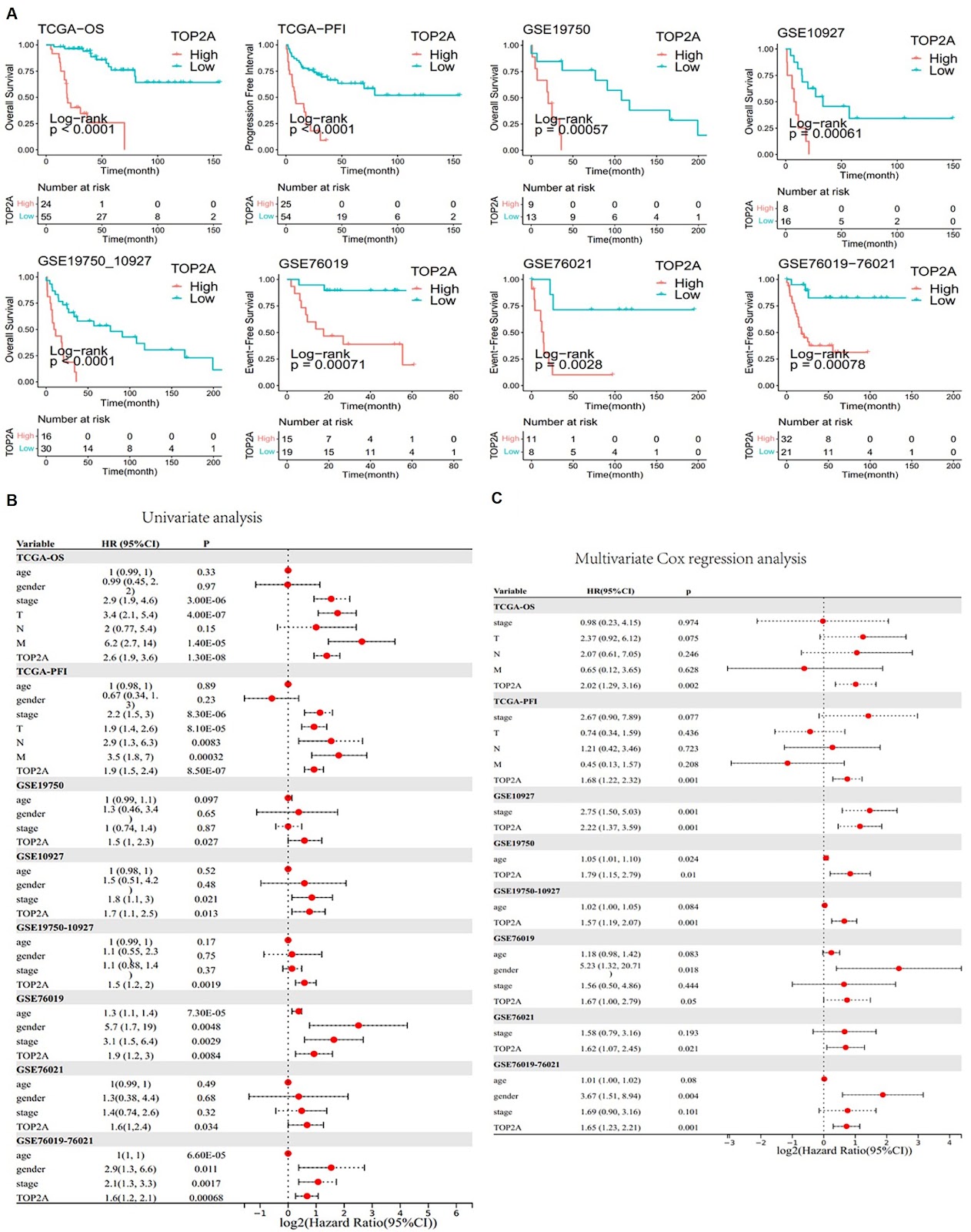
Adrenocortical carcinoma (ACC) is a rare but aggressive cancer. Recent studies identified DNA Topoisomerase II Alpha (TOP2A) as a potential biomarker for ACC, which can provide new avenues for targeted therapy and improve clinical outcomes. This study aims to elucidate the role of TOP2A in ACC by exploring its prognostic value and identifying inhibitors for ACC therapy. Utilizing RNA sequencing data, mutation data, and clinical information from The Cancer Genome Atlas (TCGA-ACC) and additional datasets from the Gene Expression Omnibus (GEO), differential expression and prognostic analyses are conducted to assess the significance of TOP2A in ACC. Immunohistochemistry and cell assays, including cell viability, colony formation, and transwell assays, are conducted to validate the oncogenic effects of TOP2A. The “IOBR” R package is used to examine the relationship between TOP2A expression and CD8+ T-cell infiltration. The CMap platform is used to identify potential TOP2A inhibitors. In vivo assays verify the therapeutic effect of TOP2A inhibitors on ACC. Our findings indicate that TOP2A is significantly overexpressed in ACC and is associated with poor prognosis. Immunohistochemistry and cell assays confirm the oncogenic role of TOP2A. Furthermore, distinct gene expression patterns related to different TOP2A expression levels are identified, influencing the response to immunotherapy. Potential inhibitors targeting TOP2A are discovered, and the therapeutic effects of resminostat and etoposide are confirmed via in vivo assays, suggesting new therapeutic strategies for ACC treatment. In conclusion, TOP2A serves as a crucial biomarker in ACC and is associated with adverse clinical outcomes and a diminished immune response. The identification of potential inhibitors against TOP2A opens new avenues for the development of targeted therapies for ACC patients.

Exploring DNA topoisomerase II alpha in adrenocortical carcinoma through multi-omics analysis: a potential biomarker and therapeutic target
Graphical Abstract
Original ResearchVol. 58, Issue 3 • pp. 574-583DOI: 10.3724/abbs.2025135

Targeting USP2 induces degradation of PML-RARα with or without drug-resistant mutations in acute promyelocytic leukemia

Authors: Jie Zhang, Wenxuan Wu, Yun Wang, Youping Zhang, Yingying Wang, Wenhui Bai, Zhenge Zhang, Chujiao Zhu, Yunzhao Wu, Ziwei Zhang, Li Yang, Hu Lei, Hanzhang Xu, Li Zhou, Yingli Wu

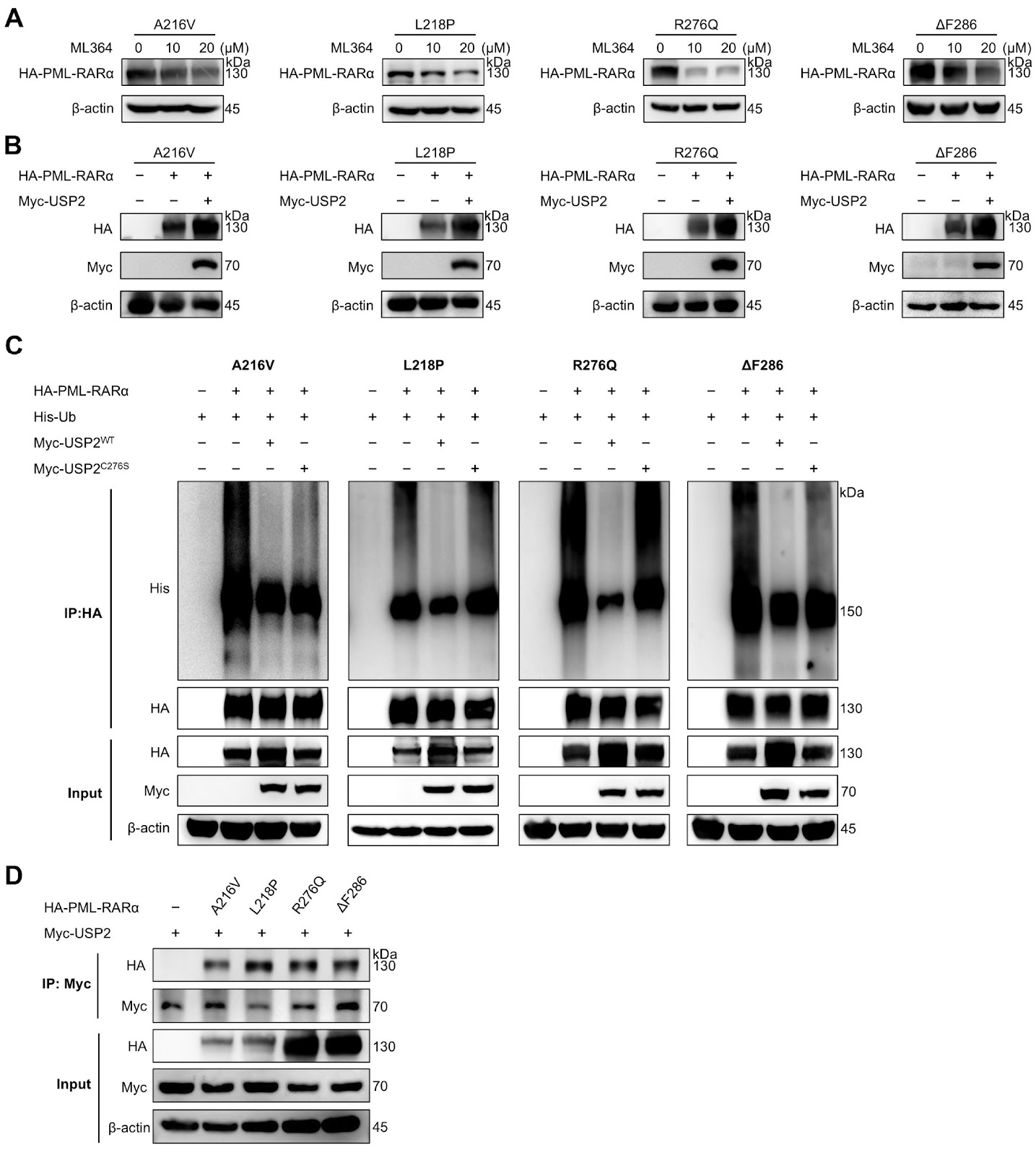
Despite the high efficacy of all-trans retinoic acid (ATRA) and arsenic trioxide (ATO) in treating acute promyelocytic leukemia (APL), approximately 10%–20% of patients develop drug resistance due to mutations in PML-RARα and other factors. Here, we find that inhibition of USP2 with ML364 or USP2 silencing reduces PML-RARα protein levels in both ATRA-sensitive and ATRA-resistant APL cells, and this effect is reversed by proteasome inhibition. Conversely, USP2 overexpression enhances PML-RARα stability. Mechanistically, USP2 interacts with and deubiquitinates PML-RARα, including its drug-resistant mutants. Consistent with PML-RARα degradation, ML364 treatment significantly induces apoptosis in APL cell lines and primary leukemia cells. In conclusion, this study identifies USP2 as a novel deubiquitinating enzyme for PML-RARα and highlights USP2 inhibition as a potential therapeutic strategy for APL with PML-RARα mutations.

Targeting USP2 induces degradation of PML-RARα with or without drug-resistant mutations in acute promyelocytic leukemia
Graphical Abstract
Original ResearchVol. 58, Issue 3 • pp. 700-703DOI: 10.3724/abbs.2025104

A novel method to increase transgene expression and the stability of gene therapy-associated episomal vectors

Authors: Xi Zhang, Rui Liu, Zimeng Han, Zihan Guo, Mengying Ji, Wen Wang, Yanlong Jia, Tianyun Wang, Xiaoyin Wang

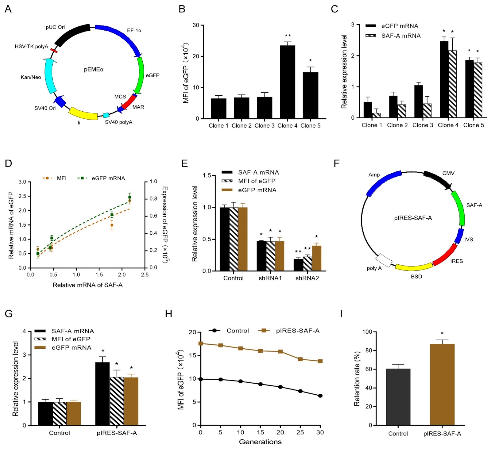
Non-viral episomal vectors offer a safe and attractive alternative to viral and integrated vectors by avoiding insertional mutagenesis and position effects, making them ideal expression vectors for gene therapy. The first non-viral episomal vector, pEPI-1, which is based on the full-length scaffold/matrix attachment region (S/MAR), was established by Piechaczek et al. The full-length S/MAR element interacts with the nuclear matrix via the matrix protein, e.g. SAF-A, thereby maintaining mitotic stability and transgene expression. Several strategies, including optimization of the vector backbone and promoter and incorporation of chromatin-modifying elements, have been used to increase expression levels and stability. In our previous work, we constructed the novel vector pEGFP-C1-M on the basis of S/MAR characteristic motifs (only 375 bp). This vector, which is shorter than the prototype episomal vector pEPI-1, resulted in relatively higher transgene expression. Building on the pEGFP-C1-M vector, we further constructed the episomal vector pEMEα with the EF-1α promoter and demonstrated that pEMEα maintained higher transgene expression, stability and copy number. The transgene expression levels of episomal vectors are correlated with gene copy number, that is, the number of plasmid episomes on the host cell chromosome. Previous studies have shown that the episomal maintenance of pEPI-1 vectors is mediated primarily by SAF-A. While the role of SAF-A in maintaining mammalian pEPI-1 episomal vectors has been well established, it remains unknown whether the overexpression of SAF-A promotes transgene expression and stability and whether the 375 bp MAR characteristic sequence retains its interaction with SAF-A. In the present study, we first evaluated whether transgene expression is positively correlated with the expression level of SAF-A. The non-viral episomal vector pEMEα was used as the gene of interest (GOI) vector and was subsequently transfected into CHO-K1 cells using the Lipofectamine 2000 reagent. The cells were cultured in medium containing 800 μg/mL geneticin (G418) 48 h post-transfection, and the G418 concentration was then reduced to 400 μg/mL to obtain monoclonal cell lines using the limiting dilution method. Five monoclonal cell clones were selected, and the eGFP expression levels, measured as the mean fluorescence intensity (MFI), were (6.5 ± 1.0) × 104, (6.8 ± 0.9) × 104, (7.0 ± 1.4) × 104, (23.5 ± 1.2) × 104 and (14.9 ± 0.17) × 104 for Clones 1–5, respectively. qPCR analysis of Clones 1–5 revealed that the relative mRNA levels of SAF-A and eGFP were 0.16 ± 0.13, 0.43 ± 0.11, 0.46 ± 0.23, 2.17 ± 0.41, 1.78 ± 0.15 and 0.51 ± 0.16, 0.71 ± 0.12, 1.05 ± 0.09, 2.47 ± 0.14, and 1.86 ± 0.10, respectively. Our results indicated that eGFP mRNA and protein expression levels are positively correlated with SAF-A mRNA level. To further verify the relationship between SAF-A expression and transgene expression, two shRNA plasmids targeting SAF-A (shRNA1: 5′-GCCACCTGTTGAAGAAGAAGA-3′, and shRNA2: 5′-GCTGGAGGAAGAGCTTCTTAT-3′) which were obtained from Shanghai GenePharma Co., Ltd. were designed and transfected into stable cell pools with the pEMEα vector. qPCR analysis revealed that the relative SAF-A mRNA levels in the shRNA1 and shRNA2 vectors were 0.47 ± 0.01 and 0.19 ± 0.02, respectively, indicating successful downregulation of SAF-A expression. Moreover, flow cytometry and qPCR revealed that, compared with those in the control group, the relative protein and mRNA levels of eGFP were reduced by 0.47- and 0.23-fold, and 0.47- and 0.40-fold in the pools of cells transfected with the shRNA1 and shRNA2 vectors, respectively. On the basis of the above results, the SAF-A overexpression vector pIRES-SAF-A was constructed and transfected into CHO-K1 cells, and the cells were cultured in blasticidin-containing medium 48 h after transfection to obtain stable cell pools. The stable cell pools overexpressing SAF-A were subsequently transfected with the pEMEα vector. Stable cell pools coexpressing SAF-A and GOI were selected, and the relative mRNA levels of SAF-A and eGFP were analyzed. qPCR analysis revealed that the relative mRNA levels of SAF-A and eGFP in the pools of cells overexpressing SAF-A were 2.69-fold and 2.05-fold higher than those in the control group, respectively. Flow cytometry also revealed a 2.07-fold increase in MFI in stable cell pools overexpressing SAF-A compared with the control group. To assess the long-term stability of transgene expression, we measured the MFI in st
A novel method to increase transgene expression and the stability of gene therapy-associated episomal vectors
Graphical Abstract
Original ResearchVol. 58, Issue 3 • pp. 584-594DOI: 10.3724/abbs.2025146

FSCN1-mediated hepatic gluconeogenesis is indispensable for neonatal mice survival

Authors: Xiangxiang Liu, Yuanzhao Hu, Liangwei Wu, Yiwen Zhang, Lei Sang, Yake Gao, Lei He, Wenyong Xiong, Shengyu Yang, Jianwei Sun

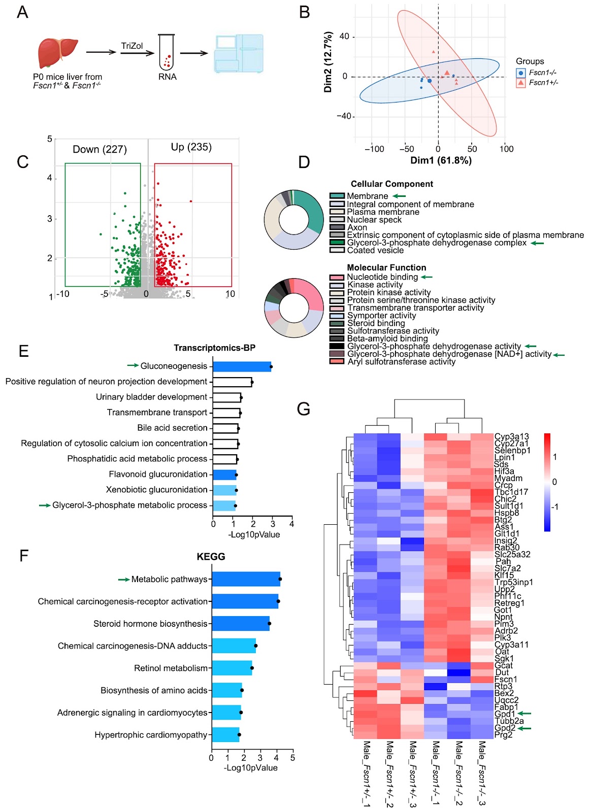
Actin-bundling protein Fascin1 (FSCN1) is encoded by the Fscn1 gene and is crucial for cytoskeletal remodeling and cellular migration. Although a previous study linked Fscn1 deficiency to neonatal lethality in mice, the underlying metabolic mechanism remains unclear. In this study, we report that systemic knockout (KO) of Fscn1 leads to 52.2% mortality within 24 h post-birth, accompanied by severe hypoglycemia in KO pups compared with their littermates. Remarkably, this lethality is fully rescued by oral glucose administration, indicating a glucose supply-dependent survival mechanism. Surviving Fscn1-KO neonates display persistent developmental deficits, including growth retardation and depleted lipid stores, despite intact canonical insulin-regulated hepatic gluconeogenic pathways. Transcriptomic profiling of P0 livers reveals that Fscn1 loss predominantly disrupts metabolic pathways, with the glycerol phosphate shuttle being the most significantly downregulated module. Mechanistically, Fscn1-KO livers exhibit markedly reduced protein levels of glycerol-3-phosphate dehydrogenase isoforms (GPD1/GPD2), key enzymes bridging glycolysis and gluconeogenesis. Consistently, glycerol tolerance tests demonstrate impaired glycerol-to-glucose conversion in Fscn1-KO mice, confirming defective glycerol-driven gluconeogenesis. Our findings establish FSCN1 as a novel cytoskeletal-metabolic integrator essential for neonatal survival by sustaining hepatic glucose production from glycerol, thus revealing an unexpected role of actin dynamics in coordinating metabolic adaptation during early postnatal development.

FSCN1-mediated hepatic gluconeogenesis is indispensable for neonatal mice survival
Graphical Abstract
Original ResearchVol. 58, Issue 3 • pp. 595-609DOI: 10.3724/abbs.2025130

Autophagy-dependent sensitization effects of PARP inhibitors on recurrent nasopharyngeal carcinoma treated with carbon ion and photon irradiation

Authors: Ziyu Le, Haojiong Zhang, Li Chen, Wanzun Lin, Qingting Huang, Shikai Geng, Wei Hu, Huaiyuan Chen, Fangzhu Wan, Xingyu Liu, Jiyi Hu, Fengtao Su, Jiade J. Lu, Lin Kong

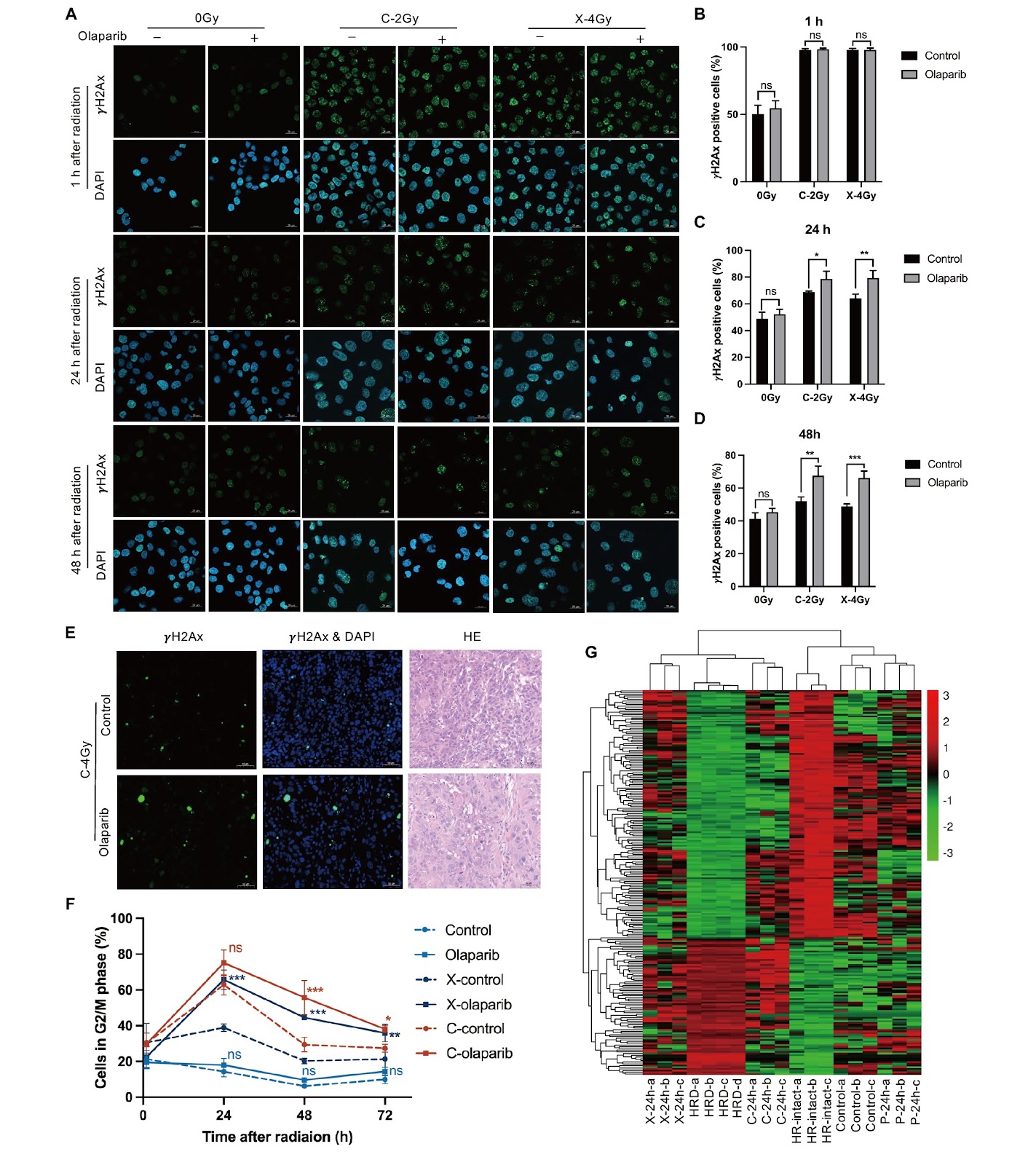
Tumor radioresistance and severe toxicity make reirradiation for recurrent nasopharyngeal carcinoma (NPC) a significant clinical challenge. This study aims to investigate the ability of the poly(ADP-ribose) polymerase (PARP) inhibitor olaparib to sensitize recurrent NPC cells irradiated with photon or carbon ion (C-ion), and to explore the underlying mechanism of the synergistic promotion of cell death by olaparib and ionizing radiation. The results show that olaparib has significant X-ray and C-ion radiosensitization effects on recurrent NPC cells and the associated HK-RR photon-resistant model. Radiation, particularly C-ion exposure, induces a homologous recombination (HR)-deficient gene signature in HR-proficient NPC cells, potentially increasing their sensitivity to PARP inhibition. C-ion and X-ray irradiation induces similar modes of cell death, and multiple cell death pathways [including apoptosis, necrosis, ferroptosis, senescence, and autophagic cell death (ACD)] contribute to the cytotoxic effects of radiation combined with olaparib, with ACD being the dominant pathway. Both the pharmacological and genetic inhibition of autophagy significantly attenuate the radiosensitization effect of olaparib. In conclusion, olaparib effectively sensitizes recurrent NPC cells to both X-ray irradiation and C-ion irradiation, with autophagy playing a central role in mediating this effect.

Autophagy-dependent sensitization effects of PARP inhibitors on recurrent nasopharyngeal carcinoma treated with carbon ion and photon irradiation
Graphical Abstract
Original ResearchVol. 58, Issue 3 • pp. 541-550DOI: 10.3724/abbs.2025179

Pervasive environmental contaminant acrolein compromises myocardial geometry and function through the induction of cuproptosis

Authors: Xiaohu Wang, Shuyi Zhu, Yipin Zhao, Xinlong Di, Lingfei Cao, Russel J. Reiter, Emily Y. He, Yuan Zhou, Bonglee Kim, Yong Cheng, Jun Ren

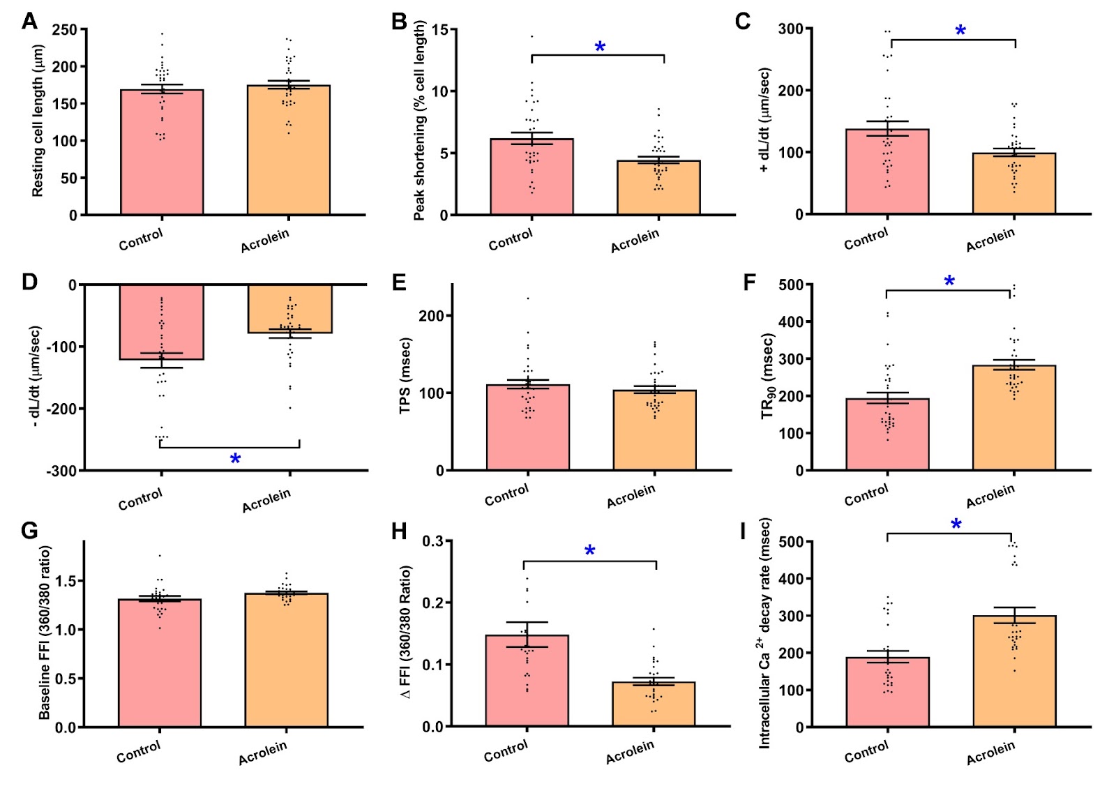
Acrolein, a highly reactive α,β-unsaturated aldehyde found in cigarette smoke, automobile exhaust, industrial emissions, combustion byproducts, cooking and cyclophosphamide chemotherapy, has raised serious health concerns, although the precise mechanism remains unclear. This study is designed to examine the impact of this pervasive environmental pollutant on myocardial geometry and function alongside the underlying cellular mechanisms. Adult C57BL/6 mice are challenged with acrolein (2.5 mg/kg/day, i.p., for 20 days) prior to the evaluation of myocardial geometry and function. Acrolein exposure evokes evident cardiac remodeling (interstitial fibrosis), compromised echocardiographic (enlarged LVESD, compromised ejection fraction and fractional shortening), cardiomyocyte contractile and intracellular Ca2+ capacities [decreased peak shortening, maximal velocity of shortening and relengthening (±dL/dt), and electrically stimulated rise in Fura-2 fluorescence intensity (ΔFFI), prolonged time-to-90% relengthening (TR90) and intracellular Ca2+ decay], accompanied by overt mitochondrial damage (ultrastructure, aconitase and mitochondrial protein contents), free radical buildup, apoptosis (Bax, Caspase-3, and Bcl2) and cuproptosis (upregulated SLC31A1, DLAT and FDX1), downregulated the Fe-S cluster proteins ACO2 and NDUFS8 alongside unchanged ATP7A and the ferroptosis markers GPX4 and SLC7A11. The levels of copper-sensing protein metal response element binding transcription factor 2 (MTF2), but not MTF1, are increased by acrolein insult. CB-DOCK2 analysis predicts an interaction between acrolein and the MTF2 dimer within its DNA-binding regions. In vivo administration of the cuproptosis inhibitor tetrathiomolybdate (TTM), the mitochondrial antioxidant mitoTEMPO or the nonselective MTF2 inhibitor actinomycin D alleviates acrolein-evoked cardiomyocyte dysfunction (decreased PS, ±dL/dt, and prolonged TR90). These findings indicate that acrolein evoked cardiac functional anomalies possibly through MTF2-related control of cuproptosis.

Pervasive environmental contaminant acrolein compromises myocardial geometry and function through the induction of cuproptosis
Graphical Abstract
Original ResearchVol. 58, Issue 3 • pp. 681-690DOI: 10.3724/abbs.2025228

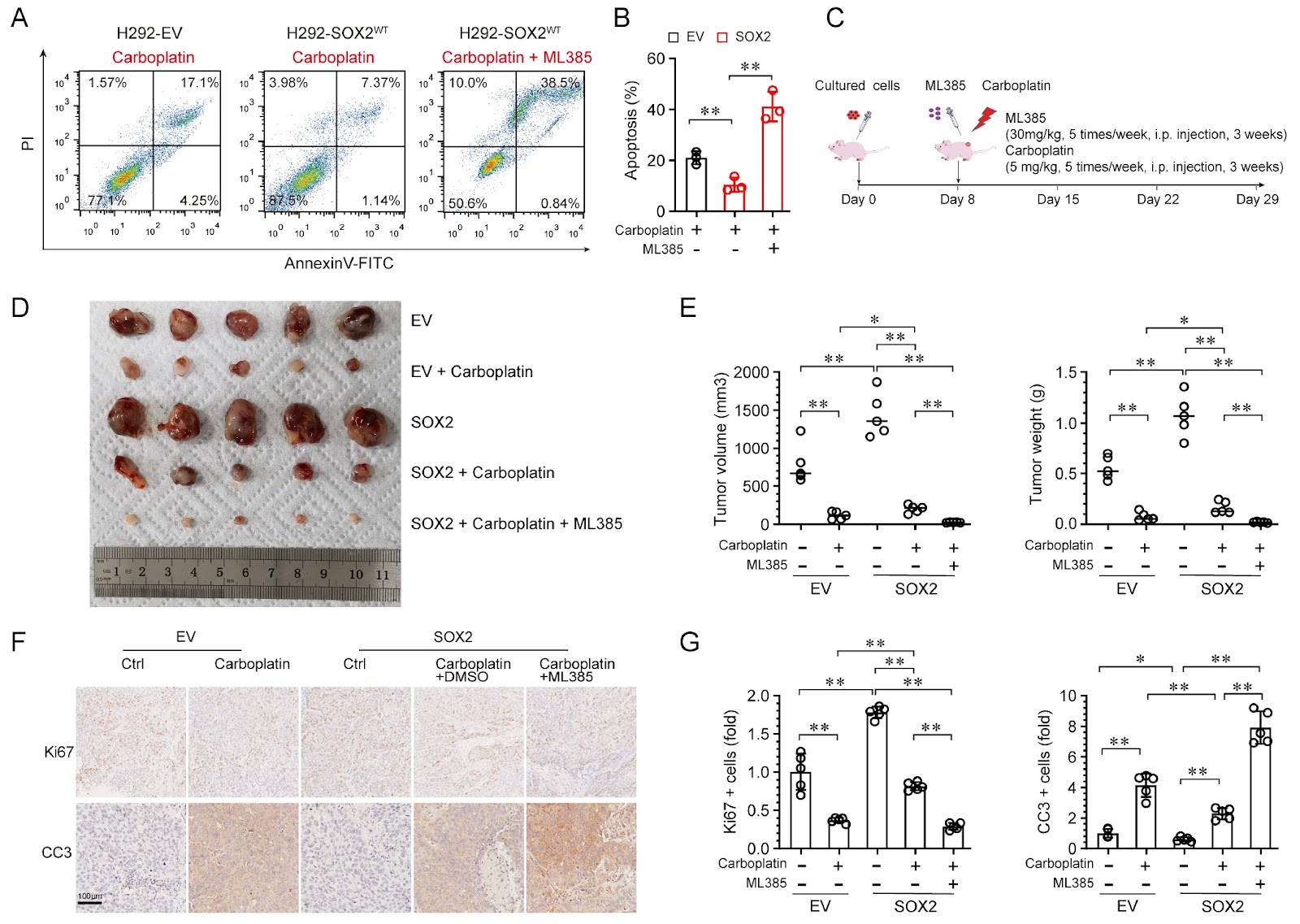
SOX2 transactivates NRF2 to promote carboplatin resistance in lung squamous cell carcinoma

Authors: Hanfei Gao, Chaomei Li, Jie Sun, Liyuan Deng, Jia Li, Zhiqiang Wu, Hu Chen

Lung squamous cell carcinoma (LUSC) remains a major therapeutic challenge because of its pronounced resistance to chemotherapy, particularly carboplatin. In this study, we investigate the role of SOX2, a lineage-survival oncogene, in mediating carboplatin resistance in LUSC. We demonstrate that SOX2 is highly expressed in LUSC and is significantly associated with poor prognosis. Our results show that SOX2 directly transactivates the expression of NRF2, a master regulator of cellular redox homeostasis, thereby increasing glutathione (GSH) synthesis and protecting cells from carboplatin-induced oxidative stress. Pharmacological or genetic inhibition of NRF2 effectively abrogates SOX2-mediated carboplatin resistance both in vitro and in vivo, resensitizing LUSC cells to chemotherapy. These findings highlight SOX2 as a critical redox regulator that modulates NRF2 signaling to promote carboplatin resistance in LUSC. The identification of the SOX2-NRF2 axis as a potential therapeutic target suggests that NRF2 inhibition may represent a promising strategy to overcome chemoresistance in LUSC.

SOX2 transactivates NRF2 to promote carboplatin resistance in lung squamous cell carcinoma
Graphical Abstract