🧬 SinoBioData Academic Portal
šŸ“š Peer-Reviewed Translated Literature

All Biomedical & Clinical Articles (Page 20)

Browse complete peer-reviewed translations from top Chinese biomedical, oncology, and genomics journals. Read verified previews and download full authentic clinical reports.

Published Research Papers

Showing 24 of 1542 peer-reviewed translated articles (Page 20 of 65)

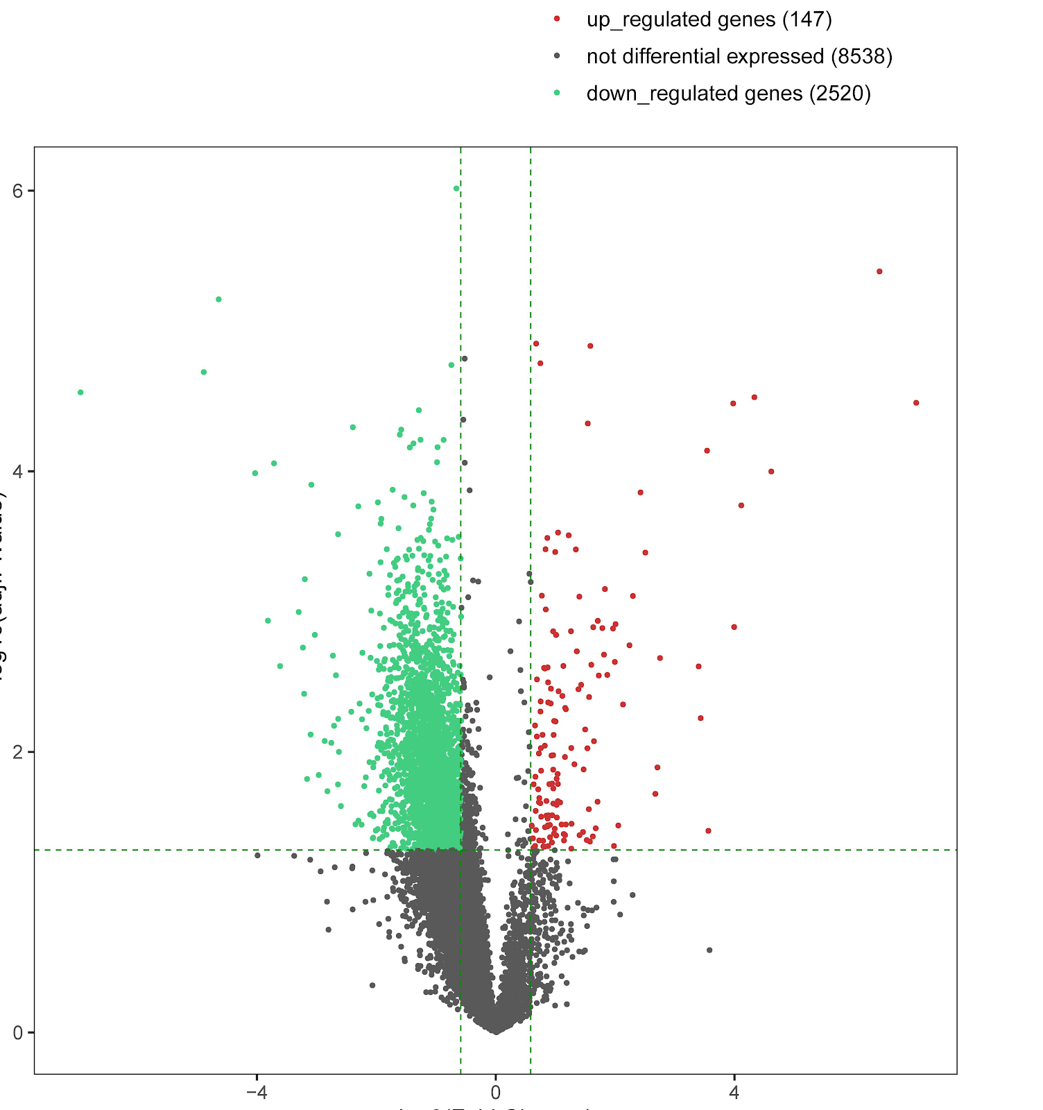
Transcriptomic analysis of expression and function of differential genes in traditional Chinese medicine syndromes of postmenopausal osteoporosisGraphical AbstractVerified
Chinese Journal of Tissue Engineering Research2026

Transcriptomic analysis of expression and function of differential genes in traditional Chinese medicine syndromes of postmenopausal osteoporosis

BACKGROUND: Kidney yin-yang deficiency syndrome in postmenopausal osteoporosis holds particular clinical significance due to its complex features of yin-yang imbalance, and analysis of its differential genes is key to revealing molecular mechanisms. OBJECTIVE: To compare differential gene expression profiles among different kidney deficiency syndromes of postmenopausal osteoporosis, screen for differential genes and signaling pathways associated with kidney yin-yang deficiency syndrome, reveal its molecular biological characteristics, and provide a basis for the objectification of traditional Chinese medicine syndromes. METHODS: Eighteen postmenopausal osteoporosis patients with kidney deficiency syndromes (kidney yang deficiency, kidney yin deficiency, and kidney yin-yang deficiency, 6 cases each) were included, and 6 healthy postmenopausal women served as healthy controls. Transcriptome sequencing was used to screen differential genes, followed by Gene Ontology and Kyoto Encyclopedia of Genes and Genomes enrichment analyses. The expression levels of four target genes (HSP90AB4P, CTU1, ST6GALNAC2, PTGS2) were validated by qRT-PCR. RESULTS AND CONCLUSION: (1) Compared with healthy controls, kidney yin deficiency, and kidney yang deficiency groups, the kidney yin-yang deficiency group had 235, 247, and 4,557 differentially expressed genes, respectively. Intersection analysis of the three comparison groups identified 22 differential genes associated with kidney yin-yang deficiency syndrome (18 up-regulated, 4 down-regulated). (2) qRT-PCR validation showed that the up/down regulation trends of target genes were consistent with transcriptome sequencing results. (3) Gene Ontology analysis showed that biological processes focused on energy metabolism (NAD synthesis and metabolism) and physiological homeostasis (thermogenesis, blood pressure regulation), molecular functions involved immune defense, metabolic regulation, inflammation and signal transduction, ion channel regulation, etc.; cellular components were related to ribosomes, endoplasmic reticulum, nucleus, and tRNA modification. (4) Kyoto Encyclopedia of Genes and Genomes enrichment identified 60 pathways, including apoptotic cell clearance, nuclear factor kappa B, tumor necrosis factor, interleukin-17, vascular endothelial growth factor, forkhead box O signaling pathways, and metabolic pathways. (5) These findings suggest that postmenopausal osteoporosis with kidney yin-yang deficiency syndrome is a comprehensive manifestation of multidimensional molecular network imbalance, related to ribosomal synthesis disorders, non-coding RNA and signal transduction and transcriptional regulation, immune-inflammatory regulation, and metabolic transport.


Read Full Abstract10.12307/2026.21521
Integrated proteomics and transcriptomics analysis of the mechanism of Buyang Huanwu Tang in protecting the acute spinal cord injury rat modelGraphical AbstractVerified
Chinese Journal of Tissue Engineering Research2026

Integrated proteomics and transcriptomics analysis of the mechanism of Buyang Huanwu Tang in protecting the acute spinal cord injury rat model

BACKGROUND: Research indicates that Buyang Huanwu Tang has positive therapeutic effects on the spinal cord injury symptoms and spinal cord function recovery, although its therapeutic mechanisms remain unclear. Spinal cord tissue contains numerous proteins and peptides that may serve as disease biomarkers. OBJECTIVE: To investigate the protective mechanism of Buyang Huanwu Tang in an acute spinal cord injury rat model by regulating proteomic- and transcriptomic-related pathways. METHODS: Thirty-six Sprague-Dawley rats were randomly divided into blank group, model group, and Buyang Huanwu Tang group. The latter two groups were used to establish acute spinal cord injury rat models using Allen's modified method. Motor function recovery was assessed by BBB score, pathological morphology was observed by Nissl staining, and differentially expressed proteins and genes were screened by isobaric tags for relative and absolute quantification (iTRAQ) proteomics and RNA-seq transcriptomics, followed by GO enrichment and KEGG pathway analyses. A PPI network was constructed using the STRING interaction database to identify key pathways, and core targets were validated by Western blot, immunohistochemistry, and RT-PCR. RESULTS AND CONCLUSION: (1) Motor function scores: Compared with the blank group, the model group showed significantly lower BBB scores (P < 0.001) and smaller inclined plane test angles (P < 0.001). Compared with the model group, the Buyang Huanwu Tang group showed higher BBB scores (P < 0.05) and larger inclined plane test angles (P < 0.001). Pathological morphology: The blank group showed relatively normal neuronal cells with intact structure, normal gaps, and clear nucleoli and nuclear membranes. The model group showed severe necrosis, disordered structure, pyknotic nuclei, disappearance of most nucleoli and nuclear membranes, numerous tissue cavities, and inflammatory cell infiltration. The Buyang Huanwu Tang group showed irregular but relatively intact neuronal cells with less swelling, reduced tissue cavities and cell necrosis. (2) GO functional annotation and KEGG pathway analysis revealed that differentially expressed proteins were mainly enriched in acute phase response, regulation of protein activation cascade, regulation of acute inflammatory response, platelet alpha granules, blood microparticles, vesicle lumen, serine-type endopeptidase inhibitor activity, and involved in pathways such as map04142 (lysosome), map04612 (antigen processing and presentation), map03013 (nucleocytoplasmic transport), map04964 (proximal tubule bicarbonate reclamation), map04610 (complement and coagulation cascades), map00511 (other glycan degradation), map03040 (spliceosome), map03410 (base excision repair), map00531 (glycosaminoglycan degradation), and coronavirus disease-COVID-19 pathway. Through PPI construction, 20 core differential proteins including FGG, FN1, FGB, HSP90B1, CASP3, and 20 core differential genes including FGG, FN1, FGB, CXCL1, CXCL13 were identified. (3) Western blot showed that Buyang Huanwu Tang inhibited the expression of myeloid differentiation factor 88 (MyD88) and P-IKBα in spinal cord tissue. Immunohistochemistry showed that Buyang Huanwu Tang inhibited the expression of BAX and Caspase-3, promoted BCL-2 expression, and inhibited apoptosis. RT-PCR results showed that Buyang Huanwu Tang inhibited the expression of MyD88 and CXCL1. These results suggest that Buyang Huanwu Tang may protect spinal cord tissue by inhibiting the Toll-like receptor 4/MyD88/nuclear factor-κB pathway to reduce inflammation, and by regulating the BAX/BCL-2 balance to inhibit Caspase-3 expression and prevent apoptosis.

Read Full Abstract10.12307/2026.21524
10-Hydroxy-2-decenoic acid facilitates osteogenic differentiation via the enhancement of autophagy and antioxidant capacityGraphical AbstractVerified
Chinese Journal of Tissue Engineering Research2026

10-Hydroxy-2-decenoic acid facilitates osteogenic differentiation via the enhancement of autophagy and antioxidant capacity

BACKGROUND: 10-Hydroxy-2-decenoic acid (10-HDA) exhibits potent anti-inflammatory, antioxidant, and immunomodulatory effects, but its role in regulating bone metabolism remains unclear. OBJECTIVE: To investigate the regulatory effects and potential mechanisms of 10-HDA in bone remodeling. METHODS: Rat bone marrow mesenchymal stem cells (BMSCs) were cultured with different concentrations of 10-HDA (0, 0.5, 1, 2, 4 mmol/L); cytoskeletal staining, live/dead staining, and CCK-8 assay were used to assess cell morphology, viability, and proliferation. For osteogenic differentiation, BMSCs were cultured with 10-HDA (0, 0.5, 1, 2 mmol/L) and osteogenic induction; alkaline phosphatase (ALP) and alizarin red staining were performed, and osteogenic-related protein expression was analyzed by western blot and immunofluorescence. Mouse bone marrow mononuclear cells were induced to differentiate into macrophages and cultured in osteoclast differentiation medium with different concentrations of 10-HDA (0, 0.5, 1, 2 mmol/L); tartrate-resistant acid phosphatase (TRAP) and F-actin staining were used to detect osteoclast formation. BMSCs were serum-starved for 6 h and then cultured normally, divided into control, 10-HDA, 10-HDA+AS1842856 (FOXO1 inhibitor), and 10-HDA+EX-527 (SIRT1 inhibitor) groups; 10-HDA concentration was 0.5 mmol/L. Western blot and immunofluorescence were used to analyze SIRT1/FOXO1 pathway activation and expression of autophagy- and osteogenesis-related proteins. BMSCs were divided into control, H2O2, and H2O2+10-HDA groups; 10-HDA concentration was 0.5 mmol/L; after H2O2 treatment for 24 h, corresponding drug interventions were applied; after osteogenic induction, ALP and alizarin red staining were performed. BMSCs were divided into five groups: control, H2O2, H2O2+10-HDA, H2O2+10-HDA+AS1842856, and H2O2+10-HDA+EX-527; 10-HDA concentration was 0.5 mmol/L; after H2O2 treatment for 24 h, corresponding drug interventions were applied; western blot was used to detect SIRT1/FOXO1 signaling pathway and antioxidant-related protein expression; TUNEL and β-galactosidase staining were used to assess apoptosis and senescence. RESULTS AND CONCLUSION: Cytoskeletal staining, live/dead staining, and CCK-8 assay showed that 0.5, 1, 2 mmol/L 10-HDA promoted proliferation of rat BMSCs; these three concentrations were selected for subsequent experiments. ALP, alizarin red staining, western blot, and immunofluorescence analysis showed that 0.5 mmol/L 10-HDA promoted osteogenic differentiation and mineralization of rat BMSCs and increased osteogenic-related protein expression. TRAP and F-actin staining showed that 0.5 mmol/L 10-HDA significantly inhibited osteoclast formation. Western blot and immunofluorescence showed that 10-HDA activated the SIRT1/FOXO1 signaling pathway, promoted FOXO1 deacetylation and nuclear translocation, and upregulated autophagy-related proteins and antioxidant enzymes. ALP and alizarin red staining showed that under oxidative stress, 10-HDA promoted osteogenic differentiation and mineralization of rat BMSCs. Western blot showed that under oxidative stress, 10-HDA enhanced the antioxidant capacity of rat BMSCs by activating the SIRT1/FOXO1 signaling pathway. TUNEL and β-galactosidase staining showed that under oxidative stress, 10-HDA reduced apoptosis and senescence of rat BMSCs via activation of the SIRT1/FOXO1 signaling pathway. These findings indicate that 10-HDA enhances autophagy and antioxidant capacity through regulation of the SIRT1/FOXO1 signaling pathway, thereby promoting osteogenic differentiation.

Read Full Abstract10.12307/2026.21520
Transcriptome sequencing analysis of tibial transverse transport in a rabbit model of diabetic foot ulcersGraphical AbstractVerified
Chinese Journal of Tissue Engineering Research2026

Transcriptome sequencing analysis of tibial transverse transport in a rabbit model of diabetic foot ulcers

BACKGROUND: Tibial transverse transport has emerged as a pivotal therapeutic approach for severe diabetic foot ulcers (Wagner grade III and above); however, its precise molecular regulatory mechanisms remain largely elusive. OBJECTIVE: To establish an animal model of tibial transverse transport for diabetic foot ulcer treatment and perform transcriptome sequencing, aimed at identify significantly differentially expressed miRNAs and key target genes, as well as construct the corresponding miRNA-mRNA regulatory network. METHODS: A diabetic foot ulcer wound model was established in rabbits and treated with tibial transverse transport. Peripheral blood samples were collected for miRNA-seq and mRNA-seq. Differentially expressed miRNAs and mRNAs were identified through bioinformatics analysis of sequencing data. Target genes of miRNAs were predicted using TargetScan and miRanda, and a miRNA-mRNA regulatory network was constructed. Kyoto Encyclopedia of Genes and Genomes enrichment analysis and protein-protein interaction analysis were performed on target genes, and core target genes were screened using MCC and Degree algorithms. RESULTS AND CONCLUSION: A total of 9 significantly differentially expressed miRNAs and 2,667 significantly differentially expressed mRNAs were identified. Based on differential analysis and predicted miRNA target genes, a miRNA-mRNA regulatory network containing 7 miRNAs and 79 target genes was constructed. Kyoto Encyclopedia of Genes and Genomes enrichment analysis revealed that these target genes were mainly involved in metabolic pathways, protein processing in endoplasmic reticulum, FoxO signaling pathway, propanoate metabolism, and N-glycan biosynthesis. Protein-protein interaction analysis revealed interactions among the proteins encoded by these target genes and identified BAG3, AKT3, PPP4C, SEC61A1, DNAJC3, USP7, DAD1, and SETD7 as core target genes. These results indicate that a series of key miRNAs, target genes, and signaling pathways related to diabetic foot ulcer treatment were identified through transcriptome sequencing, providing new candidates for therapeutic targets and opening new avenues for exploring the therapeutic mechanism of tibial transverse transport.

Read Full Abstract10.12307/2026.21522
Efficiency of anterior tooth intrusion in deep overbite cases with clear aligners: analysis via the indirect model superimposition methodGraphical AbstractVerified
Chinese Journal of Tissue Engineering Research2026

Efficiency of anterior tooth intrusion in deep overbite cases with clear aligners: analysis via the indirect model superimposition method

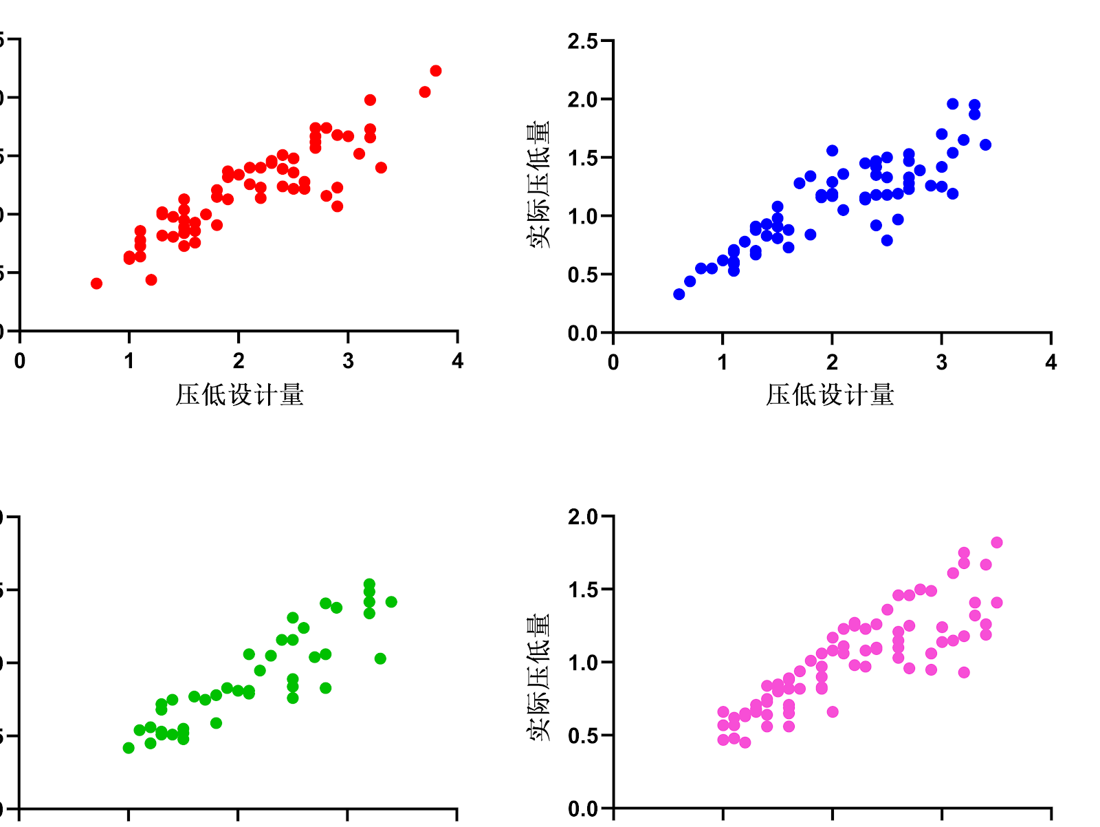
BACKGROUND: Anterior tooth intrusion is commonly used in the deep overbite cases with clear aligners. However, the intrusion efficiency varies significantly among different cases. OBJECTIVE: To analyze and compare the intrusion efficiency of the anterior teeth with clear aligners by the indirect model superimposition method based on the occlusal records. METHODS: Forty-three patients with anterior deep overbite were selected as the subjects of this study and divided into the non-extraction and extraction groups. The non-extraction group was then divided into the alignment and maxillary molar distalization subgroups. The extraction group was subdivided into the first premolar extraction and second premolar extraction subgroups. In the Geomagic 2014 software, the occlusal plane inclinations of the pre- and post-treatment models were calibrated based on the angle between the Frankfort plane and occlusal plane. The maxillary and mandibular models were superimposed by the maxillary palatine folds and occlusal records. Based on the Frankfort plane, the measurement coordinate system was established to measure the actual intrusion amount. The relative intrusion amount in the ClinCheck plan was recorded as the designed intrusion amount. Correlation and linear regression analyses were performed between the designed and actual intrusion amounts. RESULTS AND CONCLUSION: (1) The actual intrusion amount of anterior teeth in both non-extraction and extraction groups was significantly less than the designed amount (P < 0.01), indicating that overcorrection should be designed when intruding anterior teeth with clear aligners. (2) The anterior intrusion efficiency in the non-extraction group (50.69%) was significantly higher than that in the extraction group (39.73%) (P < 0.01), suggesting that more overcorrection should be designed in extraction cases. (3) The intrusion efficiency of lower anterior teeth was significantly higher than that of upper anterior teeth (P < 0.01), so more overcorrection should be designed for upper anterior teeth. (4) There was no significant difference in anterior intrusion efficiency between the maxillary molar distalization subgroup and the simple alignment subgroup (P > 0.05). (5) The anterior intrusion efficiency in the second premolar extraction subgroup was significantly higher than that in the first premolar extraction subgroup (P < 0.01), indicating that extraction of the second premolar is more favorable for vertical control of anterior teeth than extraction of the first premolar. (6) There was a linear relationship between the designed and actual intrusion amounts in all groups, and the regression equations can provide a reference for clinicians to design individualized overcorrection amounts according to different tooth positions and different treatment plans.

Read Full Abstract10.12307/2026.21525
Highly sensitive indicators of neck muscle fatigue derived from multimodal electrophysiological and metabolic coupling analysisGraphical AbstractVerified
Chinese Journal of Tissue Engineering Research2026

Highly sensitive indicators of neck muscle fatigue derived from multimodal electrophysiological and metabolic coupling analysis

BACKGROUND: Prolonged forward head posture induces neck muscle fatigue, a significant contributing factor to cervical spondylosis. Current unimodal monitoring approaches are inadequate to capture the dynamic coupling among muscle activation, metabolic activity, and motor control. OBJECTIVE: To systematically evaluate the temporal characteristics of neck muscle fatigue using multimodal monitoring technology, thereby providing a theoretical foundation for early detection and intervention of cervical fatigue. METHODS: Twenty healthy participants were recruited. Surface electromyography, near-infrared spectroscopy, and three-dimensional motion capture technology were synchronized to record electrophysiological signals, oxygenated hemoglobin concentration, and cervical kinematics during a sustained 45° static forward flexion task until subjective fatigue was reached (Borg CR-10 score ≄ 4). Temporal changes in root mean square amplitude, mean power frequency, muscle oxygen saturation, and normalized forward head angle were analyzed across fatigue stages segmented into 10% intervals of total endurance time. RESULTS AND CONCLUSION: (1) The root mean square amplitude increased significantly (P < 0.001), while mean power frequency and muscle oxygen saturation decreased significantly (P < 0.001) throughout the task, with the reduction in muscle oxygen saturation commencing from the 40% fatigue stage. (2) Linear regression analysis between mean power frequency and muscle oxygen saturation showed high explanatory power (upper trapezius R²=0.58, middle trapezius R²=0.61), and metabolic compensation preceded significant electrophysiological changes. (3) The upper trapezius entered fatigue earlier than the middle trapezius (P < 0.05). These results indicate that combined monitoring of mean power frequency and muscle oxygen saturation provides highly sensitive indicators for early warning of neck muscle fatigue.

Read Full Abstract10.12307/2026.21527
Application of novel materials in tissue repair of diabetic foot woundsGraphical AbstractVerified
Chinese Journal of Tissue Engineering Research2026

Application of novel materials in tissue repair of diabetic foot wounds

BACKGROUND: In recent years, the convergence of materials science and biomedical engineering has opened new avenues for diabetic foot wound treatment. Novel biomaterials such as multifunctional hydrogels, tissue engineering scaffolds, and nanoparticle systems have become a research hotspot. OBJECTIVE: To analyze, through bibliometric analysis, the design principles and biological functions of novel materials used in diabetic foot wound treatment, their potential in addressing the complex pathological challenges of diabetic foot, and to forecast future directions and challenges in this field. METHODS: We searched the Web of Science database for literature on novel materials for diabetic foot wounds from database inception to June 2025, and used CiteSpace software for bibliometric visualization analysis. RESULTS AND CONCLUSION: From 1979 to 2025, the number of publications in the field of novel materials for diabetic foot wound treatment showed an overall fluctuating upward trend, with explosive growth from 2022 to 2025. The United States ranked first with 602 publications, followed by China (284 publications) and India (166 publications). By publication count, Sichuan University had the most publications, followed by Tehran University of Medical Sciences; by citation impact, Sichuan University had the highest total citation impact, followed by Nankai University. West China Hospital ranked first in publication count, followed by Beth Israel Deaconess Medical Center and Thomas Jefferson University Hospital; by citation impact, West China Hospital had the highest impact, followed by Shanghai Ninth People's Hospital and Thomas Jefferson University Hospital. Among companies, Success Bio-Tech Co. Ltd, Engineering Software Research and Development Inc, and Foot and Ankle Associates of Central Illinois LLC each had 2 publications, ranking first; by citation impact, Organogenesis Inc had the highest impact, followed by Food Industry Research Co. The journal Wounds had the most publications (96), followed by Foot Ankle Int (40) and Cureus (38). Among authors, Bus, Sicco A had the most publications (12), followed by LƔzaro-Martƭnez, JosƩ Luis (8); by citation impact, GBD 2021 US Burden of Disease and Forecasting Collaborators, GBD 2021 Diabetes Collaborators, GBD 2021 US Obesity Forecasting Collaborators, GBD 2021 US Burden of Disease Collaborators, GBD 2021 Adult BMI Collaborators, GBD 2021 Adolescent BMI Collaborators, and GBD 2021 Causes of Death Collaborators had the highest impact. Keywords: diabetic foot; diabetic foot wound; diabetic foot ulcer; biomaterials; dressing; visualization analysis.

Read Full Abstract10.12307/2026.21603
Grape seed proanthocyanidin oligomers alleviate demyelination in cuprizone-fed miceGraphical AbstractVerified
Chinese Journal of Tissue Engineering Research2026

Grape seed proanthocyanidin oligomers alleviate demyelination in cuprizone-fed mice

BACKGROUND: Recent studies on the pathogenesis of multiple sclerosis suggest that intervening in glial cells may play a key role in reducing relapses and delaying disability progression. Grape seed proanthocyanidin oligomers significantly inhibit demyelination in cuprizone-treated mice. OBJECTIVE: To explore the mechanism by which grape seed proanthocyanidin oligomers protect myelin sheaths through regulating astrocytes. METHODS: (1) Animal experiment: Thirty mice were randomly divided into normal group, CPZ group, and CPZ+oligomeric proanthocyanidin group. The latter two groups were fed a diet containing 0.2% CPZ for 6 weeks to induce demyelination. From the 5th week, the normal and CPZ groups were given ddH2O by gavage, while the CPZ+oligomeric proanthocyanidin group received grape seed proanthocyanidin oligomers [50 mg/(kg·d)] once daily for 2 weeks. Behavioral changes were observed; LFB and oil red staining were used to assess myelin pathology; ELISA detected inflammatory factors in the brain; immunofluorescence staining detected related protein expression. (2) Cell experiment: In vitro, grape seed proanthocyanidin oligomers (30 μg/mL) were used to intervene in an astrocyte inflammation model induced by tumor necrosis factor α, interleukin 1α, and C1q. Conditioned medium was collected and used to culture oligodendrocytes. Cells were divided into normal, model, and model+oligomeric proanthocyanidin groups. L-lactate dehydrogenase and CCK-8 assays were used to detect oligodendrocyte damage and cell activity, and western blot was used to detect apoptosis-related protein expression. RESULTS AND CONCLUSION: (1) Grape seed proanthocyanidin oligomers significantly improved demyelination in CPZ mice, inhibited the expression of pro-inflammatory factors tumor necrosis factor α, interleukin 6, interleukin 1α, and interleukin 17 in the brain, promoted the secretion of anti-inflammatory factor transforming growth factor β, accompanied by astrocyte proliferation in the corpus callosum, significantly reduced the marker C3d of pro-inflammatory astrocytes, and inhibited the phosphorylation of JNK, a signaling molecule related to astrocyte polarization. (2) Compared with the model group, the conditioned medium after intervention with grape seed proanthocyanidin oligomers significantly reduced the apoptosis of oligodendrocytes induced by inflammatory astrocytes, promoted the expression of Bcl-2, and inhibited the expression of Bax and Caspase-3. (3) These results indicate that grape seed proanthocyanidin oligomers can inhibit demyelination in CPZ mice by inhibiting JNK phosphorylation in astrocytes, reducing the polarization of astrocytes to the pro-inflammatory A1 type, thereby inhibiting the apoptosis of oligodendrocytes induced by inflammatory astrocytes.

Read Full Abstract10.12307/2026.21523
Magnetic calcium-regulated mitochondrial sensitivity in adolescents: assessment of skeletal muscle function and body compositionGraphical AbstractVerified
Chinese Journal of Tissue Engineering Research2026

Magnetic calcium-regulated mitochondrial sensitivity in adolescents: assessment of skeletal muscle function and body composition

BACKGROUND: Magnetic mitochondrial calcium regulation technology, as a non-invasive method for promoting skeletal muscle function, has been effectively demonstrated in improving muscle function and enhancing metabolic sensitivity. Existing studies have shown that this technology has significant physiological promoting effects on adults, the elderly, and postoperative rehabilitation populations, but its intervention effects on skeletal muscle function and body composition in adolescents aged 12-13 years remain unclear. OBJECTIVE: To investigate the sensitivity of skeletal muscle function and body composition to magnetic stimulation in adolescents aged 12-13 years by evaluating these parameters. METHODS: A total of 34 junior high school students from Liaoning Experimental School were recruited and randomly divided into a control group and an experimental group. The control group maintained routine campus activities without a specific physical training program. The experimental group additionally received low-frequency pulsed magnetic field intervention twice a week (stimulation duration 10 min, magnetic field intensity 1.5 mT, frequency 3300 Hz, with an interval of 72 h between interventions) for 4 consecutive weeks. Before (pre-test) and after (post-test) the intervention, changes in maximum strength, explosive power, endurance quality, and body composition indicators of the intervention site were observed. RESULTS AND CONCLUSION: After 4 weeks of 8 sessions of low-frequency pulsed magnetic field stimulation, the participants' explosive power, aerobic endurance, muscle isometric endurance, and body composition indicators were significantly improved, indicating that adolescents aged 12-13 years have good magnetic calcium-regulated stimulation sensitivity for the above skeletal muscle functions and body composition. In terms of maximum strength improvement, the effect was not significant, and their muscle magnetic sensitivity was lower than that of adults, but it had certain advantages in maintaining maximum strength under long-term sedentary conditions. The results indicate that magnetic calcium-regulated mitochondrial technology, as a novel passive, non-invasive skeletal muscle function promotion technique, can be attempted as a new intervention means to improve adolescent physical health.

Read Full Abstract10.12307/2026.21526
Mechanism of action of extracellular vesicles loaded with biomaterials in repairing spinal cord injuryGraphical AbstractVerified
Chinese Journal of Tissue Engineering Research2026

Mechanism of action of extracellular vesicles loaded with biomaterials in repairing spinal cord injury

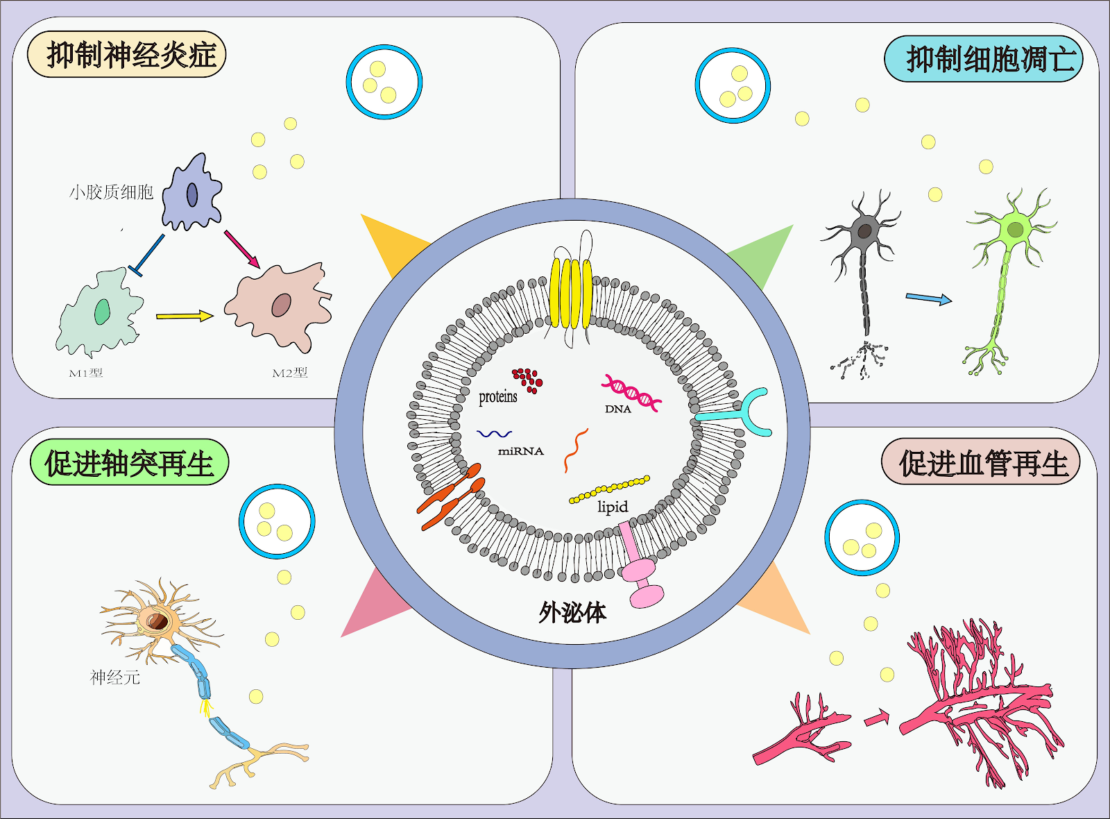
BACKGROUND: Combining exosomes with biomaterials such as hydrogels and biological scaffolds enables targeted delivery, providing effective support for damaged tissue and significantly enhancing the therapeutic efficiency of exosomes, thus positively impacting spinal cord injury repair. OBJECTIVE: To review the action mechanisms and research progress of exosomes combined with biomaterials in spinal cord injury. METHODS: Databases including CNKI, PubMed, and Web of Science were searched using the Chinese and English search terms ā€œspinal cord injury, exosomes, hydrogel, biological scaffold, neuroinflammation, oxidative stress, axonal regeneration, angiogenesis.ā€ Based on the inclusion criteria, 106 articles were finally included in this review. RESULTS AND CONCLUSION: Current research focuses on the repair of secondary injury after spinal cord injury, and how to better alleviate the damage caused by secondary injury is a key research question. Exosomes combined with biomaterials treat spinal cord injury mainly through regulating neuroinflammation, promoting axonal regeneration, and alleviating oxidative stress. This approach avoids immune rejection, solves the problem of low bioavailability of exosomes, and provides effective tissue support, thereby alleviating secondary symptoms after spinal cord injury. Most studies on exosome-loaded biomaterials for spinal cord injury are limited to cell and animal experiments, lacking clinical trial data. Future research should focus on further mechanistic studies, safety evaluations, and related clinical trials.

Read Full Abstract10.12307/2026.21600
Potential and application prospects of combined treatment of acute myocardial infarction with hydrogel cardiac patches and traditional Chinese medicineGraphical AbstractVerified
Chinese Journal of Tissue Engineering Research2026

Potential and application prospects of combined treatment of acute myocardial infarction with hydrogel cardiac patches and traditional Chinese medicine

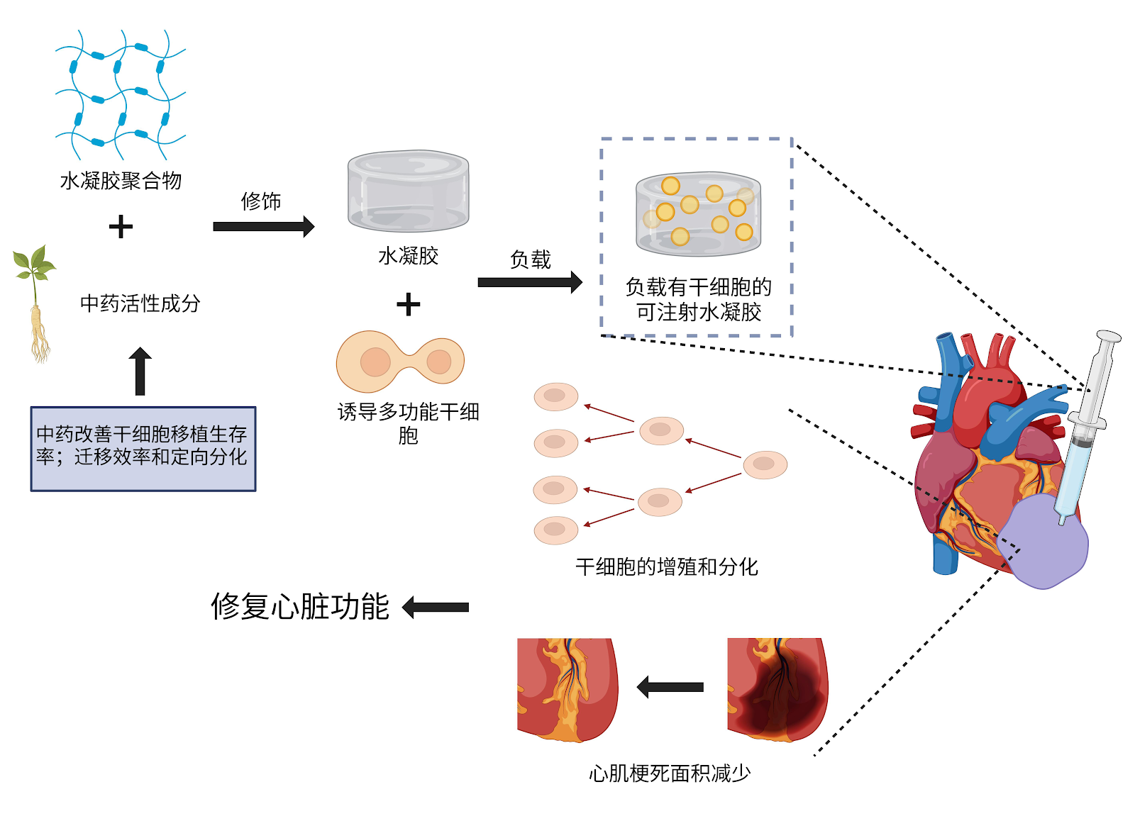
BACKGROUND: Hydrogel cardiac patches, with their excellent biocompatibility and tunable mechanical properties, demonstrate significant potential in treating acute myocardial infarction, especially when combined with stem cell technology. Hydrogels modified with active components of traditional Chinese medicine (TCM) can promote the proliferation, differentiation, migration, and homing of stem cells. This synergistic effect provides a new approach for stem cell transplantation therapy based on hydrogel cardiac patches. OBJECTIVE: To focus on the application scenarios of novel biomaterial hydrogel cardiac patches combined with TCM in the treatment of myocardial infarction, and to discuss their development prospects. METHODS: PubMed and CNKI databases were searched for literature on TCM combined with hydrogel cardiac patches for the treatment of acute myocardial infarction from January 2010 to January 2025. English search terms included "hydrogel, cardiac patch, myocardial infarction, Chinese medicine, stem cell, drug delivery system"; Chinese search terms included "ę°“å‡čƒ¶, åæƒč„č““ē‰‡, åæƒč‚Œę¢—ę­», äø­čÆ, å¹²ē»†čƒž, é€’čÆē³»ē»Ÿ". According to inclusion and exclusion criteria, 99 articles were finally included for review. RESULTS AND CONCLUSION: Hydrogel cardiac patches, with their excellent biocompatibility, tunable mechanical properties, and drug-loading capacity, provide ideal mechanical support and microenvironment for myocardial repair. As a hydrophilic polymer biomaterial, the three-dimensional network structure of hydrogel cardiac patches can highly mimic the physical properties of the natural cardiac extracellular matrix, providing a microenvironment for loaded biological cells to proliferate or maintain activity, and helping cell migration, adhesion, spreading, differentiation, and intercellular connection formation. Hydrogels modified with TCM can load stem cells, fully induce and regulate them, and more effectively perform cell regeneration therapy in the myocardial infarction area to achieve better repair. Therefore, the combined application of TCM and hydrogel cardiac patches has great development potential and prospects. Currently, the combined application mainly focuses on the following aspects: precise sustained-release therapy of TCM active ingredients via hydrogel cardiac patch loading; cell regeneration therapy via induced pluripotent stem cells delivered by TCM-modified hydrogel patches; and repair of cardiac electrophysiological function via TCM combined with conductive hydrogels. In future research, it is necessary to deeply understand the characteristics of hydrogels made from different materials and methods, and the therapeutic effects and mechanisms of TCM monomers and compound extracts, to achieve more combined applications of TCM and hydrogel cardiac patches.

Read Full Abstract10.12307/2026.21583
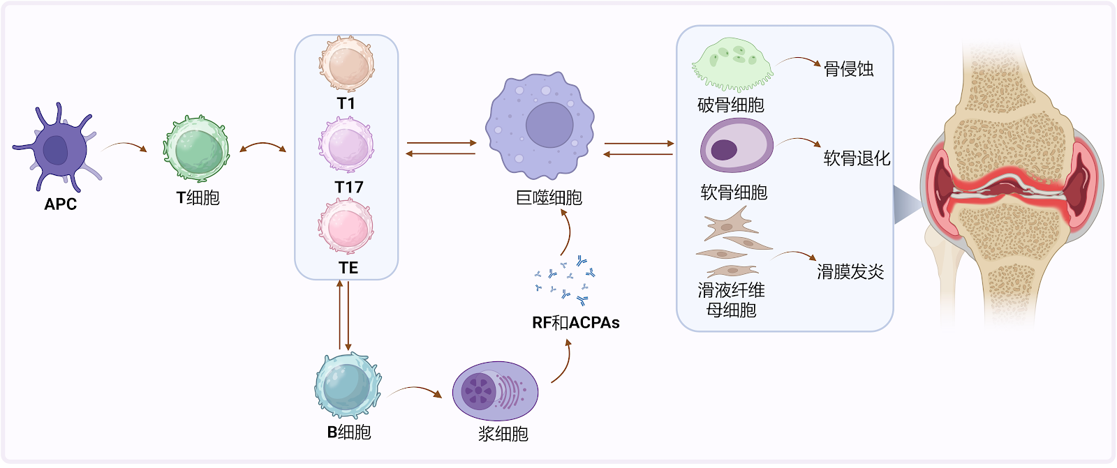
Hydrogel-based drug delivery systems for rheumatoid arthritis treatmentGraphical AbstractVerified
Chinese Journal of Tissue Engineering Research2026

Hydrogel-based drug delivery systems for rheumatoid arthritis treatment

BACKGROUND: In recent years, hydrogels have become an important research direction in the treatment of rheumatoid arthritis due to their excellent biocompatibility, controllable drug release performance, and advantages in multiple drug delivery routes. OBJECTIVE: To systematically review the application of hydrogel-based materials as drug delivery carriers in the treatment of rheumatoid arthritis, and explore the impact of different administration routes on the therapeutic effect. METHODS: Using ā€œhydrogel, rheumatoid arthritis, smart hydrogel system, injectable hydrogel, intra-articular injection, transdermal drug deliveryā€ as Chinese and English search terms, we searched PubMed, Web of Science, CNKI, WanFang Data, and VIP. Based on the inclusion criteria, 62 articles were finally included for review. RESULTS AND CONCLUSION: Hydrogels, leveraging their three-dimensional network structures and tunable physicochemical properties, not only allows drugs to accurately reach the lesion area but also significantly prolongs the retention time of drugs in the joint cavity, making them an ideal carrier in the field of drug delivery. The drug release mechanisms of hydrogels mainly include diffusion, chemical regulation, and swelling-mediated release; in addition, stimulus-responsive hydrogels can dynamically regulate drug release behavior based on environmental conditions (such as pH, temperature, enzyme concentration, etc.). In the treatment of rheumatoid arthritis, common administration routes for hydrogel drug delivery systems include parenteral administration, oral administration, transdermal administration, and intra-articular injection, which significantly reduce systemic adverse reactions, improve drug absorption efficiency, and enhance patient compliance. Although hydrogels as drug delivery carriers have shown significant application potential in the treatment of rheumatoid arthritis, long-term safety, biodegradability, and large-scale production still need breakthroughs to promote the clinical translation of hydrogel drug delivery carriers.

Read Full Abstract10.12307/2026.21586
Hydrogel scaffolds in tissue engineering for urethral repair and reconstructionGraphical AbstractVerified
Chinese Journal of Tissue Engineering Research2026

Hydrogel scaffolds in tissue engineering for urethral repair and reconstruction

BACKGROUND: In recent years, hydrogel scaffolds, as important carriers in the field of tissue engineering, have made significant progress in their application in urethral repair and reconstruction. OBJECTIVE: To systematically review the design strategies of urethral tissue engineering scaffolds, the functional modification of hydrogel scaffolds, and their application progress in urethral repair, and to explore the future development directions and clinical transformation challenges of hydrogel scaffolds. METHODS: The CNKI and PubMed databases were searched using Chinese and English keywords "hydrogel, tissue engineering scaffold, urethral tissue engineering, urethral repair, urethral reconstruction." Based on the inclusion criteria, 69 articles were finally selected for inductive analysis. RESULTS AND CONCLUSION: In urethral tissue engineering, urethral scaffolds should not only possess basic characteristics such as biocompatibility but also exhibit unique properties that adapt to the structure of the urethra and the urinary environment. Hydrogel scaffolds can be classified into natural hydrogel scaffolds (commonly protein-based, polysaccharide-based, DNA-based, and extracellular matrix-based) and synthetic polymer hydrogel scaffolds (commonly polyvinyl alcohol, polyacrylic acid, acrylate-based). Crosslinking methods mainly include physical and chemical crosslinking, and preparation methods include in situ gelation, freeze-drying, electrospinning, and 3D bioprinting. To avoid the drawbacks of single materials, composite hydrogel scaffolds are often used in urethral tissue engineering, such as composite scaffolds adapted to the urethral microenvironment, composite scaffolds utilizing decellularized extracellular matrix, multilayer composite scaffolds loaded with stem cells, and non-stem cell-based hydrogel scaffolds. With the development of tissue engineering and regenerative medicine, future hydrogel scaffolds should be designed with intelligent, engineered, and cell-free strategies, while considering current clinical translation barriers and proposing comprehensive solutions.

Read Full Abstract10.12307/2026.21594
Application strategies of DNA hydrogels for tissue repairGraphical AbstractVerified
Chinese Journal of Tissue Engineering Research2026

Application strategies of DNA hydrogels for tissue repair

BACKGROUND: DNA hydrogels possess thermal resilience, thixotropy, enzyme responsiveness, and degradability. Further functionalization with chemical modifications, peptides, aptamers, and other elements can endow hydrogels with unique responsiveness, demonstrating their immense potential as smart materials. OBJECTIVE: To summarize fabrication strategies, functional properties, and applications of DNA hydrogels. METHODS: PubMed and CNKI databases were searched using "DNA hydrogel, tissue engineering, tissue regeneration" as English and Chinese search terms. Based on inclusion criteria, 76 relevant articles were selected for in-depth analysis. RESULTS AND CONCLUSION: Current research has established diverse preparation methods for DNA hydrogels, including: nanomodule assembly, rolling circle amplification, and hybridization chain reaction for pure DNA hydrogels; grafting DNA molecules onto polymers and adding crosslinkers to form hybrid DNA hydrogels; interpenetrating polymer networks with DNA nanomodule networks; and physical crosslinking. Modular self-assembled hydrogels often exhibit shear-thinning and self-healing capabilities, making them injectable for irregular wounds and bone regeneration with good filling capacity. DNA hydrogels can load, protect, and deliver various bioactive components. They can mimic the extracellular matrix, support 3D cell culture, and serve as delivery platforms for stem cell therapy in bone repair and neural regeneration. Additionally, DNA hydrogels enable theranostics, intelligently responding to different disease requirements. However, clinical translation faces challenges: immunogenicity remains controversial, and laboratory-scale synthesis is costly and limited. Future efforts should focus on integrating bioactive molecules to enhance therapeutic efficacy and improving cost-effectiveness.

Read Full Abstract10.12307/2026.21584
4D bioprinting for regenerative medicine: a new strategy for intelligent material regulation and tissue regenerationGraphical AbstractVerified
Chinese Journal of Tissue Engineering Research2026

4D bioprinting for regenerative medicine: a new strategy for intelligent material regulation and tissue regeneration

BACKGROUND: 4D printing enables dynamic control of structure and function, allowing constructs to more closely mimic complex physiological environments and driving the development of tissue engineering and regenerative medicine towards intelligence and personalization. OBJECTIVE: To review the role and application advances of 4D printing in tissue engineering and regenerative medicine. METHODS: Relevant literature published between 1994 and 2025 was retrieved from CNKI, WanFang, PubMed, and Web of Science. Chinese and English terms were ā€œ4D printing, regenerative medicine, tissue engineering, wound healing, biological ink, intelligent materials.ā€ A total of 115 articles were systematically reviewed and analyzed. RESULTS AND CONCLUSION: 4D bioprinting represents a key advance in tissue engineering and regenerative medicine, integrating diverse fabrication methods and biomaterials to create structures that dynamically respond to environmental cues. 4D bioprinting enhances the biomimicry of natural tissues, enables customized responsiveness, and improves integration with biological systems. 4D bioprinting facilitates the development of tissues and organs, as well as intelligent implants and advanced drug delivery systems, ultimately for tissue repair. 4D bioprinting can seamlessly adapt to the physiological complexity of the human body, apply to personalized medicine, and significantly improve therapeutic outcomes in changing environments.

Read Full Abstract10.12307/2026.21602
Applications and advances of hydrogels in bone tissue engineering repair related to sports injuriesGraphical AbstractVerified
Chinese Journal of Tissue Engineering Research2026

Applications and advances of hydrogels in bone tissue engineering repair related to sports injuries

BACKGROUND: Hydrogel, as a highly biomimetic and modifiable biomaterial, shows a broad application prospect in the field of bone tissue engineering. OBJECTIVE: To review the current research status and development trend of hydrogel in bone tissue engineering. METHODS: The PubMed and CNKI databases were searched for articles on the application of hydrogels in bone tissue engineering. Chinese and English search terms were ā€œhydrogel, bone tissue engineering, nanomaterials, bone regeneration mechanism, bone defect repair.ā€ Based on the inclusion and exclusion criteria, 113 articles were included for review. RESULTS AND CONCLUSION: To improve how hydrogels work for bone tissue engineering, researchers have used different strategies to add new functions. These include changing their physical and chemical properties, adding biological components, and strengthening them with other materials. The goal is to make hydrogels more compatible with the body, stronger, able to break down at a controlled rate, and better at delivering drugs. Functionalized hydrogels help bones regenerate through several ways: by influencing how cells interact, regulating growth factors and signaling pathways, controlling mechanical signals and the surrounding environment, and acting as drug and nano-delivery systems. Future research on hydrogels will concentrate on building systems that combine multiple functions. For instance, by adding features that respond to temperature, pH, enzymes, or magnetic fields, the hydrogels can achieve spatiotemporally controlled release of bioactive factors or drugs, enhancing their dynamic intervention capability throughout the bone regeneration process. Developing polymer materials with biodegradability, tunable mechanical properties, and microenvironment adaptability will effectively improve their stability and tissue integration in complex physiological environments. With the aid of high-precision 3D printing technology, scaffolds with controlled structure, functional zoning, and personalized customization can be constructed to match the structural characteristics of individual patient defects.

Read Full Abstract10.12307/2026.21592
Material selection and manufacture method of non-porous adherent cell microcarriersGraphical AbstractVerified
Chinese Journal of Tissue Engineering Research2026

Material selection and manufacture method of non-porous adherent cell microcarriers

BACKGROUND: The key consumable item in stirred-tank bioreactors is non-porous microcarriers, which are suitable for adherent cell culture and play a significant role in the fabrication of viruses, recombinant proteins, and stem cells. OBJECTIVE: To summarize the material selection and manufacturing methods of non-porous microcarriers based on the favorable conditions for cell-microcarrier adhesion. METHODS: A computerized search of CNKI, PubMed, and Web of Science databases was performed with the search terms ā€œcell cultivation, adherent cells, microcarrier, fibronectin, bioreactor, microsphere preparationā€ in Chinese and ā€œcell adhesion, microcarrier, bioreactor, dextran, cell-matrix interaction, suspension cultureā€ in English. The search time limit was from 1967 to 2025. After screening according to the inclusion and exclusion criteria, 52 articles were finally included for summary analysis. RESULTS AND CONCLUSION: Non-porous microcarriers are suitable for adherent cell culture at a density of 109-1010 cells/L. Due to the requirements for cell adhesion, non-porous microcarriers need to provide appropriate surface positive charge or integrin binding sites. Dextran, polystyrene, and collagen-based non-porous microcarriers have been commercialized and can widely support large-scale culture of various cells. Chitosan and cellulose-based non-porous microcarriers have been reported for large-scale cell culture, but these new materials have not been commercialized. Dextran and polystyrene microcarriers have been commercialized for a long time, with preparation methods of crosslinking and polymerization, respectively, and mature preparation technology. Collagen and cellulose microcarriers are mostly prepared by crosslinking, while chitosan microcarriers can be prepared by crosslinking and phase inversion.

Read Full Abstract10.12307/2026.21597
Application of self-healing hydrogels for sports injury prevention and rehabilitationGraphical AbstractVerified
Chinese Journal of Tissue Engineering Research2026

Application of self-healing hydrogels for sports injury prevention and rehabilitation

BACKGROUND: Self-healing hydrogel, as a novel smart material, has attracted much attention in sports medicine and rehabilitation engineering due to its high water content, favorable biocompatibility, tailorable mechanical properties, and intrinsic ability to self-heal after damage. OBJECTIVE: To systematically review the progress of self-healing hydrogels in the prevention, treatment, and rehabilitation of sports injuries. METHODS: A search was conducted on authoritative domestic and international databases including CNKI, X-mol, and PubMed. Research literature involving self-healing hydrogels and sports medicine was selected. English and Chinese search terms included "self-healing hydrogel, sports injury repair, activity monitoring, smart rehabilitation, wearable sensors." Based on inclusion criteria, 136 articles were finally included in this review. RESULTS AND CONCLUSION: Self-healing hydrogels show significant advantages in the repair of ligaments, tendons, cartilage, and bone tissue, providing essential mechanical support and promoting cell adhesion, proliferation, and differentiation. By controlling the release of drugs or growth factors, they achieve anti-inflammatory, analgesic, and accelerated tissue regeneration effects. In the fields of motion monitoring and rehabilitation, self-healing hydrogels serve as core components of flexible sensors for real-time monitoring of movement posture and joint stress, and can be integrated with virtual reality, augmented reality, and telemedicine platforms to promote intelligent and precise rehabilitation training. Self-healing hydrogels are evolving toward functional integration, extending beyond simple material repair to a synergistic platform integrating monitoring, treatment, and rehabilitation. Despite significant progress in recent years, challenges remain in mechanical durability, rapid healing efficiency, scalable preparation, and clinical translation. Future research should focus on optimizing high-strength, multi-stimuli-responsive hydrogel designs, deepening the integration of sports biomechanics and artificial intelligence, constructing data-driven personalized rehabilitation models, and promoting interdisciplinary collaboration and establishment of testing standards, to provide more scientific and standardized solutions for the prevention, intervention, and rehabilitation of sports injuries.

Read Full Abstract10.12307/2026.21590
Properties of machinable resin-ceramic composites and their application in onlay restorationsGraphical AbstractVerified
Chinese Journal of Tissue Engineering Research2026

Properties of machinable resin-ceramic composites and their application in onlay restorations

BACKGROUND: Resin-ceramic composites have gradually become an important choice for onlay restoration materials due to their advantages such as good aesthetics, elastic modulus close to that of dental tissues, excellent repair performance, and high efficiency in manufacturing processes. OBJECTIVE: To review the performance of computer-aided design and computer-aided manufacturing (CAD/CAM) milled resin-ceramic composite materials and their current application in minimally invasive onlay restorations. METHODS: Using "machinable resin ceramic material, nano resin ceramic, resin-infiltrated ceramic, onlay" as Chinese keywords and "CAD/CAM resin ceramic, resin nano ceramic, polymer-infiltrated ceramic network, onlay" as English keywords, a systematic search was conducted in the CNKI and PubMed databases. The search covered relevant literature published from January 2020 to August 2025, with a small number of earlier publications also included. The titles and abstracts of the retrieved literature were initially screened, and the full texts of the selected literature were thoroughly reviewed. Studies with low relevance, incomplete data, or duplicate research were excluded, and 76 articles were finally included for review. RESULTS AND CONCLUSION: Machinable resin-ceramic composites are a class of chairside restorative materials composed of resin and inorganic ceramics, mainly divided into nano resin ceramics and resin-infiltrated ceramics. Existing research data and clinical applications indicate that the mechanical properties of machinable resin-ceramic composites are close to those of traditional glass ceramics, with elastic modulus matching dentin, good fatigue resistance and wear resistance, and significantly improved bonding strength to dentin after targeted surface treatment, showing stable performance in complex oral environments. When applied to onlay restorations, strict selection of indications, avoidance of high-risk factors, and adherence to minimally invasive preparation principles can achieve high restoration success and survival rates, as well as high patient satisfaction. Currently, there are still complications such as fracture and debonding in clinical applications, requiring more research to further improve material properties and optimize clinical outcomes.

Read Full Abstract10.12307/2026.21596
Characterization and biological performance of manganese-doped hydroxyapatite/polydopamine composite materialsGraphical AbstractVerified
Chinese Journal of Tissue Engineering Research2026

Characterization and biological performance of manganese-doped hydroxyapatite/polydopamine composite materials

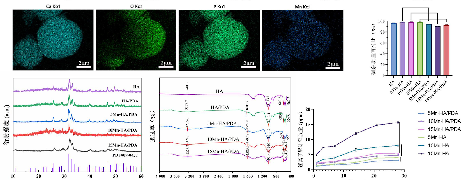
BACKGROUND: Hydroxyapatite is widely used in bone tissue engineering due to its excellent osteoconductivity. However, its limited osteoinductivity restricts its clinical application and therapeutic efficacy. OBJECTIVE: To construct manganese-doped hydroxyapatite/polydopamine composite bone graft materials and characterize their physicochemical and biological properties. METHODS: (1) Using Ca(NOā‚ƒ)ā‚‚Ā·4Hā‚‚O as the calcium source, (NH4)2HPO4 as the phosphorus source, and manganese nitrate solution as the manganese source, manganese-doped hydroxyapatite was prepared by hydrothermal homogeneous coprecipitation with manganese/(calcium+manganese) molar ratios of 5%, 10%, and 15%, respectively. The corresponding materials were denoted as 5Mn-HA, 10Mn-HA, and 15Mn-HA. Hydroxyapatite and the three manganese-doped hydroxyapatite materials were immersed in dopamine hydrochloride-Tris buffer solution to prepare manganese-doped hydroxyapatite/polydopamine composites, denoted as HA/PDA, 5Mn-HA/PDA, 10Mn-HA/PDA, and 15Mn-HA/PDA. The morphology, manganese ion release, degradation rate, and cytocompatibility were characterized. The manganese-doped material with better cytocompatibility was selected for subsequent experiments. (2) Hydroxyapatite, HA/PDA, 5Mn-HA, and 5Mn-HA/PDA were co-cultured with rat bone marrow mesenchymal stem cells. After osteogenic induction, alkaline phosphatase activity and alizarin red S staining were performed, and q-PCR was used to detect Runx2 and osteocalcin mRNA expression. (3) In 24 SD rats, two circular full-thickness bone defects of 5 mm diameter were created on each side of the calvarium. The right defects were implanted with hydroxyapatite, HA/PDA, 5Mn-HA, or 5Mn-HA/PDA (6 rats per material), while the left defects served as blank controls. At 4 and 8 weeks post-surgery, samples were harvested for Micro-CT scanning, hematoxylin-eosin and Masson staining. RESULTS AND CONCLUSION: (1) The manganese-doped hydroxyapatite/polydopamine composites were microspherical with particle sizes ranging from 9.86 to 13 μm. With increasing manganese doping, the release of manganese ions increased, and the release rate from the composites was lower than that from the corresponding manganese-doped hydroxyapatite. The composites exhibited faster degradation rates compared to manganese-doped hydroxyapatite. Based on live/dead staining and CCK-8 assays, 5Mn-HA showed no obvious cytotoxicity; therefore, 5Mn-HA and 5Mn-HA/PDA were selected for further osteogenic evaluation and in vivo verification. (2) Combined results of alkaline phosphatase activity, alizarin red S staining, and q-PCR indicated that 5Mn-HA/PDA had the strongest osteogenic ability among the four materials. (3) Micro-CT scanning showed that the blank control group had the slowest bone repair and least new bone formation, while the 5Mn-HA/PDA group had the fastest bone repair and most new bone. Hematoxylin-eosin and Masson staining further confirmed the Micro-CT results. (4) In conclusion, manganese-doped hydroxyapatite/polydopamine composites possess good physicochemical and biological properties.

Read Full Abstract10.12307/2026.21572
Postmenopausal cognitive impairment: a bibliometric analysis of developmental context and hot trendsGraphical AbstractVerified
Chinese Journal of Tissue Engineering Research2026

Postmenopausal cognitive impairment: a bibliometric analysis of developmental context and hot trends

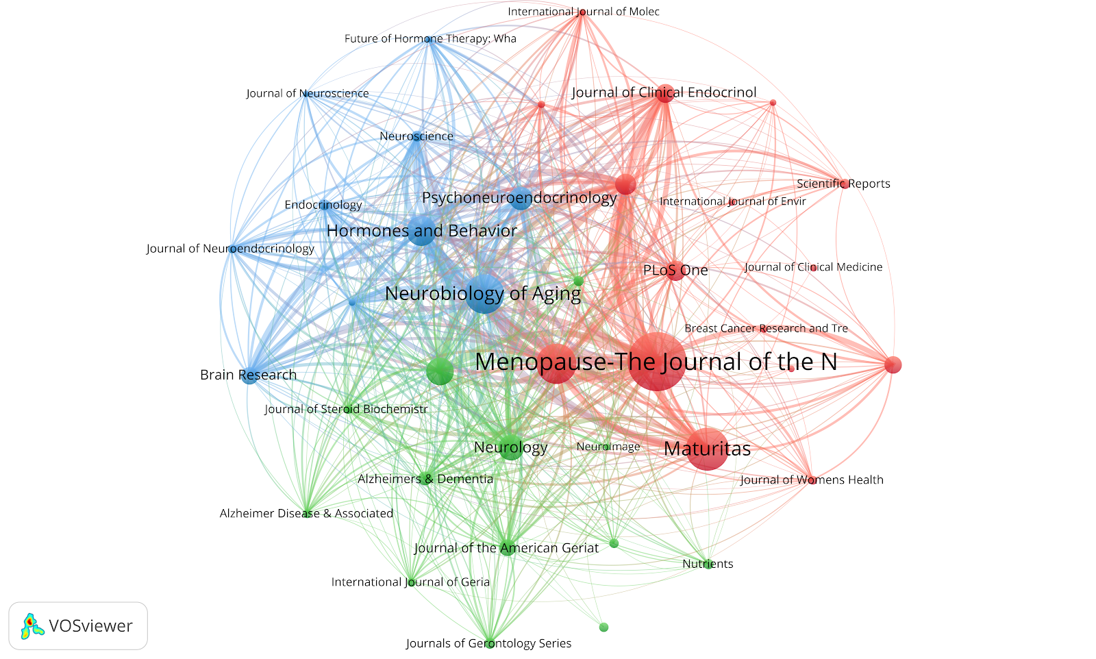
BACKGROUND: At present, there are few longitudinal studies on postmenopausal cognitive impairment, and there is no systematic bibliometric analysis of its development context and hot trends. OBJECTIVE: To systematically integrate the related research on postmenopausal cognitive impairment with the help of bibliometric tools, identify the research trends and hot trends in this field, and promote interdisciplinary cooperation and clinical transformation. METHODS: The Web of Science Core Collection database was retrieved for literature related to postmenopausal cognitive impairment published from January 1, 2005 to March 24, 2025. Visualization analysis was conducted using software such as VOSviewer, the R package ā€œBibliometrix,ā€ and CiteSpace. RESULTS AND CONCLUSION: A total of 1 452 articles were included, involving 71 countries, 1 830 institutions, 510 journals, and 6 909 authors. The annual publication volume showed a fluctuating upward trend. The United States dominated the field, with Stanford University, University of Pittsburgh, Harvard Medical School, and University of Illinois leading. Menopause-The Journal of the North American Menopause Society published the most articles (78), and JAMA-J Am Med Assoc had the highest co-citation count (3 791). Espeland, Mark A. ranked first with 55 publications, and Shumaker, Sally A. was the most co-cited author (859 times). High-frequency keyword ā€œestrogenā€ (211 times) and burst keywords ā€œplus progestinā€ (strength=15.77) and ā€œestrogen replacement therapyā€ (strength=15.57) indicated that the long-term effects and safety of hormonal interventions remained the core research focus. Keyword clustering revealed four research directions. Future research could focus on constructing a ā€œcross-life cycle-multidimensionalā€ framework, which would help comprehensively understand the mechanisms of hormones on cognitive function and neuropsychiatric status at different life stages. The shift in hot topics from single issues (e.g., magnetic resonance imaging, hormones and cognition) to multiple health issues (psychological, tumor risk, cardiovascular, metabolic diseases, physical activity, etc.) and the non-hormonal turn in intervention strategies reflected the development trend of the discipline.

Read Full Abstract10.12307/2026.21608
Spinal injury risk assessment of a double-layer cushion for ejection seats based on ABAQUSGraphical AbstractVerified
Chinese Journal of Tissue Engineering Research2026

Spinal injury risk assessment of a double-layer cushion for ejection seats based on ABAQUS

BACKGROUND: The enormous impact acceleration experienced by the human body during ejection is a major risk factor for spinal injury. Therefore, optimizing the cushioning and energy absorption design of ejection seat cushions is crucial for ensuring the safety of pilots. OBJECTIVE: To construct a human-seat cushion coupled finite element model based on ABAQUS to quantitatively evaluate the impact of a double-layer cushion, which combines the advantages of high and slow rebound, on lumbar spine biomechanical response and spinal injury risk under ejection conditions, and to quantify its safety margin. METHODS: CT imaging data of the entire spine and legs from one male pilot volunteer were selected. Three-dimensional anatomical reconstruction, geometric repair, and finite element meshing were completed to construct a highly biologically faithful digital human model encompassing the entire spine, pelvis, both femurs, and skin soft tissues. A geometric model of the dual-layer seat cushion was also established. Subsequently, a human-chair system coupled model was assembled in ABAQUS. Dynamic simulations were conducted by applying an ejection acceleration time history. Stress responses in the L4–L5 and L5–S1 intervertebral discs were compared between the high-resilience and dual-layer cushion designs. Injury probability was predicted using the Spinal Injury Dynamic Response Index. RESULTS AND CONCLUSION: The simulation results showed good agreement with experimental data, validating the model's accuracy. Performance comparison indicated that, compared with the traditional high-resilience cushion, the double-layer cushion reduced peak stresses in the L4–L5 and L5–S1 intervertebral discs, decreased the Dynamic Response Index by 1.8%, and reduced the spinal injury probability by 10.6%. While meeting relevant limits, it provided a higher safety margin and effectively reduced the risk of spinal injury under ejection impact loads.

Read Full Abstract10.12307/2026.21579
Antibacterial properties of photocrosslinkable hydrogel loaded with quercetin-silver nanoparticles for infected woundsGraphical AbstractVerified
Chinese Journal of Tissue Engineering Research2026

Antibacterial properties of photocrosslinkable hydrogel loaded with quercetin-silver nanoparticles for infected wounds

BACKGROUND: Numerous studies have confirmed that quercetin can inhibit the release of inflammatory factors and reduce local inflammatory responses in wounds, creating favorable conditions for wound healing. However, quercetin has poor water solubility and low bioavailability, which limits its application in wound treatment. OBJECTIVE: To investigate the antibacterial properties of quercetin-silver nanoparticle/methacrylated hyaluronic acid composite hydrogels. METHODS: Quercetin-silver nanoparticles (QuAgNPs) were synthesized by hydrothermal reduction method. QuAgNPs were loaded into methacrylated hyaluronic acid (HAMA) hydrogel precursor solution at different mass concentrations (1, 5, 10, 20, 40, 60, 80 μg/mL) to prepare composite hydrogels, denoted as 1, 5, 10, 20, 40, 60, 80 μg/mL QuAgNPs/HAMA hydrogels, while pure HAMA hydrogels were also prepared. Staphylococcus aureus was co-cultured with 1, 5, 10, 20, 40, 60, 80 μg/mL QuAgNPs/HAMA hydrogels, and antibacterial properties were detected by inhibition zone experiment. In vitro drug release properties of 60 and 80 μg/mL QuAgNPs/HAMA hydrogels were tested. Based on the results of the inhibition zone experiment, appropriate QuAgNPs/HAMA hydrogels were selected for subsequent experiments. The microstructure of HAMA hydrogel and 60 μg/mL QuAgNPs/HAMA hydrogel was observed under scanning electron microscope. Staphylococcus aureus (or Escherichia coli) were co-cultured with QuAgNPs, HAMA hydrogel, and 60 μg/mL QuAgNPs/HAMA hydrogel, with bacteria cultured alone as blank control, and antibacterial rates were detected by spread plate antibacterial experiment. Human umbilical vein endothelial cells were co-cultured with QuAgNPs, HAMA hydrogel extract, and 60 μg/mL QuAgNPs/HAMA hydrogel extract, with cells cultured alone as blank control, and cell viability was detected by live/dead cell staining. RESULTS AND CONCLUSION: The inhibition zone experiment showed that the antibacterial properties of 60 and 80 μg/mL QuAgNPs/HAMA hydrogels were stronger than those of 1, 5, 10, 20, 40 μg/mL QuAgNPs/HAMA hydrogels, and there was no difference between 60 and 80 μg/mL QuAgNPs/HAMA hydrogels. Both 60 and 80 μg/mL QuAgNPs/HAMA hydrogels had good in vitro drug release properties. Therefore, 60 μg/mL QuAgNPs/HAMA hydrogel was selected for subsequent experiments. Scanning electron microscopy showed that HAMA hydrogel had a loose porous structure, and approximately spherical QuAgNPs particles were scattered on the surface of 60 μg/mL QuAgNPs/HAMA hydrogel. Spread plate antibacterial experiment showed that QuAgNPs and 60 μg/mL QuAgNPs/HAMA hydrogel had significant inhibitory effects on Staphylococcus aureus and Escherichia coli. Live/dead cell staining showed that QuAgNPs, HAMA hydrogel, and 60 μg/mL QuAgNPs/HAMA hydrogel did not affect cell viability and had good cytocompatibility. These results indicate that QuAgNPs/HAMA composite hydrogel has good antibacterial properties.

Read Full Abstract10.12307/2026.21571
Regulatory effects of optimized extraction processes for chlorella-derived peptides on key pathological links in rheumatoid arthritisGraphical AbstractVerified
Chinese Journal of Tissue Engineering Research2026

Regulatory effects of optimized extraction processes for chlorella-derived peptides on key pathological links in rheumatoid arthritis

BACKGROUND: Recent studies have shown that Chlorella possesses potential value in treating rheumatoid arthritis. The pathological progression of rheumatoid arthritis is closely associated with an imbalance in oxidative stress, abnormal macrophage polarization, aggressive activation of fibroblast-like synoviocytes, and disturbances in the vascular endothelial system. However, the optimization of extraction processes for peptides derived from Chlorella and their regulatory effects on key pathological links of rheumatoid arthritis remain to be systematically validated. OBJECTIVE: To optimize the extraction process of antioxidant peptides from Chlorella, clarify their antioxidant activity and biosafety, and explore their regulatory effects on pathological phenotypes of rheumatoid arthritis-related cells (RAW 264.7 mouse monocyte macrophage leukemia cells, fibroblast-like synoviocytes, and human umbilical vein endothelial cells), providing experimental evidence for the treatment of rheumatoid arthritis with Chlorella peptides. METHODS: (1) Chlorella peptide extract was prepared by bromelain enzymatic hydrolysis combined with phosphomolybdic acid precipitation. Using peptide yield as the evaluation index, the extraction process parameters were optimized by single-factor experiments, including solid-liquid ratio, enzymatic hydrolysis time, and reaction system pH. (2) The peptide content was determined by BCA method, antioxidant capacity was detected by ABTS method, and biosafety of peptides on RAW 264.7 cells, fibroblast-like synoviocytes, and human umbilical vein endothelial cells was evaluated by CCK-8 method. (3) An inflammatory model of RAW 264.7 cells induced by lipopolysaccharide was established. The effects of peptides on intracellular reactive oxygen species levels and M1/M2 polarization phenotypes were detected by DCFH-DA staining, flow cytometry, and real-time fluorescence quantitative reverse transcription polymerase chain reaction. (4) An activation model of fibroblast-like synoviocytes induced by tumor necrosis factor-alpha was established. The effects of peptides on migration, proliferation, invasion, and related gene expression of fibroblast-like synoviocytes were detected by wound healing assay, EdU proliferation assay, Transwell invasion assay, and real-time fluorescence quantitative reverse transcription polymerase chain reaction. (5) An abnormal activation model of human umbilical vein endothelial cells induced by vascular endothelial growth factor A was established. The effects of peptides on migration, tube formation, and expression of hypoxia-inducible factor 1 alpha and vascular endothelial growth factor A genes were detected by wound healing assay, Transwell assay, tube formation assay, and real-time fluorescence quantitative reverse transcription polymerase chain reaction. RESULTS AND CONCLUSION: (1) The optimal extraction process for Chlorella peptides was solid-liquid ratio of 2:1 (g:100 mL), enzymatic hydrolysis time of 60 minutes, and reaction system pH of 6.5, yielding the highest peptide yield. (2) Chlorella peptides exhibited concentration-dependent antioxidant activity and showed no obvious toxicity to RAW 264.7 cells, fibroblast-like synoviocytes, and human umbilical vein endothelial cells in the concentration range of 1-10 μg/mL, indicating good biocompatibility. (3) Chlorella peptides dose-dependently inhibited lipopolysaccharide-induced reactive oxygen species generation in RAW 264.7 cells, downregulated M1 pro-inflammatory genes such as interleukin-1 beta and tumor necrosis factor-alpha, upregulated M2 anti-inflammatory genes such as interleukin-10 and arginase 1, and promoted macrophage polarization from M1 to M2 phenotype. (4) Chlorella peptides significantly inhibited tumor necrosis factor-alpha-induced migration, proliferation, and invasion of fibroblast-like synoviocytes, and downregulated the expression of interleukin-6, matrix metalloproteinase 13, tumor necrosis factor receptor superfamily member 11A, and C-X-C motif chemokine ligand 12. (5) Chlorella peptides effectively inhibited vascular endothelial growth factor A-induced migration and tube formation of human umbilical vein endothelial cells, and reduced the expression of hypoxia-inducible factor 1 alpha and vascular endothelial growth factor A genes. These results indicate that Chlorella peptides regulate multiple pathological links of rheumatoid arthritis through anti-oxidative stress, regulation of macrophage polarization, inhibition of aggressive phenotype of fibroblast-like synoviocytes, and improvement of vascular endothelial disorders, suggesting potential therapeutic value for rheumatoid arthritis.

Read Full Abstract10.12307/2026.21577