🧬 SinoBioData Academic Portal
šŸ“š Peer-Reviewed Translated Literature

All Biomedical & Clinical Articles (Page 18)

Browse complete peer-reviewed translations from top Chinese biomedical, oncology, and genomics journals. Read verified previews and download full authentic clinical reports.

Published Research Papers

Showing 24 of 1542 peer-reviewed translated articles (Page 18 of 65)

Impact of lesser trochanter displacement on hip function following minimally invasive intramedullary nailing for geriatric intertrochanteric fracturesGraphical AbstractVerified
Chinese Journal of Tissue Engineering Research2026

Impact of lesser trochanter displacement on hip function following minimally invasive intramedullary nailing for geriatric intertrochanteric fractures

BACKGROUND: Percutaneous minimally invasive intramedullary nailing is a common and effective treatment for intertrochanteric fractures in the elderly, effectively avoiding complications. However, intertrochanteric fractures involving the lesser trochanter remain problematic, particularly with lesser trochanteric separation. Due to the diverse types of lesser trochanteric separation, there are few reports on whether lesser trochanteric separation should be fixed. OBJECTIVE: To investigate the impact of lesser trochanter displacement distance on postoperative functional outcomes in elderly patients with intertrochanteric fractures treated with minimally invasive intramedullary nailing, providing clinical data to determine whether fixation is necessary. METHODS: A retrospective analysis was conducted on 46 elderly patients with intertrochanteric fractures and lesser trochanter separation who underwent intramedullary nailing fixation and received effective follow-up at the Department of Emergency Surgery, Guizhou Provincial People's Hospital from August 2020 to December 2023. Patients were divided into two groups based on displacement distance: group A (displacement ≄1 cm, n=23) and group B (displacement <1 cm, n=23). Postoperative functional recovery indicators were compared, including time to first straight leg raise, time to first ambulation, fracture healing time, Harris hip scores at 1, 2, 3, 6, 9, and 12 months postoperatively, and excellent rate of Harris score at final follow-up. RESULTS AND CONCLUSION: (1) No significant differences in preoperative general data between the two groups (P > 0.05). (2) Group A had significantly longer time to first straight leg raise (18.44±3.99 days vs. 15.91±3.64 days, P < 0.05) and time to first ambulation (7.83±1.92 days vs. 6.70±1.80 days, P < 0.05) compared to group B. However, fracture healing time showed no significant difference (P > 0.05). (3) Harris scores in group B were significantly higher than group A at 1, 2, 3, and 6 months postoperatively (P < 0.05), but no significant differences at 9 and 12 months (P > 0.05). (4) At final follow-up, excellent rate of Harris score (≄90) was 74% in group A and 87% in group B, with no significant difference (P > 0.05). (5) These findings indicate that elderly patients with different lesser trochanter displacement distances have different functional recovery within 6 months postoperatively, but after 6 months, the displaced lesser trochanter does not affect hip function. Therefore, routine reduction and fixation of the lesser trochanter is not recommended for elderly intertrochanteric fracture patients. However, for patients with high early functional demands, reduction and fixation may be considered after individual assessment of activity needs and physical condition.


Read Full Abstract10.12307/2026.21645
Application of finite element analysis in unicompartmental knee arthroplasty for knee osteoarthritisGraphical AbstractVerified
Chinese Journal of Tissue Engineering Research2026

Application of finite element analysis in unicompartmental knee arthroplasty for knee osteoarthritis

BACKGROUND: Single condylar knee arthroplasty is an effective method to treat unilateral compartment knee osteoarthritis. As an advanced biomechanical research tool, finite element analysis provides important support for the prosthesis design and surgical planning of single condylar knee arthroplasty and the biomechanical characteristics of knee joint after unicompartmental knee arthroplasty. OBJECTIVE: To explore the application progress of finite element analysis in unicompartmental knee arthroplasty, including the optimization of unicompartmental knee arthroplasty prosthesis design, surgical planning and biomechanical characteristics of knee joint after unicompartmental knee arthroplasty. METHODS: The first author used PubMed, CNKI, and WanFang for articles published from database inception to August 2025. English search terms were "FEA, UKA, knee osteoarthritis, knee joint, femoral component, tibia component, biomechanics, ligament of knee joint." Chinese search terms were "finite element analysis, unicompartmental knee arthroplasty, knee osteoarth..." (truncated in original). RESULTS AND CONCLUSION: ā‘ Optimization of prosthesis design: Finite element analysis can provide precise data guidance for the optimization of unicompartmental prosthesis design by constructing different unicompartmental prosthesis replacement models and analyzing stress distribution under different prosthesis structures; ā‘”Precise surgical planning: Finite element analysis can precisely predict the effects of different osteotomy schemes on knee biomechanics, including tibial posterior slope, joint line position, and lower limb alignment, providing theoretical guidance for surgeons to choose the best surgical plan, improve surgical accuracy, reduce complication risk, and enhance long-term prosthesis survival; ā‘¢Using finite element analysis to predict biomechanical characteristics after unicompartmental replacement can non-invasively and predictively simulate postoperative knee mechanical behavior, including ligament tension, cartilage contact pressure, and bone strain distribution, solving mechanical details that two-dimensional imaging cannot capture.

Read Full Abstract10.12307/2026.21685
Low-frequency repetitive transcranial magnetic stimulation improves limb motor function and upper limb muscles in stroke patients: a meta-analysisGraphical AbstractVerified
Chinese Journal of Tissue Engineering Research2026

Low-frequency repetitive transcranial magnetic stimulation improves limb motor function and upper limb muscles in stroke patients: a meta-analysis

OBJECTIVE: Low-frequency repetitive transcranial magnetic stimulation significantly improves upper limb motor function in stroke patients, but its efficacy in reducing upper limb muscle spasticity remains controversial. Therefore, this study systematically evaluated the effects of low-frequency repetitive transcranial magnetic stimulation on limb motor function and upper limb muscle spasticity in stroke patients. METHODS: Utilizing computer search technology, this study meticulously screened randomized controlled studies targeting stroke patients undergoing low-frequency repetitive transcranial magnetic stimulation treatment from databases such as PubMed, EMBASE, ScienceDirect, Cochrane Library, Web of Science, China National Knowledge Infrastructure, VIP, Wanfang, and China Biology Medicine disc. The experimental group received low-frequency repetitive transcranial magnetic stimulation alone or combined with physical therapy/conventional rehabilitation, while the control group received conventional rehabilitation, sham stimulation, or physical therapy. RevMan 5.3 software was used for meta-analysis. RESULTS: A total of 8 high-quality studies involving 502 patients were included. Meta-analysis showed that compared with the control group, the experimental group had significantly higher scores on the Brunnstrom hand motor function scale, Fugl-Meyer motor function scale, modified Barthel index, and Berg Balance Scale [MD=1.51, 95%CI(1.22, 1.80), P < 0.000 01; MD=5.69, 95%CI(3.18, 8.20), P < 0.000 01; MD=8.55, 95%CI(3.27, 13.84), P=0.002; MD=5.72, 95%CI(3.13, 8.32), P < 0.000 1], while there was no significant difference in the modified Ashworth score between the two groups [MD=-0.11, 95%CI(-0.17, 0.85), P=0.82]. CONCLUSION: Low-frequency repetitive transcranial magnetic stimulation can effectively improve limb motor function in stroke patients, especially hand motor function, overall limb motor function, activities of daily living, and balance ability, but it did not show a significant advantage in improving upper limb muscle spasticity. This conclusion still needs to be further verified by more studies with higher methodological quality, longer intervention duration, and follow-up.

Read Full Abstract10.12307/2026.21681
Deep learning in bone imaging diagnosisGraphical AbstractVerified
Chinese Journal of Tissue Engineering Research2026

Deep learning in bone imaging diagnosis

BACKGROUND: Deep learning methods have made breakthrough progress in the field of bone imaging diagnosis. They have overcome the problems of easy misdiagnosis and low efficiency in traditional bone imaging diagnosis methods, and are conducive to the popularization of intelligent diagnosis methods in orthopedics. OBJECTIVE: To review the application, advantages and disadvantages of deep learning in the diagnosis of common bone diseases. METHODS: Literature published from January 2021 to June 2025 on deep learning-assisted skeletal image diagnosis was retrieved from CNKI, WanFang, PubMed, and Web of Science databases. Chinese and English search terms included ā€œartificial intelligence, deep learning, machine learning, computer-aided diagnosis, skeletal imaging, fracture, bone tumor, osteoporosis, osteoarthritis, synovitis, spinal, cartilage, classification, detection, segmentation.ā€ According to the inclusion criteria, 76 articles were finally included in this review. RESULTS AND CONCLUSION: Deep learning models have become powerful tools for bone imaging diagnosis and are gradually gaining recognition from clinicians, improving the efficiency of bone imaging diagnosis. Deep learning technology uses its image feature capture ability to help improve the clinical diagnosis of fractures, bone tumors, osteoporosis, osteoarthritis, synovitis, and spinal lesions, providing a reference for clinical decision-making. Although deep learning diagnostic applications have great potential, they are prone to insufficient model generalization and rely heavily on large amounts of annotated data, which reduces model credibility and hinders clinical translation. Future research should focus on improving the robustness and generalization of deep learning models. In summary, deep learning has certain reference value in clinical bone imaging diagnosis.

Read Full Abstract10.12307/2026.21692
3D-printed personalized osteotomy guides affect mid- to long-term efficacy of unicompartmental knee arthroplasty: mechanisms, evidence, and prospectsGraphical AbstractVerified
Chinese Journal of Tissue Engineering Research2026

3D-printed personalized osteotomy guides affect mid- to long-term efficacy of unicompartmental knee arthroplasty: mechanisms, evidence, and prospects

BACKGROUND: Unicompartmental knee arthroplasty is a commonly used surgical method for treating localized knee osteoarthritis, and its success relies on precise osteotomy and prosthesis positioning. Traditional techniques depend on the surgeon's experience and two-dimensional imaging, which makes it difficult to completely avoid alignment errors; these errors may affect the prosthesis survival rate and long-term efficacy. OBJECTIVE: To systematically summarize the factors determining the mid- and long-term efficacy of unicompartmental knee arthroplasty, analyze the technical principles of personalized osteotomy guides based on 3D printing combined with digital design, artificial intelligence planning, and physical manufacturing, providing evidence to improve surgical accuracy. Furthermore, this article elaborates on enhancing surgical precision by optimizing biomechanics and reducing abnormal prosthesis wear, thereby improving prosthesis survival rate, maintaining joint function, and reducing the risk of adverse reactions. METHODS: Relevant literature published in PubMed and China National Knowledge Infrastructure was searched, using Chinese and English search terms including ā€œ3D printing, unicompartmental knee arthroplasty, patient-specific instrumentation, mid-term, long-term, outcome.ā€ After screening, a total of 65 articles were included for analysis. RESULTS AND CONCLUSION: ā‘ 3D-printed personalized osteotomy guides, created through CT scanning, three-dimensional reconstruction, and 3D printing, significantly improve the surgical accuracy of unicompartmental knee arthroplasty. ā‘”Compared with traditional surgery, 3D-printed personalized osteotomy guides reduce coronal and sagittal prosthesis placement errors, improve lower limb alignment, increase prosthesis survival rate, and reduce mid- to long-term adverse reactions such as aseptic loosening. However, there are controversies including suboptimal tibial rotational alignment, increased costs, and no significant advantage in short-term functional scores. ā‘¢Compared with robotic assistance, 3D-printed personalized osteotomy guides are slightly less accurate but have a shorter learning curve and lower cost. Currently, there is a lack of long-term randomized controlled evidence in this field, and the technique relies on high-quality imaging data with a long preoperative preparation period. ā‘£By analyzing existing research on personalized osteotomy guides, this article summarizes the role of this technology in improving surgical precision, while also discussing its current status and limitations in terms of cost-effectiveness, clinical workflow integration, and insufficient high-quality evidence. ⑤To promote the widespread application of personalized guides in precise unicompartmental knee arthroplasty treatment, future efforts should include multicenter long-term randomized controlled trials, integration of artificial intelligence and intraoperative dynamic navigation, and the establishment of predictive models based on real-world data, thereby providing a more solid theoretical basis for its clinical application.

Read Full Abstract10.12307/2026.21686
Digital measurement and correlation analysis of anatomical CT for clinical application in the hip jointGraphical AbstractVerified
Chinese Journal of Tissue Engineering Research2026

Digital measurement and correlation analysis of anatomical CT for clinical application in the hip joint

BACKGROUND: The complexity of hip joint anatomy makes it difficult to quantitatively assess by parameter indexes, and real-image rendering technology is important for the accurate display of hip joint microstructure and detection of diseases. OBJECTIVE: To evaluate the anatomical and morphological parameters of the hip joint through three-dimensional digital measurement by CT multi-planar reconstruction and cinematic rendering, compare the differences and analyze the correlation, so as to provide a precise anatomical parameter basis for the diagnosis and evaluation of hip-related diseases and hip replacement. METHODS: A total of 156 subjects (312 hips) aged ≄18 years without hip trauma or disease were included and divided into 6 groups by 10-year age intervals. CT images were processed with cinematic rendering and multi-planar reconstruction. Bilateral hip joint parameters including acetabular abduction angle, femoral head center distance, acetabular coverage, acetabular depth, neck-shaft angle, femoral offset, center-edge angle, acetabular anterior sector angle, acetabular posterior sector angle, and acetabular anteversion angle were measured. SPSS software was used to compare differences between different age groups and analyze correlations among anatomical parameters and with clinical baseline data. RESULTS AND CONCLUSION: (1) Significant differences were found between left and right sides for neck-shaft angle and femoral offset (P < 0.05). (2) By gender, acetabular abduction angle, femoral offset, and acetabular anteversion angle were greater in females than males (P < 0.05). (3) By age group, the young group had smaller acetabular maximum depth, acetabular coverage, acetabular anterior sector angle, acetabular posterior sector angle, and center-edge angle than the elderly group (P < 0.05). (4) No significant differences were found between traditional and innovative methods for acetabular coverage, and between coronal and axial positions for acetabular maximum depth, but significant correlations existed (r=0.76, 0.95, P < 0.01). Acetabular abduction angle was negatively correlated with body mass index, acetabular coverage, acetabular maximum depth, center-edge angle, acetabular anterior sector angle, and acetabular posterior sector angle (P < 0.05), and the latter five indicators were positively correlated with each other (P < 0.01). (5) Cinematic rendering and multi-planar reconstruction are beneficial for precise measurement and evaluation of hip joint anatomical parameters. There are significant differences in parameters among different genders and age groups, showing certain characteristic trends, and significant correlations exist among different anatomical parameters.

Read Full Abstract10.12307/2026.21616
Impact of HoloLens 2–assisted navigation for precision acetabular cup placement on the learning curve of surgeons with different levels of experienceGraphical AbstractVerified
Chinese Journal of Tissue Engineering Research2026

Impact of HoloLens 2–assisted navigation for precision acetabular cup placement on the learning curve of surgeons with different levels of experience

BACKGROUND: There is a long surgical learning curve for safe and accurate placement of the acetabular prosthesis in hip replacement surgery. Mixed reality technology can fuse virtual image data models with real patient entities, which can help operations such as virtual surgery and intraoperative navigation, potentially improving the accuracy of acetabular prosthesis placement and shortening the learning curve for junior surgeons. OBJECTIVE: To use HoloLens 2 to develop a system that overlays the correct placement data of the acetabular prosthesis onto a 3D-printed pelvic model, and to evaluate the effect of mixed reality technology in training and guiding hip replacement surgery. METHODS: Senior orthopedic surgeons with experience in joint replacement and junior surgeons without experience in cup placement were selected. They performed simulated hip replacement surgery on 3D-printed pelvic models either freehand or with the guidance of HoloLens 2 head-mounted device. The abduction and anteversion angles of the acetabular cup were measured and recorded, and the accuracy of cup placement was compared between groups. RESULTS AND CONCLUSION: (1) Under freehand conditions, the experienced group had cup abduction angle of (41.52±3.76)° and anteversion angle of (21.35±3.63)°; the inexperienced group had abduction angle of (44.29±7.62)° and anteversion angle of (21.55±7.82)°, with significant differences between the two groups (P < 0.05). (2) With HoloLens assistance, the inexperienced group achieved abduction and anteversion angles of (41.10±1.28)° and (20.09±0.53)°, respectively, while the experienced group achieved (41.08±1.09)° and (20.28±0.65)°, with no significant difference between the two groups. (3) These findings indicate that for total hip replacement, the use of mixed reality technology significantly improves the accuracy of cup placement compared with freehand operation, and has obvious clinical auxiliary value in precise reaming and cup placement, shortening the learning curve for orthopedic surgeons.

Read Full Abstract10.12307/2026.21620
Application of anatomical biomimetic reconstruction of the external rotators combined with repair of the zona orbicularis in unilateral artificial femoral head replacementGraphical AbstractVerified
Chinese Journal of Tissue Engineering Research2026

Application of anatomical biomimetic reconstruction of the external rotators combined with repair of the zona orbicularis in unilateral artificial femoral head replacement

BACKGROUND: The current focus of related research on surgical techniques for elderly hip fractures has shifted from mechanical structural repair to biomechanically precise reconstruction based on the principles of bionic medicine. Although the theory of anatomical reduction and repair is widely accepted, how to achieve an optimal balance between repair stability and joint range of motion through personalized treatment plans still requires further investigation. OBJECTIVE: To investigate the application effect of anatomical bionic reconstruction of the external rotator muscles combined with repair of the zona orbicularis in unilateral artificial femoral head replacement. METHODS: A retrospective cohort study was conducted, including 124 elderly patients with unilateral femoral neck fractures admitted to the Fifth Affiliated Hospital of Xinjiang Medical University from June 2021 to March 2023. All patients underwent artificial femoral head replacement via the posterolateral approach. Based on comprehensive analysis of clinical data and preoperative doctor-patient communication, patients were divided into two groups: the experimental group (65 cases) received anatomical biomimetic reconstruction of the external rotators combined with repair of the zona orbicularis, while the control group (59 cases) received conventional suture method (fixing the joint capsule and external rotator tendon to the greater trochanter). Surgical time, intraoperative blood loss, incision length, incision healing, hospital stay, complication rate, and readmission rate were recorded for both groups. Postoperative visual analog scale (VAS) scores, Harris scores, hip flexion-extension range of motion, and internal-external rotation range of motion were compared between the two groups to evaluate the clinical value of the anatomical biomimetic reconstruction technique. RESULTS AND CONCLUSION: (1) There were no significant differences in general data between the two groups (P > 0.05). (2) Perioperative indicators: The experimental group and control group had 1 and 2 cases of grade B wound healing, respectively, which healed well after active anti-infection and regular dressing changes. The experimental group had significantly better intraoperative blood loss and incision length than the control group (P < 0.05). The surgical time in the experimental group was longer than that in the control group, but the difference was not statistically significant (P > 0.05). There were no significant differences in incision healing and hospital stay between the two groups (P > 0.05). (3) There was no significant difference in the time to full weight-bearing activity between the two groups (P > 0.05). Compared with the control group, the experimental group had significantly better postoperative VAS scores (except at 1 month postoperatively), Harris scores, hip flexion-extension range of motion, and internal-external rotation range of motion, indicating that the long-term prognosis of the experimental group was significantly better than that of the control group. (4) There was no significant difference in postoperative acetabular abduction angle between the two groups (P > 0.05). However, the postoperative anteversion angle in the experimental group was significantly larger than that in the control group (P < 0.05). (5) During regular follow-up, no prosthesis loosening, periprosthetic infection, or fracture occurred in either group. No dislocation occurred during hospitalization, but after discharge, 1 case in the experimental group and 3 cases in the control group experienced dislocation, which were corrected by closed reduction under anesthesia; the difference was not statistically significant. (6) The results indicate that anatomical biomimetic reconstruction can not only ensure joint stability but also achieve better functional outcomes, providing a new reference dimension for clinical surgical selection.

Read Full Abstract10.12307/2026.21618
Supercapsular percutaneously assisted total hip approach combined with local and intravenous tranexamic acid reduces perioperative hidden blood loss in hemiarthroplastyGraphical AbstractVerified
Chinese Journal of Tissue Engineering Research2026

Supercapsular percutaneously assisted total hip approach combined with local and intravenous tranexamic acid reduces perioperative hidden blood loss in hemiarthroplasty

BACKGROUND: Intraoperative blood loss in hip hemiarthroplasty is reduced with the supercapsular percutaneously assisted total hip approach compared with the posterolateral approach, but the difference in hidden blood loss between the two approaches and the effect of tranexamic acid on it has not been fully investigated. OBJECTIVE: To investigate whether the supercapsular percutaneously assisted total hip approach reduces perioperative hidden blood loss in hip hemiarthroplasty for unstable femoral neck fractures in advanced age compared with the posterolateral approach and to analyze the effect of combined local and intravenous tranexamic acid on it. METHODS: This study retrospectively analyzed a total of 200 elderly unstable femoral neck fracture patients who underwent hip hemiarthroplasty in the Department of Orthopedics, First Affiliated Hospital, Soochow University from January 1, 2020 to December 31, 2024. They were divided into four groups (n=50 per group) according to the surgical approach and whether tranexamic acid was used in the perioperative period: (1) posterolateral approach group; (2) posterolateral approach + tranexamic acid group (combined local and intravenous tranexamic acid); (3) supercapsular percutaneously assisted total hip approach group; (4) supercapsular percutaneously assisted total hip approach + tranexamic acid group (combined local and intravenous tranexamic acid). General data including age, sex, height, weight, and surgical side, as well as preoperative hemoglobin, hematocrit, prothrombin time, activated partial thromboplastin time, and fibrinogen were collected. Hemoglobin and hematocrit were measured on postoperative day 3, and total blood loss and hidden blood loss were calculated. RESULTS AND CONCLUSION: (1) There were no statistically significant differences in preoperative general data among the four groups. (2) The total blood loss and hidden blood loss on postoperative day 3 in the supercapsular percutaneously assisted total hip approach group were significantly lower than those in the posterolateral approach group (P < 0.05). (3) The total blood loss and hidden blood loss in the posterolateral approach + tranexamic acid group were significantly lower than those in the posterolateral approach group without tranexamic acid (P < 0.05). (4) The total blood loss and hidden blood loss in the supercapsular percutaneously assisted total hip approach + tranexamic acid group were significantly lower than those in the supercapsular percutaneously assisted total hip approach group without tranexamic acid (P < 0.05). (5) The incidence of lower extremity venous thrombosis was very low in all groups, with no statistically significant difference among groups. (6) These findings suggest that compared with the traditional posterolateral approach, the supercapsular percutaneously assisted total hip approach for hip hemiarthroplasty in elderly patients with unstable femoral neck fractures can reduce perioperative total blood loss and hidden blood loss; combined local and intravenous tranexamic acid can reduce perioperative blood loss in both traditional and supercapsular percutaneously assisted total hip approaches without increasing the risk of thrombosis.

Read Full Abstract10.12307/2026.21619
Relationship between range of motion during distal femoral fracture release surgery and postoperative knee joint function: validation of a multivariate prediction modelGraphical AbstractVerified
Chinese Journal of Tissue Engineering Research2026

Relationship between range of motion during distal femoral fracture release surgery and postoperative knee joint function: validation of a multivariate prediction model

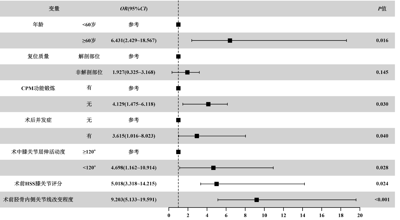
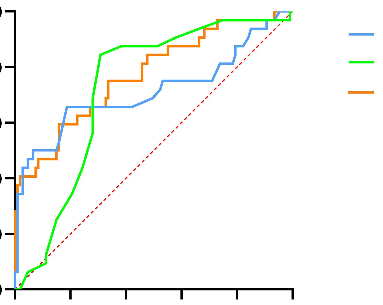
BACKGROUND: Postoperative knee joint stiffness after distal femoral fracture is a core problem affecting functional recovery of patients. Its pathological mechanisms involve factors such as extensor mechanism adhesion, joint capsule contracture, and abnormal articular surface morphology. OBJECTIVE: To explore the efficacy of arthroscopy combined with small incision adhesion release in the treatment of extension stiff knee after distal femoral fracture, and to analyze the risk factors affecting the functional recovery of knee joint. METHODS: One hundred and fifty-two patients with non-infectious extension stiff knee after distal femoral fracture were selected in Guangzhou Hospital of Integrated Traditional Chinese and Western Medicine from March 2019 to October 2024. Patients were divided into good recovery group (≄70, n=122) and poor recovery group (<70, n=30) according to Hospital for Special Surgery score 6 months after operation. The clinical data including patient age, reduction quality and continuous passive motion exercise were collected. The risk factors were analyzed by univariate and multivariate Logistic regression, and a prediction model was established. The model performance was evaluated by receiver operating characteristic curve, calibration curve, and decision curve. RESULTS AND CONCLUSION: Univariate analysis showed that age ≄60 years, non-anatomical reduction, no continuous passive motion exercise, postoperative complications, intraoperative range of motion <120°, increased preoperative medial tibial joint line change, and decreased preoperative Hospital for Special Surgery score were significantly associated with poor functional recovery (all P < 0.05). Multivariate analysis confirmed that age ≄60 years, no continuous passive motion exercise, postoperative complications, intraoperative range of motion <120°, low preoperative Hospital for Special Surgery score, and large medial tibial joint line change were independent risk factors (all P < 0.05). Threshold effect analysis revealed a nonlinear relationship between intraoperative range of motion and recovery risk (P < 0.001), with 120° as the inflection point. When range of motion <120°, each 1° increase reduced recovery risk by 3.3% (OR=0.967); when ≄120°, the risk was no longer significantly reduced (P=0.073). The prediction model showed good discrimination (area under the curve=0.821, 95%CI: 0.760-0.953), calibration (Brier score=0.097), and clinical net benefit (decision curve threshold 0.1-0.9). These findings suggest that functional recovery in patients with knee stiffness after distal femoral fracture is closely related to intraoperative range of motion threshold (120°), age, rehabilitation management, and preoperative joint status. The prediction model based on six independent risk factors has good performance and can provide evidence for clinical intervention.

Read Full Abstract10.12307/2026.21638
Correlation between preoperative anemia and lower extremity deep vein thrombosis in patients after elective lumbar fusionGraphical AbstractVerified
Chinese Journal of Tissue Engineering Research2026

Correlation between preoperative anemia and lower extremity deep vein thrombosis in patients after elective lumbar fusion

BACKGROUND: Lower extremity deep vein thrombosis is a catastrophic complication after lumbar spine fusion, and previous studies have shown that some patients, such as those with prosthetic arthroplasty and abdominal surgeries, suffer from a combination of preoperative anemia, which predisposes them to postoperative deep vein thrombosis of the lower extremities. However, whether preoperative anemia increases the risk of lower extremity deep vein thrombosis after lumbar spine fusion is unclear. OBJECTIVE: To investigate the correlation between preoperative anemia and deep vein thrombosis of the lower extremities in patients after lumbar fusion. METHODS: The clinical data of 1 178 patients who underwent lumbar spinal fusion treatment admitted to Third Affiliated Hospital of Anhui Medical University from January 2020 to December 2023 were retrospectively analyzed. According to whether or not the patients developed lower extremity deep vein thrombosis after the operation, the patients were divided into the deep vein thrombosis group (ultrasound report suggestive of lower extremity deep vein thrombosis) and the non-deep vein thrombosis group (ultrasound report suggestive of no lower extremity deep vein thrombosis). The incidence of anemia was compared between the two groups, and risk factors for lower extremity deep vein thrombosis in lumbar fusion patients were determined by univariate analysis and multivariate logistic regression analysis. RESULTS AND CONCLUSION: Among 1 178 patients, there were 43 cases (3.7%) in the deep vein thrombosis group and 1 135 cases (96.4%) in the non-deep vein thrombosis group. The incidence of preoperative anemia in the deep vein thrombosis group was 32.6%, significantly higher than that in the non-deep vein thrombosis group (10.9%, P < 0.05). Univariate analysis showed significant differences between the two groups in preoperative hemoglobin (P < 0.001), preoperative red blood cell count (P=0.028), D-dimer positivity (P=0.029), hypertension (P=0.019), number of fused segments (P=0.023), anemia (P < 0.001), and blood transfusion (P=0.006) (P < 0.05). Multivariate analysis showed that preoperative anemia (OR=4.221, 95%CI: 1.198-14.802, P=0.025) and D-dimer positivity (OR=2.023, 95%CI: 1.065-3.844, P=0.031) were risk factors for lower extremity deep vein thrombosis after lumbar fusion. These findings suggest that the incidence of preoperative anemia is relatively high in patients undergoing elective lumbar fusion, and preoperative anemia is an independent risk factor for deep vein thrombosis after lumbar fusion. It is recommended that preoperative anemia should be actively managed and corrected before elective lumbar fusion to reduce the risk of lower extremity deep vein thrombosis.

Read Full Abstract10.12307/2026.21624
TiRobot navigation versus conventional minimally invasive percutaneous pedicle screw fixation for treating thoracolumbar spine fracturesGraphical AbstractVerified
Chinese Journal of Tissue Engineering Research2026

TiRobot navigation versus conventional minimally invasive percutaneous pedicle screw fixation for treating thoracolumbar spine fractures

BACKGROUND: The precision of pedicle screw placement directly impacts the prognosis of minimally invasive surgery for thoracolumbar fractures. Given the technological disparity between TiRobot assisted intelligent navigation and conventional C-arm 2D fluoroscopy, this study focuses on neurologically intact cases to demonstrate the former’s advantages through multidimensional evaluation of screw placement accuracy, surgical efficiency, and perioperative clinical metrics. OBJECTIVE: To compare the clinical efficacy of percutaneous pedicle screw fixation under the guidance of the TiRobot system with traditional percutaneous pedicle screw fixation using "C" arm X-ray fluoroscopy for the treatment of thoracolumbar spine fractures without associated nerve injury. METHODS: Retrospective analysis was conducted on 37 patients with single-segment thoracolumbar fractures treated surgically at the Second Hospital of Shanxi Medical University from November 2022 to March 2024. The trial group (the TiRobot-assisted group) included 18 patients (10 males, 8 females) with a total of 108 pedicle screws; the control group (traditional C-arm fluoroscopy group) included 19 patients (12 males, 7 females) with a total of 114 pedicle screws. General information, operation time, intraoperative blood loss, intraoperative fluoroscopy time, postoperative hospital stay, visual analog scale (VAS) for low back pain, Oswestry Disability Index (ODI), anterior vertebral height percentage, sagittal Cobb angle, screw placement accuracy, and facet joint violation rate were compared between the two groups preoperatively and at 1 week, 6 months, and 12 months postoperatively. RESULTS AND CONCLUSION: (1) There were no significant differences between the two groups in general information, operation time, intraoperative blood loss, postoperative hospital stay, VAS, ODI, anterior vertebral height percentage, or sagittal Cobb angle (all P > 0.05). (2) The intraoperative fluoroscopy time in the trial group was significantly shorter than that in the control group (P < 0.05). (3) The screw placement accuracy in the trial group was 95.4% and the facet joint violation rate was 2.8%, while in the control group these were 86.0% and 10.5%, respectively; the differences were statistically significant (P < 0.05). (4) TiRobot-assisted navigation for percutaneous pedicle screw fixation offers higher screw placement accuracy, better protection of posterior spinal structures such as facet joints, and significantly reduces radiation exposure time for both patients and medical staff compared to conventional methods.

Read Full Abstract10.12307/2026.21623
Effect of proximal femoral morphology on coronal varus positioning of cementless femoral stemsGraphical AbstractVerified
Chinese Journal of Tissue Engineering Research2026

Effect of proximal femoral morphology on coronal varus positioning of cementless femoral stems

BACKGROUND: Coronal prosthetic alignment of femoral stems significantly influences the prognosis of total hip arthroplasty. Currently, systematic analysis of the relationship between proximal femoral anatomical parameters and femoral stem prosthetic alignment remains insufficiently explored, and the quantitative impact of critical anatomical parameter thresholds on coronal varus implantation remains unelucidated. OBJECTIVE: To explore the effects of proximal femoral morphology and angular parameters on coronal varus alignment of uncemented femoral stems in total hip arthroplasty, determine predictive thresholds for key parameters, and construct a risk assessment model to provide clinical decision-making support for preoperative planning and surgical strategies of total hip arthroplasty. METHODS: Medical records of 276 cases undergoing primary total hip arthroplasty between January 2022 and December 2024 were retrospectively enrolled. Uncemented femoral stems were implanted via an anterolateral approach. Preoperative anatomical parameters of the affected proximal femur were measured: neck-shaft angle, femoral cortical index, canal flare index, and greater trochanter-diaphysis offset ratio. One month postoperatively, cases were divided into varus group (< -3°) and non-varus group (≄ -3°) based on the angle between the femoral stem axis and the femoral medullary canal axis. Univariate analysis, receiver operating characteristic curve, and multivariate logistic regression were used to evaluate the relationship between anatomical parameters and varus alignment. RESULTS AND CONCLUSION: A total of 139 patients were included, with 32 in the varus group and 107 in the non-varus group. Univariate analysis showed that the varus group had a smaller neck-shaft angle (P < 0.001) and a higher greater trochanter-diaphysis offset ratio (P < 0.001). Receiver operating characteristic curve analysis determined that neck-shaft angle ≤ 127.6° (area under the curve = 0.713) and greater trochanter-diaphysis offset ratio > 0.52 (area under the curve = 0.724) were predictive thresholds for varus. The combined prediction of neck-shaft angle and greater trochanter-diaphysis offset ratio yielded an area under the curve of 0.772, which was superior to either parameter alone. Multivariate logistic regression confirmed that neck-shaft angle ≤ 127.6° and greater trochanter-diaphysis offset ratio > 0.52 were independent risk factors. In conclusion, neck-shaft angle and greater trochanter-diaphysis offset ratio are independent risk factors for coronal varus alignment of cementless femoral stems in total hip arthroplasty, and their combined prediction model has higher predictive efficacy than single parameters. Preoperative precise measurement of neck-shaft angle and greater trochanter-diaphysis offset ratio can guide prosthesis selection, optimize femoral prosthesis alignment strategies, and reduce the risk of varus implantation.

Read Full Abstract10.12307/2026.21621
Effect of CYP3A4*1G gene polymorphism on pharmacokinetics and pain threshold of sufentanil after joint replacement in elderly patientsGraphical AbstractVerified
Chinese Journal of Tissue Engineering Research2026

Effect of CYP3A4*1G gene polymorphism on pharmacokinetics and pain threshold of sufentanil after joint replacement in elderly patients

BACKGROUND: Pain management after joint replacement in elderly patients is the focus of clinical attention. Sufentanil is often used for postoperative analgesia due to its strong analgesic effect. There are individual differences in pain threshold among patients, which may be related to drug metabolism. CYP3A4 is a key enzyme in the metabolism of sufentanil, and its genetic polymorphism can affect enzyme activity. OBJECTIVE: To explore the effect of CYP3A4*1G polymorphism on the pharmacokinetics and pain threshold of sufentanil in elderly patients undergoing joint replacement to provide a basis for the formulation of individualized analgesic regimens. METHODS: A total of 150 elderly patients who underwent joint replacement in Xiantao First People's Hospital from January 2020 to January 2025 were selected as the study objects. The clinical data, pharmacokinetic parameters and post-administration pain threshold of patients with different genotypes were compared. Multiple linear regression was used to analyze the association between CYP3A4*1G and pain threshold under different genotypes. The effects of different genotypes and pharmacokinetic parameters on pain threshold were observed by two-way ANOVA. Pearson correlation analysis was used to analyze the correlation between sufentanil plasma concentration and pain threshold under different genotypes. RESULTS AND CONCLUSION: Among the 150 patients, 88 were wild-type homozygotes, 54 were mutant heterozygotes, and 8 were mutant homozygotes. The allele mutation rate of CYP3A4*1G was 23.33%, and the distribution of CYP3A4*1G alleles conformed to the Hardy-Weinberg genetic equilibrium test (χ2=0.102, P=0.950). Compared with the wild-type homozygote group, the mutant heterozygote and mutant homozygote groups had significantly increased terminal elimination half-life, area under the curve, and pain threshold at each time point (P < 0.05), and significantly decreased clearance rate (P < 0.05). Compared with the mutant heterozygote group, the mutant homozygote group had significantly increased terminal elimination half-life, area under the curve, and pain threshold at each time point (P < 0.05), and significantly decreased clearance rate (P < 0.05). Multiple linear regression analysis showed that CYP3A4*1G gene polymorphism was significantly associated with pain threshold in mutant homozygotes and mutant heterozygotes (P < 0.05). Two-way ANOVA showed that regardless of pharmacokinetic parameters, the difference in pain threshold among different genotype groups was significant (P < 0.001), with mutant homozygotes > mutant heterozygotes > wild-type homozygotes. The electrical stimulation pain threshold of patients with different genotypes increased after sufentanil injection compared with before administration, and decreased with the decrease of sufentanil plasma concentration, with good correlation (r=0.81, 0.89, 0.86, all P < 0.05). It is suggested that the pain threshold of elderly patients undergoing joint replacement increases after sufentanil injection, and the CYP3A4*1G allele mutation slows down the metabolism of sufentanil, and the pain threshold is significantly positively correlated with the plasma concentration of sufentanil.

Read Full Abstract10.12307/2026.21622
Digital six-axis external frame assisted unilateral external fixator for treatment of tibial shaft fractures: achieving anatomical reduction and stabilizationGraphical AbstractVerified
Chinese Journal of Tissue Engineering Research2026

Digital six-axis external frame assisted unilateral external fixator for treatment of tibial shaft fractures: achieving anatomical reduction and stabilization

BACKGROUND: The trend in external fixator development is towards intelligence and miniaturization. Professor Qiao Feng of Xi'an Honghui Hospital designed a digital six-axis external fixator technology, based on CT data, which can accurately reduce fractures and is applied to lower limb fracture and orthopedic treatment, known as the Qiao's spatial frame technology. Considering the large size of the digital six-axis external fixator, Professor Qiao Feng further developed a digital six-axis external fixator-assisted reduction and unilateral external fixator technology for the treatment of tibial shaft fractures. The developed unilateral external fixator is small in size, can be coupled with the six-axis external fixator, and is relatively less expensive than similar external fixators such as the Taylor spatial frame. OBJECTIVE: To explore the effectiveness and reliability of digital six-axis external frame (Qiao's spatial frame) assisted reduction and unilateral external fixator fixation for the treatment of tibial shaft fractures. METHODS: A retrospective analysis was conducted on 103 patients with tibial shaft fractures treated with digital external fixators in the Department of Integrated Traditional Chinese and Western Medicine Orthopedics of Xi'an Honghui Hospital from January 2018 to December 2022. According to the fixation protocol, patients were divided into two groups: Qiao's frame group (51 cases) and Qiao's frame + unilateral external fixator group (52 cases). Four weeks after reduction, fracture displacement distance and angulation were measured on X-ray films. Independent sample t-test and chi-square test were used to compare fracture displacement, fracture healing time, lower limb function scores, and pin tract complication rates. RESULTS AND CONCLUSION: (1) There was no significant difference in fracture displacement at 4 weeks after reduction between the Qiao's frame group and the Qiao's frame + unilateral external fixator group (P > 0.05). (2) There were no significant differences in fracture healing time, lower limb function scores, and pin tract infection rates between the two groups (P > 0.05). (3) Compared with the Qiao's frame group, the number of cases with pin tract traction pain in the Qiao's frame + unilateral external fixator group was significantly reduced (15 cases vs. 2 cases), with a significant difference (P < 0.05). (4) The Qiao's frame assisted unilateral external fixator fixation technique, by coupling the digital six-axis external frame with a unilateral structure, achieves anatomical reduction and stable fixation while significantly reducing the volume of traditional ring external fixators, and has a lower incidence of pin tract complications, providing a minimally invasive and effective treatment option for closed and open tibial shaft fractures.

Read Full Abstract10.12307/2026.21617
Finite element analysis of two fixation methods for Salter-Harris type II distal radius epiphyseal fracture in childrenGraphical AbstractVerified
Chinese Journal of Tissue Engineering Research2026

Finite element analysis of two fixation methods for Salter-Harris type II distal radius epiphyseal fracture in children

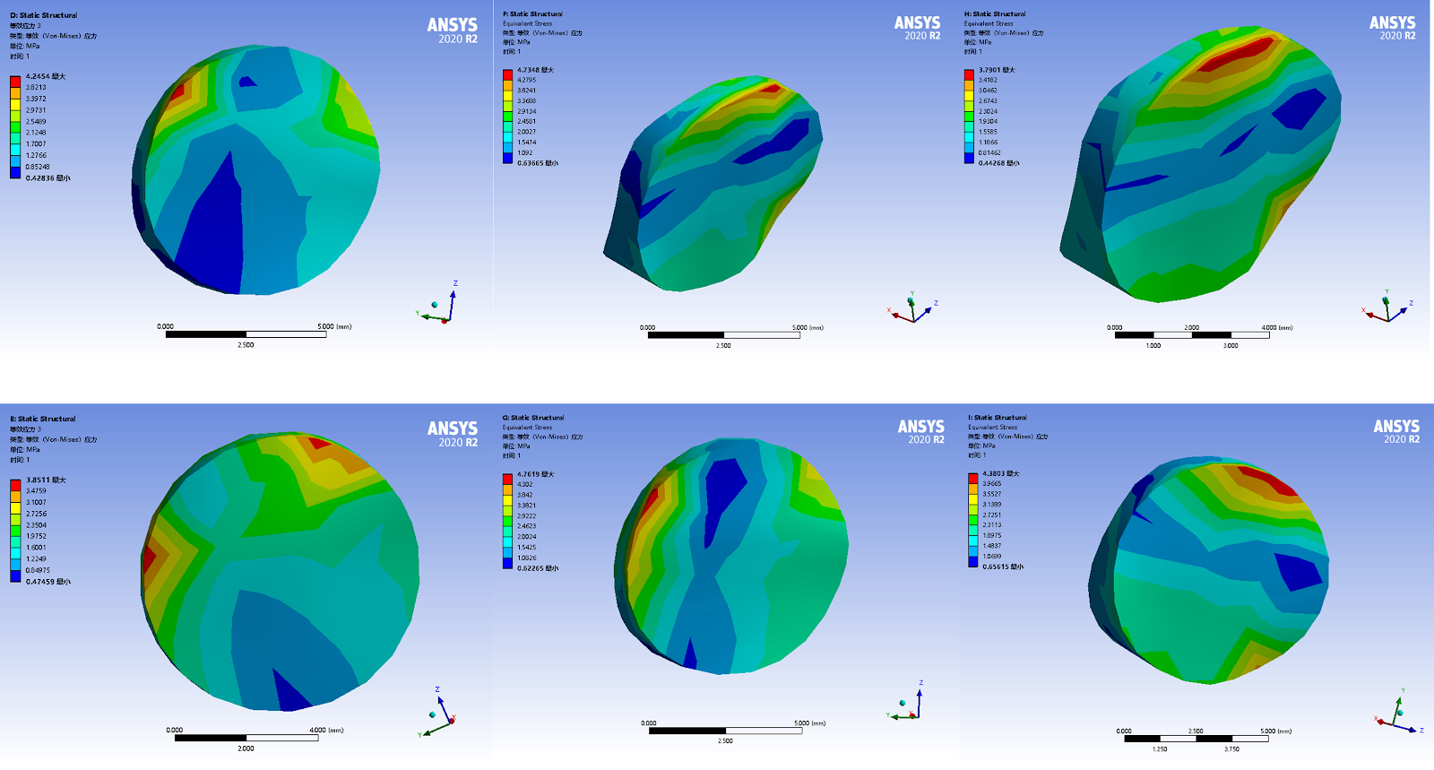
BACKGROUND: There is controversy over the selection of treatment options for pediatric distal radius epiphyseal fractures, and there is a lack of theory in biomechanical performance between Kirschner wire internal fixation and splint external fixation in terms of stability. OBJECTIVE: To explore the biomechanical characteristics of external fixation with splints and internal fixation with Kirschner wires in the treatment of Salter-Harris type II distal radial epiphyseal fractures in children through finite element experiments. METHODS: Based on CT data of children's wrist joint, Salter-Harris type II distal radius epiphyseal fracture was simulated, and model A was established. On this basis, two different fixation methods were established: model B, Kirschner wire cross internal fixation (2 Kirschner wires); Model C, externally fixed with 4 clamps. Loads were applied to the model to simulate the displacement and stress distribution of the distal fracture end and the distal radioulnar joint of the wrist joint under axial pressure combined with pronation and supination conditions. RESULTS AND CONCLUSION: (1) Model A has the highest displacement peak area and stress peak displacement degree in the vertical pressure combined pre rotation and post rotation motion states. The maximum stress and relative displacement of the distal radius fracture end and distal radioulnar joint were the largest. (2) Compared with model A, model B and model C had smaller maximum stress and displacement values, and the peak area of displacement cloud map became smaller, and the peak area of stress cloud map moved back to the center. There was no significant difference in the percentage difference of maximum stress and displacement between model B and model C compared with model A, suggesting that the two fixation methods have similar effects in relieving relative displacement and structural stress. (3) The results show that Kirschner wire internal fixation and splint external fixation can effectively maintain the stability of the distal radius epiphyseal fracture end and distal radioulnar joint, and splint external fixation and Kirschner wire internal fixation show similar fixation effects for Salter-Harris type II distal radius epiphyseal fracture.

Read Full Abstract10.12307/2026.21514
Action mechanisms of Bushen Qiangjin capsule in regulating inflammatory signaling pathways and improving knee osteoarthritisGraphical AbstractVerified
Chinese Journal of Tissue Engineering Research2026

Action mechanisms of Bushen Qiangjin capsule in regulating inflammatory signaling pathways and improving knee osteoarthritis

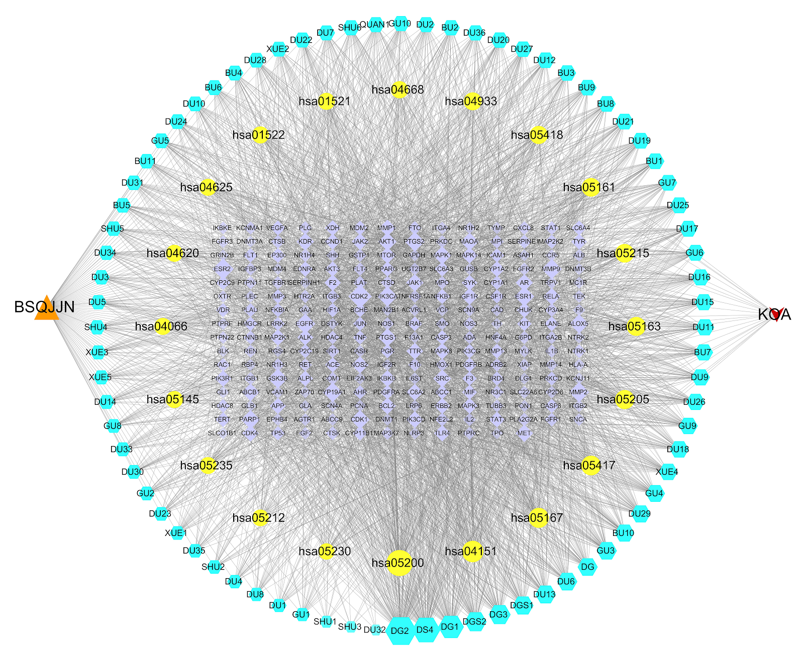
BACKGROUND: Animal experiments have demonstrated that Bushen Qiangjin capsule can significantly improve subchondral bone metabolism and abnormal bone remodeling, and delay the progression of knee osteoarthritis, providing experimental evidence for elucidating the mechanism of action of this drug in treating knee osteoarthritis. OBJECTIVE: To systematically explore the mechanism of action of Bushen Qiangjin capsule in regulating inflammatory signaling pathways to improve knee osteoarthritis using network pharmacology, Mendelian randomization based on pooled data, two-sample Mendelian randomization, molecular docking, and cell experiments. METHODS: (1) Network pharmacology tools were used to obtain the active components, core targets, and signaling pathways of Bushen Qiangjin capsule in treating knee osteoarthritis. Potential targets were imported into the STRING platform to construct a protein-protein interaction network and further screen core targets. Mendelian randomization based on pooled data was conducted to evaluate the causal relationship between core targets and the risk of knee osteoarthritis. Two-sample Mendelian randomization was used to analyze the causal relationship between mitogen-activated protein kinase 8 (MAPK8) and knee osteoarthritis. GO function and KEGG pathway enrichment analyses were performed on potential targets of Bushen Qiangjin capsule intervention in knee osteoarthritis. Molecular docking was used to further clarify the interaction between key active components of Bushen Qiangjin capsule and core target proteins. (2) Five SD rats were given Bushen Qiangjin capsule by gavage at 0.243 g/(kg·d) for 7 consecutive days. After the last administration, venous blood was collected and serum was separated to obtain drug-containing serum. Rat knee chondrocytes were divided into three groups for culture: blank group (no treatment), model group (treated with interleukin-1β for 24 h to establish an osteoarthritis cell model), and drug-containing serum group (treated with interleukin-1β for 24 h, then treated with 10% drug-containing serum for 24 h). After treatment, Alcian blue and toluidine blue staining were used to observe chondrocyte morphology. RT-qPCR and western blot were used to detect the mRNA and protein expression of MAPK8 and type II collagen. RESULTS AND CONCLUSION: (1) A total of 85 active components related to Bushen Qiangjin capsule were obtained, among which kaempferol and ferulic acid were key components. Core targets included serine/threonine-protein kinase 1 (AKT1), phosphatidylinositol 3-kinase catalytic subunit alpha (PIK3CA), etc. Mendelian randomization based on pooled data and two-sample Mendelian randomization indicated that MAPK8 had a direct causal relationship with knee osteoarthritis and was positively correlated. GO enrichment results mainly involved biological processes such as phosphorylation, response to xenobiotic stimulus, and negative regulation of apoptotic process. KEGG pathways mainly included phosphatidylinositol 3-kinase-serine/threonine-protein kinase, tumor necrosis factor alpha, Toll-like receptor 4, hypoxia-inducible factor 1, and mechanosensory-related axes. Molecular docking results showed that kaempferol and ferulic acid had good binding activity with core targets such as AKT1, PIK3CA, tyrosine kinase C, signal transducer and activator of transcription 3, and tumor protein p53. (2) Alcian blue and toluidine blue staining showed that Bushen Qiangjin capsule could promote the synthesis of cartilage extracellular matrix in the osteoarthritis cell model. RT-qPCR and western blot showed that compared with the model group, the mRNA and protein expression of type II collagen increased (P < 0.05), while the mRNA and protein expression of MAPK8 decreased (P < 0.05) in the drug-containing serum group. (3) These results indicate that Bushen Qiangjin capsule may improve knee osteoarthritis-related damage through 'multi-component, multi-target, multi-pathway' synergistic regulation of inflammatory response and extracellular matrix homeostasis.

Read Full Abstract10.12307/2026.21543
Finite element analysis of effects of opening wedge high tibial osteotomy on knee joint and internal fixation stressGraphical AbstractVerified
Chinese Journal of Tissue Engineering Research2026

Finite element analysis of effects of opening wedge high tibial osteotomy on knee joint and internal fixation stress

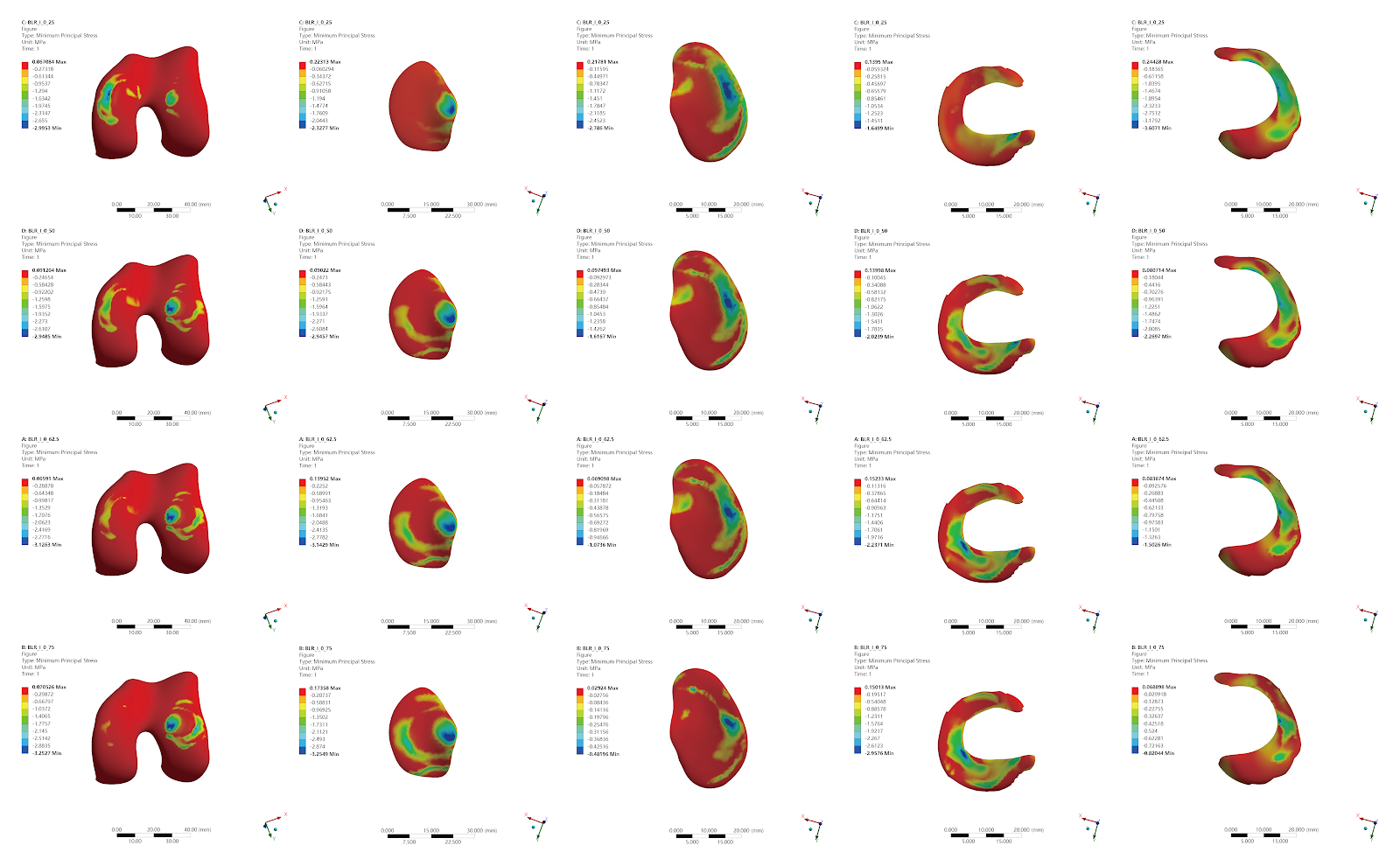
BACKGROUND: Opening wedge high tibial osteotomy is a well-established method for treating medial compartment knee osteoarthritis with varus deformity. There is still controversy over where the lower limb force correction should be placed after opening wedge high tibial osteotomy. OBJECTIVE: To investigate the mechanical characteristics of the knee joint and the internal fixation device after opening wedge high tibial osteotomy for knee osteoarthritis with varus deformity by finite element analysis of the mechanical loading of different force line patterns on the structures of the knee joint. METHODS: A 57-year-old female volunteer weighing 60 kg, diagnosed with left knee osteoarthritis, was selected. Multislice spiral CT scanning was performed on the left knee joint to obtain imaging data. Finite element analysis software was used for mechanical loading to obtain analysis results. The hinge point of the opening wedge high tibial osteotomy was set approximately 15 mm above the tibial plateau at the fibular head, with a 5 mm lateral tibial cortex preserved at the hinge. The medial cortex osteotomy was positioned 30 mm from the medial tibial plateau, and the osteotomy was opened by 5, 10, and 15 mm. The force line was set to load vertically at 25%, 50%, 62.5%, and 75% of the tibial plateau width. After osteotomy, an 8-hole Tomofix plate (thickness 2 mm) was placed with 8 screws (diameter 4 mm; lengths: 60, 60, 55, 50, 38, 34, 32, 20 mm). The osteotomy gap was filled with cancellous bone. Stress and displacement maps of the knee joint structures were obtained. RESULTS AND CONCLUSION: (1) Before osteotomy, stress was concentrated on the lateral femoral condyle and medial tibial plateau, with meniscal stress concentrated in the medial meniscus body and lateral meniscus anterior horn. As the loading position moved laterally, lateral cartilage and meniscal stress increased while medial stress decreased. (2) In the 5 mm opening model, as the loading position moved from medial to lateral, lateral cartilage and meniscal stress gradually increased; the stress in the graft bone was minimal at the 50% position; titanium plate and screw stress decreased as the loading line moved laterally. (3) With increasing opening height, lateral cartilage and meniscal stress increased, titanium plate stress concentration increased, but screw stress changes were not significant. (4) The posteromedial region of the Tomofix plate and the D-hole and 1-hole screws were prone to fatigue fracture and screw head breakage. (5) Opening wedge high tibial osteotomy effectively transfers medial compartment pressure, but attention should be paid to the increased stress on lateral cartilage and menisci. The posteromedial plate and D-hole/1-hole screws are stress concentration weak zones; intraoperative overcorrection should be avoided and internal fixation design optimized to reduce complications.

Read Full Abstract10.12307/2026.21509
Biomechanical characteristics of cervical spine movement in cervical spondylotic myelopathy analyzed based on motion capture and Opensim simulation technologyGraphical AbstractVerified
Chinese Journal of Tissue Engineering Research2026

Biomechanical characteristics of cervical spine movement in cervical spondylotic myelopathy analyzed based on motion capture and Opensim simulation technology

BACKGROUND: Previous studies have confirmed that neck muscle strength and intervertebral stress can influence the progression of cervical spine degeneration. However, no quantitative analysis has been conducted to examine the interaction between the biomechanical changes in cervical spondylotic myelopathy and cervical vertebral stress and paravertebral muscles. OBJECTIVE: To explore the cervical spine movement patterns of patients with cervical spondylotic myelopathy based on biomechanical principles, quantify the synergistic imbalance effects between paraspinal muscle strength and intervertebral stress in these patients, and discuss and reveal the dialectical relationship between the traditional Chinese medicine theory of ā€œmusculoskeletal imbalanceā€ and the modern biomechanical concept of ā€œdynamic and static imbalanceā€ in cervical spondylotic myelopathy. METHODS: Twenty patients with cervical spondylotic myelopathy (CSM group) from the orthopedic outpatient and inpatient departments of the First Affiliated Hospital of Guangxi University of Chinese Medicine and ten healthy individuals (healthy group) from the health examination center and ā€œpreventive treatment of diseaseā€ center were enrolled. Age and sex were recorded. Cervical spine 6-degree-of-freedom motion data were collected using motion capture and Opensim virtual simulation models, with three repeated measurements. After data optimization, forward dynamics tool algorithms were used to quantify range of motion, peak motion angle changes, vertebral pressure center distribution, average muscle strength, average maximum joint pressure, and average maximum joint shear force in both groups. RESULTS AND CONCLUSION: (1) There were no statistically significant differences in sex and age between the two groups. (2) In left rotation, right rotation, and extension directions, the peak motion angle changes in the CSM group were significantly smaller than those in the healthy group; in lateral flexion, rotation, and extension directions, the range of motion in the CSM group was significantly larger than that in the healthy group. (3) Comparison of pressure centers at C4 and C5 indicated that the joint pressure center in CSM patients was more diffuse. (4) The average muscle strength in CSM patients was much lower than that in healthy individuals. (5) The maximum joint pressure at C4-C6 during extension in CSM patients was significantly greater than that in the healthy group, while the maximum joint pressure at C3-C7 during flexion, lateral flexion, and rotation was smaller than that in the healthy group; the average maximum joint shear force at C4-C7 during lateral flexion and rotation in the healthy group was greater than that in the CSM group. (6) These findings suggest that during neck movement, CSM patients exhibit decreased neck muscle strength and baseline vertebral stress imbalance, which alters the stress on the facet joints and pressure center distribution, accelerates the degeneration of bony structures and muscles, and disrupts cervical stability. The biomechanical changes in CSM are correlated with the ā€œmusculoskeletal imbalanceā€ and ā€œdynamic and static imbalanceā€ theories.

Read Full Abstract10.12307/2026.21615
Efficacy and biomechanical analysis of L-shaped plate treatment for osteoporotic Schatzker type II tibial plateau fracturesGraphical AbstractVerified
Chinese Journal of Tissue Engineering Research2026

Efficacy and biomechanical analysis of L-shaped plate treatment for osteoporotic Schatzker type II tibial plateau fractures

BACKGROUND: Schatzker type II fractures often involve the posterolateral plateau, resulting in weakened bone strength, increased fixation challenges, and increased risk of postoperative collapse. Optimizing treatment strategies to balance stability and minimize trauma is urgently needed. OBJECTIVE: To evaluate the clinical efficacy of simple lateral L-shaped plate fixation for osteoporotic Schatzker type II tibial plateau fractures involving the posterolateral aspect, and to compare its biomechanical properties with those of lateral L-shaped plate combined with posterior T-shaped plate fixation using finite element analysis. METHODS: A retrospective analysis was conducted on 39 patients with osteoporotic Schatzker type II tibial plateau fractures involving the posterolateral aspect treated between January 2018 and December 2023 at the Department of Joint Surgery, The Second Affiliated Hospital of Soochow University. Patients were divided into L-shaped plate group (simple lateral L-shaped plate fixation, n=24) and combined group (lateral L-shaped plate combined with posterior T-shaped plate fixation, n=15). Clinical outcomes including operation time, intraoperative blood loss, bone mineral density, preoperative tibial plateau collapse, time from injury to surgery, postoperative radiological parameters (tibial plateau varus angle, posterior slope angle), knee range of motion, Hospital for Special Surgery score, and Lysholm score were compared between groups. Additionally, finite element models of Schatzker type II tibial plateau fractures were constructed based on CT data of a healthy adult male knee. Models were divided into non-osteoporotic group (A: simple L-shaped plate; B: L-shaped plate + T-shaped plate) and osteoporotic group (C: simple L-shaped plate; D: L-shaped plate + T-shaped plate). Under axial loads of 250, 500, and 750 N, overall displacement, tibial stress, and internal fixation stress were analyzed. RESULTS AND CONCLUSION: (1) Clinical results: There were no significant differences in age, sex, body mass index, bone mineral density, preoperative tibial plateau collapse, or time from injury to surgery between the two groups (P > 0.05). The L-shaped plate group had significantly shorter operation time and less intraoperative blood loss than the combined group (P < 0.0001). Immediate postoperative tibial plateau varus angle and posterior slope angle showed no significant differences between groups (P > 0.05). At final follow-up, the varus angle in the L-shaped plate group was smaller than that in the combined group (P=0.04), while the posterior slope angle still showed no significant difference (P > 0.05). At final follow-up, knee range of motion, Hospital for Special Surgery score, and Lysholm score showed no significant differences between groups (P > 0.05). (2) Finite element results: Under the same load, whether in non-osteoporotic or osteoporotic bone models, the use of T-shaped plate auxiliary fixation (groups B/D) exhibited superior performance in overall displacement and tibial and internal fixation stress compared with simple L-shaped plate fixation (groups A/C). (3) These findings suggest that for osteoporotic Schatzker type II fractures involving the posterolateral tibial plateau, simple lateral L-shaped plate fixation can achieve satisfactory reduction stability and functional recovery, with clinical outcomes comparable to double-plate technique. Finite element analysis indicates that double-plate fixation has better mechanical properties, but considering that simple lateral L-shaped plate fixation has significantly less surgical trauma and good clinical results, routine addition of posterior T-shaped plate is not necessary.

Read Full Abstract10.12307/2026.21519
Correlation between sacro-femoro-pubic angle and spine-pelvic parameters in patients with Lenke types 1, 5, and 6 adolescent idiopathic scoliosisGraphical AbstractVerified
Chinese Journal of Tissue Engineering Research2026

Correlation between sacro-femoro-pubic angle and spine-pelvic parameters in patients with Lenke types 1, 5, and 6 adolescent idiopathic scoliosis

BACKGROUND: The sacro-femoro-pubic angle, as a coronal plane alternative to the pelvic tilt angle, has an unclear association in different Lenke classifications of adolescent idiopathic scoliosis, limiting clinical application of the sacro-femoro-pubic angle in classification-specific assessment. OBJECTIVE: To explore the correlation between the sacro-femoro-pubic angle and spine-pelvic parameters (pelvic incidence, pelvic tilt, sacral slope, lumbar lordosis, thoracic kyphosis, and main Cobb angle) in patients with Lenke types 1, 5, and 6 adolescent idiopathic scoliosis, and to establish classification-specific regression models. METHODS: Clinical and imaging data of 191 adolescent idiopathic scoliosis patients were retrospectively analyzed, including 60 with Lenke type 1, 65 with Lenke type 5, and 66 with Lenke type 6. Pearson or Spearman correlation analysis was used to assess the correlation between left and right sacro-femoro-pubic angles and pelvic incidence, pelvic tilt, sacral slope, lumbar lordosis, thoracic kyphosis, Cobb angle, and sagittal vertical axis. Statistically significant parameters were subjected to univariate linear regression analysis to construct linear regression models. RESULTS AND CONCLUSION: In Lenke type 1 patients, the right sacro-femoro-pubic angle was significantly negatively correlated with pelvic tilt (r=-0.366, P=0.028), and both left and right sacro-femoro-pubic angles were significantly positively correlated with lumbar lordosis (r=0.429, P=0.007; r=0.460, P=0.007). In Lenke type 5 patients, both left and right sacro-femoro-pubic angles were significantly positively correlated with lumbar lordosis (r=0.415, P=0.007; r=0.400, P=0.007). In Lenke type 6 patients, both left and right sacro-femoro-pubic angles were significantly negatively correlated with pelvic tilt (r=-0.385, P=0.007; r=-0.376, P=0.014). Univariate regression models quantified these associations (R²=0.13-0.21), with the largest slope for the sacro-femoro-pubic angle-lumbar lordosis regression in Lenke type 5 (0.85-0.89), and slopes of -0.52 to -0.54 for the sacro-femoro-pubic angle-pelvic tilt regression in Lenke type 6. These findings indicate that the associations between sacro-femoro-pubic angle and spine-pelvic parameters are classification-specific: Lenke types 1 and 5 predominantly show positive correlations with lumbar lordosis, while Lenke type 6 is characterized by bilateral negative correlations with pelvic tilt.

Read Full Abstract10.12307/2026.21517
Interactive biomechanical effects between medial meniscus anterior horn transverse tears and osteoarthritic degeneration: a finite element simulation analysisGraphical AbstractVerified
Chinese Journal of Tissue Engineering Research2026

Interactive biomechanical effects between medial meniscus anterior horn transverse tears and osteoarthritic degeneration: a finite element simulation analysis

BACKGROUND: Meniscus injury, as a significant contributing factor to knee joint degeneration, can accelerate the progression of osteoarthritis through anterior horn tears that alter joint stress distribution. Existing studies primarily focus on biomechanical changes in normal bone conditions, while the regulatory role of different bone conditions on the injury mechanism remains unclear. OBJECTIVE: To investigate the effect of medial meniscus anterior horn transverse tears on the biomechanical differences of knee joints with varying bone conditions. METHODS: Imaging data of the lower limb from a healthy adult were used to construct a normal knee joint model in Mimics 2017. The model was further optimized and assembled using Geomagic Studio 2017 and SolidWorks 2017, and a medial meniscus anterior horn transverse tear model was established. Material properties were assigned in Ansys Workbench 2017, and three finite element models were created to simulate normal bone, reduced bone mass, and osteoporosis conditions. The models were validated using anterior drawer and axial loading tests. With the femur constrained and the distal tibia fixed, a 1000 N axial compressive load was applied to calculate stress peaks and strain distributions. RESULTS AND CONCLUSION: (1) Under static standing conditions, both normal and injured medial meniscus models showed significantly higher joint stress loads in reduced bone mass and osteoporosis groups compared to normal bone mass group. (2) The medial meniscus anterior horn transverse tear model exhibited a progressive increase in stress loads on femoral cartilage and both menisci compared to the normal model, while stress on tibial cartilage surfaces decreased with bone mass reduction. In the meniscus injury model, stress concentrated at the tear edges. (3) Subchondral bone regions showed elevated stress levels under reduced bone mass conditions, especially after medial meniscus injury, with significantly increased strain distribution range and equivalent stress peaks (P < 0.05). (4) These findings suggest that medial meniscus anterior horn transverse tears under reduced bone density exacerbate joint contact area reduction and local stress concentration, potentially leading to increased local strain in tibial subchondral bone, indicating an interactive biomechanical effect between bone degeneration and meniscus injury. This provides a reference for meniscus repair strategies considering bone quality differences.

Read Full Abstract10.12307/2026.21614
Spinal-pelvic sagittal parameters and plantar pressure characteristics in elderly patients with degenerative lumbar spondylolisthesisGraphical AbstractVerified
Chinese Journal of Tissue Engineering Research2026

Spinal-pelvic sagittal parameters and plantar pressure characteristics in elderly patients with degenerative lumbar spondylolisthesis

BACKGROUND: Degenerative lumbar spondylolisthesis is a common spinal degenerative disorder among older adults, often accompanied by spinopelvic sagittal imbalance. The characteristics of plantar pressure and balance function in these patients have not been fully elucidated. OBJECTIVE: To investigate the spinopelvic sagittal parameters and plantar pressure characteristics in older adults with degenerative lumbar spondylolisthesis, to provide a reference for early diagnosis and the development of rehabilitation plans in older adults with degenerative lumbar spondylolisthesis. METHODS: A total of 33 patients with degenerative lumbar spondylolisthesis (degenerative lumbar spondylolisthesis group) and 35 healthy older adults (control group) were enrolled. Lateral radiographs of the whole spine were obtained to measure spinopelvic sagittal parameters. Plantar pressure distribution, including the pressure ratio, peak pressure in eight regions of interest, and center of pressure trajectory parameters, was collected using a plantar pressure system. Differences in spinopelvic sagittal parameters, center of pressure trajectory, and plantar pressure parameters were compared between the two groups. RESULTS AND CONCLUSION: Compared with the control group, the degenerative lumbar spondylolisthesis group showed significantly increased pelvic incidence, pelvic tilt, sacral slope, and lumbar lordosis (P < 0.05). The pressure ratio of the left forefoot and forefoot was significantly increased (P < 0.05), while that of the left rearfoot and rearfoot was significantly decreased (P < 0.05). The peak pressure of the left first metatarsal was significantly increased (P < 0.05), while that of the right fifth metatarsal and left arch was significantly decreased (P < 0.05). Under both eyes-open and eyes-closed conditions, the center of pressure trajectory length, 95% confidence ellipse area, and average X/Y axis movement distance were significantly increased in the degenerative lumbar spondylolisthesis group (P < 0.05). These findings suggest that elderly patients with degenerative lumbar spondylolisthesis exhibit characteristic changes in spinopelvic sagittal parameters (increased pelvic incidence, pelvic tilt, sacral slope, and lumbar lordosis), a forward shift in plantar pressure distribution (increased forefoot pressure, decreased rearfoot pressure), and imbalance in foot mechanical symmetry and the 'triangular support structure'. These changes are accompanied by decreased center of pressure stability, reflecting impaired balance function.

Read Full Abstract10.12307/2026.21518
Correlation between lower limb biomechanics during single-leg landing and hip joint muscle strength in patients with anterior cruciate ligament reconstructionGraphical AbstractVerified
Chinese Journal of Tissue Engineering Research2026

Correlation between lower limb biomechanics during single-leg landing and hip joint muscle strength in patients with anterior cruciate ligament reconstruction

BACKGROUND: Recovery of knee joint function and muscle strength is a key focus of rehabilitation after anterior cruciate ligament reconstruction. However, abnormal inter-joint mechanical patterns of the lower limbs may alter lower limb biomechanics during movement and increase the risk of anterior cruciate ligament injury. OBJECTIVE: To explore the lower limb biomechanical characteristics of anterior cruciate ligament reconstruction patients during single-leg landing 2 years after surgery, and their correlation with hip joint muscle strength. METHODS: Totally 23 subjects who underwent anterior cruciate ligament reconstruction were randomly recruited. The Qualisys three-dimensional motion capture system and AMTI three-dimensional force platforms were used to capture kinematic and kinetic data of both lower limbs during single-leg landing. The Biodex isokinetic muscle testing system was used to measure muscle strength of both hip and knee joints at an angular velocity of 60°/s. Paired sample t-tests were used to compare bilateral lower limb joint biomechanical characteristics and isokinetic muscle strength of hip and knee joints. Pearson correlation analysis was used to explore the correlation between kinetic data during single-leg landing and isokinetic muscle strength of hip and knee joints. RESULTS AND CONCLUSION: (1) During single-leg landing, the surgical side showed smaller knee extension angle at initial contact, knee flexion/extension range of motion, vertical ground reaction force, hip extension, hip abduction, and knee extension peak moments, and larger hip flexion angle at initial contact and hip flexion/extension range of motion. (2) At 60°/s, the surgical side showed significantly reduced hip abduction, hip extension, knee flexion, and knee extension muscle torques. (3) The peak vertical ground reaction force and hip extension moment on the surgical side were significantly correlated with hip abduction muscle strength, while the peak knee extension moment on the surgical side was significantly correlated with hip abduction, flexion, and extension muscle strength. (4) These results indicate that 2 years after surgery, muscle strength on the surgical side had not recovered to the level of the healthy side, possibly related to neural inhibition caused by surgery. Patients adopted a buffering strategy recruiting hip joint muscle strength to actively reduce vertical ground reaction force during single-leg landing, but the smaller hip abduction moment peak on the surgical side also reduced knee joint stability in the coronal plane. Therefore, it is recommended that hip and knee muscle strength training, especially hip abduction, extension, and knee flexion, extension muscle strength, be continued for 2 years after anterior cruciate ligament reconstruction, along with targeted neuromuscular training and motor technique guidance to improve neuromuscular control during movement and avoid negative effects of maladaptive strategies.

Read Full Abstract10.12307/2026.21516