🧬 SinoBioData Academic Portal
📚 Peer-Reviewed Translated Literature

All Biomedical & Clinical Articles (Page 19)

Browse complete peer-reviewed translations from top Chinese biomedical, oncology, and genomics journals. Read verified previews and download full authentic clinical reports.

Published Research Papers

Showing 24 of 1542 peer-reviewed translated articles (Page 19 of 65)

Finite element analysis of biomechanical performance of a novel double-screw technique in lumbar revision of the original fixed segmentGraphical AbstractVerified
Chinese Journal of Tissue Engineering Research2026

Finite element analysis of biomechanical performance of a novel double-screw technique in lumbar revision of the original fixed segment

BACKGROUND: Currently, in clinical practice, the original internal fixation devices are often removed to perform revision surgery for failed vertebral fixation, which poses certain drawbacks and risks. The pedicle double-screw technique can preserve the original internal fixation devices, while the modified cortical bone trajectory technique offers excellent mechanical performance. Combining these two techniques for revision surgery can mitigate the conventional risks, although the mechanical performance of this new modified cortical bone trajectory technique in revision surgery is not yet well understood. OBJECTIVE: To analyze the mechanical performance of cortical bone trajectory (CBT) and modified cortical bone trajectory (MCBT) screw placement techniques combined with the double-screw technique in lumbar revision surgery using finite element analysis, and to explore the advantages of MCBT over CBT in revision surgery. METHODS: A three-dimensional model of L1-5 vertebrae, endplates, and intervertebral discs was established based on computed tomography data. Screws were placed following the traditional trajectory pedicle screw technique, and the models were divided into traditional trajectory initial group and loosening group based on different screw-bone contact forms. Revision was performed on the traditional trajectory loosening group using MCBT and CBT screws to re-fix the lumbar spine. Finite element analysis was used to evaluate the mechanical performance of MCBT and CBT in revision surgery. RESULTS AND CONCLUSION: (1) Under flexion, extension, lateral bending, and axial rotation, the CBT revision group showed reductions in range of motion (ROM) of 31.97%, 29.15%, 15.12%, and 29.63%, and reductions in intervertebral disc stress of 15.44%, 78.67%, 54.36%, and 40.55%, respectively, compared with the control group. (2) The MCBT revision group showed reductions in ROM of 32.16%, 29.33%, 15.47%, and 31.42%, and reductions in intervertebral disc stress of 16.25%, 83.00%, 64.82%, and 45.83%, respectively, compared with the control group. (3) Compared with the CBT revision group, the MCBT revision group showed reductions in ROM of 0.28%, 0.25%, 0.40%, and 2.54%, reductions in intervertebral disc stress of 0.96%, 20.25%, 22.91%, and 8.88%, reductions in vertebral body stress of 15.78%, 4.75%, 11.22%, and 7.42%, and reductions in screw-rod system stress of 0.15%, 9.80%, 1.04%, and 0.84%, respectively. (4) Both CBT and MCBT techniques effectively enhance the mechanical stability of the fixed segment in lumbar revision surgery, with MCBT showing superior overall performance, providing a new technical option for clinical lumbar revision.

Read Full Abstract10.12307/2026.21511
Diabetes mellitus and ferroptosis: a visual analysis of related research literatureGraphical AbstractVerified
Chinese Journal of Tissue Engineering Research2026

Diabetes mellitus and ferroptosis: a visual analysis of related research literature

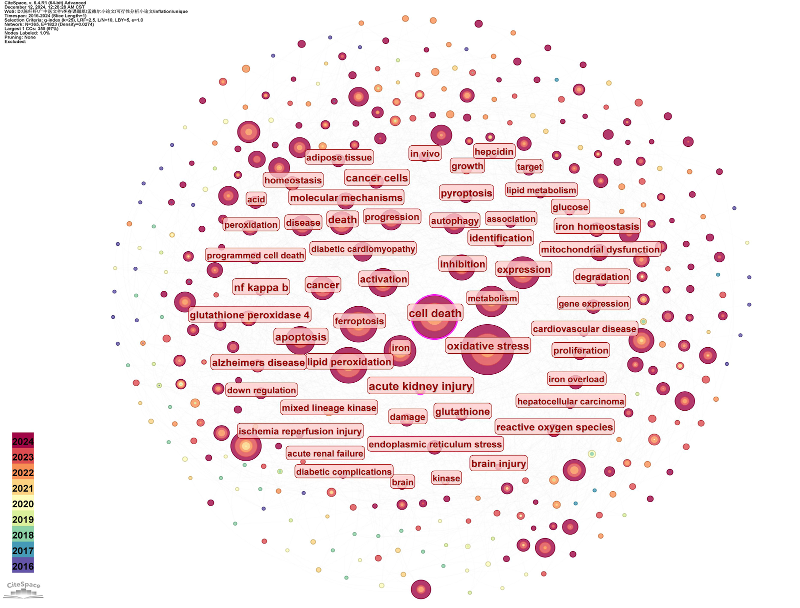
BACKGROUND: Oxidative stress induced by chronic hyperglycemia and impaired antioxidant systems are one of the core mechanisms underlying the onset and progression of diabetes mellitus. Ferroptosis, a new type of programmed cell death caused by iron-dependent lipid peroxidation, has received considerable academic interest due to its association with diabetes. OBJECTIVE: To reveal the current status and trends of ferroptosis-related research in the field of diabetes mellitus using bibliometric methods, aiming to offer academic resources to further develop this research area. METHODS: Based on the Web of Science Core Collection database, with a time span set from January 1, 2015 to January 1, 2025, 797 articles related to ferroptosis in the field of diabetes were retrieved. After deduplication, 758 high-quality articles were analyzed using CiteSpace (6.2.R1) for visualization of publication output, country/institution collaboration, high-impact authors/reference co-citation, keyword co-occurrence/clustering/burst detection, and international frontier trends. RESULTS AND CONCLUSION: Bibliometric analysis showed a substantial growth in ferroptosis-related research in diabetes. Among the 758 records, Linkermann, Andreas was the most prolific author, while Dixon SJ established an academic influence benchmark with 420 citations. The journal Cell served as a key knowledge dissemination hub. The most frequent keywords included oxidative stress, cell death, and lipid peroxidation, with clusters mainly focusing on diabetic nephropathy and cardiomyopathy, glutathione peroxidase 4, and necroptosis. The application of ferroptosis regulatory networks is deepening and has become an emerging paradigm for targeted therapy research in diabetes and its complications.

Read Full Abstract10.12307/2026.21558
Biomechanical finite element analysis of different ulnar shortening osteotomy techniques in treatment of ulnar impaction syndromeGraphical AbstractVerified
Chinese Journal of Tissue Engineering Research2026

Biomechanical finite element analysis of different ulnar shortening osteotomy techniques in treatment of ulnar impaction syndrome

BACKGROUND: Ulnar impaction syndrome is a common wrist disorder, and ulnar shortening osteotomy is one of the definitive surgical interventions for its treatment. Although various ulnar shortening osteotomy techniques exist, numerous clinical comparative studies have focused on pairwise comparisons, while biomechanical simulations comparing the efficacy of different osteotomy methods via finite element analysis remain unreported. OBJECTIVE: To simulate and compare the biomechanical characteristics of ulnar impaction syndrome under different osteotomy treatment modalities employing finite element method so as to provide references and evidence for clinical decision-making. METHODS: CT data of the intact ulna and radius from a healthy adult male volunteer were utilized. Modeling and finite element software platforms — Mimics 19.0, Geomagic Studio 2013, SolidWorks 2019, and Ansys 17.0 — were sequentially applied to construct five ulnar osteotomy models: (1) distal ulnar V-shaped osteotomy; (2) distal ulnar transverse osteotomy; (3) ulnar metaphyseal transverse osteotomy; (4) distal ulnar trapezoidal osteotomy; (5) distal ulnar oblique osteotomy. According to the experimental design and internal fixation principles, plates and screws were assembled. Subsequently, three motion modes of wrist joint axial compression, pronation, and supination were simulated, and corresponding boundary conditions and loads were applied to each group to obtain stress distribution and displacement at the osteotomy site and internal fixation devices. Finally, the results were compared with established experimental data standards to draw relevant conclusions. RESULTS AND CONCLUSION: (1) Under three different motions and loads, the five different ulnar shortening osteotomy methods all maintained stable osteotomy ends without significant relative differences. (2) There were certain differences in the stress and deformation of internal fixation devices among the five methods under three simulated motion states: under simulated pronation, the internal fixation plate of ulnar metaphyseal transverse osteotomy was at risk of fracture; under simulated supination, the plate also exhibited deformation risk. (3) Regarding stress and displacement of internal fixation, the distal ulnar V-shaped osteotomy showed relative advantages in stability under all three simulated states.

Read Full Abstract10.12307/2026.21515
Finite element analysis of four Kirschner wire fixation methods for treating patellar transverse fracturesGraphical AbstractVerified
Chinese Journal of Tissue Engineering Research2026

Finite element analysis of four Kirschner wire fixation methods for treating patellar transverse fractures

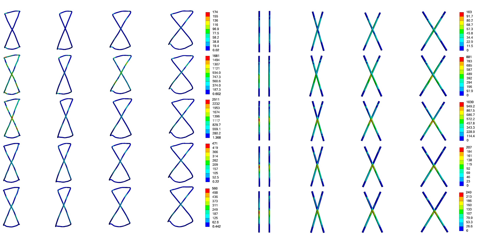
BACKGROUND: Kirschner wire and tension band internal fixation is the preferred surgical procedure for treating transverse patellar fractures, but it is often associated with postoperative instability, nonunion, and internal fixation failure. Therefore, optimizing the internal fixation method is of great clinical significance. OBJECTIVE: To investigate the effect of crossed Kirschner wire placement on the fixation of transverse patellar fractures. METHODS: A patellar model was constructed using normal lower limb CT scan data. A transverse patellar fracture and Kirschner wire model was further constructed. Parallel, 30°, 45°, and 60° crossed Kirschner wire placement models were designed. Finite element analysis was performed to analyze the fracture surface stress, fracture surface displacement, Kirschner wire stress, and wire stress under five different working conditions (neutral knee position, 5° flexion, 15° flexion, 45° flexion, and 60° flexion). RESULTS AND CONCLUSION: The fracture surface stresses in the four internal fixation models ranged from 2.06 to 40.00 MPa. The parallel Kirschner wire fixation group had the highest fracture surface stress among all five conditions. The crossed 30° Kirschner wire fixation group had lower fracture surface stress at 15° of knee flexion than the crossed 45° and crossed 60° Kirschner wire fixation groups. The fracture surface displacements in the four internal fixation models ranged from 0.03 to 0.61 mm. The crossed 60° Kirschner wire fixation group had the largest fracture surface displacement at 5° and 15° of knee flexion, while the parallel group had the smallest at 5° and the crossed 30° group had the smallest at 15°. The wire stresses ranged from 56.80 to 2511.00 MPa. The parallel group had the largest wire stress at 5° and 15° of knee flexion, while the crossed 30° group had the smallest. The Kirschner wire stresses ranged from 65.67 to 1018.00 MPa. The crossed 60° group had the largest Kirschner wire stress at 5° of knee flexion, the parallel group had the largest at 15°, and the crossed 30° group had the smallest at both 5° and 15°. The results indicate that 30° crossed Kirschner wire placement provides the best fracture stability and stress distribution, demonstrating superior biomechanical advantages.

Read Full Abstract10.12307/2026.21512
Finite element analysis of five internal fixation strategies for Schatzker IV tibial plateau fracturesGraphical AbstractVerified
Chinese Journal of Tissue Engineering Research2026

Finite element analysis of five internal fixation strategies for Schatzker IV tibial plateau fractures

BACKGROUND: Schatzker IV tibial plateau fractures are highly challenging due to their involvement of the primary weight-bearing area and high rate of soft tissue complications. Although traditional double plating provides mechanical stability, it violates the minimally invasive principle and is associated with more postoperative complications, especially in elderly patients or those with high-energy trauma. Currently, there is a lack of an internal fixation strategy that can meet both mechanical stability and minimally invasive requirements. OBJECTIVE: To establish a three-dimensional model of Schatzker IV tibial plateau fractures using the finite element method and compare the biomechanical stability of five fixation methods to provide an optimal surgical option for the treatment of Schatzker IV tibial plateau fractures. METHODS: A healthy male volunteer underwent knee CT scanning, and a Schatzker IV tibial plateau fracture model was constructed using finite-element software. Five internal-fixation configurations were defined as Groups A, B, C, D, and E. Group A: isolated medial plate; Group B: medial plate plus two posteromedial tension screws; Group C: medial plate plus two lateral tension screws; Group D: posteromedial double plating; Group E: medial-lateral double plating. Under identical boundary and constraint conditions, finite-element analysis software was employed to evaluate the biomechanical performance of five internal fixation models. RESULTS AND CONCLUSION: Finite element analysis showed that minimally invasive combinations (Groups B and C) had comparable overall biomechanical performance to traditional double plating. Group B was an ideal choice for elderly patients, as it had the lowest fracture fragment stress (9.0392 MPa), which could effectively prevent osteoporosis-related collapse, and the percutaneous screw technique reduced the risk of soft tissue complications. Group C showed potential in young patients, benefiting from the smallest implant displacement (4.388 mm), providing excellent stability, and the lateral tension screws avoided neurovascular injury associated with the posteromedial approach.

Read Full Abstract10.12307/2026.21513
Force analysis of three-dimensional finite element models for single radius and multi radius prostheses during flexion and extension in total knee arthroplastyGraphical AbstractVerified
Chinese Journal of Tissue Engineering Research2026

Force analysis of three-dimensional finite element models for single radius and multi radius prostheses during flexion and extension in total knee arthroplasty

BACKGROUND: For patients with end-stage knee osteoarthritis, total knee arthroplasty is often necessary in clinical practice to address issues such as pain and limited mobility. Although knee replacement surgery has achieved good clinical results in treating severe osteoarthritis, there is still controversy over the clinical efficacy of single radius and multi radius prostheses. OBJECTIVE: To compare the stress characteristics of single radius and multi radius prostheses at different flexion angles in total knee arthroplasty using finite element analysis, and provide a basis for clinical selection. METHODS: Based on normal adult CT data, a three-dimensional skeletal model was established and optimized using Mimics, Geomagic, and SolidWorks. Single radius and multi radius prostheses were assembled, and the 0°-120° flexion state was simulated in Ansys. The peak von Mises stress was used as the observation index to observe the Mises stress distribution and contact area on the tibial prosthesis. RESULTS AND CONCLUSION: (1) In the range of 0°-90°, the contact stress on the tibial insert of both single radius and multi radius prostheses increased with increasing flexion angle; at high flexion angles of 90°-120°, the stress of the multi radius prosthesis gradually decreased, while that of the single radius prosthesis slightly increased. (2) At 0° flexion, the posterior capsular stress of the single radius prosthesis was lower than that of the multi radius prosthesis. (3) In the low and medium flexion range of 0°-90°, the contact area gradually decreased with increasing flexion angle for both prostheses, and the contact area on the polyethylene insert of the single radius prosthesis was always larger than that of the multi radius prosthesis. (4) Both single radius and multi radius knee prostheses can meet clinical needs well; clinically, the appropriate femoral prosthesis should be selected based on patient age, activity level, and functional requirements.

Read Full Abstract10.12307/2026.21510
Bibliometric analysis of trends and hotspots in immune cells for fibrotic diseasesGraphical AbstractVerified
Chinese Journal of Tissue Engineering Research2026

Bibliometric analysis of trends and hotspots in immune cells for fibrotic diseases

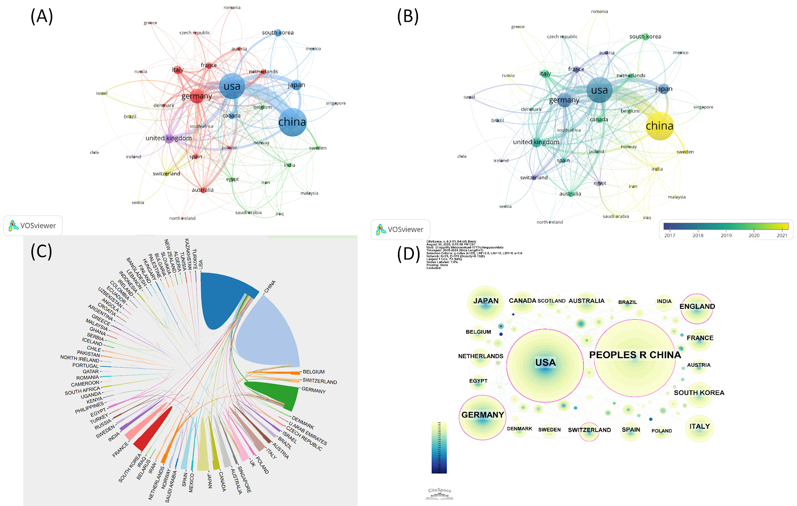
BACKGROUND: Research on multi-organ fibrotic diseases has gained increasing prominence in recent years. Immune cells play a crucial regulatory role in the pathogenesis of fibrotic diseases across various organs; however, a comprehensive bibliometric analysis in this specific research field is currently lacking. OBJECTIVE: To systematically analyze the current research status, hotspots, and emerging trends in the field of immune cells and fibrotic diseases using bibliometric methods. METHODS: Publications on immune cells and fibrotic diseases of the liver, lungs, kidneys, and heart were collected from the Web of Science Core Collection database spanning January 1, 2000 to December 31, 2024. Bibliometric and visual knowledge mapping analyses were performed on the extracted data using VOSviewer, CiteSpace, and the R package "bibliometrix". RESULTS AND CONCLUSION: A total of 1 777 relevant articles were identified. These publications were contributed by 11 347 authors from 2 239 institutions across 73 countries and were published in 637 academic journals. From January 1, 2000 to December 31, 2024, the annual publication volume showed an overall increasing trend. China, the United States, and Germany were the major contributing countries. The most prolific institutions were Zhejiang University and Huazhong University of Science and Technology in China. The most cited institution was RWTH Aachen University Hospital in Germany. The most productive journal was Frontiers in Immunology. The most prolific authors were Tacke, Frank and Trautwein, Christian. Core keywords included liver fibrosis, pulmonary fibrosis, macrophages, expression, and activation. The bibliometric analysis revealed a paradigm shift from single-organ studies to shared immune mechanisms, with the field evolving from basic research on liver fibrosis to molecular and cellular targeted regulation of multi-organ fibrosis including heart, lung, and kidney. KEYWORDS: fibrosis; immune cells; bibliometrics; visual analysis; VOSviewer software; CiteSpace software; single-cell sequencing technology; macrophages

Read Full Abstract10.12307/2026.21555
Neutrophils and the repair of hard-to-heal woundsGraphical AbstractVerified
Chinese Journal of Tissue Engineering Research2026

Neutrophils and the repair of hard-to-heal wounds

BACKGROUND: Dysfunction of neutrophils in the microenvironment of refractory wounds and their abnormal interactions with other immune cells and repair cells have become a central research focus for understanding the pathophysiology of chronic wounds and developing novel intervention strategies. OBJECTIVE: To investigate the current research status, hotspots, frontiers, and development trends regarding neutrophils in the field of hard-to-heal wound repair. METHODS: Relevant literature regarding neutrophils in the repair of hard-to-heal wounds published between 2004 and 2024 was retrieved from the Web of Science Core Collection database. Bibliometric visualization analysis methods were employed for analysis and visualization, revealing the research landscape, major hotspots, and frontier trends from dimensions including publication volume, countries/regions, institutions, authors, journals, references, and keywords. RESULTS AND CONCLUSION: A total of 1 062 relevant articles concerning neutrophils in hard-to-heal wound repair were included. The annual publication output in this field showed a significant growth trend, especially after 2014, with the number of publications exceeding 100 in 2023, indicating increasing academic attention. The United States led globally with 378 publications and extensive international collaboration networks; China ranked second with 189 publications, showing strong research vitality, though international collaboration and citations per paper still have room for improvement. Shanghai Jiao Tong University in China was the institution with the most publications, while the University of Illinois and Harvard University in the United States were centers of citation frequency. Journal analysis showed that Wound Repair and Regeneration was the core journal with the highest publication and citation counts. Research hotspots focused on inflammation, angiogenesis, diabetic foot ulcers, etc. Keyword clustering timeline and burst analysis revealed that neutrophil extracellular trap formation and neutrophil-macrophage interactions are frontier research trends. The results indicate that the field has formed a clear development path from clinical problems to molecular mechanisms and then to immune intervention strategies, and future precise regulation of neutrophil function is expected to open new avenues for the treatment of hard-to-heal wounds.

Read Full Abstract10.12307/2026.21552
Gut microbiota and short-chain fatty acids: mechanisms of aerobic exercise regulation in type 2 diabetesGraphical AbstractVerified
Chinese Journal of Tissue Engineering Research2026

Gut microbiota and short-chain fatty acids: mechanisms of aerobic exercise regulation in type 2 diabetes

BACKGROUND: Recent studies have shown that exercise modulates gut microbiota and glucose metabolism; however, the mechanism linking exercise to gut microbiota and short-chain fatty acid production in type 2 diabetes remains unclear. OBJECTIVE: To investigate the mechanism by which exercise modulates gut microbiota composition and short-chain fatty acid metabolism to treat type 2 diabetes. METHODS: Twenty male Sprague-Dawley rats were randomly divided into a control group (n=6) and a model group (n=14). The rats in the model group were fed a high-sugar, high-fat diet for 8 weeks to induce insulin resistance. Following 12 hours of fasting (water allowed), rats received a tail vein injection of 1% streptozotocin solution (35 mg/kg) to damage pancreatic β-cells and elevate blood glucose, establishing type 2 diabetes models. Following successful modeling, feeding protocols remained unchanged. Twelve type 2 diabetes rats were divided into a model group (n=6) and an exercise group (n=6), and the exercise group were subjected to 12 weeks of aerobic exercise. Following the final aerobic exercise intervention, blood samples were collected for glucose metabolism indicators, and fresh feces were collected for short-chain fatty acid measurement by gas chromatography. Total microbial DNA was extracted from fresh feces, PCR amplified, purified, and sequenced using NovaSeq high-throughput sequencing. Species annotation was performed using the SILVA database, differential flora screening based on linear discriminant analysis effect size, and MetaCyc functional pathway prediction to explore associations between exercise intervention and glycolipid metabolism pathways. Heatmaps and visualization network diagrams were used to analyze correlations between key flora and biochemical indicators. RESULTS AND CONCLUSION: After 12 weeks of aerobic exercise, the exercise group showed significant improvement in glycolipid metabolism disorders, reduced inflammation, enhanced insulin sensitivity, and significantly decreased fasting blood glucose (P < 0.01). α-diversity analysis showed that the exercise group had significantly higher richness (Chao index), coverage (Coverage index), diversity (Shannon index), and evenness (Simpson index) of gut microbiota compared to the model group (P < 0.05). Abundance statistics, correlation heatmaps, and network diagrams showed that exercise significantly increased the abundance of short-chain fatty acid-producing genera such as Colidextribacter and Intestinimonas within Firmicutes, positively correlated with hexanoic acid and valeric acid levels; while inhibiting pathogenic bacteria such as Klebsiella and Turicibacter within Proteobacteria, negatively correlated with short-chain fatty acid levels. Lactobacillus promoted the production of butyric acid and other short-chain fatty acids, and was negatively correlated with glycolipid metabolism and inflammatory markers, fasting blood glucose, and interleukin-6 (P < 0.05). KEGG and MetaCyc metabolic function prediction showed that aerobic exercise reshaped energy homeostasis by bidirectionally regulating microbial metabolic pathways: significantly downregulating pathways related to excessive glycolipid catabolism and pro-inflammatory metabolism, while upregulating key pathways for short-chain fatty acid synthesis and glycolytic homeostasis. This functional remodeling was highly synergistic with the restoration of short-chain fatty acid-producing genera in Firmicutes and inhibition of pathogenic bacteria in Proteobacteria (P < 0.05). These results suggest that the dynamic balance of microbial metabolic pathways and host glycolipid metabolism improvement and inflammation alleviation form a closed-loop regulation, indicating that microbiota, short-chain fatty acid synthesis, and metabolic function remodeling are core mechanisms by which exercise improves the pathological process of diabetes.

Read Full Abstract10.12307/2026.21537
Causal relationship between immune cell-mediated circulating inflammatory proteins and rheumatoid arthritisGraphical AbstractVerified
Chinese Journal of Tissue Engineering Research2026

Causal relationship between immune cell-mediated circulating inflammatory proteins and rheumatoid arthritis

BACKGROUND: Studies have shown that circulating inflammatory proteins and immune cells are associated with rheumatoid arthritis, but the causal relationship is unclear. OBJECTIVE: To explore the causal relationships between circulating inflammatory proteins and rheumatoid arthritis mediated by immune cells. METHODS: We downloaded data on circulating inflammatory proteins and immune cell phenotypes from the GWAS Catalog database (a publicly accessible database jointly established and maintained by the National Human Genome Research Institute and the European Bioinformatics Institute), and genome-wide association study data for rheumatoid arthritis from the FinnGen database (a genomics project resulting from collaboration between Finnish research institutions, biobanks, and international industry partners, also publicly accessible). Two-step Mendelian randomization analyses were performed: inverse variance weighting was used to assess the causal effects of 91 circulating inflammatory proteins and 731 immune cell phenotypes on rheumatoid arthritis risk, supplemented by MR-Egger, weighted median, weighted mode, simple mode, and sensitivity analyses. The mediating role of identified immune cells in the relationship between circulating inflammatory proteins and rheumatoid arthritis was evaluated. RESULTS AND CONCLUSION: Inverse variance weighting analysis showed that four circulating inflammatory proteins were significantly associated with rheumatoid arthritis risk, of which one was a risk factor and three were protective factors. Forty-six immune cell phenotypes were significantly associated with rheumatoid arthritis, of which 20 were risk factors and 26 were protective factors. Reverse Mendelian randomization analysis found no causal association between rheumatoid arthritis and the four identified circulating inflammatory proteins. Sensitivity analyses revealed no significant heterogeneity or horizontal pleiotropy. Further mediation analysis showed that CD19 on IgD- CD38br partially mediated the causal effect of interleukin-18 (β=0.064, OR=1.066, P=0.044) on rheumatoid arthritis, with a mediation effect of 0.004, a mediation proportion of 5.7%, and a direct effect of 0.060. The results reveal causal associations between circulating inflammatory proteins and immune cells with rheumatoid arthritis, and identify that CD19 on IgD- CD38br partially mediates the causal relationship between interleukin-18 and rheumatoid arthritis. For the Chinese biomedical research field, reference can be made to the integrated analysis framework of international multi-omics platforms and cross-ethnic cohort data to construct a combined database of epigenomics, proteomics, and metabolomics specific to the Chinese population, revealing the molecular regulatory networks underlying complex diseases, and facilitating disease subtyping and early diagnostic biomarker development. By learning from transnational collaborative research mechanisms, establish natural population cohorts of multiple ethnicities and regions in China, systematically analyze the impact of environmental exposure and gene interactions on health, and provide scientific evidence for formulating localized disease prevention strategies.

Read Full Abstract10.12307/2026.21554
Visualization analysis on research literature about animal models for osteonecrosis of the femoral headGraphical AbstractVerified
Chinese Journal of Tissue Engineering Research2026

Visualization analysis on research literature about animal models for osteonecrosis of the femoral head

BACKGROUND: Osteonecrosis of the femoral head is a refractory disorder characterized by osteocyte apoptosis and structural collapse of the femoral head. Its pathogenesis is closely associated with vascular injury, dysregulated bone metabolism, and aberrant mechanical stress. In recent years, animal models have served as indispensable tools for simulating pathological processes, playing an irreplaceable role in elucidating molecular mechanisms of osteonecrosis of the femoral head and evaluating novel interventions. Nevertheless, the standardization of model development and their clinical translational value require systematic investigation. OBJECTIVE: To analyze the research landscape in the field of osteonecrosis of the femoral head using bibliometric approaches, with an emphasis on evaluating the application characteristics, limitations, and future optimization directions of animal models in study design, providing a reference for advancing mechanistic understanding and therapeutic development. METHODS: Literature published between January 2015 and March 2025 was retrieved from the Web of Science Core Collection (SCI-Expanded), China National Knowledge Infrastructure (CNKI), and Wanfang databases. The search strategy for English literature was TS=(osteonecrosis of the femoral head) AND TS=(mouse OR mice OR rat OR rabbit OR dog OR swine OR pig OR sheep OR monkey OR "laboratory animal" OR "experiment animal"). For Chinese literature, the search was SU=股骨头坏死 AND SU=鼠+兔+犬+猪+羊+猴+实验动物+动物实验. CiteSpace 6.3.R1 software was used to perform visualization analysis of countries, institutions, authors, keywords, and co-cited references. Trends were summarized based on animal model classification and application characteristics. RESULTS AND CONCLUSION: (1) In English literature, China contributed over 80% of the research (458 articles). Hot topics focused on steroid-induced necrosis mechanisms (e.g., oxidative stress-autophagy axis), stem cell/tissue engineering therapies, and traditional Chinese medicine interventions. The research on animal models of osteonecrosis of the femoral head exhibited a "high output-low collaboration" characteristic. (2) In Chinese literature, the publishing institutions were mainly traditional Chinese medicine-related institutions, and traditional Chinese medicine and steroid-induced osteonecrosis of the femoral head were important research hotspots. (3) Future efforts should deepen cross-species validation platforms, multi-omics integration, and collaborative strategies for the development of Chinese and Western medicine to accelerate clinical translation.

Read Full Abstract10.12307/2026.21545
Druggable gene and single cell analyses reveal potential therapeutic targets for osteoporosisGraphical AbstractVerified
Chinese Journal of Tissue Engineering Research2026

Druggable gene and single cell analyses reveal potential therapeutic targets for osteoporosis

BACKGROUND: Genetic factors play an important role in the pathophysiology of osteoporosis, and Mendelian randomization can be used to infer causal associations between specific genes and diseases using eQTLs. OBJECTIVE: To identify potential therapeutic targets for osteoporosis based on druggable genes-related Mendelian randomization and colocalization analysis, to explore the potential biological mechanisms in the treatment of osteoporosis using bioinformatics analysis, and to predict the binding activity of drug targets using drug enrichment analysis and molecular docking. METHODS: (1) Data sources: Druggable genes were sourced from the DGIdb database (a public database constructed by the University of Washington School of Medicine, widely used for drug target discovery) and information provided in the literature. Expression quantitative trait locus (eQTL) data for druggable genes were obtained from eQTLGen (a large-scale blood eQTL database jointly constructed by multiple international research institutions, including the University of Groningen). Osteoporosis genome-wide association study (GWAS) data were obtained from FinnGen R12 (a large genomic database led by the University of Helsinki), including 10,461 osteoporosis cases and 473,264 controls. GEO datasets GSE230665 (microarray) and GSE169396 (single-cell) were also used. (2) Methods: Genes closely related to osteoporosis were screened; expression of genes in osteoporosis was evaluated via microarray data; single-cell analysis further observed the regulatory role of genes in cell communication; enrichment analysis was used to elucidate biological functions, and protein-protein interaction networks were constructed to analyze potential associations; drug enrichment and molecular docking predicted and simulated the binding of small molecule drugs to targets. RESULTS AND CONCLUSION: The study identified 37 druggable genes associated with osteoporosis, among which troponin C2 and CXC chemokine receptor 6 (CXCR6) had protective effects and shared causal genetic variants with the disease. Microarray data analysis showed that CXCR6 expression was significantly lower in osteoporosis than in normal controls, suggesting a weakened protective effect. Single-cell analysis further revealed that CXCR6 was mainly expressed on T cells, and CXCR6+ T cells exhibited stronger cell communication capabilities. Drug enrichment analysis found that NSC95397 could target CXCR6, and molecular docking showed good binding activity. These findings not only provide clues for the development of new drugs for osteoporosis but also facilitate the translation of research results.

Read Full Abstract10.12307/2026.21550
Role of bone–blood axis in bone mass regulation and hematopoietic function maintenanceGraphical AbstractVerified
Chinese Journal of Tissue Engineering Research2026

Role of bone–blood axis in bone mass regulation and hematopoietic function maintenance

BACKGROUND: The bone marrow serves not only as a primary hematopoietic organ but also as an essential component of bone tissue. The bone marrow microenvironment is a critical niche for maintaining hematopoietic stem cell function, while the hematopoietic process itself can regulate bone remodeling and maintain bone mass stability. The precise synergistic interaction between the skeletal and hematopoietic systems maintains the health of both blood and bone, yet a systematic summary of these interactions is lacking. OBJECTIVE: To systematically review the research progress on the interactions between the skeletal and hematopoietic systems, aiming to provide a reference for their mutual regulation and to explore potential therapeutic targets for blood diseases such as anemia and leukemia, and bone diseases such as osteoporosis and osteoarthritis. METHODS: A search of CNKI, Wanfang, and PubMed databases was conducted for literature published from January 2000 to July 2025 using keywords including 'bone mass regulation', 'hematopoietic function', 'bone marrow microenvironment', and 'bone and blood axis'. A total of 115 articles were included for analysis. RESULTS AND CONCLUSION: (1) The bidirectional regulatory network of the 'bone-blood axis' in the bone marrow microenvironment and its core mechanisms were systematically elaborated. (2) The bone marrow microenvironment, as a dynamic system composed of multiple cellular and non-cellular components, precisely regulates the quiescence, self-renewal, and differentiation of hematopoietic stem cells through core signaling pathways such as Wnt/β-catenin, Notch, RANK/RANKL/OPG, and Hippo-YAP, while also receiving reverse regulation from the hematopoietic system. (3) This bidirectional dialogue also dominates bone remodeling, with immune cells (e.g., macrophages and T lymphocytes) serving as key bridges connecting the skeletal and hematopoietic systems by secreting specific factors. (4) Imbalance in this dialogue network is an important pathological basis for the occurrence of cross-system diseases such as osteoporosis, myelofibrosis, and leukemia. (5) This article provides a new perspective for understanding the bone marrow microenvironment through the framework of the 'bone-blood axis', revealing the co-pathogenesis of blood and bone diseases. Targeting key signaling nodes of this axis or utilizing synergistic intervention strategies (e.g., denosumab, enasidenib) may open new avenues for integrated treatment of cross-system diseases in the future.

Read Full Abstract10.12307/2026.21540
Effects of blood flow restriction training and aerobic exercise on energy expenditure in young menGraphical AbstractVerified
Chinese Journal of Tissue Engineering Research2026

Effects of blood flow restriction training and aerobic exercise on energy expenditure in young men

BACKGROUND: In recent years, high-intensity interval exercise has been widely used and gained attention due to its short duration. However, its safety has been seriously questioned because of its high intensity and the controversial effects on the heart. Therefore, exploring time-efficient, intensity-controllable exercise interventions with good compliance has become a research hotspot in the field of exercise science. OBJECTIVE: To investigate the effects of blood flow restriction combined with aerobic exercise on energy expenditure. METHODS: Fifteen male college students were recruited. A repeated-measures crossover design was used to design two exercise protocols: low-intensity aerobic exercise (non-blood flow restriction group) and blood flow restriction training combined with low-intensity aerobic exercise (blood flow restriction group). Both protocols were performed at 40% maximal oxygen uptake, with 10 minutes of running per session, 1 minute rest between sessions, for a total of five sessions and an exercise duration of 54 minutes. In the blood flow restriction group, a cuff was placed at the most proximal end of both lower limbs and pressurized to 50% of the arterial occlusion pressure before exercise, and the pressure was released during each exercise interval. The interval between the two protocols was at least 72 hours. Blood lactate, energy expenditure during exercise, excess post-exercise oxygen consumption, ratings of perceived exertion, heart rate, and blood pressure were measured. RESULTS AND CONCLUSION: (1) Total energy expenditure in the blood flow restriction group was significantly greater than that in the non-blood flow restriction group (P < 0.05). (2) There was no significant difference in the proportional contribution of the three energy systems (aerobic, anaerobic lactic, and anaerobic alactic) between the two exercise modes (P > 0.05). (3) The total excess post-exercise oxygen consumption within 40 minutes of recovery was significantly greater in the blood flow restriction group than in the non-blood flow restriction group (P < 0.05). At 1 minute of recovery, heart rate was significantly higher in the blood flow restriction group than in the non-blood flow restriction group (P < 0.05). At other time points, there were no significant differences in ratings of perceived exertion, heart rate, or blood pressure between the two groups. (4) These findings suggest that blood flow restriction training combined with low-intensity aerobic exercise can increase energy expenditure and excess post-exercise oxygen consumption without additional increases in ratings of perceived exertion, providing an additional training option for individuals seeking to increase physical activity levels, but attention should be paid to the elevated heart rate during recovery.

Read Full Abstract10.12307/2026.21538
A new approach to intervene in Alzheimer's disease through regulating the silencing information regulator 1 signaling pathway with traditional Chinese medicineGraphical AbstractVerified
Chinese Journal of Tissue Engineering Research2026

A new approach to intervene in Alzheimer's disease through regulating the silencing information regulator 1 signaling pathway with traditional Chinese medicine

BACKGROUND: Silent information regulator 1, as a deacetylase, can regulate the transcriptional activity of various proteins and many Alzheimer’s disease related pathological processes after activation, including regulating energy metabolism, oxidative stress, neuroinflammation, autophagy, and cell apoptosis. It is closely related to the occurrence, development, and prognosis of Alzheimer’s disease. In recent years, a large number of studies have found that the active ingredients and formulas of traditional Chinese medicine monomers can delay the development of Alzheimer's disease by activating the silent information regulator 1 signaling pathway, reducing cell apoptosis, protecting neurons, and inhibiting the formation of amyloid plaque. OBJECTIVE: To explore the relationship between silent information regulator 1 and Alzheimer’s disease, as well as the action mechanism of traditional Chinese medicine in regulating the silent information regulator 1 signaling pathway for the treatment of Alzheimer’s disease. METHODS: The CNKI, WanFang, VIP, SinoMed, and PubMed databases were searched for literature published from January 2015 to March 2025 using Chinese and English search terms including 'Alzheimer’s disease, dementia, SIRT1, inflammation, oxidative stress, inflammatory factors, signaling pathways, TCM monomers, compounds'. A total of 4,921 relevant articles were retrieved, and 154 were finally included for review. RESULTS AND CONCLUSION: A large number of experimental studies have confirmed that silent information regulator 1 plays an important role in Alzheimer’s disease. Traditional Chinese medicine formulas can regulate the silent information regulator 1 signaling pathway in various ways. For example, sodium ferulate improves the learning and memory ability of Alzheimer’s disease rats by increasing the expression of silent information regulator 1 in the prefrontal cortex, thereby ameliorating nerve cell damage caused by ischemia and hypoxia. Oyster peptide can enhance reactive oxygen species, reduce oxidative damage in hippocampal tissue, alleviate neuroinflammation, enhance synaptic function, reduce neuronal damage and death, exert brain protective effects, and improve cognitive dysfunction.

Read Full Abstract10.12307/2026.21541
Functional near-infrared spectroscopy analysis of prefrontal cortex hemodynamics during dual tasks under cognitive loadsGraphical AbstractVerified
Chinese Journal of Tissue Engineering Research2026

Functional near-infrared spectroscopy analysis of prefrontal cortex hemodynamics during dual tasks under cognitive loads

BACKGROUND: In daily life, individuals often operate in a dual-task mode that requires simultaneous execution of motor and cognitive functions. This situation necessitates the coordination of both motor and cognitive functions, placing higher demands on brain workload. Theoretically, the difficulty in cognitive tasks can differentially impact dual-task performance. However, the mechanisms underlying changes in prefrontal cortex hemodynamic responses during motor-cognitive dual tasks under varying cognitive loads remain unclear. Elucidating the hemodynamic responses of the prefrontal cortex under different cognitive loads is highly important for optimizing motor-cognitive training intervention strategies and enhancing training effectiveness and safety. OBJECTIVE: To observe the effects of dual tasks combining narrow-base walking with logical subtraction under different cognitive loads on prefrontal cortex hemodynamics using a portable functional near-infrared spectroscopy device. METHODS: Thirty college students were recruited to complete three task conditions: narrow-base walking, narrow-base walking with subtracting 3, and narrow-base walking with subtracting 7. Changes in prefrontal cortex oxyhemoglobin, subjective cognitive load, dual-task cost, and spatiotemporal gait parameters were recorded. After Shapiro-Wilk test, Friedman and Wilcoxon tests were used to analyze prefrontal cortex activation differences, repeated-measures ANOVA compared cognitive load and gait performance, and Pearson correlation assessed the relationship between prefrontal cortex activation and cognitive load. RESULTS AND CONCLUSION: Compared with narrow-base walking, both subtracting 3 and subtracting 7 dual tasks significantly increased multi-channel activation in the prefrontal cortex (P < 0.05), but dorsolateral prefrontal cortex activation was higher under subtracting 3 than subtracting 7 (P < 0.05). As task difficulty increased, accuracy significantly decreased (P < 0.01), and gait parameters such as step length, stance phase, and swing phase further declined (P < 0.05-0.01), with dual-task cost increasing (P < 0.01). Significant prefrontal cortex activation channels were positively correlated with mental demand, temporal demand, effort, frustration, and overall task load, and negatively correlated with task performance. These findings suggest that selecting a moderate cognitive load during dual-task interventions is beneficial to fully activate prefrontal cortex resources without overloading, thereby achieving a better balance between cognitive and motor tasks. The results reveal the interaction mechanism between motor and cognitive functions in dual tasks, providing scientific support for optimizing the effectiveness and safety of motor-cognitive dual-task training.

Read Full Abstract10.12307/2026.21536
Establishment and validation of a Sprague-Dawley rat model of aging-related knee osteoarthritisGraphical AbstractVerified
Chinese Journal of Tissue Engineering Research2026

Establishment and validation of a Sprague-Dawley rat model of aging-related knee osteoarthritis

BACKGROUND: Knee osteoarthritis is an age-related disease, and aging is closely related to its occurrence and development. Chondrocyte senescence plays a crucial role in the pathological progression of knee osteoarthritis. OBJECTIVE: To establish a stable induced knee osteoarthritis model in SD rats. METHODS: (1) Animal experiment: Forty Sprague-Dawley rats were randomly divided into four groups: blank control group (no modeling), D-galactose group (intra-articular injection of D-galactose solution once a week for 2 months), anterior cruciate ligament transection (ACLT) group (ACLT to establish knee osteoarthritis model), and D-galactose+ACLT group (ACLT followed by intra-articular injection of D-galactose solution once a week for 2 months). One week after modeling, all rats underwent running exercise for 30 min every other day. At 4 and 8 weeks after modeling, behavioral tests (Lequesne MG score) were performed, and then samples were collected for detection of inflammatory factors in synovial fluid, histopathological morphology of knee cartilage, transmission electron microscopy observation, and immunohistochemical staining of type II collagen and aggrecan. (2) Cell experiment: At 4 and 8 weeks after modeling, knee chondrocytes were isolated from each group for flow cytometry cell cycle analysis, β-galactosidase staining, and γ-H2AX immunofluorescence staining. RESULTS AND CONCLUSION: (1) Animal experiment: At 8 weeks after modeling, Lequesne MG scores in the three model groups were higher than those in the blank control group (P < 0.05), and the score in the D-galactose+ACLT group was higher than that in the D-galactose and ACLT groups (P < 0.05). At 4 and 8 weeks, levels of interleukin-1β, interleukin-6, and tumor necrosis factor-α in synovial fluid were higher in the three model groups than in the blank control group (P < 0.05), and these levels were higher in the D-galactose+ACLT group than in the D-galactose and ACLT groups (P < 0.05). Hematoxylin-eosin and safranin O/fast green staining and transmission electron microscopy at 4 and 8 weeks showed that cartilage damage and chondrocyte mitochondrial damage were more severe in the D-galactose+ACLT group than in the D-galactose and ACLT groups. Immunohistochemical staining showed that the expression of type II collagen and aggrecan was highest in the blank control group, and lowest in the D-galactose+ACLT group among the model groups. (2) Cell experiment: At 4 and 8 weeks, the proportion of chondrocytes in G0/G1 phase was higher, and the proportions in S and G2/M phases were lower in the D-galactose+ACLT group than in the other three groups (P < 0.05). The positive rate of β-galactosidase staining and γ-H2AX immunofluorescence intensity were higher in the D-galactose+ACLT group than in the other three groups (P < 0.05). (3) These results indicate that the D-galactose+ACLT method can establish an SD rat model of aging-related knee osteoarthritis, which can better simulate the pathological state of aging and degeneration in knee osteoarthritis.

Read Full Abstract10.12307/2026.21529
Yanggan Roujin Decoction delays intervertebral disc degeneration: network pharmacological analysis and experimental validation in rat modelsGraphical AbstractVerified
Chinese Journal of Tissue Engineering Research2026

Yanggan Roujin Decoction delays intervertebral disc degeneration: network pharmacological analysis and experimental validation in rat models

BACKGROUND: The clinical efficacy of Yanggan Roujin Decoction in delaying intervertebral disc degeneration is significant; however, its underlying mechanism remains unclear. OBJECTIVE: To validate the potential mechanisms by which Yanggan Roujin Decoction delays intervertebral disc degeneration using network pharmacology techniques and animal experiments. METHODS: (1) The effective components of Yanggan Roujin Decoction and their action targets were screened using the Traditional Chinese Medicine Systems Pharmacology Database and High-Throughput Experiment- and Reference-Guided Database of Traditional Chinese Medicine. Gene sets related to intervertebral disc degeneration were obtained from the Genecard, CTD, and DisGeNet databases. A protein-protein interaction network was constructed using the STRING database, and core targets were identified. The drug-component-target-disease network was constructed using Cytoscape 3.9.1 software. Gene Ontology and Kyoto Encyclopedia of Genes and Genomes enrichment analyses were performed on the Ouyi Biological Cloud Platform to obtain potential therapeutic mechanisms. (2) A rat model of intervertebral disc degeneration was established by percutaneous puncture of the annulus fibrosus. Different doses of Yanggan Roujin Decoction and diclofenac sodium were administered. Histopathological staining was used to assess structural changes in intervertebral disc tissue before and after intervention. Western blot and quantitative real-time PCR were used to detect the expression of core targets and related pathways. RESULTS AND CONCLUSION: Network pharmacology analysis identified 187 active components of Yanggan Roujin Decoction, mainly including β-sitosterol, sitosterol, stigmasterol, quercetin, and kaempferol. There were 298 potential targets for treating intervertebral disc degeneration, mainly including tumor protein p53, transcription factor c-Jun, interleukin-6, tumor necrosis factor α, protein kinase B, and insulin. These targets were mainly involved in enzyme binding, nitric oxide synthase regulatory activity, and signaling pathways such as mitogen-activated protein kinase, hypoxia-inducible factor 1, tumor necrosis factor, and interleukin-17. Animal experiments showed that Yanggan Roujin Decoction effectively alleviated pathological structural changes in intervertebral disc tissue caused by annulus fibrosus puncture. Compared with the blank control group, the model group showed increased protein and mRNA expression of tumor protein p53, transcription factor c-Jun, interleukin-6, and tumor necrosis factor α (P < 0.01), while protein kinase B and insulin protein and mRNA expression decreased (P < 0.01). Meanwhile, the expression of representative genes in the mitogen-activated protein kinase signaling pathway (P38, extracellular regulated protein kinase) and tumor necrosis factor signaling pathway (interleukin-1β, tumor necrosis factor α) increased, while the expression of representative genes in the hypoxia-inducible factor 1 signaling pathway (hypoxia-inducible factor 1α, vascular endothelial growth factor A) decreased. After intervention with different doses of Yanggan Roujin Decoction, the protein and mRNA expression of tumor protein p53, transcription factor c-Jun, interleukin-6, and tumor necrosis factor α decreased (P < 0.01), while protein kinase B and insulin protein and mRNA expression increased (P < 0.01). Simultaneously, the expression of representative genes in the mitogen-activated protein kinase signaling pathway (P38, extracellular regulated protein kinase) and tumor necrosis factor signaling pathway (interleukin-1β, tumor necrosis factor α) decreased, while the expression of representative genes in the hypoxia-inducible factor 1 signaling pathway (hypoxia-inducible factor 1α, vascular endothelial growth factor A) increased. After diclofenac sodium intervention, the protein and mRNA expression of interleukin-6 and tumor necrosis factor α decreased (P < 0.01), and the protein and mRNA expression of representative genes in the tumor necrosis factor signaling pathway (interleukin-1β, tumor necrosis factor α) decreased, but there were no significant differences in other indicators. These results indicate that Yanggan Roujin Decoction can effectively delay the progression of intervertebral disc degeneration, and the mechanism may be related to the regulation of tumor protein p53, transcription factor c-Jun, interleukin-6, tumor necrosis factor α, protein kinase B, insulin target genes, inhibition of mitogen-activated protein kinase and tumor necrosis factor signaling pathways, and activation of hypoxia-inducible factor 1 signaling pathway.

Read Full Abstract10.12307/2026.21530
Effects and mechanisms of glycemic variability on apoptosis in mouse hippocampal neuronal HT-22 cellsGraphical AbstractVerified
Chinese Journal of Tissue Engineering Research2026

Effects and mechanisms of glycemic variability on apoptosis in mouse hippocampal neuronal HT-22 cells

BACKGROUND: Previous studies have confirmed that the "metabolic memory" effect induced by a sustained high-glucose environment can significantly exacerbate damage in mouse hippocampal neuronal cell lines HT-22. OBJECTIVE: To investigate the effects of glycemic variability and sustained high glucose on apoptosis and the expression of histone deacetylase 4 (HDAC4) and silent information regulator 1 (SIRT1) in mouse hippocampal neuronal HT-22 cells. METHODS: Passage 6 HT-22 cells were cultured in three groups after adherence: control group (25 mmol/L glucose for 3 or 5 days), high glucose group (55 mmol/L glucose for 3 or 5 days), and glycemic variability group (alternating 25 mmol/L and 55 mmol/L glucose every 12 hours for 3 or 5 days). After 3 days of culture, cell morphology was observed under an optical microscope. After 5 days, apoptosis was detected by flow cytometry. Cell viability was measured by CCK-8 assay at 3, 4, and 5 days. Reactive oxygen species (ROS) levels were detected using 2,7-dichlorofluorescein diacetate fluorescent probe at 3 and 5 days. Histone deacetylase (HDAC) content in the supernatant was measured by ELISA. Protein expression of Bax, Bcl-2, Caspase-3, Cleaved Caspase-3, SIRT1, and HDAC4 was detected by western blot, and mRNA expression of Bax, Bcl-2, Caspase-3, SIRT1, and HDAC4 was detected by RT-qPCR. RESULTS AND CONCLUSION: (1) Under the optical microscope, control cells grew well, forming a dense network with interconnected synapses; high glucose and glycemic variability groups showed inhibited growth and reduced synaptic connections. Apoptosis rate was higher in the high glucose group than in the control and glycemic variability groups (P < 0.05), and higher in the glycemic variability group than in the control group (P < 0.05). (2) At 3, 4, and 5 days, cell viability was lower in the high glucose group than in the control and glycemic variability groups (P < 0.05), and lower in the glycemic variability group than in the control group (P < 0.05). (3) At 3 and 5 days, ROS levels were higher in the high glucose group than in the control and glycemic variability groups (P < 0.05), and higher in the glycemic variability group than in the control group (P < 0.05). HDAC content in the supernatant was higher in the high glucose and glycemic variability groups than in the control group (P < 0.05). (4) At 3 and 5 days, protein and mRNA expression of HDAC4, Bax, and Caspase-3 were higher in the high glucose and glycemic variability groups than in the control group (P < 0.05), while SIRT1 and Bcl-2 expression were lower (P < 0.05). Cleaved Caspase-3 protein expression was higher in the high glucose and glycemic variability groups than in the control group (P < 0.05). (5) These results indicate that glycemic variability may induce apoptosis in HT-22 cells by upregulating HDAC4 expression and downregulating SIRT1 expression.

Read Full Abstract10.12307/2026.21533
Matrine promotes macrophage polarization to repair myocardial tissue injury in ratsGraphical AbstractVerified
Chinese Journal of Tissue Engineering Research2026

Matrine promotes macrophage polarization to repair myocardial tissue injury in rats

BACKGROUND: Matrine exerts therapeutic effects on pneumonia, sepsis, and hepatitis B because of its significant anti-inflammatory and anti-tumor properties. However, its role and mechanisms in macrophage-mediated inflammation post myocardial infarction remain poorly understood. OBJECTIVE: To investigate the effects of matrine on inflammation and myocardial tissue repair following myocardial infarction, and to clarify its potential molecular mechanism. METHODS: (1) In vivo experiment: 32 Sprague-Dawley rats were randomly divided into sham operation group, myocardial infarction group, low-dose matrine [200 mg/(kg·d)] group, and high-dose matrine [300 mg/(kg·d)] group, with 8 rats in each group. Myocardial infarction model was established in rats. Matrine was continuously gavaged for 3 days after surgery. On day 4, echocardiography and serum inflammatory cytokine levels were detected, and heart tissues were collected for histological analysis (hematoxylin-eosin staining, Masson staining) to assess myocardial injury. (2) In vitro experiment: Mouse bone marrow-derived macrophages were isolated and induced with lipopolysaccharide to establish an inflammation model, then treated with 10 μmol/L or 20 μmol/L matrine. Western blot, RT-qPCR, and flow cytometry were used to detect M1/M2 macrophage markers (inducible nitric oxide synthase, CD86, arginase 1, CD206) expression and cell proportions. Western blot was used to detect phosphorylation levels of Janus kinase 2 (JAK2) and signal transducer and activator of transcription 3 (STAT3). Additionally, H9C2 cells were used to construct a hypoxia injury model to evaluate the effect of matrine on cellular reactive oxygen species levels. RESULTS AND CONCLUSION: (1) In vivo: Matrine significantly improved cardiac function in myocardial infarction rats. Compared with the myocardial infarction group, both low-dose and high-dose matrine groups significantly increased left ventricular ejection fraction and left ventricular fractional shortening, with the high-dose group showing more pronounced improvement. Furthermore, matrine significantly inhibited the elevation of peripheral inflammatory cytokines (interleukin-1β and interleukin-6) after myocardial infarction. Histopathological analysis showed that matrine effectively reduced myocardial inflammatory cell infiltration, collagen deposition area, tissue edema, and necrosis range. (2) In vitro: RT-qPCR and Western blot results showed that matrine significantly inhibited the expression of M1 markers (inducible nitric oxide synthase, CD86) while upregulating M2 markers (arginase 1, CD206). Flow cytometry showed a decrease in CD86-positive cells and a significant increase in CD206-positive cells. Additionally, matrine significantly reduced intracellular reactive oxygen species levels. (3) Signaling pathway: Western blot results showed that matrine significantly inhibited the phosphorylation of JAK2 and STAT3 proteins, blocking pro-inflammatory signal transduction and promoting macrophage phenotype switch, thereby exerting anti-inflammatory effects. These results indicate that matrine can promote macrophage polarization toward M2 phenotype by inhibiting the JAK/STAT pathway, attenuate post-myocardial infarction inflammation, and protect cardiomyocytes in a dose-dependent manner.

Read Full Abstract10.12307/2026.21534
Exploring the characteristics of neck muscle strength and activation in patients with cervical spondylotic radiculopathy using motion capture-Opensim digital simulation technologyGraphical AbstractVerified
Chinese Journal of Tissue Engineering Research2026

Exploring the characteristics of neck muscle strength and activation in patients with cervical spondylotic radiculopathy using motion capture-Opensim digital simulation technology

BACKGROUND: Modern research suggests that the “muscle-bone imbalance” in cervical spondylotic radiculopathy is associated with changes in the performance of cervical muscles due to biomechanical alterations of the head and neck. However, most studies have focused on static analysis of the cervical spine, lacking dynamic quantitative data on neck muscle strength and activation. OBJECTIVE: To investigate the differences in muscle strength and muscle activation during cervical spine movement between patients with cervical spondylotic radiculopathy and healthy individuals from a biomechanical perspective. METHODS: From October 1, 2023 to March 1, 2024, 10 volunteers were recruited from the orthopedic outpatient or inpatient department and health examination center of the First Affiliated Hospital of Guangxi University of Chinese Medicine, including 5 patients with cervical spondylotic radiculopathy (cervical spondylotic radiculopathy group) and 5 healthy individuals (healthy control group). Inertial motion capture sensors were used to capture the motion data of the neck in six degrees of freedom: flexion, extension, left lateral flexion, right lateral flexion, left rotation, and right rotation. Each movement was repeated three times, yielding 18 sets of data per subject. Based on MRI-derived muscle parameters of patients and healthy individuals, OpenSim head-neck musculoskeletal models were respectively established. After preprocessing, the captured neck motion data were imported into the OpenSim simulation model to calculate and compare the neck muscle strength and muscle activation levels between the two groups. RESULTS AND CONCLUSION: (1) The healthy control group showed orderly synergistic activation of muscle groups, while the cervical spondylotic radiculopathy group exhibited disordered synergistic activation patterns, characterized by insufficient activation of the affected side muscles, excessive compensation of the healthy side, and significantly reduced synergy of core muscles such as the sternocleidomastoid, middle and upper trapezius, and longus colli. In the cervical spondylotic radiculopathy group, during extension, the muscle strength of the sternocleidomastoid, middle trapezius, longus capitis, and upper trapezius was lower than that of the healthy control group (P < 0.05). During flexion, the muscle strength of the sternocleidomastoid, longus capitis, longus colli, levator scapulae, multifidus, splenius cervicis, splenius capitis, and middle/upper trapezius was lower than that of the healthy control group (P < 0.05). During left lateral flexion, the muscle strength of the sternocleidomastoid, longus capitis, longus colli, scalene, levator scapulae, multifidus, splenius cervicis, splenius capitis, and middle/upper trapezius was lower than that of the healthy control group (P < 0.05). During right lateral flexion, the muscle strength of the longus colli and upper trapezius was lower than that of the healthy control group (P < 0.05). During left rotation, the muscle strength of the levator scapulae was lower than that of the healthy control group (P < 0.05). During right rotation, the muscle strength of the longus colli was lower than that of the healthy control group (P < 0.05). (2) These findings indicate that patients with cervical spondylotic radiculopathy exhibit muscle synergy imbalance in all directions of cervical spine movement. The imbalance is characterized by degeneration of the affected side muscles and nerves, reduced muscle strength and coordination, while the healthy side muscles and nerves compensate for the functional insufficiency of the affected side, leading to over-control. Among the muscle groups involved in cervical spine movement, the degeneration of the sternocleidomastoid and middle/upper trapezius is the most significant, which is one of the important features causing cervical spondylotic radiculopathy.

Read Full Abstract10.12307/2026.21528
Mechanism by which astragalus-peach kernel alleviates renal fibrosis in chronic kidney disease ratsGraphical AbstractVerified
Chinese Journal of Tissue Engineering Research2026

Mechanism by which astragalus-peach kernel alleviates renal fibrosis in chronic kidney disease rats

BACKGROUND: Our previous studies have indicated that astragalus–peach kernel alleviates the progression of chronic kidney disease yet its precise mechanism remains to be elucidated. OBJECTIVE: To investigate the therapeutic effects and underlying mechanisms of astragalus-peach kernel in preventing renal fibrosis in chronic kidney disease rats. METHODS: (1) Gene expression profile chip datasets related to chronic kidney disease were retrieved and filtered via the Gene Expression Omnibus (GEO) database. Combined with network pharmacology, core targets of chronic kidney disease were screened, and molecular docking was performed to validate key genes. (2) Forty Sprague-Dawley rats were randomly divided into four groups: blank group (n=10) without modeling, model group (n=10), dapagliflozin group (n=10), and astragalus-peach kernel group (n=10). Chronic kidney disease was induced by intragastric administration of 2% adenine solution. After successful modeling, the blank and model groups received normal saline, the dapagliflozin group received dapagliflozin, and the astragalus-peach kernel group received astragalus-peach kernel (1:1) by gavage once daily for 8 weeks. After the last administration, serum creatinine and blood urea nitrogen levels were measured; renal tissue was examined by hematoxylin-eosin staining, Masson staining, and immunohistochemistry for α-smooth muscle actin and type I collagen; RT-qPCR detected mRNA expression of α-smooth muscle actin, type I collagen, transforming growth factor β, and c-Myc; western blot detected protein expression of α-smooth muscle actin, type I collagen, transforming growth factor β, c-Myc, Smad3, and p-Smad3. RESULTS AND CONCLUSION: (1) GEO database combined with network pharmacology identified 9 active components of astragalus-peach kernel and 7 core disease targets (c-Myc, RB1, CHUK, MAPK14, DPEP1, NR1I3, NQO2). KEGG enrichment analysis showed that the Ras-MAPK-c-Myc signaling pathway was related to chronic kidney disease. Molecular docking indicated strong binding ability between c-Myc and core drug components. (2) Compared with the blank group, the model group showed reduced glomerular structure and number, tubular inflammatory cell infiltration, fibroblast proliferation, abnormal collagen fiber deposition in the interstitium, and increased serum creatinine, blood urea nitrogen, and mRNA and protein expression of α-smooth muscle actin and type I collagen (P < 0.05). Compared with the model group, the astragalus-peach kernel group showed more intact renal morphology, reduced inflammatory infiltration and fibroblast proliferation, and decreased serum creatinine, blood urea nitrogen, and mRNA and protein expression of α-smooth muscle actin and type I collagen (P < 0.05). Compared with the blank group, the model group had increased mRNA and protein expression of c-Myc and transforming growth factor β, and increased protein expression of Smad3 and p-Smad3 (P < 0.05). Compared with the model group, these expressions were decreased in the astragalus-peach kernel group (P < 0.05). (3) These results indicate that astragalus-peach kernel can delay the progression of chronic kidney disease in SD rats, and the therapeutic mechanism may be related to the c-Myc/transforming growth factor β/Smad3 signaling pathway.

Read Full Abstract10.12307/2026.21532
Combined Proteomics and Metabolomics Analysis of Pathological Mechanisms in Mouse Models of Coronary Heart DiseaseGraphical AbstractVerified
Chinese Journal of Tissue Engineering Research2026

Combined Proteomics and Metabolomics Analysis of Pathological Mechanisms in Mouse Models of Coronary Heart Disease

BACKGROUND: The pathogenesis of coronary heart disease is complex. A single omics approach is limited in elucidating its biological pathways, whereas multi-omics integration helps reveal molecular interaction networks across different levels, addressing the limitations of single-omics methods. OBJECTIVE: To investigate the pathological mechanisms of coronary heart disease in a mouse model using proteomics and metabolomics. METHODS: Healthy SPF-grade 8-week-old male C57BL/6 mice were randomly divided into a sham operation group and a model group. The mouse model of coronary heart disease was established by ligation of the left anterior descending coronary artery, while the sham operation group underwent threading without ligation. At 28 days post-surgery, cardiac function was assessed by echocardiography, and myocardial infarct size was evaluated by TTC staining. Ultra-high-performance liquid chromatography-tandem mass spectrometry was used to screen differentially expressed proteins and metabolites between groups, followed by integrated omics analysis. RESULTS AND CONCLUSION: Compared with the sham group, the model group exhibited reduced cardiac function, with significantly decreased left ventricular ejection fraction and left ventricular fractional shortening (P < 0.05), and significantly increased myocardial infarct size (P < 0.01). Proteomics identified 420 differentially expressed proteins, including 282 upregulated (e.g., Serum amyloid A protein, protein kinase D) and 138 downregulated (e.g., Protein YIPF5, E3 ubiquitin-protein ligase). KEGG pathway enrichment revealed involvement in ATP-dependent chromatin remodeling and renin-angiotensin system pathways. Metabolomics identified 155 differential metabolites, including 56 upregulated (e.g., Thromboxane, Tromethamine) and 99 downregulated (e.g., N-Acetyl-D-Tryptophan, D-Xylulose 5-Phosphate). KEGG analysis linked these to purine metabolism and glycerophospholipid metabolism. Integrated analysis found correlations between 26 differentially expressed proteins and 16 differential metabolites, involving proteins such as ATP1A3 and Hexokinase, and metabolites such as Cytochalasin B and Gluconasturtiin. CONCLUSION: The pathological mechanisms of coronary heart disease are closely related to disturbances in energy metabolism networks, activation of inflammatory-coagulation cascades, and dysregulation of ion homeostasis.

Read Full Abstract10.12307/2026.21531
Application and development of polyetheretherketone material in skull defect repairGraphical AbstractVerified
Chinese Journal of Tissue Engineering Research2026

Application and development of polyetheretherketone material in skull defect repair

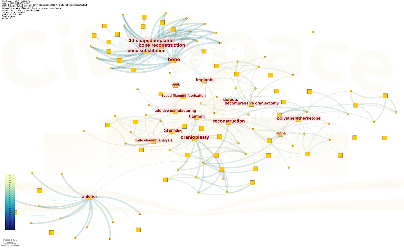
BACKGROUND: Polyetheretherketone (PEEK) synthetic material has become one of the preferred materials for repairing skull defects due to its low density, high strength, good toughness, excellent processing performance, and good biocompatibility, but there are few bibliometric analyses of PEEK for skull repair. OBJECTIVE: To explore the overall research trends, development context, research focuses, and hotspots of PEEK materials in the field of international skull defect repair using bibliometric methods. METHODS: The Web of Science Core Collection database was systematically searched for literature on PEEK materials for skull defect repair published from 1995 to 2024. On this basis, bibliometric methods were used to conduct quantitative statistics and visual analysis from the aspects of temporal dynamics of publication volume, country/region contribution, core research institution cooperation network, highly cited papers, high-yield journals, and keyword co-occurrence. RESULTS AND CONCLUSION: This study analyzed 105 studies on PEEK for skull defect repair published from 2009 to 2024. The development process was roughly divided into three stages: 2009-2014 traditional materials, 2015-2019 clinical research on PEEK, and 2020-2024 3D printing and finite element analysis, with the 2020-2024 stage accounting for 42%. Meanwhile, "3D printing, finite element analysis" and "PEEK, titanium alloy" were high-frequency technology combinations. China (21 articles), the United States (17 articles), and Germany (12 articles) were the main research countries. PEEK has been used in more than 200,000 clinical applications worldwide, with an infection rate of 3.7%, lower than that of polymethyl methacrylate (9.2%). PEEK research has shifted from "passive repair" to "active bioactivity promotion". Europe and the United States lead in clinical translation of 3D printing (equipment rate 82%, while China's domestic rate is 39%), but there is a lag of about 2 years between literature and clinical application for 3D-printed PEEK. It is predicted that conductive PEEK will accelerate translation in 2026-2027. The results indicate that PEEK has achieved a transformation from "passive repair" to "active bioactivity promotion", with 3D printing, surface modification, and intelligent integration as core directions. Global PEEK development is uneven; underdeveloped regions have high demand but less research (12%). China focuses on clinical research (68%) but lacks basic innovation (15%). It is necessary to promote low-cost 3D printing technology, establish translation hubs, support interdisciplinary research teams, and build a 10-year multicenter follow-up system.

Read Full Abstract10.12307/2026.21604